Bone-resorbing Mediator actinomycetemcomitans Is...
10
The Potent Bone-resorbing Mediator of Actinobacillus actinomycetemcomitans Is Homologous to the Molecular Chaperone GroEL Alun C. Kirby,* Sajeda Meghji,* Sean P. Nair,** Peter White," Krisanavane Reddi,** Tatsuji Nishihara,' Keisuke Nakashima,'1 Anthony C. Willis,1 Robert Sim,' Michael Wilson,* and Brian Hendersont *Department of Microbiology and Maxillofacial Surgery Research Unit, Eastman Dental Institute for Oral Health Care Sciences, University of London, London, WCIX 8LD, United Kingdom; *Department of Oral Science, National Institute of Health, Tokyo, Japan; Department of Periodontology, Tokyo Medical and Dental University, Tokyo, Japan; and 1MRC Immunochemistry Unit, Department of Biochemistry, University of Oxford, Oxford, United Kingdom Abstract Actinobacillus actinomycetemcomitans is a Gram-negative bacterium implicated in the pathology of localized juvenile periodontitis, a condition involving rapid destruction of al- veolar bone. We have established that gentle extraction of this bacterium in saline releases a proteinaceous fraction (which we have termed surface-associated material [SAM]) which has potent osteolytic activity in the murine calvarial bone resorption assay. Fractionation of the SAM has now revealed that activity is associated with a 62-kD protein. This bone-resorbing activity can be blocked by a monoclonal antibody (raised to the whole bacterium) that is claimed to recognize a protein homologous to the Escherichia coil molecular chaperone GroEL. Purification of this bone-re- sorbing protein to homogeneity has been achieved by a com- bination of anion exchange, gel filtration, and ATP-affinity chromatography and the NH2-terminal sequence shows > 95% homology to E. coli GroEL. This GroEL homologue is found in the SAM of A. actinomycetemcomitans but is not found in the osteolytically active SAM from other Gram- negative or Gram-positive bacteria. The GroEL protein from E. coli, but not from Mycobacterium tuberculosis and Mycobacterium leprae, also showed activity in the bone re- sorption assay. We believe this to be the first observation that a molecular chaperone has the capacity to stimulate the breakdown of connective tissue. (J. Clin. Invest. 1995. 96:1185-1194.) Key words: periodontal disease * chaper- onin 60 * heat shock proteins * bone resorption Introduction The chronic inflammatory periodontal diseases (CIPDs)' are the most prevalent of the persistent inflammatory diseases of Address correspondence to Professor Brian Henderson, Maxillofacial Surgery Research Unit, Eastman Dental Institute for Oral Health Care Sciences and University College Hospital London, University of Lon- don, 256 Gray's Inn Road, London WC1X 8LD, United Kingdom. Phone and FAX: 0171-915-1190. Receivedfor publication 15 November 1994 and accepted in revised form 25 May 1995. 1. Abbreviations used in this paper: CIPD, chronic inflammatory peri- odontal disease; LJP, localized juvenile periodontitis; SAM, surface- associated material. humans. They are characterized by inflammation of the gingivae and the resorption of the alveolar bone supporting the teeth, a process which can lead to tooth loss. Evidence strongly suggests that the presence of certain Gram-negative oral bacteria in peri- odontal pockets (which are formed between the gum and the tooth in this disease) is a major factor in the development of tissue pathology, and this relationship has been best documented with the bacterium Actinobacillus actinomycetemcomitans and its role in localized juvenile periodontitis (LJP) (1, 2). How- ever, the precise mechanisms responsible for the resorption of alveolar bone in this disease remain to be established. Bacteria may invade the periodontal tissues (3), but the accepted para- digm is that the resorption mechanism involves factors released by the bacteria, which either directly stimulate bone breakdown or generate the synthesis and/or release of osteolytic mediators within host tissues (3, 4). These mediators may include prostan- oids and/or certain proinflammatory cytokines such as IL-1 and TNFa, all of which have potent osteolytic activity. Using a simple saline extraction procedure we have isolated a fraction from the surface (termed surface-associated material [SAM]) of a number of bacteria implicated in the pathology of the CIPDs. This fraction contains the capsule and other com- ponents loosely associated with the outer membrane of the bac- teria, and electron microscopic examination of extracted organ- isms demonstrates the removal of extracellular material but fails to show evidence of cell lysis (5, 6). In the case of A. actinomy- cetemcomitans this highly soluble fraction consists largely of protein (> 60%) and an extremely small amount of LPS (6). There is now evidence that the SAM is shed by oral bacteria in situ, being found on the tooth roots of extracted teeth from patients with CIPD (7). The SAM from A. actinomycetemcomi- tans, has a number of biological activities including inhibition of cellular proliferation (8) and of collagen synthesis (9). How- ever, its most potent action is the stimulation of murine calvarial bone breakdown, with activity being seen at concentrations as low as 1 ng/ml (5, 6). In this assay the SAM is 10-100 times more potent than the LPS prepared from the same organism (5). Thus, the active moiety in this fraction could be a major factor in the pathogenesis of bone resorption in LJP. A number of bacterial components including LPS, teichoic acids, muramyl dipeptide, and certain protein fractions have been shown to be capable of stimulating bone resorption ( 10-13). However, with the possible exception of a toxin from the bacterium Pasteurella multocida (14), the SAM from A. actinomycetemcomitans is the most potent bacterial osteolytic agent reported. As described in this paper we have established that the active moiety is a 62- kD protein and thus its molar potency is in the picomolar range, similar to that reported for potent osteolytic cytokines such as IL-1 or TNF. Bone-resorbing Activity of a Molecular Chaperone 1185 J. Clin. Invest. X The American Society for Clinical Investigation, Inc. 0021-9738/95/09/1185/10 $2.00 Volume 96, September 1995, 1185-1194
-
Upload
phungnguyet -
Category
Documents
-
view
213 -
download
0
Transcript of Bone-resorbing Mediator actinomycetemcomitans Is...
The Potent Bone-resorbing Mediator of Actinobacillus actinomycetemcomitansIs Homologous to the Molecular Chaperone GroELAlun C. Kirby,* Sajeda Meghji,* Sean P. Nair,** Peter White," Krisanavane Reddi,** Tatsuji Nishihara,'Keisuke Nakashima,'1 Anthony C. Willis,1 Robert Sim,' Michael Wilson,* and Brian Hendersont*Department of Microbiology and Maxillofacial Surgery Research Unit, Eastman Dental Institute for Oral Health Care Sciences,University of London, London, WCIX8LD, United Kingdom; *Department of Oral Science, National Institute of Health, Tokyo, Japan;Department of Periodontology, Tokyo Medical and Dental University, Tokyo, Japan; and 1MRCImmunochemistry Unit, Department of
Biochemistry, University of Oxford, Oxford, United Kingdom
Abstract
Actinobacillus actinomycetemcomitans is a Gram-negativebacterium implicated in the pathology of localized juvenileperiodontitis, a condition involving rapid destruction of al-veolar bone. Wehave established that gentle extraction ofthis bacterium in saline releases a proteinaceous fraction(which we have termed surface-associated material [SAM])which has potent osteolytic activity in the murine calvarialbone resorption assay. Fractionation of the SAMhas nowrevealed that activity is associated with a 62-kD protein.This bone-resorbing activity can be blocked by a monoclonalantibody (raised to the whole bacterium) that is claimedto recognize a protein homologous to the Escherichia coilmolecular chaperone GroEL. Purification of this bone-re-sorbing protein to homogeneity has been achieved by a com-bination of anion exchange, gel filtration, and ATP-affinitychromatography and the NH2-terminal sequence shows> 95% homology to E. coli GroEL. This GroEL homologueis found in the SAMof A. actinomycetemcomitans but is notfound in the osteolytically active SAMfrom other Gram-negative or Gram-positive bacteria. The GroEL proteinfrom E. coli, but not from Mycobacterium tuberculosis andMycobacterium leprae, also showed activity in the bone re-sorption assay. Webelieve this to be the first observationthat a molecular chaperone has the capacity to stimulatethe breakdown of connective tissue. (J. Clin. Invest. 1995.96:1185-1194.) Key words: periodontal disease * chaper-onin 60 * heat shock proteins * bone resorption
Introduction
The chronic inflammatory periodontal diseases (CIPDs)' arethe most prevalent of the persistent inflammatory diseases of
Address correspondence to Professor Brian Henderson, MaxillofacialSurgery Research Unit, Eastman Dental Institute for Oral Health CareSciences and University College Hospital London, University of Lon-don, 256 Gray's Inn Road, London WC1X8LD, United Kingdom.Phone and FAX: 0171-915-1190.
Receivedfor publication 15 November 1994 and accepted in revisedform 25 May 1995.
1. Abbreviations used in this paper: CIPD, chronic inflammatory peri-odontal disease; LJP, localized juvenile periodontitis; SAM, surface-associated material.
humans. They are characterized by inflammation of the gingivaeand the resorption of the alveolar bone supporting the teeth, aprocess which can lead to tooth loss. Evidence strongly suggeststhat the presence of certain Gram-negative oral bacteria in peri-odontal pockets (which are formed between the gum and thetooth in this disease) is a major factor in the development oftissue pathology, and this relationship has been best documentedwith the bacterium Actinobacillus actinomycetemcomitans andits role in localized juvenile periodontitis (LJP) (1, 2). How-ever, the precise mechanisms responsible for the resorption ofalveolar bone in this disease remain to be established. Bacteriamay invade the periodontal tissues (3), but the accepted para-digm is that the resorption mechanism involves factors releasedby the bacteria, which either directly stimulate bone breakdownor generate the synthesis and/or release of osteolytic mediatorswithin host tissues (3, 4). These mediators may include prostan-oids and/or certain proinflammatory cytokines such as IL-1 andTNFa, all of which have potent osteolytic activity.
Using a simple saline extraction procedure we have isolateda fraction from the surface (termed surface-associated material[SAM]) of a number of bacteria implicated in the pathologyof the CIPDs. This fraction contains the capsule and other com-ponents loosely associated with the outer membrane of the bac-teria, and electron microscopic examination of extracted organ-isms demonstrates the removal of extracellular material but failsto show evidence of cell lysis (5, 6). In the case of A. actinomy-cetemcomitans this highly soluble fraction consists largely ofprotein (> 60%) and an extremely small amount of LPS (6).There is now evidence that the SAMis shed by oral bacteriain situ, being found on the tooth roots of extracted teeth frompatients with CIPD (7). The SAMfrom A. actinomycetemcomi-tans, has a number of biological activities including inhibitionof cellular proliferation (8) and of collagen synthesis (9). How-ever, its most potent action is the stimulation of murine calvarialbone breakdown, with activity being seen at concentrations aslow as 1 ng/ml (5, 6). In this assay the SAMis 10-100 timesmore potent than the LPS prepared from the same organism(5). Thus, the active moiety in this fraction could be a majorfactor in the pathogenesis of bone resorption in LJP. A numberof bacterial components including LPS, teichoic acids, muramyldipeptide, and certain protein fractions have been shown to becapable of stimulating bone resorption ( 10-13). However, withthe possible exception of a toxin from the bacterium Pasteurellamultocida (14), the SAMfrom A. actinomycetemcomitans isthe most potent bacterial osteolytic agent reported. As describedin this paper we have established that the active moiety is a 62-kD protein and thus its molar potency is in the picomolar range,similar to that reported for potent osteolytic cytokines such asIL-1 or TNF.
Bone-resorbing Activity of a Molecular Chaperone 1185
J. Clin. Invest.X The American Society for Clinical Investigation, Inc.0021-9738/95/09/1185/10 $2.00Volume 96, September 1995, 1185-1194

Wehave now isolated this osteolytic protein by use of anionexchange, gel filtration, and affinity chromatography and havedetermined its NH2-terminal sequence. Care has been taken toexclude the possibility that LPS is contributing to the osteolyticactivity of this protein. Isolation was aided by the use of mono-clonal antibodies which neutralize the bone-resorbing activity ofthe SAM. These studies have established that the osteolyticallyactive protein of A. actinomycetemcomitans is a member of theGroEL family of molecular chaperones.
Methods
Growth of bacteria. A. actinomycetemcomitans NCTC9710 was grownon brain-heart infusion agar (Oxoid, Basingstoke, United Kingdom) at370C in a C02-enriched atmosphere for 3 d. Eikenella corrodens NCTC10596 and Porphyromonas gingivalis W50were grown under anaerobicconditions at 370C in a medium consisting of brain-heart infusion agarsupplemented with 0.375 grams/liter cysteine-HCl (BDH), 0.25 grams/liter haemin (BDH), and 0.05 grams/liter menadione (BDH Poole,Dorset, United Kingdom). Escherichia coli Y1090 was grown on LuriaBertani agar, consisting of 10 grams/liter Bacto-Tryptone (Difco Labo-ratories Inc., Detroit, MI), 5 grams/liter bacto-yeast extract (Difco),and 5 grams/liter NaCl (Sigma Immunochemicals, St. Louis, MO), for24 h under aerobic conditions at 37TC. Staphylococcus aureus (Oxfordstrain NCTC6571) was grown under aerobic conditions on brain-heartinfusion agar at 370C for 48 h.
Extraction of SAM. All cells were harvested in sterile saline, centri-fuged, washed briefly in saline, and lyophilized. SAMwas removedfrom the various bacteria by gentle saline extraction as described (5).Briefly, lyophilized bacteria were resuspended in sterile saline andstirred gently at 40C for 1 h. The bacteria were removed by centrifugationand the soluble SAMwas dialyzed extensively against distilled waterand lyophilized. The protein content of the SAMwas determined bythe method of Lowry et al. ( 15), the carbohydrate content by the methodof Dubois et al. (16), and the nucleic acid content by absorption at260/280 nm. The LPS content was measured by use of a commercialchromogenic Limulus amebocyte lysate assay (Pyrogent, Byk-Mallinck-rodt, London, United Kingdom) according to the manufacturer's instruc-tions. In all studies the activity of the SAMwas related to dry weight.
Monoclonal antibodies. Hybridomas were raised in Balb/c miceimmunized with A. actinomycetemcomitans ATCC (#43718) wholecells (17). Briefly, spleen cells from intracutaneously immunized andintraperitoneally boosted mice were fused with SP2/0-Ag-14 myelomacells. Hybridomas were screened by ELISA for immunoglobulin secre-tion and antibody producers were cloned by limiting dilution. Three ofthese, P1, P2, and P3, secreted antibodies which have been shown tobe specific for 81-, 62-, and 62-kD proteins, respectively, and this wasconfirmed by Western blotting against SAMfrom A. actinomycetem-comitans NCTC9710. Hybridoma cells secreting mAbs P1, P2, and P3were grown in Iscoves modified Dulbecco's medium (Gibco Labora-tories, Grand Island, NY) containing 10% Seraclone FCS (Sera-LabLtd., Crawley Down, United Kingdom) at 37°C in an atmosphere of5% C02/air. When cell death began to occur, the cells were removedby centrifugation and the supernatant was collected. This was filter-sterilized and stored at -20°C until needed. Monoclonal antibody A5was raised against the SAMextracted from A. actinomycetemcomitansNCTC9710. Balb/c mice were immunized subcutaneously and boostedintravenously. Hybridoma cells were produced as described above andscreened by ELISA against SAM. mAbA5 has been shown to be specificfor a 66-kD protein component of the SAMby Western blot. Hybridomacells secreting A5 were cultured in RPMI 1640 medium (Gibco Labora-tories) containing 10%FCS (Sera-Lab). Tissue culture supernatant wascollected as described above. All mAbs were of the IgGI subclass.
All four mAbs were purified from their respective tissue culturesupernatants using a protein A column (Sepharose CL 4B-linked pro-
tein A; Bio Rad Laboratories, Hercules, CA) (18) and dialyzed exten-sively against PBS.
Murine calvarial bone resorption assay. Bone resorption was quan-tified by measuring calcium release from MF1 strain mouse calvaria invitro (19). In some experiments the LPS-unresponsive strain C3H/HeJwas used. Calvaria were removed from 5-d-old mice and halved, witheach half cultured separately on stainless steel grids. Calvaria werecultured in 30-mm dishes with 1.5 ml of BGJ medium (Flow Labora-tories Inc., McLean, VA) containing 5% heat-inactivated rabbit serum(Gibco Laboratories) and 5 mg/ 100 ml ascorbic acid (Sigma Immuno-chemicals). The calvaria were treated in groups of five. After 24 h, themedia were replaced with media containing the test substances. PGE2at 1 jiM was used as a positive control in all assays (except those inwhich the C3H/HeJ strain mice were used, when IL-la was used) toshow that the bone was responsive to osteolytic mediators. Calvariawere cultured for a further 48 h before calcium release was measuredby automated colorimetric analysis (20). The significance of the resultswas calculated using Student's t-test. Batches of SAMwere assayed forbone resorbing activity over the concentration range 10 ng/ml to 10jig/ml.
To assess the capacity of mAbs to inhibit the bone resorbing activityof the SAM, individual mAbswere added to calvarial cultures stimulatedto resorb by the presence of 1 jig/ml SAMor 1 jig/ml LPS from A.actinomycetemcomitans. Each antibody was used at various dilutions todetermine its inhibitory dose response. Nonspecific mouse immunoglob-ulin G, containing all four IgG subclasses (Sigma Immunochemicals),was used as a control in all the antibody studies. To determine if themAbs were able to deplete the SAMof its osteolytic activity, solutionsof the SAMwere incubated with an excess (1:10 wt/wt) of antibodyP3 or with nonspecific mouse IgG (Sigma Immunochemicals), or withmAbA5 overnight at 4°C with constant mixing. Antibody, along withany bound antigen, was then removed by the addition of S. aureus(Cowan strain) heat-killed/formalin-fixed whole cells (Sigma Immuno-chemicals) for 1 h at room temperature, again with constant mixing,followed by centrifugation and filter sterilization. The depleted fractionwas added to the calvarial assay at 1 or 10 jtg/ml and activity wascompared with that of untreated SAM. SAMwas also directly incubatedwith S. aureus at room temperature for 1 h to control for any possiblenonspecific binding events between bacteria and SAM, this treated frac-tion being tested in the bone resorption assay.
To determine if LPS was contributing to the osteolytic activity ofthe SAM, the crude and purified GroEL preparations or LPS from A.actinomycetemcomitans were exposed to heat (100°C for 30 min) ortrypsin (0.25% trypsin; Sigma Immunochemicals) for 1 h followed byexcess (threefold molar excess) of soya-bean trypsin inhibitor (SigmaImmunochemicals). Control cultures exposed to the trypsin/trypsin in-hibitor complex showed no increase in bone resorption, and the formedcomplex when added to bone cultures stimulated with PGE2 did notinhibit osteolysis. Polymyxin B (20 jsg/ml) was also added to bonecultures stimulated with either SAMor LPS from A. actinomycetemcom-itans to determine if it could inhibit bone resorption.
SDS-PAGE. The components of the SAMwere analyzed by SDS-PAGEusing 12% gels according to the method of Laemmli (21). Sam-ples were diluted 1:1 with sample buffer and boiled for 5 min beforeloading. Gels were run using a MiniProtean I system (Bio Rad Labora-tories) and stained with Coomassie brilliant blue (Sigma Immunochemi-cals). The molecular weight markers used were Dalton VIIL (SigmaImmunochemicals). Gels were also silver stained using a commercialkit (Gelcode@ silver stain kit; Pierce, Rockford, IL) to detect both thepresence of protein and carbohydrate.
Two-dimensional PAGE. Two-dimensional PAGEgels were run ac-
cording to the method of O'Farrell (22). Gels were run using a MiniPro-tean II system and stained with Coomassie blue, with similar molecularweight markers as above. The first dimension, isoelectric focusing, wasover the pI range of 3-10. Second dimension separation was by molecu-lar mass using a 12% SDS-PAGEgel.
Immunoblotting. Samples separated on one- or two-dimensional
1186 Kirby et al.
SDS-PAGEwere electroblotted onto Immobilon P polyvinyldifluoridemembranes (Millipore Corp., Bedford, MA) overnight (23). Mem-branes were washed with PBS containing 0.1% Triton X-100 (SigmaImmunochemicals) (PBS-T) and blocked with PBS-T containing 2%FCS (blocking buffer) (Sera-Lab). Blocked membranes were then incu-bated with the test antibody (in blocking buffer) for 1 h and washedwith PBS-T. Bound mouse IgG was detected using peroxidase-labeledgoat anti-mouse IgG (y-chain specific) (Sigma Immunochemicals) at1:1,000 in PBS-T-2% FCS. After a final wash the blots were developedwith a solution of 1 mg/ml 3,3'-diaminobenzidine tetrahydrochloride(Sigma Immunochemicals) in 50 mMTris (Sigma Immunochemicals),pH 7.6, containing 150 mMNaCl (BDH) and 0.05% hydrogen peroxide(Sigma Immunochemicals). Each reaction was terminated by extensiverinsing with distilled water.
Protein purification. Crude SAMwas fractionated at 40C on a Q-Sepharose anion exchange column (50 cm X 1.6 cm). The column wasequilibrated in 20 mMTris-HCI, pH 8.5 (buffer A), and the SAM(generally 100-400 mg) was loaded on in the same buffer. The columnwas washed with 500 ml of buffer A and then eluted with a 1,000-mllinear gradient of 0-2 MNaCl in buffer A. 10-ml fractions were col-lected, and the absorbance at 280 nm was monitored. The location ofthe osteolytic protein was determined by a combination of activity assay,SDS-PAGE, and Western blot analysis. Fractions containing osteolyticactivity were dialyzed against deionized water to remove salt and lyophi-lized. The fraction with the highest specific activity and the least numberof protein bands on SDS-PAGEwas then further fractionated at roomtemperature on a second anion exchange column. There was some evi-dence of proteolytic clipping of the 62-kD protein and so aliquots of thisfraction were dissolved at 1 mg/ml in 20 mMTris, pH 7.2, containingproteinase inhibitors (1 mMPMSF, 1 mMEDTA, and 1 mMbenzami-dine) (buffer B) and fractionated on an EconoPak Q column (BioRad Laboratories) equilibrated in buffer B. Fractions were eluted byapplication of a gradient of 0-1.5 MNaCl in buffer B, and an ab-sorbance profile at 280 nmwas obtained. Fractions were again analyzedfor osteolytic activity and Western immunoblotted with mAb P3 toconfirm the presence of the immunogenic 62-kD protein. The purity ofthe fractions was again assessed visually by SDS-PAGE, and 100 Igof the cleanest fraction was dialyzed against 50 mMof Tris buffer, pH7.6, containing 10 mMKC1 and 10 mMMgCl2 (buffer C). This samplewas run on a 5-ml ATP-Sepharose (Sigma Immunochemicals) column.The column was washed with 10 column volumes of buffer C and boundprotein eluted in 5 column volumes of 5 mMATP (Sigma Immuno-chemicals), also in buffer C. Protein was located by SDS-PAGEandvisualized using a silver stain kit (Sigma Immunochemicals). Gel filtra-tion was used to determine the molecular mass range of the osteolyticprotein isolated by ATP-affinity chromatography. This was done byrunning the purified protein on a Bio-Sil TSK250 (Bio Rad Labora-tories) column in 0.1 Msodium phosphate buffer, pH 6.7, and measuringabsorption at 205/280 nm.
Immunoaffinity purification. Affinity columns were prepared usingmAbP2, mAbP3, and both P2 and P3 together. Briefly, in each case,5 mg of antibody was linked to 3.5 ml of swollen cyanogen bromide-activated Sepharose 4B (Sigma Immunochemicals) in bicarbonatebuffer at pH 8.3. After washing, the column was blocked with 1 Methanolamine at pH 8.0, washed, and equilibrated in PBS. Crude SAM(5 mg) dissolved at 1 mg/ml in PBS was loaded on, the column waswashed extensively, and bound protein was eluted using 0.1 Mglycine,pH 2.5. Eluted fractions of 1 ml were concentrated by using Miniconmicroconcentrators with cutoff membranes of 10 kD (Amicon, Inc.,Beverly, MA), to a volume of - 50 ,1. These were analyzed by SDS-PAGEand immunoblotting and tested for osteolytic activity in the cal-varial bone resorption assay.
Protein sequencing. Material eluting from the ATP-Sepharose col-umn was run on a 10% SDS-PAGEgel according to the method ofLaemmli (21) and electroblotted onto ProBlott membrane (AppliedBiosystems Inc., Foster City, CA). The band of interest, at 62 kD, wasexcised and run on an ABI 470A protein sequencer (Applied Biosystems
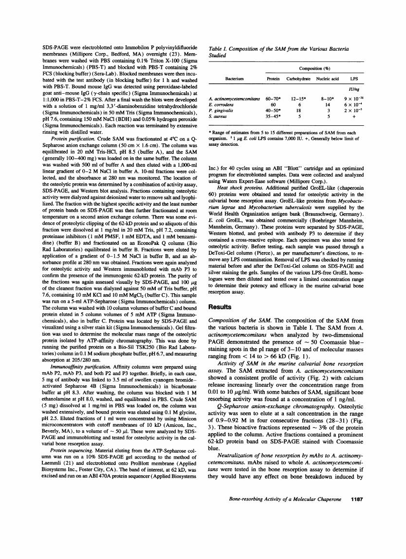
Table l. Composition of the SAMfrom the Various BacteriaStudied
Composition (%)
Bacterium Protein Carbohydrate Nucleic acid LPS
IUing
A. actinomycetemcomitans 60-70* 12-15* 8-10* 9 X io-3tE. corrodens 60 6 14 6 X 10⁻⁴P. gingivalis 40-50* 18 3 2 X iOnsS. aureus 35-45* 5 5 +
* Range of estimates from 5 to 15 different preparations of SAMfrom eachorganism. * 1 ug E. coli LPS contains 7,000 IU. +, Generally below limit ofassay detection.
Inc.) for 40 cycles using an ABI "Blott" cartridge and an optimizedprogram for electroblotted samples. Data were collected and analyzedusing Waters Expert-Ease software (Millipore Corp.).
Heat shock proteins. Additional purified GroEL-like (chaperonin60) proteins were obtained and tested for osteolytic activity in thecalvarial bone resorption assay. GroEL-like proteins from Mycobacte-rium leprae and Mycobacterium tuberculosis were supplied by theWorld Health Organization antigen bank (Braunschweig, Germany).E. coli GroEL, was obtained commercially (Boehringer Mannheim,Mannheim, Germany). These proteins were separated by SDS-PAGE,Western blotted, and probed with antibody P3 to determine if theycontained a cross-reactive epitope. Each specimen was also tested forosteolytic activity. Before testing, each sample was passed through aDeToxi-Gel column (Pierce), as per manufacturer's directions, to re-move any LPS contamination. Removal of LPS was checked by runningmaterial before and after the DeToxi-Gel column on SDS-PAGEandsilver staining the gels. Samples of the various LPS-free GroEL homo-logues were then diluted and tested over a limited concentration rangeto determine their potency and efficacy in the murine calvarial boneresorption assay.
Results
Composition of the SAM. The composition of the SAMfromthe various bacteria is shown in Table I. The SAMfrom A.actinomycetemcomitans when analyzed by two-dimensionalPAGEdemonstrated the presence of 50 Coomassie blue-staining spots in the pI range of 3-10 and of molecular massesranging from < 14 to > 66 kD (Fig. 1).
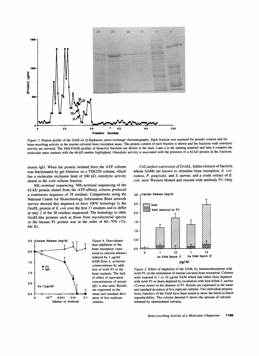
Activity of SAMin the murine calvarial bone resorptionassay. The SAM extracted from A. actinomycetemcomitansshowed a consistent profile of activity (Fig. 2) with calciumrelease increasing linearly over the concentration range from0.01 to 10 jg/ml. With some batches of SAM, significant boneresorbing activity was found at a concentration of 1 ng/ml.
Q-Sepharose anion-exchange chromatography. Osteolyticactivity was seen to elute at a salt concentration in the rangeof 0.9-0.92 M in four consecutive fractions (28-31) (Fig.3). These bioactive fractions represented 3% of the proteinapplied to the column. Active fractions contained a prominent62-kD protein band on SDS-PAGE stained with Coomassieblue.
Neutralization of bone resorption by mAbs to A. actinomy-cetemcomitans. mAbs raised to whole A. actinomycetemcomi-tans were tested in the bone resorption assay to determine ifthey would have any effect on bone breakdown induced by
Bone-resorbing Activity of a Molecular Chaperone 1187
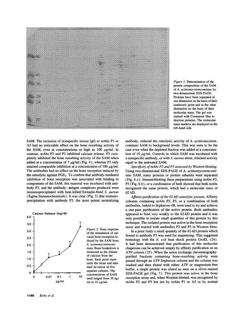
Figure 1. Determination of theprotein composition of the SAMof A. actinomycetemcomitans bytwo-dimensional SDS-PAGE.Proteins have been separated inone dimension on the basis of theirisoelectric point and in the otherdimension on the basis of theirmolecular mass. The gel wasstained with Coomassie blue todisclose proteins. The molecularmass markers are displayed on theleft-hand side.
SAM. The inclusion of nonspecific mouse IgG or mAbs PI orA5 had no noticeable effect on the bone resorbing activity ofthe SAM, even at concentrations as high as 100 ,tg/ml. Incontrast, mAbs P2 and P3 inhibited calcium release. P3 com-pletely inhibited the bone resorbing activity of the SAMwhenadded at a concentration of 7 pg/ml (Fig. 4), whereas P2 onlyattained comparable inhibition at a concentration of 100 jsg/ml.The antibodies had no effect on the bone resorption induced bythe osteolytic agonist PGE2. To confirm that antibody-mediatedinhibition of bone resorption was associated with binding tocomponents of the SAM, this material was incubated with anti-body P3, and the antibody-antigen complexes produced wereimmunoprecipitated with heat-killed/formalin-fixed S. aureus(Sigma Immunochemicals). It was clear (Fig. 5) that immuno-precipitation with antibody P3, the most potent neutralizing
Calcium Release (mg/dl)4.0 -
3.5 -
3.0 - Figure 2. Dose responseof the stimulation of cal-
2.5 - varial bone resorption in-duced by the SAMfrom
2.0 - A. actinomycetemcom-itans. Bone breakdown is
1.5 measured as the releaseof calcium from the
1.0 - bone. Each point repre-sents the mean and stan-
0.5 - dard deviation of five
07/ separate cultures. The
0 0.01 0.1 1 1 0 concentrations of SAMused ranged from 10 ng/
.ig/ml ml to 10 ,g/ml.
antibody, reduced the osteolytic activity of A. actinomycetem-comitans SAMto background levels. This was seen to be thecase even when the depleted fraction was added at a concentra-tion of 10 Atg/ml. Controls in which SAMwas incubated witha nonspecific antibody, or with S. aureus alone, retained activityequal to the untreated SAM.
Specificity of mAbsP2 and P3 assessed by Western blotting.Using two-dimensional SDS-PAGEof A. actinomycetemcomi-tans SAM, many proteins or protein subunits were separated(Fig. 6 a). Immunoblotting these preparations using mAbs P2,P3 (Fig. 6 b), or a combination of both showed that both mAbsrecognized the same protein, which had a molecular mass of62 kD.
Affinity purification of the 62-kD osteolytic protein. Affinitycolumns containing mAbs P2, P3, or a combination of bothantibodies, linked to Sepharose 4B, were used to try and achievea one-pass purification of the active protein. Both antibodiesappeared to bind very weakly to the 62-kD protein and it wasonly possible to isolate small quantities of this protein by thistechnique. The isolated protein was active in the bone resorptionassay and reacted with antibodies P2 and P3 in Western blots.
In a prior study a small quantity of the 62-kD protein whichbound to antibody P3 was used for sequencing. This suggestedhomology with the E. coli heat shock protein GroEL (24).It had been demonstrated that purification of this molecularchaperone can be achieved simply by affinity purification on anATP column (25). When the anion exchange chromatography-purified fractions containing bone-resorbing activity werepassed through an ATP-Sepharose column and the column waswashed and then eluted with either ATP or magnesium-freebuffer, a single protein was eluted as seen on a silver-stainedSDS-PAGEgel (Fig. 7). This protein was active in the boneresorption assay and, when Western blotted, was recognized bymAbs P2 and P3 but not by mAbs P1 or A5 or by normal
1188 Kirby et al.
C
00.
0'0 20 40 7 60 80 100
Fraction Number
Figure 3. Elution profile of the SAMon Q-Sepharose anion exchange chromatography. Each fraction was assessed for protein content and forbone-resorbing activity in the murine calvarial bone resorption assay. The protein content of each fraction is shown and the fractions with osteolyticactivity are arrowed. The SDS-PAGEprofiles of bioactive fractions are shown in the inset. Lane a is the starting material and lane b contains the
molecular mass markers with the 66-kD marker highlighted. Osteolytic activity is associated with the presence of a 62-kD protein in the fractions.
mouse IgG. When the protein isolated from the ATP columnwas fractionated by gel filtration on a TSK250 column, whichhas a molecular exclusion limit of 300 kD, osteolytic activityeluted in the void volumn fraction.
NH2-terminal sequencing. NH2-terminal sequencing of the62-kD protein eluted from the ATP-affinity column produceda continuous sequence of 38 residues. Comparisons using theNational Centre for Biotechnology Information Blast networkservice showed this sequence to have 100% homology to theGroEL protein of E. coli over the first 17 residues and to differat only 2 of the 38 residues sequenced. The homology to otherGroEl-like proteins such as those from mycobacterial speciesor the human P1 protein was in the order of 60-70% (Ta-ble II).
2.5 Calcium Release (mg/dl) Figure 4. Dose-depen-dent inhibition of the
2.0 4 kzZIbone resorption (mea-
2.0 sured as calcium release)induced by 1 yg/ml
1.5 - SAMfrom A. actinomy-* P3 cetemcomitans by addi-A IgG tion of mAbP3 to the
1.0 - bone explants. The lackof effect of equivalent
0.5 - concentrations of mouseAa (1pg/ml) IgG is also seen. Results
are expressed as the0.0 -1 mean and standard devi-
0 10-4 0.001 0.01 0.1 ation of five replicateDilution of Antibody cultures.
Cell surface expression of GroEL. Saline extracts of bacteriawhose SAMs are known to stimulate bone resorption, E. cor-rodens, P. gingivalis, and S. aureus, and a crude extract of E.coli, were Western blotted and reacted with antibody P3. Only
3.6 1 Calcium Release (mg/dl)
3.0
2.4 -
1.8 -
1.2 -
0.6 -
0.0 -
SAMSAM depleted by P3
0 1 10 1 10Aa SAM (batch 1) Aa SAM (batch 2)
pg/miFigure 5. Effect of depletion of the SAM, by immunoabsorption withmAbP3, on the stimulation of murine calvarial bone resorption. Cultureswere exposed to 1 or 10 pig/ml SAMwhich had either been depletedwith mAbP3 or sham-depleted by incubation with heat-killed S. aureus(Cowan strain) in the absence of P3. Results are expressed as the meanand standard deviation of five replicate cultures. Two individual prepara-tions (batches) of the SAMhave been tested to show the batch-to-batchreproducibility. The column denoted 0 shows the amount of calciumreleased by unstimulated calvaria.
Bone-resorbing Activity of a Molecular Chaperone 1189
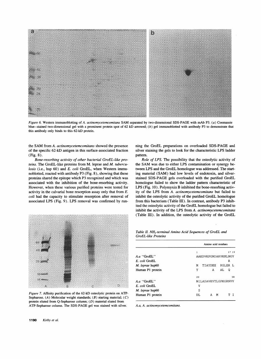
Figure 6. Western immunoblotting of A. actinomycetemcomitans SAMseparated by two-dimensional SDS-PAGEwith mAbP3. (a) Coomassieblue-stained two-dimensional gel with a prominent protein spot of 62 kD arrowed; (b) gel immunoblotted with antibody P3 to demonstrate thatthis antibody only binds to this 62-kD protein.
the SAMfrom A. actinomycetemcomitans showed the presenceof the specific 62-kD antigen in this surface-associated fraction(Fig. 8).
Bone-resorbing activity of other bacterial GroEL-like pro-teins. The GroEL-like proteins from M. leprae and M. tubercu-losis (i.e., hsp 60) and E. coli GroEL, when Western immu-noblotted, reacted with antibody P3 (Fig. 8), showing that theseproteins shared the epitope which P3 recognized and which wasassociated with the inhibition of the bone-resorbing activity.However, when these various purified proteins were tested foractivity in the calvarial bone resorption assay only that from E.coli had the capacity to stimulate resorption after removal ofassociated LPS (Fig. 9). LPS removal was confirmed by run-
Figure 7. Affinity purification of the 62-kD osteolytic protein on ATP-Sepharose. (A) Molecular weight standards; (B) starting material; (C)protein eluted from Q-Sepharose column; (D) material eluted fromATP-Sepharose column. The SDS-PAGEgel was stained with silver.
ning the GroEL preparations on overloaded SDS-PAGEandsilver staining the gels to look for the characteristic LPS ladderpattern.
Role of LPS. The possibility that the osteolytic activity ofthe SAMwas due to either LPS contamination or synergy be-tween LPS and the GroEL homologue was addressed. The start-ing material (SAM) had low levels of endotoxin, and silver-stained SDS-PAGEgels overloaded with the purified GroELhomologue failed to show the ladder pattern characteristic ofLPS (Fig. 10). Polymyxin B inhibited the bone-resorbing activ-ity of the LPS from A. actinomycetemcomitans but failed toinhibit the osteolytic activity of the purified GroEL homologuefrom this bacterium (Table III). In contrast, antibody P3 inhib-ited the osteolytic activity of the GroEL homologue but failed toinhibit the activity of the LPS from A. actinomycetemcomitans(Table III). In addition, the osteolytic activity of the GroEL
Table I. NH2-terminal Amino Acid Sequences of GroEL andGroEL-like Proteins
Amino acid residues
1 17 1 9
A.a "GroEL" AAKDVKFGNDARVKMLNGVE. coli GroEL RM. leprae hsp6O M TIAYDEE RGLER LHuman PI protein Y A AL Q
20 38
A.a "GroEL" NILADAVKVTLGPKGRNVVE. coli GroEL VM. leprae hsp6O SHuman PI protein DL A M T I
A.a, A. actinomycetemcomitans.
1190 Kirby et al.
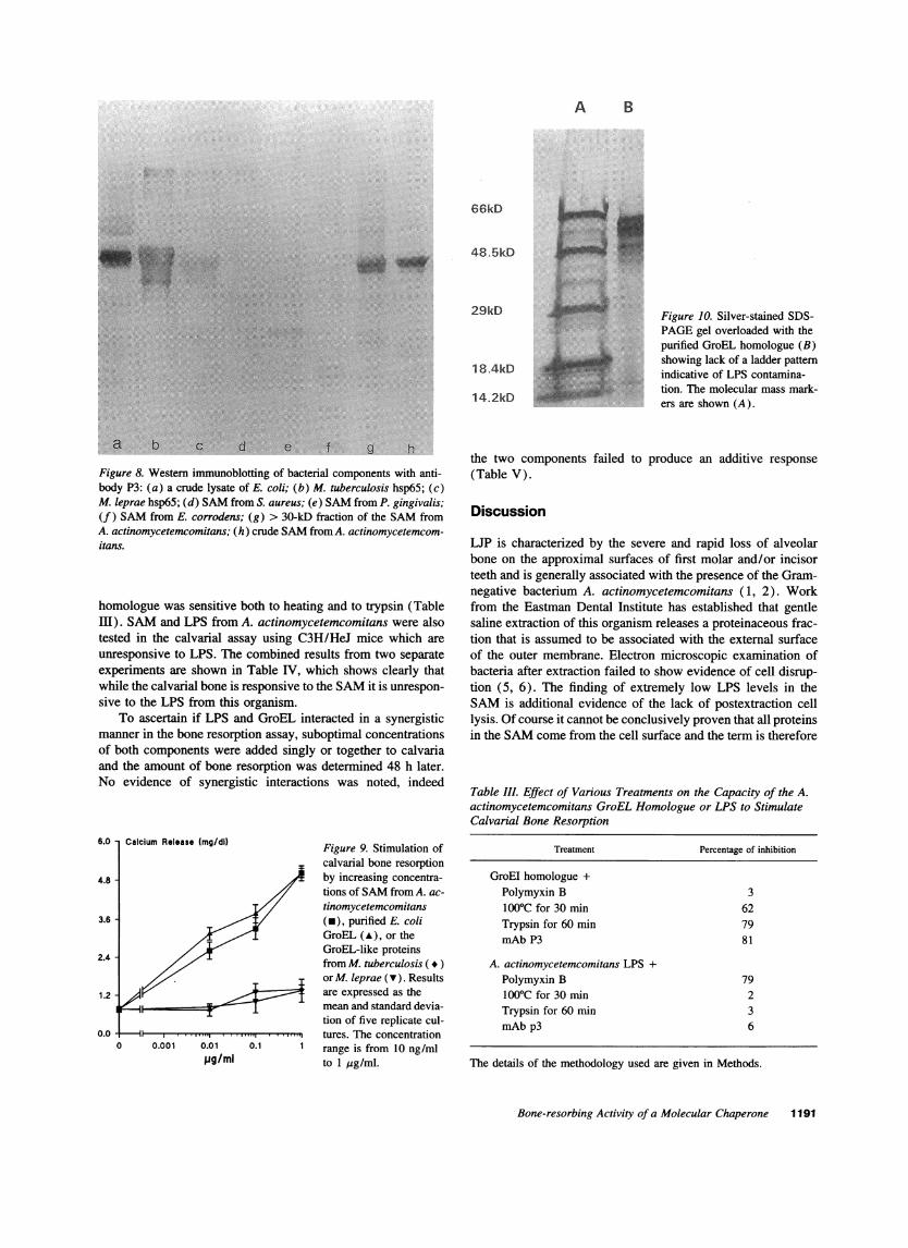
Figure 10. Silver-stained SDS-PAGEgel overloaded with thepurified GroEL homologue (B)showing lack of a ladder patternindicative of LPS contamina-tion. The molecular mass mark-ers are shown (A).
Figure 8. Western immunoblotting of bacterial components with anti-body P3: (a) a crude lysate of E. coli; (b) M. tuberculosis hsp65; (c)M. leprae hsp65; (d) SAMfrom S. aureus; (e) SAMfrom P. gingivalis;(f ) SAMfrom E. corrodens; (g) > 30-kD fraction of the SAMfromA. actinomycetemcomitans; (h) crude SAMfrom A. actinomycetemcom-itans.
homologue was sensitive both to heating and to trypsin (TableIll). SAMand LPS from A. actinomycetemcomitans were alsotested in the calvarial assay using C3H/HeJ mice which are
unresponsive to LPS. The combined results from two separateexperiments are shown in Table IV, which shows clearly thatwhile the calvarial bone is responsive to the SAMit is unrespon-
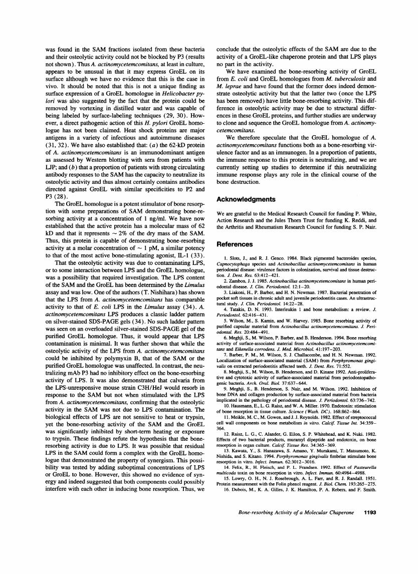
sive to the LPS from this organism.To ascertain if LPS and GroEL interacted in a synergistic
manner in the bone resorption assay, suboptimal concentrationsof both components were added singly or together to calvariaand the amount of bone resorption was determined 48 h later.No evidence of synergistic interactions was noted, indeed
6.0 Calcium Release (mg/di) Figure 9. Stimulation ofcalvarial bone resorption
4.8 - by increasing concentra-tions of SAMfrom A. ac-
tinomycetemcomitans3.6 (*), purified E. coli
GroEL (A), or the
2.4 - GroEL-like proteinsfrom M. tuberculosis ( *)
or M. leprae (v). Results1.2 are expressed as the
mean and standard devia-
tion of five replicate cul-0.0 -1 ,, . .tures. The concentration
0 0.001 0.01 0.1 1 range is from 10 ng/ml
pg/mi to 1 ,ug/ml.
the two components failed to produce an additive response
(Table V).
Discussion
LUP is characterized by the severe and rapid loss of alveolarbone on the approximal surfaces of first molar and/or incisorteeth and is generally associated with the presence of the Gram-negative bacterium A. actinomycetemcomitans (1, 2). Workfrom the Eastman Dental Institute has established that gentlesaline extraction of this organism releases a proteinaceous frac-tion that is assumed to be associated with the external surfaceof the outer membrane. Electron microscopic examination ofbacteria after extraction failed to show evidence of cell disrup-tion (5, 6). The finding of extremely low LPS levels in theSAMis additional evidence of the lack of postextraction celllysis. Of course it cannot be conclusively proven that all proteinsin the SAMcome from the cell surface and the term is therefore
Table III. Effect of Various Treatments on the Capacity of the A.actinomycetemcomitans GroEL Homologue or LPS to StimulateCalvarial Bone Resorption
Treatment Percentage of inhibition
GroEl homologue +Polymyxin B 3100'C for 30 min 62Trypsin for 60 min 79mAbP3 81
A. actinomycetemcomitans LPS +Polymyxin B 791000C for 30 min 2Trypsin for 60 min 3mAbp3 6
The details of the methodology used are given in Methods.
Bone-resorbing Activity of a Molecular Chaperone 1191
A B
66kD
48.5kD
29kD
18.4kD
14.2kD
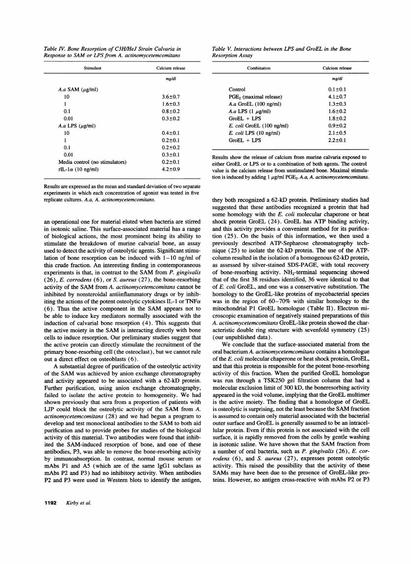
Table IV. Bone Resorption of C3H/HeJ Strain Calvaria inResponse to SAMor LPS from A. actinomycetemcomitans
Stimulant Calcium release
mg/dl
A.a SAM(pg/ml)10 3.6±0.71 1.6±0.30.1 0.8±0.20.01 0.3±0.2
A.a LPS (/g/ml)10 0.4±0.11 0.2±0.10.1 0.2±0.20.01 0.3±0.1
Media control (no stimulators) 0.2±0.1rIL-la (10 ng/ml) 4.2±0.9
Results are expressed as the mean and standard deviation of two separateexperiments in which each concentration of agonist was tested in fivereplicate cultures. A.a, A. actinomycetemcomitans.
an operational one for material eluted when bacteria are stirredin isotonic saline. This surface-associated material has a rangeof biological actions, the most prominent being its ability tostimulate the breakdown of murine calvarial bone, an assayused to detect the activity of osteolytic agents. Significant stimu-lation of bone resorption can be induced with 1-10 ng/ml ofthis crude fraction. An interesting finding in contemporaneousexperiments is that, in contrast to the SAMfrom P. gingivalis(26), E. corrodens (6), or S. aureus (27), the bone-resorbingactivity of the SAMfrom A. actinomycetemcomitans cannot beinhibited by nonsteroidal antiinflammatory drugs or by inhib-iting the actions of the potent osteolytic cytokines IL- I or TNFa(6). Thus the active component in the SAMappears not tobe able to induce key mediators normally associated with theinduction of calvarial bone resorption (4). This suggests thatthe active moiety in the SAMis interacting directly with bonecells to induce resorption. Our preliminary studies suggest thatthe active protein can directly stimulate the recruitment of theprimary bone-resorbing cell (the osteoclast), but we cannot ruleout a direct effect on osteoblasts (6).
A substantial degree of purification of the osteolytic activityof the SAMwas achieved by anion exchange chromatographyand activity appeared to be associated with a 62-kD protein.Further purification, using anion exchange chromatography,failed to isolate the active protein to homogeneity. We hadshown previously that sera from a proportion of patients withUP could block the osteolytic activity of the SAMfrom A.actinomycetemcomitans (28) and we had begun a program todevelop and test monoclonal antibodies to the SAMto both aidpurification and to provide probes for studies of the biologicalactivity of this material. Two antibodies were found that inhib-ited the SAM-induced resorption of bone, and one of theseantibodies, P3, was able to remove the bone-resorbing activityby immunoabsorption. In contrast, normal mouse serum ormAbs PI and A5 (which are of the same IgG1 subclass asmAbs P2 and P3) had no inhibitory activity. When antibodiesP2 and P3 were used in Western blots to identify the antigen,
Table V. Interactions between LPS and GroEL in the BoneResorption Assay
Combination Calcium release
mg/dl
Control 0.1±0.1PGE2 (maximal release) 4.1±0.7A.a GroEL (100 ng/ml) 1.3±0.3A.a LPS (1 ,ug/ml) 1.6±0.2GroEL + LPS 1.8±0.2E. coli GroEL (100 ng/ml) 0.9±0.2E. coli LPS (10 ng/ml) 2.1±0.5GroEL + LPS 2.2±0.1
Results show the release of calcium from murine calvaria exposed toeither GroEL or LPS or to a combination of both agents. The controlvalue is the calcium release from unstimulated bone. Maximal stimula-tion is induced by adding 1 gg/ml PGE2. A.a, A. actinomycetemcomitans.
they both recognized a 62-kD protein. Preliminary studies hadsuggested that these antibodies recognized a protein that hadsome homology with the E. coli molecular chaperone or heatshock protein GroEL (24). GroEL has ATP binding activity,and this activity provides a convenient method for its purifica-tion (25). On the basis of this information, we then used apreviously described ATP-Sepharose chromatography tech-nique (25) to isolate the 62-kD protein. The use of the ATP-column resulted in the isolation of a homogenous 62-kD protein,as assessed by silver-stained SDS-PAGE, with total recoveryof bone-resorbing activity. NH2-terminal sequencing showedthat of the first 38 residues identified, 36 were identical to thatof E. coli GroEL, and one was a conservative substitution. Thehomology to the GroEL-like proteins of mycobacterial specieswas in the region of 60-70% with similar homology to themitochondrial PI GroEL homologue (Table II). Electron mi-croscopic examination of negatively stained preparations of thisA. actinomycetemcomitans GroEL-like protein showed the char-acteristic double ring structure with sevenfold symmetry (25)(our unpublished data).
Weconclude that the surface-associated material from theoral bacterium A. actinomycetemcomitans contains a homologueof the E. coli molecular chaperone or heat shock protein, GroEL,and that this protein is responsible for the potent bone-resorbingactivity of this fraction. When the purified GroEL homologuewas run through a TSK250 gel filtration column that had amolecular exclusion limit of 300 kD, the boneresorbing activityappeared in the void volume, implying that the GroEL multimeris the active moiety. The finding that a homologue of GroELis osteolytic is surprising, not the least because the SAMfractionis assumed to contain only material associated with the bacterialouter surface and GroEL is generally assumed to be an intracel-lular protein. Even if this protein is not associated with the cellsurface, it is rapidly removed from the cells by gentle washingin isotonic saline. Wehave shown that the SAMfraction froma number of oral bacteria, such as P. gingivalis (26), E. cor-rodens (6), and S. aureus (27), expresses potent osteolyticactivity. This raised the possibility that the activity of theseSAMsmay have been due to the presence of GroEL-like pro-teins. However, no antigen cross-reactive with mAbs P2 or P3
1192 Kirby et al.
was found in the SAMfractions isolated from these bacteriaand their osteolytic activity could not be blocked by P3 (resultsnot shown). Thus A. actinomycetemcomitans, at least in culture,appears to be unusual in that it may express GroEL on itssurface although we have no evidence that this is the case invivo. It should be noted that this is not a unique finding assurface expression of a GroEL homologue in Helicobacter py-lori was also suggested by the fact that the protein could beremoved by vortexing in distilled water and was capable ofbeing labeled by surface-labeling techniques (29, 30). How-ever, a direct pathogenic action of this H. pylon GroEL homo-logue has not been claimed. Heat shock proteins are majorantigens in a variety of infectious and autoimmune diseases(31, 32). Wehave also established that: (a) the 62-kD proteinof A. actinomycetemcomitans is an immunodominant antigenas assessed by Western blotting with sera from patients withLJP; and (b) that a proportion of patients with strong circulatingantibody responses to the SAMhas the capacity to neutralize itsosteolytic activity and thus almost certainly contains antibodiesdirected against GroEL with similar specificities to P2 andP3 (28).
The GroEL homologue is a potent stimulator of bone resorp-tion with some preparations of SAMdemonstrating bone-re-sorbing activity at a concentration of 1 ng/ml. Wehave nowestablished that the active protein has a molecular mass of 62kD and that it represents - 2% of the dry mass of the SAM.Thus, this protein is capable of demonstrating bone-resorbingactivity at a molar concentration of -1 pM, a similar potencyto that of the most active bone-stimulating agonist, IL-i (33).
That the osteolytic activity was due to contaminating LPS,or to some interaction between LPS and the GroEL homologue,was a possibility that required investigation. The LPS contentof the SAMand the GroEL has been determined by the Limulusassay and was low. One of the authors (T. Nishihara) has shownthat the LPS from A. actinomycetemcomitans has comparableactivity to that of E. coli LPS in the Limulus assay (34). A.actinomycetemcomitans LPS produces a classic ladder patternon silver-stained SDS-PAGEgels (34). No such ladder patternwas seen on an overloaded silver-stained SDS-PAGEgel of thepurified GroEL homologue. Thus, it would appear that LPScontamination is minimal. It was further shown that while theosteolytic activity of the LPS from A. actinomycetemcomitanscould be inhibited by polymyxin B, that of the SAMor thepurified GroEL homologue was unaffected. In contrast, the neu-tralizing mAbP3 had no inhibitory effect on the bone-resorbingactivity of LPS. It was also demonstrated that calvaria fromthe LPS-unresponsive mouse strain C3H/HeJ would resorb inresponse to the SAMbut not when stimulated with the LPSfrom A. actinomycetemcomitans, confirming that the osteolyticactivity in the SAMwas not due to LPS contamination. Thebiological effects of LPS are not sensitive to heat or trypsin,yet the bone-resorbing activity of the SAMand the GroELwas significantly inhibited by short-term heating or exposureto trypsin. These findings refute the hypothesis that the bone-resorbing activity is due to LPS. It was possible that residualLPS in the SAMcould form a complex with the GroEL homo-logue that demonstrated the property of synergism. This possi-bility was tested by adding suboptimal concentrations of LPSor GroEL to bone. However, this showed no evidence of syn-ergy and indeed suggested that both components could possiblyinterfere with each other in inducing bone resorption. Thus, we
conclude that the osteolytic effects of the SAMare due to theactivity of a GroEL-like chaperone protein and that LPS playsno part in the activity.
Wehave examined the bone-resorbing activity of GroELfrom E. coli and GroEL homologues from M. tuberculosis andM. leprae and have found that the former does indeed demon-strate osteolytic activity but that the latter two (once the LPShas been removed) have little bone-resorbing activity. This dif-ference in osteolytic activity may be due to structural differ-ences in these GroEL proteins, and further studies are underwayto clone and sequence the GroEL homologue from A. actinomy-cetemcomitans.
We therefore speculate that the GroEL homologue of A.actinomycetemcomitans functions both as a bone-resorbing vir-ulence factor and as an immunogen. In a proportion of patients,the immune response to this protein is neutralizing, and we arecurrently setting up studies to determine if this neutralizingimmune response plays any role in the clinical course of thebone destruction.
Acknowledgments
Weare grateful to the Medical Research Council for funding P. White,Action Research and the Jules Thorn Trust for funding K. Reddi, andthe Arthritis and Rheumatism Research Council for funding S. P. Nair.
References
1. Slots, J., and R. J. Genco. 1984. Black pigmented bacteroides species,Capnocytophaga species and Actinobacillus actinomycetemcomitans in humanperiodontal disease: virulence factors in colonization, survival and tissue destruc-tion. J. Dent. Res. 63:412-421.
2. Zambon, J. J. 1985. Actinobacillus actinomycetemcomitans in human peri-odontal disease. J. Clin. Periodontol. 12:1-20.
3. Liakoni, H., P. Barber, and H. N. Newman. 1987. Bacterial penetration ofpocket soft tissues in chronic adult and juvenile periodontitis cases. An ultrastruc-tural study. J. Clin. PeriodontoL 14:22-28.
4. Tatakis, D. N. 1993. Interleukin 1 and bone metabolism: a review. J.Periodontol. 62:416-431.
5. Wilson, M., S. Kamin, and W. Harvey. 1985. Bone resorbing activity ofpurified capsular material from Actinobacillus actinomycetemcomitans. J. Peri-odontal. Res. 20:484-491.
6. Meghji, S., M. Wilson, P. Barber, and B. Henderson. 1994. Bone resorbingactivity of surface-associated material from Actinobacillus actinomycetemcomi-tans and Eikenella corrodens. J. Med. Microbiol. 41:197-203.
7. Barber, P. M., M. Wilson, S. J. Challacombe, and H. N. Newman. 1992.Localization of surface-associated material (SAM) from Porphyromonas gingi-valis on extracted periodontitis affected teeth. J. Dent. Res. 71:552.
8. Meghji, S., M. Wilson, B. Henderson, and D. Kinane 1992. Anti-prolifera-tive and cytotoxic activity of surface-associated material from periodontopatho-genic bacteria. Arch. Oral. BioL 37:637-644.
9. Meghji, S., B. Henderson, S. Nair, and M. Wilson. 1992. Inhibition ofbone DNAand collagen production by surface-associated material from bacteriaimplicated in the pathology of periodontal disease. J. Periodontol. 63:736-742.
10. Hausmann, E., L. G. Raisz, and W. A. Miller. 1970. Endotoxin: stimulationof bone resorption in tissue culture. Science (Wash. DC). 168:862-864.
11. Meikle, M. C., M. Gowen, and J. J. Reynolds. 1982. Effect of streptococcalcell wall components on bone metabolism in vitro. Calcif. Tissue Int. 34:359-364.
12. Raisz, L. G., C. Alander, G. Eilon, S. P. Whitehead, and K. Nuki. 1982.Effects of two bacterial products, muramyl dipeptide and endotoxin, on boneresorption in organ culture. Calcif Tissue Res. 34:365-369.
13. Kawata, Y., S. Hanazawa, S. Amano, Y. Murakami, T. Matsumoto, K.Nishida, and S. Kitano. 1994. Porphyromonas gingivalis fimbriae stimulate boneresorption in vitro. Infect. Immun. 62:3012-3016.
14. Felix, R., H. Fleisch, and P. L. Frandsen. 1992. Effect of Pasteurellamulticoda toxin on bone resorption in vitro. Infect. Immun. 60:4984-4988.
15. Lowry, 0. H., N. J. Rosebrough, A. L. Farr, and R. J. Randall. 1951.Protein measurement with the Folin phenol reagent. J. Biol. Chem. 193:265-275.
16. Dubois, M., K. A. Gilles, J. K. Hamilton, P. A. Rebers, and F. Smith.
Bone-resorbing Activity of a Molecular Chaperone 1193
1956. Colorimetric method for the determination of sugars and related substances.Anal. Chem. 28:350-356.
17. Nakashima, K. 1990. Analysis of antigens of Actinobacillus actinomycet-emcomitans with monoclonal antibodies. Nippon Shishubyo Gakkai Kaishi.32:71-92.
18. Ey, P. L., S. J. Prowse, and C. R. Jenkin. 1978. Isolation of pure IgGj,IgG2. and IgG2b immunoglobulins from mouse serum using protein A-Sepharose.Biochemistry. 15:429-436.
19. Zanelli, J. M., D. J. Lea, and J. A. Nisbet. 1969. A bioassay method invitro for parathyroid hormone. J. Endocrinol. 43:33-46.
20. Gitelman, H. J. 1967. An improved automated procedure for the determina-tion of calcium in biological specimens. Anal. Biochem. 18:521-531.
21. Laemmli, U. K. 1970. Cleavage of structural proteins during assembly ofthe head of bacteriophage T4 Nature (Lond.). 227:680-685.
22. O'Farrell, P. H. 1975. High resolution two-dimensional electrophoresis ofproteins. J. Biol. Chem. 250:4007-4021.
23. Towbin, H., T. Staehelin, and J. Gordon. 1979. Electrophoretic transferof gels to nitrocellulose sheets: procedure and some applications. Proc. Natl.Acad. Sci. USA. 76:4350-4354.
24. Koga, T., T. Kusuzaki, H. Asakawa, H. Senpuku, T. Nishihara, and T.Noguchi. 1993. The 64kDa GroEL-like protein of Actinobacillus actinomycetem-comitans. J. Periodontal. Res. 28:475-477.
25. Zahn, R., J. R. Harris, G. Pfeifer, A. Pluckthun, and W. Baumeister. 1993.Two dimensional crystals of the molecular chaperone GroEL reveal structuralplasticity. J. Mol. Biol. 229:579-584.
26. Wilson, M., S. Meghji, P. Barber, and B. Henderson. 1993. Biologicalactivites of surface-associated material from Porphyromonas gingivalis. FEMSImmunol. Med. Microbiol. 6:147-156.
27. Nair, S., Y. Song, S. Meghji, K. Reddi, M. Harris, A. Ross, S. Poole, M.Wilson, and B. Henderson. 1995. Surface-associated proteins from Staphylococcusaureus demonstrate potent bone resorbing activity. J. Bone Miner. Res. 10:726-734.
28. Meghji, S., B. Henderson, and M. Wilson. 1993. High-titer antisera frompatients with periodontal disease inhibit bacterial capsule-induced bone break-down. J. Periodontal. Res. 28:115-121.
29. Dunn, B. E., G. I. Perez-Perez, and M. J. Blaser. 1989. Characterizationof Campylobacter pylori proteins by two-dimensional gel electrophoresis andimmunoblotting. Infect. Immun. 57:1825-1833.
30. Dunn, B. E., R. Martin Roop, C.-C. Sung, S. A. Sharma, G. I. Perez-Perez, and M. J. Blaser. 1992. Identification and purification of a cpn60 heatshock protein homolog from Helicobacter pylori. Infect. Immun. 60:1946-1951.
31. Young, B. D. 1990. Chaperonins and the immune response. Semin. CellBiol. 1:27-35.
32. Kaufmann, S. H. E., B. Schoel, J. D. A. van Embden, T. Koga, A. Wand-Wurttenberger, M. E. Munk, and U. Steinhoff. 1991. Heat shock protein 60:implications for pathogenesis of and protection against bacterial infections. Immu-nol. Rev. 121:67-90.
33. Seckinger, P., J. Klein-Nulend, C. Alander, R. C. Thompson, J.-M. Dayer,and L. G. Raisz. 1990. Natural and recombinant human IL-1 receptor antagonistsblock the effects of IL-1 on bone resorption and prostaglandin production. J.Immunol. 145:4181-4184.
34. Nishihara, T., T. Fujiwara, and S. Hamada. 1986. Chemical compositionand immunobiological properties of lipopolysaccharide and lipid-associated pro-teoglycan from Actinobacillus actinomycetemcomitans. J. Periodontal. Res.21:521 -530.
1194 Kirby et al.