Bone modeling at fresh extraction sockets: … modeling at fresh extraction sockets: immediate...
6
Bone modeling at fresh extraction sockets: immediate implant placement vs spontaneous healing. An experimental study in the beagle dog Rimodellamento osseo negli alveoli da fresca estrazione: posizionamento di impianti immediati vs. guarigione spontanea: studio sperimentale nel cane beagle Discepoli N.,Vignoletti F., Muller A., de Sanctis M., Muñoz F., Sanz M. Universidad Complutense de Madrid, Spain PROCEEDINGS BOOK RESEARCH SESSION . “HENRY M. GOLDMAN PRIZE” 2011 – ATTI DELLA SESSIONE DI RICERCA “PREMIO H.M. GOLDMAN” 2011 Italian Society of Periodontology Summary Eight beagle dogs were included in the study. Implants were installed into the distal socket of third and fourth mandibular premolars while the mesial sockets were left for spontaneous healing. Each animal provided four socket (control group) and four implant sites (test group). The vertical resorption of the buccal plate was less pronounced at socket sites when compared to implant sites. The difference between overall edentulous sites and implant sites was statistically significant. *This study was partially supported by a research grant from Thommen Medical. Riassunto Otto cani beagle sono stati inclusi nello studio. Sono stati posizionati impianti nell’alveolo distale del terzo e quarto pre- molare mandibolare, mentre gli alveoli mesiali sono stati lasciati guarire spontaneamente. Ogni animale ha fornito quindi quattro alveoli (gruppo di controllo) e quattro siti implantari (gruppo test). La differenza tra i siti edentuli e i siti implantari è risultata statisticamente significativa. * Questo studio è stato in parte supportato da un finanziamento di ricerca dellla Thommen Medical. Introduction In recent years, immediate implant placement after tooth extraction (Type 1 implant placement protocol; Hammerle et al. 2004) has become a common clinical therapeutic approach. This surgical implant approach was introduced in 1976 (Schulte & Heimke 1976) as an alternative to the classical delayed surgical protocol proposed by Branemark (Branemark et al. 1977). Histological studies regarding the incorporation of implants placed into extraction sockets or into healed ridges have documented that similar patterns of osseointegration occur in both humans (Wilson et al. 1998; Paolantonio et al. 2001) and animals (Anneroth et al. 1985; Barzilay et al. 1996; Karabuda et al. 1999). Recently different experimental studies in animals were conducted in order to describe i. the healing dynamics within the socket walls after tooth extraction (Cardaropoli et al 2003), ii. the dimensional changes that occurred on the outer portion of the socket (Araujo & Lindhe 2005) and iii. the influence of implant placement on this healing process (Araujo et al. 2006 a, b, Vignoletti et al 2009 a, b, c). The studies evaluating the impact of immediate implant placement on the bone healing dynamics have reported heterogeneous results, with a mean resorption (mm) of the buccal bone plate ranging between 3.14 mm (Botticelli et al. 2006) to 0.5 mm (Covani et al. 2010). This high variability may be explained by the use of different pre-clinical models, different healing times, different implant diameters and geometries, as well as different surgical protocols. Another confounding variable that may interfere in the observed results and needs to be taken into consideration is the impact of raising a flap and exposing the underlying crestal bone. In this sense literature investigating the morphologic changes at fresh extraction sockets, have demonstrated contrasting data (Fickl et al. 2008;
Transcript of Bone modeling at fresh extraction sockets: … modeling at fresh extraction sockets: immediate...
Bone modeling at fresh extraction sockets: immediate implant placement vs spontaneous healing. An experimental study in the beagle dogRimodellamento osseo negli alveoli da fresca estrazione: posizionamento di impianti immediati vs. guarigione spontanea: studio sperimentale nel cane beagle
Discepoli N.,Vignoletti F., Muller A., de Sanctis M., Muñoz F., Sanz M.Universidad Complutense de Madrid, Spain
PROCEEDINGS BOOK RESEARCH SESSION . “HENRY M. GOLDMAN PRIZE” 2011 – ATTI DELLA SESSIONE DI RICERCA “PREMIO H.M. GOLDMAN” 2011
Italian Society of Periodontology
SummaryEight beagle dogs were included in the study. Implants were installed into the distal socket of third and fourth mandibular premolars while the mesial sockets were left for spontaneous healing. Each animal provided four socket (control group) and four implant sites (test group). The vertical resorption of the buccal plate was less pronounced at socket sites when compared to implant sites. The difference between overall edentulous sites and implant sites was statistically significant.*This study was partially supported by a research grant from Thommen Medical.
RiassuntoOtto cani beagle sono stati inclusi nello studio. Sono stati posizionati impianti nell’alveolo distale del terzo e quarto pre-molare mandibolare, mentre gli alveoli mesiali sono stati lasciati guarire spontaneamente.Ogni animale ha fornito quindi quattro alveoli (gruppo di controllo) e quattro siti implantari (gruppo test). La differenza tra i siti edentuli e i siti implantari è risultata statisticamente significativa.* Questo studio è stato in parte supportato da un finanziamento di ricerca dellla Thommen Medical.
IntroductionIn recent years, immediate implant placement after tooth extraction (Type 1 implant placement protocol; Hammerle et al. 2004) has become a common clinical therapeutic approach. This surgical implant approach was introduced in 1976 (Schulte & Heimke 1976) as an alternative to the classical delayed surgical protocol proposed by Branemark (Branemark et al. 1977). Histological studies regarding the incorporation of implants placed into extraction sockets or into healed ridges have documented that similar patterns of osseointegration occur in both humans (Wilson et al. 1998; Paolantonio et al. 2001) and animals (Anneroth et al. 1985; Barzilay et al. 1996; Karabuda et al. 1999). Recently different experimental studies in animals were conducted in order to describe i. the healing dynamics within the socket walls after tooth extraction (Cardaropoli et al 2003), ii. the dimensional changes that occurred on the outer portion of the socket (Araujo & Lindhe 2005) and iii. the influence of implant placement on this healing process (Araujo et al. 2006 a, b, Vignoletti et al 2009 a, b, c). The studies evaluating the impact of immediate implant placement on the bone healing dynamics have reported heterogeneous results, with a mean resorption (mm) of the buccal bone plate ranging between 3.14 mm (Botticelli et al. 2006) to 0.5 mm (Covani et al. 2010). This high variability may be explained by the use of different pre-clinical models, different healing times, different implant diameters and geometries, as well as different surgical protocols. Another confounding variable that may interfere in the observed results and needs to be taken into consideration is the impact of raising a flap and exposing the underlying crestal bone. In this sense literature investigating the morphologic changes at fresh extraction sockets, have demonstrated contrasting data (Fickl et al. 2008;
Araujo & Lindhe 2009; Blanco et al. 2009; Caneva et al. 2010). However there is an overall agreement that the placing of an implant immediately after tooth extraction failed to preserve the bone remodeling process that occurs mainly at the buccal bone plate after loosing one tooth. Not withstanding the question still remains whether the placement of an implant interferes with this process. There is just one animal study reported in the literature (Araujo et al. 2005) where the healing of fresh extraction sockets was compared in the same jaws with and without implant placement. In this study, the amount of buccal bone height reduction after 3 months of heal-ing was similar at implant sites and edentulous sites in contralateral jaws, being both adjacent to natural teeth. It is still unknown, whether in adjacent sites, the process of healing of an implant immediately placed at fresh extraction sockets can be comparable with sockets with an undisturbed healing process. It was, therefore, the purpose of this investigation to describe the healing of adjacent fresh extraction sockets and compare the dimensional alterations of the alveolar ridge that occurred 6 weeks after either immediate implant placement, or undisturbed healing.
Materials and MethodsThe Regional Ethics Committee for Animal Research of Madrid approved the protocol of this investigation. Eight adult beagle dogs participated in this prospective study. Each animal provided four-test immediate implant sites and four edentulous sites. All animals were sacrificed 6 weeks after implant placement. The four implant sites were filled with four different commercially available implant systems: 3i (Biomet 3i, Palm Springs FL, USA) Osseotite Certain straight Ø 3.25 mm / L= 8.5, 11.0 mm; Astra (Astra Tech, Sweden) MicroThread™- OsseoSpeed™ Ø 3.5 mm / L= 9.0, 11 mm; Thommen (Thommen Medical, AG, Switzerland) SPI ELEMENT®Ø 3.5 mm / L = 9.5 mm; Straumann (Straumann AG, Switzerland) ITI standard Ø 3.3 mm / L = 8, 12 mm, and were compared with the adjacent edentulous sites that underwent an undis-turbed healing of the socket during the same period. A total of 32 implant sites and 32 edentulous sites were assessed.Surgical procedureOnce the animals were anesthetized, intrasulcular incisions were performed and full thickness flaps were reflected in order to gain access to the alveolar crest. The third and fourth mandibular premolars were extracted. The distal socket of each premolar was selected as the implant-recipient site (implant site) while the mesial sockets were allowed to fill with blood and heal without any further intervention (edentulous site). The four-implant types were randomly placed into the center of the distal sockets of 3P3 and 4P4, on each side of the mandible. Healing abutments were then secured and the flaps were sutured. At the mesial roots of the premolars (edentulous sites) the sockets were left undisturbed and the flaps closed with simple sutures. Six weeks after the extraction and implant placement the animals were sacrificed and specimens were retrieved for histological evaluation.Histological MethodsAnimals were euthanatized and tissue blocks containing the implant sites and the edentulous socket sites, were dissected and prepared for ground sectioning according to methods described by (Donath & Breuner 1982). From each implant and edentulous site, one buccal–lingual section representing the central area of the site was prepared. The histological ex-amination was performed in a Nikon Eclipse Ti microscope (Nikon, Heidelberg Germany) equipped with an image analysis software (Q-500 MC; Nikon, Heidelberg, Germany). Histometric measurementsThe histometric evaluation of the implant sites was reported in a previous publication (de Sanctis et al. 2009). For the comparisons carried out in this study, the distance between the implant shoulder (I) and the marginal bone crest (Bc) was utilized. The mean I-Bc value was calculated and reported for each implant system (de Sanctis et al. 2009). Since im-plants with different macroscopic designs were used in this study, the average for each macroscopic design (one-piece and two-piece) per dog was also calculated. Furthermore the mean of the four-implant systems was calculated per dog and an overall mean value of the implant sites was reported. At the edentulous sites four bucco-lingual sections per animal were assessed with the method described by Araujo & Lindhe (2005) using the landmarks depicted in Fig. 1, on the buccal and lingual walls of the sockets. A line parallel to the long axis of the healed socket was drawn (C–C) in the centre of the section separating the socket in buccal and lingual compartments. Subsequently, horizontal lines perpendicular to C–C were drawn to project the most coronal portions of the buccal (B, buccal) and lingual bone crest (L, lingual). The vertical distance (LB) between the buccal and lingual intersections with C–C was measured and expressed in mil-limeters. Means of the i. the edentulous sites, ii. the alveoli adjacent to their respective one or two-piece implants and iii. the alveoli adjacent to each implant system, were calculated per each dog.Statistical analysis The Mann-Whitney test was used to evaluate the differences between the mean values of the implant sites (implant sys-
tem, implant geometry and implant site) and their respective adjacent edentulous sites. The dog was used as the statisti-cal unit and differences between test and control groups were considered significant when the p<0.05.
ResultsHistological observationsEdentulous sites:These sites were filled with variable proportions of woven bone. A bridge of cortical bone was always observed in the coro-nal part of the socket. In some specimens bundle bone was still observed in the inner part of the socket six weeks after the tooth extraction. Although in most of the specimens a clear vertical bone resorption was observed at the buccal bone wall there were a few where the original vertical dimension of the buccal bone crest was partially preserved. This bone resorption was less pronounced at the lingual bone wall.Implant sites:The void between the implant surface and the bony walls was filled with varying proportions of woven bone. Bundle bone was also observed in the inner portion of the socket. A marked vertical bone resorption was observed at the buccal aspect of all the implant sites. This finding was not observed at the lingual bone wall.
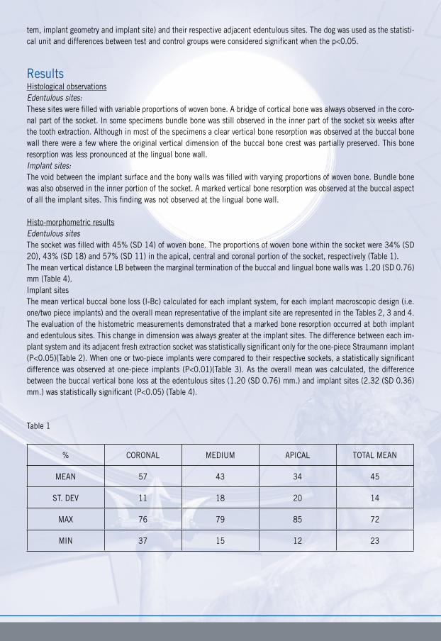
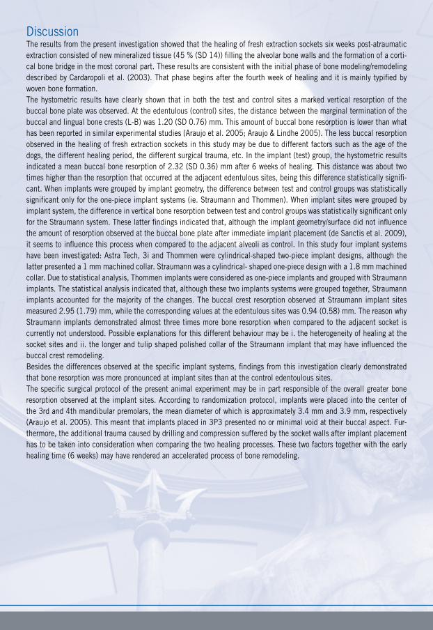
Histo-morphometric resultsEdentulous sitesThe socket was filled with 45% (SD 14) of woven bone. The proportions of woven bone within the socket were 34% (SD 20), 43% (SD 18) and 57% (SD 11) in the apical, central and coronal portion of the socket, respectively (Table 1).The mean vertical distance LB between the marginal termination of the buccal and lingual bone walls was 1.20 (SD 0.76) mm (Table 4). Implant sitesThe mean vertical buccal bone loss (I-Bc) calculated for each implant system, for each implant macroscopic design (i.e. one/two piece implants) and the overall mean representative of the implant site are represented in the Tables 2, 3 and 4. The evaluation of the histometric measurements demonstrated that a marked bone resorption occurred at both implant and edentulous sites. This change in dimension was always greater at the implant sites. The difference between each im-plant system and its adjacent fresh extraction socket was statistically significant only for the one-piece Straumann implant (P<0.05)(Table 2). When one or two-piece implants were compared to their respective sockets, a statistically significant difference was observed at one-piece implants (P<0.01)(Table 3). As the overall mean was calculated, the difference between the buccal vertical bone loss at the edentulous sites (1.20 (SD 0.76) mm.) and implant sites (2.32 (SD 0.36) mm.) was statistically significant (P<0.05) (Table 4).
Table 1
% CORONAL MEDIUM APICAL TOTAL MEAN
MEAN 57 43 34 45
ST. DEV 11 18 20 14
MAX 76 79 85 72
MIN 37 15 12 23
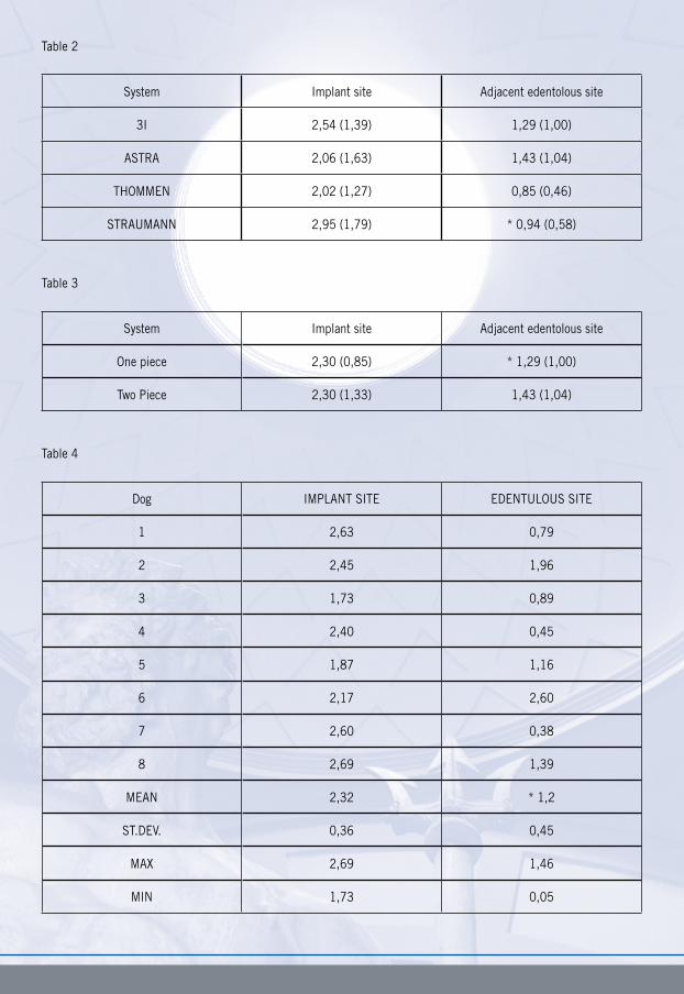
Table 2
System Implant site Adjacent edentolous site
3I 2,54 (1,39) 1,29 (1,00)
ASTRA 2,06 (1,63) 1,43 (1,04)
THOMMEN 2,02 (1,27) 0,85 (0,46)
STRAUMANN 2,95 (1,79) * 0,94 (0,58)
Table 3
System Implant site Adjacent edentolous site
One piece 2,30 (0,85) * 1,29 (1,00)
Two Piece 2,30 (1,33) 1,43 (1,04)
Table 4
Dog IMPLANT SITE EDENTULOUS SITE
1 2,63 0,79
2 2,45 1,96
3 1,73 0,89
4 2,40 0,45
5 1,87 1,16
6 2,17 2,60
7 2,60 0,38
8 2,69 1,39
MEAN 2,32 * 1,2
ST.DEV. 0,36 0,45
MAX 2,69 1,46
MIN 1,73 0,05
DiscussionThe results from the present investigation showed that the healing of fresh extraction sockets six weeks post-atraumatic extraction consisted of new mineralized tissue (45 % (SD 14)) filling the alveolar bone walls and the formation of a corti-cal bone bridge in the most coronal part. These results are consistent with the initial phase of bone modeling/remodeling described by Cardaropoli et al. (2003). That phase begins after the fourth week of healing and it is mainly typified by woven bone formation.The hystometric results have clearly shown that in both the test and control sites a marked vertical resorption of the buccal bone plate was observed. At the edentulous (control) sites, the distance between the marginal termination of the buccal and lingual bone crests (L-B) was 1.20 (SD 0.76) mm. This amount of buccal bone resorption is lower than what has been reported in similar experimental studies (Araujo et al. 2005; Araujo & Lindhe 2005). The less buccal resorption observed in the healing of fresh extraction sockets in this study may be due to different factors such as the age of the dogs, the different healing period, the different surgical trauma, etc. In the implant (test) group, the hystometric results indicated a mean buccal bone resorption of 2.32 (SD 0.36) mm after 6 weeks of healing. This distance was about two times higher than the resorption that occurred at the adjacent edentulous sites, being this difference statistically signifi-cant. When implants were grouped by implant geometry, the difference between test and control groups was statistically significant only for the one-piece implant systems (ie. Straumann and Thommen). When implant sites were grouped by implant system, the difference in vertical bone resorption between test and control groups was statistically significant only for the Straumann system. These latter findings indicated that, although the implant geometry/surface did not influence the amount of resorption observed at the buccal bone plate after immediate implant placement (de Sanctis et al. 2009), it seems to influence this process when compared to the adjacent alveoli as control. In this study four implant systems have been investigated: Astra Tech, 3i and Thommen were cylindrical-shaped two-piece implant designs, although the latter presented a 1 mm machined collar. Straumann was a cylindrical- shaped one-piece design with a 1.8 mm machined collar. Due to statistical analysis, Thommen implants were considered as one-piece implants and grouped with Straumann implants. The statistical analysis indicated that, although these two implants systems were grouped together, Straumann implants accounted for the majority of the changes. The buccal crest resorption observed at Straumann implant sites measured 2.95 (1.79) mm, while the corresponding values at the edentulous sites was 0.94 (0.58) mm. The reason why Straumann implants demonstrated almost three times more bone resorption when compared to the adjacent socket is currently not understood. Possible explanations for this different behaviour may be i. the heterogeneity of healing at the socket sites and ii. the longer and tulip shaped polished collar of the Straumann implant that may have influenced the buccal crest remodeling. Besides the differences observed at the specific implant systems, findings from this investigation clearly demonstrated that bone resorption was more pronounced at implant sites than at the control edentoulous sites. The specific surgical protocol of the present animal experiment may be in part responsible of the overall greater bone resorption observed at the implant sites. According to randomization protocol, implants were placed into the center of the 3rd and 4th mandibular premolars, the mean diameter of which is approximately 3.4 mm and 3.9 mm, respectively (Araujo et al. 2005). This meant that implants placed in 3P3 presented no or minimal void at their buccal aspect. Fur-thermore, the additional trauma caused by drilling and compression suffered by the socket walls after implant placement has to be taken into consideration when comparing the two healing processes. These two factors together with the early healing time (6 weeks) may have rendered an accelerated process of bone remodeling.
References
Araujo, M.G., Sukekava, F., Wennstrom, J.L. & Lindhe, J. (2005) Ridge alterations following implant placement in fresh extraction sockets:an experimental study in the dog. Journal of Clinical Periodontology 32, 65-652
Araujo, M.G., Sukekava, F., Wennstrom, J.L. & Lindhe, J. (2006)Tissue modeling following implant placement in fresh extraction sockets. Clinical Oral Implants Research 17, 615-624
Blanco,J., Nunez, V., Aracil, L., Muñoz, F. & Ramos, I. (2008) Ridge alterations following immediate implant placement in the dog: flap versus flapless surgery. Journal of Clinical Periodontology 35, 640-648
Botticelli, D., Berglundh, T.& Lindhe, J. (2004) Hard tissue alterations following immediate implant placement in extrac-tion sites. Journal of Clinical Periodontology 31, 820-828
Caneva M, Salata LA, de Souza SS, Baffone G, Lang NP, Botticelli D. Influence of implant positioning in extraction sock-ets on osseointegration: histomorphometric analyses in dogs.Clin. Oral Impl. Res. 21, 2010; 43–49.
de Sanctis, M., Vignoletti F., Discepoli N., Zucchelli, G & Sanz, M (2009) Immediate implants at fresh extraction sockets: bone healing in four different implant systems. Journal of Clinical Periodontology 36, 705-711
Donath,K & Breuner, G (1982) A method for the study of undecalcified bones and teeth with attached soft tissues. The Sage-Schliff (sawing and grinding) technique. Journal of Oral Pathology 11, 318–326.
Vignoletti, F., de Sanctis, M., Berglundh, T., Abrahamsson, I. & Sanz, M. (2009a) Early healing of implants placed into fresh extraction sockets: an experimental study in the beagle dog II: ridge alterations. Journal of Clinical Periodontology 36, 688–697.