Bone marrow biopsy as prognostic indicator in childhood acute lymphoblastic leukemia—another...
10
Letter to the Editor: Acute Basal Ganglia Necrosis Associated With Cytarabine Therapy To the Editor: Cytarabine has been recognized as an important drug in the treatment of acute leukemia over the last two decades [1]. High-dose schedules carry a well-known risk of various neurologic toxicities, of which the acute cerebellar syndrome is the most common [2]. We encountered isolated acute basal ganglia necrosis associated with cytarabine therapy, which we believe to be the first reported instance. It occurred in a 35-month- old boy who was treated in October 1995 for combined bone marrow and testicular relapse of acute lymphoblas- tic leukemia (ALL) without central nervous system (CNS) involvement. Neurologic examination was normal at that time. On the third day of the second course of salvage therapy using the VANDA protocol [dexameth- asone (20 mg/m 2 orally on days 1–5), cytarabine (1,000 mg/m 2 intravenously every 12 h on days 1 and 2), eto- poside (150 mg/m 2 intravenously on days 3–5), mitox- antrone (8 mg/m 2 intravenously on days 3 and 4), and asparaginase (10,000 U/m 2 intravenously on days 7, 9, 11, and 13), plus intrathecal cytarabine (20 mg), metho- trexate (8 mg), and prednisone (6 mg) on day 5], the patient suddenly deteriorated neurologically. He became lethargic, hypotonic, and mute. Cerebellar examination was normal. Mild horizontal nystagmus was present on lateral gaze. Cranial nerves were intact in function. Deep- tendon reflexes were present, but weak. Serum and CSF chemistries were normal. Centrifugation and staining for malignant cells were negative. Cerebral CT scan was normal. Cranial MRI on day 5 showed abnormal signal intensity involving primarily gray matter of the basal ganglia. Over the next several days, his neurologic status improved gradually, but the patient developed clinical features of secondary parkinsonism. By day 21, cerebral MRI showed features compatible with basal ganglia ne- crosis. Two years later, his neurologic status has steadily improved. There is no cognitive impairment. Cerebral MRI shows unchanged findings without cerebellar atro- phy. Basal ganglia necrosis is rare in children. The main causes are toxic (carbon monoxide, methanol, or cya- nide), hypoxo-ischemic, metabolic (sulfite oxydase defi- ciency), or postinfectious (e.g., measles, mycoplasma pneumoniae) [3]. Neuroleptic-induced persistent parkin- sonism has also been associated with putaminal hypoin- tensity in young patients [4]. We are aware of one previous report of parkinsonism in a child associated with cytarabine therapy [3]. Cranial MRI revealed signal intensity involving the gray matter of the basal ganglia, adjacent temporal and frontal lobes, and cerebellum. Our patient did not present any of the risk factors associated with cytarabine-induced CNS tox- icity: age, abnormal renal or hepatic function, or prior central nervous system involvement. The total dose of cytarabine, which appears for several authors to be the chief cause of neurologic toxicity, was low (8 g/m 2 ) [2]. The importance of the cumulative dose of cytarabine as a factor has been suggested, but there has been great variation from case to case in the dose required to induce neurologic complication [5]. In our patient (cumulative dose from the beginning of the treatment, 17.8 g), we cannot rule out that there was subclinical damage after the first course of cytarabine that became clinically ap- parent only after the second course. Thus, a careful re- view of all the etiologies, the previous similar report in a child, and the temporal relationship between the admin- istration of cytarabine and the onset of neurologic symp- toms support the probable etiologic role of cytarabine or its metabolites as the cause of this neurologic toxicity. We must underline that the CNS toxicity in our patient is restricted to the basal ganglia, without cerebellar toxicity or atrophy of the vermis. Nicolas Sirvent, MD Fabrice Monpoux, MD Laurent Benet, MD Christian Richelme, MD Roger Mariani, MD Unite ´ d’Onco-He ´matologie De ´partement de Pe ´diatrie Bernard Diaine, MD De ´partement de Radiologie Ho ˆpital de l’Archet Nice, France REFERENCES 1. Ellison RR, Holland JF, Weil M, et al.: Arabinosyl cytosine: A useful agent in the treatment of acute leukemia in adults. Blood 32:507–523, 1968. 2. Baker JW, Royer GL, Weiss RB: Cytosine arabinoside and neu- rologic toxicity. J Clin Oncol 9:679–693, 1991. 3. Pranzatelli MR, Mott SH, Pavlakis SG, et al.: Clinical spectrum of secondary parkinsonism in childhood: A reversible disorder. Pe- diatr Neurol 10:131–140, 1994. 4. Lazarus HM, Herzig RH, Herzig GP: Central nervous system toxicity of high-dose systemic cytosine arabinoside. Cancer 48: 2577–2582, 1981. 5. Benger A, Browman GP, Walker IR: Clinical evidence of a cu- mulative effect of high-dose cytosine arabinoside on the cerebel- lum in patients with acute leukemia: A Leukemia Intergroup re- port. Cancer Treat Rep 69:240–241, 1985. Medical and Pediatric Oncology 30:308–317 (1998) © 1998 Wiley-Liss, Inc.
Transcript of Bone marrow biopsy as prognostic indicator in childhood acute lymphoblastic leukemia—another...

Letter to the Editor: Acute Basal Ganglia Necrosis Associated WithCytarabine Therapy
To the Editor: Cytarabine has been recognized as animportant drug in the treatment of acute leukemia overthe last two decades [1]. High-dose schedules carry awell-known risk of various neurologic toxicities, ofwhich the acute cerebellar syndrome is the most common[2]. We encountered isolated acute basal ganglia necrosisassociated with cytarabine therapy, which we believe tobe the first reported instance. It occurred in a 35-month-old boy who was treated in October 1995 for combinedbone marrow and testicular relapse of acute lymphoblas-tic leukemia (ALL) without central nervous system(CNS) involvement. Neurologic examination was normalat that time. On the third day of the second course ofsalvage therapy using the VANDA protocol [dexameth-asone (20 mg/m2 orally on days 1–5), cytarabine (1,000mg/m2 intravenously every 12 h on days 1 and 2), eto-poside (150 mg/m2 intravenously on days 3–5), mitox-antrone (8 mg/m2 intravenously on days 3 and 4), andasparaginase (10,000 U/m2 intravenously on days 7, 9,11, and 13), plus intrathecal cytarabine (20 mg), metho-trexate (8 mg), and prednisone (6 mg) on day 5], thepatient suddenly deteriorated neurologically. He becamelethargic, hypotonic, and mute. Cerebellar examinationwas normal. Mild horizontal nystagmus was present onlateral gaze. Cranial nerves were intact in function. Deep-tendon reflexes were present, but weak. Serum and CSFchemistries were normal. Centrifugation and staining formalignant cells were negative. Cerebral CT scan wasnormal. Cranial MRI on day 5 showed abnormal signalintensity involving primarily gray matter of the basalganglia. Over the next several days, his neurologic statusimproved gradually, but the patient developed clinicalfeatures of secondary parkinsonism. By day 21, cerebralMRI showed features compatible with basal ganglia ne-crosis. Two years later, his neurologic status has steadilyimproved. There is no cognitive impairment. CerebralMRI shows unchanged findings without cerebellar atro-phy.
Basal ganglia necrosis is rare in children. The maincauses are toxic (carbon monoxide, methanol, or cya-nide), hypoxo-ischemic, metabolic (sulfite oxydase defi-ciency), or postinfectious (e.g., measles, mycoplasmapneumoniae) [3]. Neuroleptic-induced persistent parkin-sonism has also been associated with putaminal hypoin-tensity in young patients [4].
We are aware of one previous report of parkinsonismin a child associated with cytarabine therapy [3]. CranialMRI revealed signal intensity involving the gray matterof the basal ganglia, adjacent temporal and frontal lobes,
and cerebellum. Our patient did not present any of therisk factors associated with cytarabine-induced CNS tox-icity: age, abnormal renal or hepatic function, or priorcentral nervous system involvement. The total dose ofcytarabine, which appears for several authors to be thechief cause of neurologic toxicity, was low (8 g/m2) [2].The importance of the cumulative dose of cytarabine asa factor has been suggested, but there has been greatvariation from case to case in the dose required to induceneurologic complication [5]. In our patient (cumulativedose from the beginning of the treatment, 17.8 g), wecannot rule out that there was subclinical damage afterthe first course of cytarabine that became clinically ap-parent only after the second course. Thus, a careful re-view of all the etiologies, the previous similar report in achild, and the temporal relationship between the admin-istration of cytarabine and the onset of neurologic symp-toms support the probable etiologic role of cytarabine orits metabolites as the cause of this neurologic toxicity.We must underline that the CNS toxicity in our patient isrestricted to the basal ganglia, without cerebellar toxicityor atrophy of the vermis.
Nicolas Sirvent,MD
Fabrice Monpoux,MD
Laurent Benet,MD
Christian Richelme,MD
Roger Mariani,MD
Unite d’Onco-HematologieDepartement de Pe´diatrie
Bernard Diaine,MD
Departement de RadiologieHopital de l’Archet
Nice, France
REFERENCES1. Ellison RR, Holland JF, Weil M, et al.: Arabinosyl cytosine: A
useful agent in the treatment of acute leukemia in adults. Blood32:507–523, 1968.
2. Baker JW, Royer GL, Weiss RB: Cytosine arabinoside and neu-rologic toxicity. J Clin Oncol 9:679–693, 1991.
3. Pranzatelli MR, Mott SH, Pavlakis SG, et al.: Clinical spectrum ofsecondary parkinsonism in childhood: A reversible disorder. Pe-diatr Neurol 10:131–140, 1994.
4. Lazarus HM, Herzig RH, Herzig GP: Central nervous systemtoxicity of high-dose systemic cytosine arabinoside. Cancer 48:2577–2582, 1981.
5. Benger A, Browman GP, Walker IR: Clinical evidence of a cu-mulative effect of high-dose cytosine arabinoside on the cerebel-lum in patients with acute leukemia: A Leukemia Intergroup re-port. Cancer Treat Rep 69:240–241, 1985.
Medical and Pediatric Oncology 30:308–317 (1998)
© 1998 Wiley-Liss, Inc.

Letter to the Editor: Experience Treating a Patient With Bloom Syndrome andAcute Myelogenous Leukemia
To the Editor: Bloom syndrome is an autosomal re-cessive disorder of genomic instability and predisposi-tion to malignant tumors. Its characteristics are severeprenatal and postnatal growth retardation, variable sun-sensitive telangiectatic facial erythema, and a narrowface [1]. Bloom syndrome leads to malignant tumors inabout 25% of patients [2], frequently at more than oneprimary site [3]. Therapy of malignant disorders in theserare patients is hampered by extremely poor tolerance forcytostatic drugs. Our detailed experience in treating sucha patient may be helpful to others faced with this difficultproblem.
In our female patient, Bloom syndrome was diagnosedat age 4 years. (See ref. 4 for details.) When 15, acutemyelo-monocytic leukemia (FAB-type M4) was diag-nosed; blood count showed Hb of 6.5 g/dl, WBC of39,700/mm3 with 86% blasts, and 33,000 platelets/mm3.Bone marrow smear revealed 80% monoblasts (80% per-oxidase-positive, 50% esterase-positive, 70% of mono-cytic immunophenotype). We tried to induce a remissionwith multiagent chemotherapy, using the consolidationregimen of the current German Acute Myelogenous Leu-kemia (AML) treatment study (AML-BFM-93). Thedoses for cytarabine and doxorubicin were reduced to50% (Table I). The first block of chemotherapy was in-terrupted on day 24 due to fever (39.8°C). Under i.v.antibiotic and antimycotic treatment the patient devel-oped a bartholinitis. Marsupialization led to deferves-cence and relief of local symptoms. Bone marrow biopsyon day 41 revealed reduced cellularity with persistenceof blasts (86%). A second course of chemotherapy wasstarted on day 42 after admission (Table I). Severe mu-cositis and massive gastrointestinal bleeding (day 53)required interruption of chemotherapy. During the next 2weeks the patient lost 5 kg of weight, and was periodi-
cally febrile with episodes of tachycardia and tachydys-pnea. Blood pressure remained stable. Due to recurrentgastrointestinal bleeding and hematopoietic insuffi-ciency, 14 units of red blood cells and 72 units of plate-lets were given. In view of the poor tolerance of cyto-static treatment and the inadequate results, further thera-peutic attempts were restricted to symptom control on anoutpatient basis. At home she remained stable and didfairly well for a month. She finally developed pneumo-nia. At this time she had a WBC of 14,000/mm3 with80% blasts. She died from respiratory failure. An autopsywas not done.
Poor tolerance of cytostatic treatment is known in pa-tients with Bloom syndrome. However, some of themhave successfully been treated, partly with standard drugdoses, for acute lymphoblastic leukemia (ALL) [2,5],non-Hodgkin lymphoma of B-cell type (B-NHL) [6] or
TABLE I. Chemotherapy Administered for Acute MyelogenousLeukemia to a 15-Year-Old-Girl With Bloom Syndrome*
Drug Dose in mg/m2 Route Day
Block 1 Cytarabine 45 i.th. 7Prednisone 40 p.o. 1–15Thioguanine 60 p.o. 1–15Doxorubicin 17 i.v. 3, 10Vincristine 1.5 i.v. 3, 10Cytarabine 39 i.v. 16–19
Block 2 Cytarabine 45 i.th. 9Prednisone 40 p.o. 1–16Thioguanine 60 p.o. 3–16Doxorubicin 22 i.v. 8Vincristine 1.5 i.v. 8Cytarabine 51 i.v. 3–16
10, 11, 14
*Block 1 was started on day 12, and block 2 on day 42 after admission.p.o., per os; i.v., intravenous injection; i.th., intrathecal injection.
TABLE II. Cumulative Doses of Cytostatic Agents Given to 2 Patients WithBloom Syndrome Within 8 Weeks of Treatment for Malignancy*
Drug (mg/m2)
Patient Y.M. (reference [6]),B-non-Hodgkin
lymphoma
Our patient,acute myeloid leukemia
FAB M4
Doxorubicin 60 56Vincristine 1.5 4.5Cytarabine 800 603Thioguanine 1,640Cyclophospamide 2,200Methotrexate 3,000Etoposide 1,200Outcome Treatment continued,
long-term remissionTreatment discontinued,
no remission, death
*Corticosteroids omitted.
Letters to the Editor 309

unclassified [7], and for nephroblastoma [8]. By contrast,all reported attempts to induce remission of AML inpatients with Bloom syndrome have failed, as did ours[2], perhaps because drug doses had to be reduced. Incontrast to ALL, NHL, or solid tumors, treatment ofAML requires intensive chemotherapy to achieve remis-sion, and this almost always lead to severe bone marrowaplasia [9].
It is true that a Japanese girl with Bloom syndromeand B-NHL tolerated higher cumulative doses [6] thandid our patient (see Table II), a fact that might be ex-plained by interindividual differences between patientsor by different drug scheduling. Our AML patient re-ceived daily oral thioguanine continuously over 14 days,while the Japanese B-NHL protocol consisted of intra-venous applications of drugs on 5–6 out of 14–16 days.Spreading the drug doses over time possibly augmentsside effects in Bloom syndrome.
In conclusion, successful therapy of AML in patientswith Bloom syndrome has so far been prevented by thediscrepancy between the treatment intensity needed toinduce remission, and poor tolerance of these patients forcytostatic drugs.
ACKNOWLEDGMENTS
We thank Professor Werner Havers, Department ofPediatric Hematology-Oncology, University of Essen,Germany, and Professor James German, Laboratory ofHuman Genetics, New York Blood Center, New York,for helpful advice in the care of this patient.
Hartmut Grasemann,MD
Bernhard Kremens,MD
KinderklinikEberhard Passarge,MD
Institut fur HumangenetikUniversitatsklinikum Essen
Essen, Germany
REFERENCES
1. German J, Passarge E: Bloom’s syndrome: XII. Report from theregistry for 1987. Clin Genet 35:57–69, 1989.
2. German J, Gardin C: Bloom’s syndrome. XV. The instances ofacute myelogenous leukemia in the Bloom’s syndrome registry. InGale RP (ed): ‘‘Acute Myelogenous Leukemia: Progress and Con-troversies.’’ New York: Wiley-Liss, Inc., 1990, pp. 35–49.
3. German J: Bloom syndrome: A Mendelian prototype of somaticmutational disease. Medicine (Baltimore) 72:393–406, 1993.
4. Passarge E: Bloom’s syndrome: The German experience. AnnGenet (Paris) 34:179–197, 1991.
5. Festa RS, Meadows AT, Boshes RA: Leukemia in a black childwith Bloom’s syndrome. Cancer 44:1507–1510, 1979.
6. Oto S, Miyamoto S, Kudoh F, et al.: Treatment for B-cell-typelymphoma in a girl associated with Bloom’s syndrome. ClinGenet 41:46–50, 1992.
7. Arase S, Takahashi O, Ishizaki K, et al.: Bloom’s syndrome in aJapanese boy with lymphoma. Clin Genet 18:123–127, 1980.
8. Cairney AEL, Andrews M, Greenberg M, et al.: Wilms tumor inthree patients with Bloom syndrome. J Pediatr 111:414–416,1987.
9. Mayer RJ: Current chemotherapeutic treatment approaches to themanagement of previously untreated adults with de novo acutemyelogenous leukemia. Semin Oncol 14:384–396, 1987.
310 Letters to the Editor

Letter to the Editor: Chemotherapy for Spinal Cord Astrocytoma
To the Editor: We are writing in response to the casereport by Bouffet et al. [1] regarding treatment of a spinalcord astrocytoma with carboplatin and vincristine. Theypresent a 30-year-old woman whose spinal astrocytomashowed a dramatic response to these chemotherapeuticagents. At the end of their report, they say that furtherreports of such therapy in spinal cord astrocytomas mayhelp clarify whether chemotherapy has a role in the man-agement of these tumors. We therefore thought it perti-nent to share an adverse experience with carboplatin andvincristine in a child with a spinal cord astrocytoma.
He presented in 1995, when he was 8 years old, withback pain and stiffness. MRI showed a midline tumorextending from C7 to T4, with an extensive syrinx ros-trally and caudally. A radical subtotal operation was per-formed in May 1995. Postoperative MRI scan showed asmall amount of residual tumor, which was less than 5%of the original mass. Institutional pathology was reportedas low-grade astrocytoma. He remained neurologically
normal postoperatively. Serial MRI scans obtained regu-larly from May 1995 through August 1996 showed noprogression of the tumor or syrinx, and he developed nonew neurological signs. He did have progressive kypho-scoliosis. In December 1996, there was evidence of pro-gression on a routine MRI scan. Reoperation and alter-natives to reoperation were discussed with the parents.They sought second opinions and then expressed a pref-erence for chemotherapy. Due to the time taken to getsecond opinions, chemotherapy with carboplatin and vin-cristine did not begin until February 1997.
A repeat scan on March 5, 1997 (Fig. 1) suggestedtumor progression compared to the MRI from December1996. Given that chemotherapy had only begun the pre-vious month, a decision was made to continue the car-boplatin and vincristine and scan him in a short period oftime. Over the next few weeks, he developed backacheand complained of abnormal sensation in the lower limb
Fig. 1. MRI, performed March 5, 1997, showing tumor at the cer-vical thoracic junction. Vertebral bodies are numbered.
Fig. 2. MRI, performed April 15, 1997, showing an increase intumor volume compared to Figure 1. Vertebral bodies are numbered.
Letters to the Editor 311
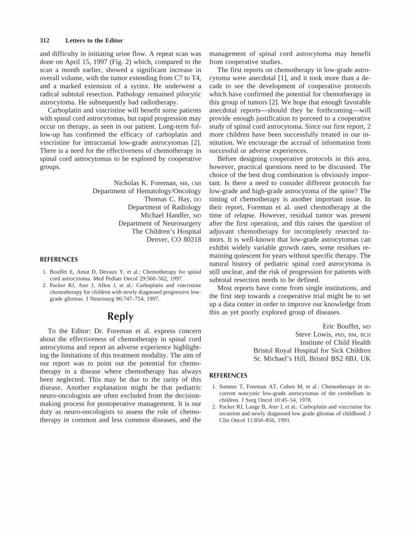
and difficulty in initiating urine flow. A repeat scan wasdone on April 15, 1997 (Fig. 2) which, compared to thescan a month earlier, showed a significant increase inoverall volume, with the tumor extending from C7 to T4,and a marked extension of a syrinx. He underwent aradical subtotal resection. Pathology remained pilocyticastrocytoma. He subsequently had radiotherapy.
Carboplatin and vincristine will benefit some patientswith spinal cord astrocytomas, but rapid progression mayoccur on therapy, as seen in our patient. Long-term fol-low-up has confirmed the efficacy of carboplatin andvincristine for intracranial low-grade astrocytomas [2].There is a need for the effectiveness of chemotherapy inspinal cord astrocytomas to be explored by cooperativegroups.
Nicholas K. Foreman,MB, ChB
Department of Hematology/OncologyThomas C. Hay,DO
Department of RadiologyMichael Handler,MD
Department of NeurosurgeryThe Children’s Hospital
Denver, CO 80218
REFERENCES
1. Bouffet E, Amat D, Devaux Y, et al.: Chemotherapy for spinalcord astrocytoma. Med Pediatr Oncol 29:560–562, 1997.
2. Packer RJ, Ater J, Allen J, et al.: Carboplatin and vincristinechemotherapy for children with newly diagnosed progressive low-grade gliomas. J Neurosurg 86:747–754, 1997.
ReplyTo the Editor: Dr. Foreman et al. express concern
about the effectiveness of chemotherapy in spinal cordastrocytoma and report an adverse experience highlight-ing the limitations of this treatment modality. The aim ofour report was to point out the potential for chemo-therapy in a disease where chemotherapy has alwaysbeen neglected. This may be due to the rarity of thisdisease. Another explanation might be that pediatricneuro-oncologists are often excluded from the decision-making process for postoperative management. It is ourduty as neuro-oncologists to assess the role of chemo-therapy in common and less common diseases, and the
management of spinal cord astrocytoma may benefitfrom cooperative studies.
The first reports on chemotherapy in low-grade astro-cytoma were anecdotal [1], and it took more than a de-cade to see the development of cooperative protocolswhich have confirmed the potential for chemotherapy inthis group of tumors [2]. We hope that enough favorableanecdotal reports—should they be forthcoming—willprovide enough justification to porceed to a cooperativestudy of spinal cord astrocytoma. Since our first report, 2more children have been successfully treated in our in-stitution. We encourage the accrual of information fromsuccessful or adverse experiences.
Before designing cooperative protocols in this area,however, practical questions need to be discussed. Thechoice of the best drug combination is obviously impor-tant. Is there a need to consider different protocols forlow-grade and high-grade astrocytoma of the spine? Thetiming of chemotherapy is another important issue. Intheir report, Foreman et al. used chemotherapy at thetime of relapse. However, residual tumor was presentafter the first operation, and this raises the question ofadjuvant chemotherapy for incompletely resected tu-mors. It is well-known that low-grade astrocytomas canexhibit widely variable growth rates, some residues re-maining quiescent for years without specific therapy. Thenatural history of pediatric spinal cord astrocytoma isstill unclear, and the risk of progression for patients withsubtotal resection needs to be defined.
Most reports have come from single institutions, andthe first step towards a cooperative trial might be to setup a data center in order to improve our knowledge fromthis as yet poorly explored group of diseases.
Eric Bouffet, MD
Steve Lowis,PhD, BM, BCH
Institute of Child HealthBristol Royal Hospital for Sick ChildrenSt. Michael’s Hill, Bristol BS2 8BJ, UK
REFERENCES
1. Sumner T, Freeman AT, Cohen M, et al.: Chemotherapy in re-current noncystic low-grade astrocytomas of the cerebellum inchildren. J Surg Oncol 10:45–54, 1978.
2. Packer RJ, Lange B, Ater J, et al.: Carboplatin and vincristine forrecurrent and newly diagnosed low grade gliomas of childhood. JClin Oncol 11:850–856, 1993.
312 Letters to the Editor
Letter to the Editor: The Importance of Molecular Screening of 11q23Abnormalities in Childhood Acute Lymphoblastic Leukemia: Has the
t(11;19)(q23;p13) a Higher Frequency Than That Revealed by ConventionalCytogenetic Techniques?
To the Editor: We have read with interest the report byIda et al. [1] on the use of reverse transcriptase-polymerase chain reaction (RT-PCR) to detect 11q23 ab-normalities that produce rearrangement of the MLL gene.We consider it valuable to carry out a molecular study ondiagnosis of all cases of childhood acute lymphoblasticleukaemia (ALL), first with Southern blot and in cases inwhich rearrangement takes place with PCR, since, asdemonstrated here in two cases, these abnormalities canremain undetected by conventional cytogenetic tech-niques. Their detection can be considered of special im-portance if one takes into account the poor prognosis ofpatients with translocations involving chromosome band11q23 [2] in contrast to deletions and inversions at thisband that have a favourable prognosis and lack MLLgene rearrangement [3].
In April 1996, we started a prospective study of pae-diatric ALL that included analysis of the hybrid genesTEL/AML1, E2A/PBX1, and BCR/ABL by RT-PCR to-gether with a study of the rearrangements of the MLLgene and p16 delections by Southern blot. Until June1997, we had studied 15 patients with PCR and 11 ofthese with Southern blot (9 B-precursor ALLs and 3T-cell ALLs).
For the study of the 11q23 rearrangements, DNA wasextracted from bone marrow mononuclear cells, digestedwith both Bam HI and Bgl II restriction enzymes, andhybridized with the B859 probe, containing MLL exon5-11 sequences, labelled with P32 using the publishedmethods [4].
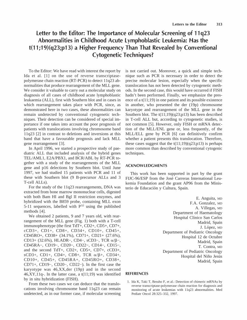
We obtained 2 patients, 9 and 7 years old, with rear-rangement of the MLL gene (Fig. 1) both with a T-cellimmunophenotype (the first TdT+, CD2+, CD5+, CD7+,cCD3+, CD1+, CD8+, CD34+, CD10+, CD45+,CD45RO+, CD38+ (34.1%), CD71+, CD21+ (27.6%),CD13+ (32.6%), HLADR−, CD4−, sCD3−, TCRa/b−,CD45RA−, CD19−, CD20−, CD22−, CD14−, CD15−,and the second TdT+, CD2+, CD5+, CD7+, cCD3+,sCD3+, CD1+, CD4+, CD8+, TCRa/b+, CD34+,CD10+, CD45+, CD45RA+, CD45RO+, CD38+,CD71+, CD19−, CD20−, CD22−). In the first case thekaryotype was 46,XX,der (19p) and in the second46,XY,11q-. In the latter case, a t(11;19) was identifiedby in situ hybridization (FISH).
From these two cases we can deduce that the translo-cations involving chromosome band 11q23 can remainundetected, as in our former case, if molecular screening
is not carried out. Moreover, a quick and simple tech-nique such as PCR is necessary in order to detect theprecise molecular lesion, especially when the specifictranslocation has not been detected by cytogenetic meth-ods. In the second case, this would have occurred if FISHhadn’t been performed. Finally, we emphasize the pres-ence of a t(11;19) in one patient and its possible existencein another, who presented the der (19p) chromosomekaryotype and rearrangement of the MLL gene in theSouthern blot. The t(11;19)(q23;p13) has been describedin T-cell ALL but, according to cytogenetic studies, isnot common [5]. However, only FISH or mRNA detec-tion of the MLL/ENL gene or, less frequently, of theMLL/ELL gene by PCR [6] can definitively confirmwhether a patient presents this translocation. Therefore,these cases suggest that the t(11;19)(q23;p13) is perhapsmore common than described by conventional cytogenictechniques.
ACKNOWLEDGMENTS
This work has been supported in part by the grantFIJC-96/ESP from the Jose´ Carreras International Leu-kemia Foundation and the grant AP96 from the Minis-terio de Educacio´n y Cultura, Spain.
E. Anguita,MD
F.A. Gonzalez,MD
A. Villegas, MD
Department of HaematologyHospital Clınico San Carlos
Madrid, SpainJ. Lopez,MD
Department of Pediatric OncologyHospital 12 de Octubre
Madrid, SpainT. Contra,MD
Department of Pediatric OncologyHospital del Nino Jesu´s
Madrid, Spain
REFERENCES
1. Ida K, Taki T, Bessho F, et al.: Detection of chimeric mRNAs byreverse transcriptase-polymerase chain reaction for diagnosis andmonitoring of acute leukemias with 11q23 abnormalities. MedPediatr Oncol 28:325–332, 1997.
Letters to the Editor 313

2. Behm FG, Raimondi SC, Frestedt JL, et al.: Rearrangement of theMLL gene confers a poor prognosis in childhood acute lympho-blastic leukemia, regardless of presenting age. Blood 87:2870–2877, 1996.
3. Raimondi SC, Frestedt JL, Pui CH, et al.: Acute lymphoblas-tic leukemias with deletion of 11q23 or a novel inversion(11)(p13q23) lack MLL gene rearrangements and have favorablefeatures. Blood 86:1881–1886, 1995.
4. Cimino G, Rapanotti MC, Elia L, et al.: ALL-1 gene rearrange-ments in acute myeloid leukemia: Association with M4-M5French-American-British classification subtypes and young age.Cancer Res 55:1625–1628, 1995.
5. Thandla S, Aplan PD: Molecular biology of acute lymphocyticleukemia. Semin Oncol 24:45–53, 1997.
6. Rubnitz JE, Behm FG, Curcio-Brint AM, et al.: Molecular analy-sis of t(11;19) breakpoints in childhood acute leukemias. Blood87:4804–4808, 1996.
ReplyTo the Editor: Anguita et al. reported that t(11;19)
(q23;p13) may be more common than described by con-ventional cytogenetic techniques. We agree with theiropinion because we had one acute lymphoblastic leuke-mia (ALL) patient who had an initially normal karyotypeand then had the t(11;19) abnormality at relapse. Inter-estingly, theMLL gene was found to be rearranged atboth diagnosis and relapse, suggesting that the leukemiccells of this patient at diagnosis were assumed to havehad the t(11;19). It is difficult to analyze the precisebreakpoint of the t(11;19) because it is a subtle abnor-mality [1]. In this regard,MLL-ENL andMLL-ELL/MENmRNA were identified in t(11;19)(q23;p13.3) andt(11;19)(q23;p13.1), respectively. The former was foundin ALL and acute myeloid leukemia (AML), and thelatter only in AML [2].
In 11q23 chromosomal abnormalities other thant(11;19), cytogenetic del(11)(q23) was considered to bet(6;11)(q27;q23) by fluorescence in situ hybridization(FISH) [3] or reverse transcriptase-polymerase chain re-action (RT-PCR) [4]. The t(9;11) is also difficult to ana-
lyze cytogenetically, and is often determined as being ofnormal karyotype by conventional G- or Q-bandingmethods [5]. One of the 4 patients whose karyotypeswere unsuccessfully analyzed was identified to be t(9;11)by RT-PCR in our study [6]. In infant leukemia, thefrequency ofMLL gene rearrangements was reported tobe approximately 80%, higher than theMLL gene rear-rangement found by cytogenic analysis (50–60%) in ourstudy [5]. ALL infants with theMLL gene rearrangementhad a significantly poor clinical outcome [5,7,8].
These results combined with those in the literaturesuggest that t(11;19) as well as t(6;11) and t(9;11) aredifficult to identify cytogenetically if good metaphasesare not obtained. Recently we reported thatMLL-CBPand MLL-p300 were involved in t(11;16) and t(11;22)therapy-related leukemia [9,10]. These abnormalitieswere also difficult to identify cytogenetically. Patientswith MLL gene rearrangement had a poor prognosis inchildhood ALL, regardless of presenting age [11]. Nota-bly, Behm et al. [12] reported that cytogenetics did notdetect an 11q23 abnormality in 13 (33%) of 39 childhoodAML patients withMLL gene rearrangements. Not onlyALL patients, as suggested by Anguita et al., but alsoAML children should be examined forMLL rearrange-ments by Southern blotting first, and by either FISH orRT-PCR to predict clinical outcome.
Yasuhide HayashiTomohiko Taki
Kohmei IdaDepartment of Pediatrics
University of TokyoTokyo 113, Japan
REFERENCES
1. Hayashi Y, Kobayashi Y, Hirai H, et al.: Immunoglobulin heavychain gene rearrangements and mixed lineage characteristics in
Fig. 1. Filter containing ALL DNAs hy-bridized with the B859 probe, which ex-plores the MLL breakpoint region at chro-mosome 11q23.Lane 1: Molecular weightmarker (lambda phage/Hind III digest la-beled with32P) with bands of 23.1, 9.4, 6.6,4.4, 2.3, and 2.0 Kb.Lanes 2–10:Bam HIdigested ALL DNAs.Lanes 11–19:DNAsfrom the same cases digested with Bgl II.Lanes 5 and 14 and 9 and 18, respectively,are the cases described in the text.
314 Letters to the Editor

acute leukemias with the 11;19 translocation. Cancer 61:712–720,1988.
2. Rubnitz JE, Behm FG, Curcio-Brint AM, et al.: Molecular analy-sis of t(11;19) breakpoints in childhood acute leukemias. Blood87:4804–4808, 1996.
3. Kobayashi H, Espinosa R, Thirman MJ, et al.: Do terminal dele-tions of 11q23 exist? Identification of undetected translocationswith fluorescence in situ hybridization. Gene Chromosomes Can-cer 7:204–208, 1993.
4. Taki T, Hayashi Y, Taniwaki M, et al.: Fusion of theMLL genewith two different genes,AF-6 and AF-5a, by a complex trans-location involving chromosomes 5, 6, 8, and 11 in infant leuke-mia. Oncogene 13:2121–2130, 1996.
5. Taki T, Ida K, Bessho F, et al.: Frequency and clinical significanceof the MLL gene rearrangements in infant acute leukemia. Leu-kemia 10:1303–1307, 1996.
6. Ida K, Taki T, Bessho F, et al.: Detection of chimeric mRNAs byreverse transcriptase-polymerase chain reaction for diagnosis andmonitoring of acute leukemias with 11q23 abnormalities. MedPediatr Oncol 28:325–332, 1997.
7. Rubnitz JE, Link MP, Shuster JJ, et al.: Frequency and prognosticsignificance of HRX rearrangements in infant acute lymphoblasticleukemia: A Pediatric Oncology Group study. Blood 84:570–574,1994.
8. Pui C-H, Kane JR, Crist WM: Biology and treatment of infantleukemias. Leukemia 9:762–769, 1995.
9. Taki T, Sako M, Tsuchida M, et al.: The t(11;16)(q23;p13) trans-location in myelodysplastic syndrome fuses theMLL gene to theCBP gene. Blood 89:3945–3950, 1997.
10. Ida K, Kitabayashi I, Taki T, et al.: Adenoviral E1A-associatedprotein p300 is involved in acute myeloid leukemia witht(11;22)(q23;q13). Blood 90:4699–4704, 1997.
11. Behm FG, Raimondi SC, Frestedt JL, et al.: Rearrangement of theMLL gene confers a poor prognosis in childhood acute lympho-blastic leukemia, regardless of presenting age. Blood 87:2870–2877, 1996.
12. Behm FG, Shurtleff SA, Raimondi SC, et al.: A retrospectivemolecular study of the frequency of AML1/ETO, CBFB/MYH11,PML/RARA, and rearrangement of MLL in childhood de novoAML and comparison with survival. Blood 90:63, 1997 (abstract).
Letter to the Editor: Bone Marrow Biopsy as Prognostic Indicator in ChildhoodAcute Lymphoblastic Leukemia—Another Opinion
To the Editor: In a recent issue of this journal, Schultzet al. retrospectively evaluated the day 7 bone marrow(BM) biopsy as a prognostic measure of outcome in 88children with acute lymphoblastic leukemia (ALL) en-rolled in five different CCG protocols and treated at theirinstitution [1]. The authors concluded that the informa-tion gained from the day 7 BM biopsy can improve pre-diction of outcome in children with ALL but also that aprospective confirmation with larger studies is needed.
BM biopsy, however, as acknowledged by Schultz etal. [1], basically remains an invasive procedure requiring‘‘conscious sedation or general anesthetic.’’ In addition,BM biopsy reproducibility is hampered by sampling er-ror, inadequate yield, and variability of evaluation, ac-cording to the expertise of the pathologist. Laboratorytime and costs are also relatively high, making this tech-nique neither easily available nor recommendable forlarge cooperative studies.
Also to be considered is that the lowest disease-freesurvival (DFS) found after the patients’ stratificationbased on the ABI-aspirate value </ù.06 was 51%. Thisinformation was obtained in a retrospective study and ona limited number of patients, who underwent differenttypes of treatment. These data may not be especiallyuseful, since it is possible to recognize subsets of patientswith DFS even much lower than 51% simply by usingwhite blood cell count and age [2], steroid response(alone or with additional features), and marrow residualblast infiltration after 7 or 14 days of treatment or delayin achieving complete remission [3–5].
More sophisticated techniques such as the polymerasechain reaction may also allow the detection of minimalresidual disease (MRD) and the early recognition of pa-tients at very high risk of relapse [6]. Detection of MRDat the beginning of maintenance therapy in T-cell ALLhas been very recently reported to predict virtually allrelapses in this subset of patients [7].
Results in childhood ALL can vary greatly, dependingon the choice of stratification criteria and treatment mo-dalities. Simplification in the stratification of patients andin the report of results is thus considered important for abetter understanding of overall results in clinical trials[2]. It is generally acknowledged that a prognostic factor,to be clinically useful, should be feasible, reproducible,specific, sensitive, and widely available, especially in thecontext of national or international cooperative trials.The addition of any new prognostic factors to those al-ready available should thus be carefully evaluated beforeincorporation into front-line clinical trials.
ACKNOWLEDGMENTS
The authors were supported by the ‘‘Comitato MariaLetizia Verga per lo Studio e la Cura della Leucemia delBambino.’’
Carmelo Rizzari,MD
Valentino Conter,MD
Department of PediatricsUniversity of MilanHospital of Monza
20052 Monza, Italy
Letters to the Editor 315

REFERENCES
1. Schultz KR, Massing B, Spinelli JJ, et al.: Importance of the day7 bone marrow biopsy as a prognostic measure of the outcome inchildren with acute lymphoblastic leukemia. Med Pediatr Oncol29:16–22, 1997.
2. Smith M, Arthur D, Camitta B, et al.: Uniform approach to riskclassification and treatment assignment for children with acutelymphoblastic leukemia. J Clin Oncol 14:18–24, 1996.
3. Reiter A, Schrappe M, Sauter S, et al.: Chemotherapy in 998unselected childhood acute lymphoblastic leukemia patients. Re-sults and conclusions of the multicenter trial ALL-BFM 86. Blood34:3122–3133, 1994.
4. Steinherz PG, Gaynon PS, Breneman JC, et al.: Cytoreduction andprognosis in acute lymphoblastic leukemia. The importance ofearly marrow response: Report from the Children’s Cancer Group.J Clin Oncol 14:389–398, 1996.
5. Arico M, Basso G, Mandelli F, et al.: Good steroid response invivo predicts a favorable outcome in children with T-cell acutelymphoblastic leukemia. Cancer 75:1684–1693, 1995.
6. Brisco MJ, Condon J, Hughes E, et al.: Outcome prediction inchildhood acute lymphoblastic leukaemia by molecular quantifi-cation of residual disease at the end of induction. Lancet 343:196–200, 1994.
7. Dibenedetto SP, Lo Nigro L, Mayer SP, et al.: Detectable mo-lecular residual disease at the beginning of maintenance therapyindicates poor prognosis in children with T-cell acute lymphoblas-tic leukemia. Blood 90:1226–1232, 1997.
ReplyTo the Editor: We appreciate the amount of attention
that Dr. Rizzari has given to our recent article entitled‘‘Importance of the Day 7 Bone Marrow Biopsy as aPrognostic Measure of the Outcome in Children WithAcute Lymphoblastic Leukemia’’ [1]. Dr. Rizzari makes6 points we will address.
His first area of concern is that a bone marrow biopsyrequires either conscious sedation or general anesthetic.We previously had stated that we think botha bone mar-row aspirate and biopsy require either conscious sedationor general anesthetic in the pediatric population. Per-forming both a biopsy and aspirate does not increase theneed for this type of sedation, although the duration maybe increased by 5 to 10 minutes. In addition, more centersin North America are beginning to use short outpatientgeneral anesthetic, making it even easier to perform thecombination of aspirate and biopsy.
The second point is that bone marrow biopsy repro-ducibility is an issue. Sampling variability is a theoreticalproblem that applies to all methodologies used to mea-sure early response including bone marrow aspirate, bi-opsy, and peripheral blood count. Many hematopatholo-gists feel that both a marrow aspirate and biopsy arerequired to fully assess the status of a marrow. As westated in our paper [1], 5% of patients had inadequateaspirates, 7% had inadequate biopsies and 2% had bothan inadequate aspirate and biopsy. The bone marrow as-pirate is an established prognostic indicator for large
multicenter studies within the Children’s Cancer Group(CCG) in North America. Since the bone marrow biopsyhas a similar variability to aspirates, we feel that it isacceptable that the bone marrow biopsy be investigatedin large studies in combination with the aspirate as aprognostic factor as a method to potentially improve thepredictability of the bone marrow aspirate. The advan-tage of the biopsy is that accurate assessment of marrowcellularity and leukemic burden can be made. The sameassessment of cellularity cannot be made, if a dilute apar-ticulate aspirate is obtained.
The third point made is that the laboratory cost andtime is high. Within our center a bone marrow biopsy isnot a costly procedure, but we understand that a bonemarrow biopsy can be expensive at other centers. On theother hand, relapse of leukemia is even more expensive.Improved prognostication for patients with ALL in orderto alter therapy in patients with a poorer prognosis, isconsidered acceptable by most pediatric oncologists.This is evidenced by continued evaluations using, cyto-gentics, RT-PCR, PCR, and clonogenic assays. The costsassociated with these complex investigations are nottrivial.
The fourth point made by Dr. Rizzari is that the bonemarrow cellularity estimated by the biopsy and the aspi-rate does not identify a sufficiently poor prognosticgroup to be useful. He states that age and WBC, thesteroid response measured by peripheral blast counts af-ter 7 days, the day 7 or 14 bone marrow aspirate, or aninduction failure are better prognostic factors. We agreethat a day 28 bone marrow aspirate with >5% blast con-veys a poor prognosis, but do not consider this to be anearly prognosticator. Since we can only evaluate patientstreated by CCG protocols using vincristine, prednisone,L-asparaginase ± daunomycin induction, we cannot ad-dress the early Prednisone response as a prognostic in-dicator. Recent evaluations of age and WBC as prognos-tic indicators for the outcome of patients treated on CCGprotocols revealed that these can no longer identify pa-tients whose EFS is <60% when patients with the t(9;22)translocation are excluded [unpublished data]. In fact,cytogenetic indicators appear to be the only factors thatcan identify patient with a very poor outcome treated oncurrent CCG trials. On the other hand, the day 7 and 14bone marrow aspirate continues to be able to identifypatients who have a 10–30% poorer outcome in slowearly responders [2]. Our point was that our preliminarystudy demonstrated an improved predictive value by thebone marow aspirate when the bone marrow cellularityfrom the biopsy was also considered [Fig. 3]. To sum-marize, current CCG protocols have improved patientoutcomes and decreased the predictive value of previ-ously published factors such as age and WBC.
Dr. Rizzari’s fifth point is that the use of PCR willallow the identification of MRD in patients later in
316 Letters to the Editor

therapy and will be a more accurate prognostic marker.The limitations of this approach is that evaluations usingPCR or RT-PCR for detection of ALL have only beendone at the end of induction and not at earlier time points[3,4]. In addition, minimal residual disease measured bythese techniques is many times present at the end ofinduction and their prognostic significance at the end ofinduction has not been established by large studies. Thepoint of using the day 7 bone marrow aspirate and biopsyis to determine a population of patients who will benefitfrom an early alteration in therapy with the goal to de-crease the evidence of MRD at a later time point. Thus,a day 7 bone marrow aspirate and biopsy detects diseaseat an early time point and more expensive molecularmethods can be reserved to detect disease at later timepoints or after therapy has finished. In addition, the util-ity of RT-PCR or PCR detection for MRD and its prog-nostic value still needs to be determined. An example isthat detection of ALL cells expressing p210 BCR/ABLafter marrow transplantation does not predict relapse [5].
The last and sixth point made by Dr. Rizzari is that‘‘ . . . a prognostic factor to be clinically useful should befeasible, reproducible, specific, sensitive, and widelyavailable.’’ We completely agree with Dr. Rizzari. Abone marrow biopsy on day 7 is both feasible and widelyavailable and it appears to increase the specificity andpossibly the sensitivity of the day 7 bone marrow aspi-rate. As we have already stated [1] the value of the day7 bone marrow biopsy in combination with the aspirateneeds to be validated in larger prospective studies. Stud-
ies to evaluate in vivo responses to therapy will providecritical insights into disease/host biology and are worthyof serious study.
Kirk R. Schultz,MD
Bonnie Massing,MD
John J. Spinelli,PhD
Paul S. Gaynon,MD
Louis Wadsworth,MB, ChB
B.C.’s Children’s HospitalVancouver, BC Canada
REFERENCES
1. Schultz KR, Massing B, Spinelli JJ, et al: Importance of the day7 bone marrow biopsy as a prognostic measure of the outcome inchildren with acute lymphoblastic leukemia. Med Ped Oncol29:16–22, 1997
2. Gaynon PS, Desai AA, Bostrom BC, et al: Early response totherapy and outcome in childhood acute lymphoblastic leuke-mia—A review. Cancer 80(9):1717–1726, 1997.
3. Brisco MJ, Sykes PJ, Dolman G, et al: Effect of the Philadelphiachromosome on minimal residual disease in acute lymphoblasticleukemia. Leukemia 11(9):1497–1500, 1997.
4. Kiyoi H, Naoe T, Yamauchi T, et al: Minimal residual diseasestatus in pre-B acute lymphoblastic leukemia patients after che-motherapy and bone marrow transplatation: Assessment of theanti-leukemic effects of chemotherapy and BMT. Leukemia Re-search 17(8):677–684, 1993.
5. Radich J, Gehly G, Lee A, et al: Detection of Bcr-Abl transcriptsin Philadelphia chromosome-positive acute lymphoblastic leuke-mia after marrow transplantation. Blood 89:2602–2609, 1997.
Letters to the Editor 317