Bone graft substitutes presentation
60
BONE GRAFT SUBSTITUTES ‘ Ideal bone graft substitute’-should be biocompatible, bioresorbable,osteoconductive,osteoinductive, structurally similar to bone ,easy to use and cost effective..
-
Upload
santoshi-tanabuddi -
Category
Health & Medicine
-
view
400 -
download
3
Transcript of Bone graft substitutes presentation

BONE GRAFT SUBSTITUTES‘ Ideal bone graft substitute’-should be biocompatible, bioresorbable,osteoconductive,osteoinductive, structurally similar to bone ,easy to use and cost effective..

Need for graft substitutes Limitations of Autogenous bone graft:
Increased morbidity of surgical procedure.Increased anaesthesia time,Increased blood loss.Post op donor site complicationsLimited amount of graft material.

LAURENCIN classificationBone graft substitutes classified into 5 major
cetegories1. Allograft based.2. Factor based.3. Cell based.4. Ceramic based.5. Polymer based.
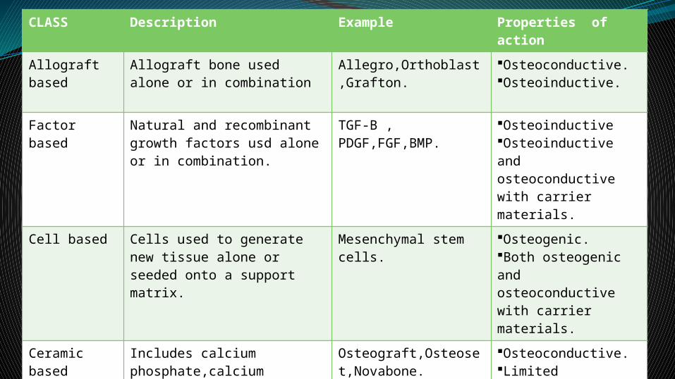
CLASS Description Example Properties of action
Allograft based
Allograft bone used alone or in combination
Allegro,Orthoblast,Grafton.
Osteoconductive.Osteoinductive.
Factor based Natural and recombinant growth factors usd alone or in combination.
TGF-B , PDGF,FGF,BMP.
OsteoinductiveOsteoinductive and osteoconductive with carrier materials.
Cell based Cells used to generate new tissue alone or seeded onto a support matrix.
Mesenchymal stem cells.
Osteogenic.Both osteogenic and osteoconductive with carrier materials.
Ceramic based
Includes calcium phosphate,calcium sulfate,and bioactive glass used alone or in combination.
Osteograft,Osteoset,Novabone.
Osteoconductive.Limited osteoinductive when mixed bonemarrow.
Polymer based
Includes degradable and nondegradable polymers used alone and in combination with other materials.
Cortoss,OPLA,Immix.
Osteoconductive.Bioresorbable in degradable polymer.

ALLOGRAFT BASED substitutesUses allograft bone with or without other elements.Comes in many forms and many preparations-
freeze dried,irradiated and decalcified.DEMINERALISED BONE MATRIX (DBM)-
chemosterilised,antigen extracted,surface demineralised autolysed-allogeneic bone .
DBM is generally mixed with a carrier –glycerol,calcium sulfate powder,sodium hyaluronate ,gelatin.

DBM sterilised by gamma irradiation and ethylene oxide-1.decreases the risk of disease transmission.
2.decreases the osteoinduvtive activity. Contraindications to DBM-1.Severe vascular or neurological disease.2.fever.3.Uncontrolled DM.

4.Severe Degenerative Bone disease.5.Pregnancy.6.Hypercalcemia.7.Renal compromise.8.Pott disease,or osteomyelitis or sepsis at the
surgical site.

Complications of allograft: Transmission of disease. Variable osteoinductive strength. Infection of graft.(large allografts for
structural replacement have the greatest risk of disease transmission.)
DBM is much less likely to transmit infection .

GROWTH FACTOR BASED substitutesURIST first discovered BMP in 1965,when he
recognised its ability to induce enchondral bone formation.
Growth factors are a part of a very large group of cytokines.
Growth factors commonly involved are:1.TGF-β.2.IGF.3.PDGF.4.VEGF.5.b FGF.Most of the BMP’s used today are in the bone
super family transforming growth factor –β.

This super family includes the inhibin/activin family, mulleraian-inhibiting substance family, and the decapentaloplegic family.
IGF and TGF-β mostly modulate the synthesis of cartilage matrix.
bFGF has a powerful mitogenic factor which stimulates the differentiation of chondrocytes.
bFGF is produced locally in bone during the initial phase of fracture healing and is known to stimulate cartilage and bone forming cells.

BMP’s shown to have osteogenic properties are-1.BMP 2,7-key role in osteoblast differentiation.2.BMP-3 – induces bone formation.3.BMP-4 – regulates the formation of teeth,limbs and
bone from mesoderm.4.BMP-5 –functions in cartilage develoment.6.BMP-6- role in joint integrity in adults.7.BMP -8a – involved in Bone and cartilage
development.BMP’s are group of noncollagenous glycoproteins
that belong to the TGF-β super family.
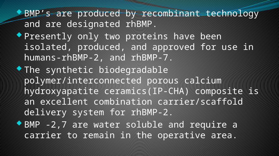
BMP’s are produced by recombinant technology and are designated rhBMP.
Presently only two proteins have been isolated, produced, and approved for use in humans-rhBMP-2, and rhBMP-7.
The synthetic biodegradable polymer/interconnected porous calcium hydroxyapatite ceramics(IP-CHA) composite is an excellent combination carrier/scaffold delivery system for rhBMP-2.
BMP -2,7 are water soluble and require a carrier to remain in the operative area.

Representation of BMP action sites
Bb BMP 2,6,9 BMP
2,4,7,9Most BMP’s
Pluripotent MSC
Osteoprogenitor cell
Osteoblast
Osteocyte

MOA of rhBMP’s is :chemotaxis,mitogenesis, and cell differentiation.
rhBMP’s differ in the type of cell induced to differentiate:
-rhBMP-2 acts on the mesenchymal stem cell and preosteoblast to differentiate into osteoblasts while rhBMP-7 acts only on the preosteoblast.
Uses of rhBMP’s: 1.Spinal fusion 2.Treatment of open tibial
fractures. 3.Maxillofacial surgeries.

Complications of rhBMP’sProblems with the implant-bending,breaking
subsidence or migration, and loosening,-neurological complications-paralysis,nerve and
spinal cord damage,dural tears,sexual dysfunction,bowel and bladder dysfunction.
-General organ complications-respiratory failure, GIT problems.

In anterior cervical spine-anterior soft tissue swelling, -dysphagia,tracheostomies -airway related complicationsIn posterior cervical spine-seroma pressing on the cordIn posterior lumbar spine-osteolysis, -neurological deterioration -ectopic and hypertrophic bone formation. -wound complications.

GROWTH FACTOR
SOURCE FUNCTIONS
1.TGF-β Platelets,T-Cells, macrophages,endothelial cells,fibroblasts.
1.Chemotactic for PMN’s,macrophages, lymphocytes,fibroblasts.2.StimulatesTIMPsynthesis,angiogenesis,fibroplasia.3.Inhibits production of MMP’s.
2.FGF-1-Acidic.-2-Basic
Macrophages ,mast cells,T-cells, endothelial cells, fibroblasts
1.Chemotactic for fibroblasts.2.Mitogenic for fibroblasts & keratinocytes.3.Stimulates keratinocyte migration.4.Angiogenesis ,wound contraction.
3.VEGFIsoforms-A,B,C,D.
Many types of cells
1.Increased vascular permeability.2.Mitogenic for endothelial cells.3.Angiogenesis.
4.PDGFIsoforms-A,B,C,D.
Platelets,macrophages,endothelial cells,keratinocytes,
1.Chemotactic for PMN’s,macrophages,fibroblasts.2.Mitogenic for fibroblasts,endothelial cells.3.Stimulates production of MMP’s,fibronectin,HA.

CELL BASED substitutesMost frequently used cell based graft is autologous
bone marrow.Bone marrow contains hemopoietic stem cells as well
as ‘mesenchymal stem cells’ or ‘stromal cells.’BM stromal cells depending on the tissue environment
can generate- osteoblasts,chondrocytes,adipocytes, myoblasts,endothelial cell precursors,hemopoietic stem cells.

Collection of Stem cellsBone marrow aspiration.Mesenchymal stem cells(MSC’s) are isolated and
cultured in flasks.After several passages ,a sufficient number of
MSC are collected.Trephination and collection of MSC from the flasks.MSC are then loaded in the scaffold.
Aspiration should be done at multiple sites to decrease dilution by blood.

Role of stem cells in orthopaedics1.Nonunion.2.Delayed union.3.Stabilisation of
fracture .4.Segemental bone
defects.5.Femoral head
osteonecrosis.6.Spinal fusion
7.Physeal and bone cysts.
8.Osteochondral defects.
9.Articular cartilage defects.

BM aspiration procedureDone under aseptic condition and general anaesthesia.3mm incision at anterior iliac crests on both sides and
needles (16 or 18 guaze) passed deep into iliac crests.BM is aspirated with 10ml syringes ,rinsed with a buffer
solution containing 400ml of phosphate buffered saline solution,25000 u of heparin and 100ml of albumin,to avoid clotting.
Contents in syringes are transferred to BM collection unit,to obtain final volume of 400 ml of BM.


Percutaneous Autologous bone grafting for nonunionMSC are aspirated from BM iliac crest.Centrifugation of aspirate is done on cell separator.Centrifugation produces a buffy coat,that contains
the ‘progenitor cells’,the source of angiogenic and osteogenic cytokines.
Buffy coat is taken into a syringe for intraosseous injection and using a trocar placed in nonunion gap.

CERAMIC BASED substitutesBioceramics – specially designed ceramics for
the repair and reconstruction of diseased or damaged parts of the body.
Types-1.single crystals. 2.polycrystalline. 3.glass. 4.glass – ceramics. 5.composites.

-Clinical success requires 1.Stable interface with connective tissue .2.Matching of mechanical behaviour of implant with the
tissue to be replaced.No material implanted is inert and all elicit a response:1.Material – toxic - surrounding tissue dies.2.Material –nontoxic,biologically inactive-fibrous tissue
forms.3.Material –nontoxic,biologically active-interfacial bond
forms.4.material-nontoxic dissolves-surrounding tissue replaces
it.

Type of ceramic
Type of aattachment Example
1.Dense,nonporous,nearly inert.
Attach by bone growth into surface irregularities,by press fitting into a defect.(MORPHOLOGICAL FIXATION.)
•Al2O3(single crystal and polycrystalline).
2.Porous inert implant.
Bone ingrowth occurs,which mechanically attaches the bone to the material.(BIOLOGICAL FIXATION.)
•Al2O3(porous polycrystalline),•HA coated porous materials.
3.Dense,nonporous surface reactive ceramics
Attach directly by chemical bonding with bone.-(BIOACTIVE FIXATION.)
•Bioactive glasses.•Bioactive glass ceramics.•HA.
4.Dense ,porous/ nonporous resorbable
Slowly replaced by bone. •Calcium sulfate•TCP•Calcium phosphate

Level of reactivity of an implant influences the thickness of the interfacial zone (layer between the material and the tissue).
Inert biomaterial-Interface is not chemically / biologically bonded, leading to relative movement at interface thus decreasing the function of the implant.
‘Bioactive Material’-that elicits a specific biological response at the interface resulting in formation of a bond between tissue and the material.
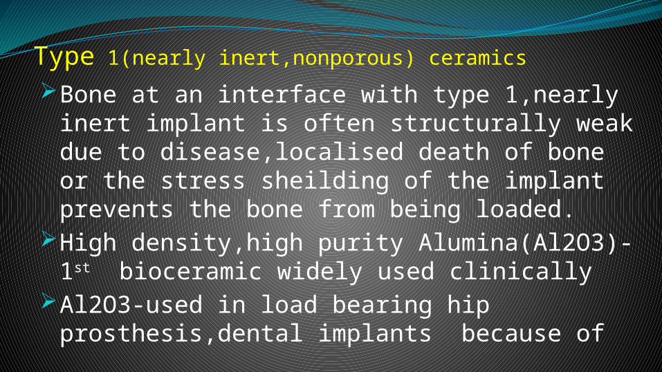
Type 1(nearly inert,nonporous) ceramicsBone at an interface with type 1,nearly inert
implant is often structurally weak due to disease,localised death of bone or the stress sheilding of the implant prevents the bone from being loaded.
High density,high purity Alumina(Al2O3)-1st bioceramic widely used clinically
Al2O3-used in load bearing hip prosthesis,dental implants because of

1. Excellent corrosion resistance2. Good biocompatibility and very thin capsule
formation permitting cementless fixation of prostheses.
3. High wear resistance.4. High strength.- Most alumina devices are very fine grained
polycrystalline alpha Al2O3.- Alumina with an average grain size of <4 µm
and >99.7% purity exhibits good flexural strength and excellent compressive strength.

An average increase in grain size to >7µm can decrease the mechanical properties by about 20%.
The primary use of alumina is for the ball of the hip joint with the acetabular component being ultra high molecular wt PE.
Other clinical applications of alumina include-1.Knee prostheses.2.bone screws.3.alveolar
ridge of jaw bone .4.maxillofacial reconstruction.5.ossicular bone reconsruction.

Medical grade alumina used as femoral balls in total hip replacement.

Type 2(porous ceramics)Potential advantage-inertness combined with
mechanical stability of highly convoluted interface developed when bone grows into the pores of ceramic.
The micro structure of certain corals makes an ideal investment material for the casting of stuctures with controlled pore sizes.
The most promising coral gene PORITES has pores with size of 140-160 µm with all pores interconnected.

Another coral gene ‘GONIOPORA’ has larger pore size of 200-1000µm.
REPLAMINEFORM process- duplicating the porous microstructure of corals that have a high degree of uniform pore size and interconnection.
The advantage of the above process is that the pore size and microstucture are uniform,contolled with interconnections of pores.
Procedure-1.To machine the coral with proper microstructure into desired shape.

2.The machined coral shape is fired to drive off carbondioxide from lime stone forming ‘CALCIA’.
3.Calcia structure serves as investment material for forming the porous material.
The limitation with type-2 porous implants is that for the tissue to remain viable and healthy, it is necessary for the pores to be >100-150µm in diameter to provide a blood supply to the ingrown tissue.
Ageing of porous ceramics leads to decrease in strength ,posing question as to the successful longterm application of porous material.

INGROWTH OF BONE WITHIN> 100µm OF ALUMINA BICERAMIC

Bioactive glasses glass ceramicsCertain compositions of glasses,ceramics ,glass
ceramics and composites have been shown to bond to bone-Bioactive ceramics.
A common characteristic of bioactive glasses and bioactive ceramics is a time dependent,kinetic modification of the surface that occurs upon implantation.
The surface forms a biologically active ‘Hydroxycarbonate Apatite’ (HCA) layer which provides the bonding interface with tissues.

The HCA phase that forms on bioactive implants is equivalent chemically and structurally to the mineral phase in bone.
The interfacial strength of adhesion is equivalent or greater than the cohesive strength of implant or tissue.
Therefore failure occurs either in the implant or in the bone but almost never in the interface.
Many bioactive silica glasses are based upon the formula-’ 45 S 5 ‘: 45 – wt % of SiO2
S – networker former 5 – 5 to 1 molar ratio of Ca to P

The collagenous constitutent of soft tissue can strongly adhere to the bioactive silica glasses.
The dense HCA – Collagen agglomerates mimic the nature of bonding between tendons and ligaments composed entirely of collagen fibrils and bone which is a composite of HCA crystals and collagen.
Three key compositional features of these glasses distinguish them from traditional Na2O-CaO-SiO2 glasses:
1.<60 mol% SiO2

2.High Na2O and high CaO content3.High Cao/P2O5 ratio.

1.SEM micrograph of collagen fibrills incorporated within the HCA layer growing on a 45S5 Bioglass invitro.2.Close up of the HCA crystals bonding to a collagen fibrill.

Stage
Reaction
1. Rapid exchange of Na+ or K+ with H+ or H3O+ from solution.(Ion exchange)
2. Loss of soluble silica in the form of Si(OH)4 to the solution,resulting from breaking of Si-O-Si bonds and formation of Si-OH at the glass solution interface.(Silica network dissolution)
3. Condensation and repolymerisation of a SiO2 rich layer on the surface
4. Migration of Ca2+ and PO4 groups to the surface through the SiO2 rich layer forming a CaO-P2O5 rich film
5. Crystallisaton of the amorphous CaO-P2O5 film by incorporation of OH-, CO3,or F- anions from solution to form a mixed Hydroxyl,Carbonate,Fluoroapatite layer.
Reaction stages of a bioactive implant

Sequence of interfacial reactions involved in forming a bond between tissue and bioactive ceramics.

Calcium phosphate ceramicsThe stable phase of calcium phosphate
ceramics depend considerably upon temperature and presence of water.
At pH<4.2 the stable phase is CaHPO4.2H2O. (Dicalcium phosphate or Brushite).
At pH>4.2 the stable phase is Ca 10(PO4)6(OH)2. (Hyroxyapatite).
At higher temperatures-other phases are stable.
1.Ca3(PO4)2- Tricalcium phosphate.

2.Ca4P2O9 – tetracalcium phosphate.The unhydrated high temperature calcium
phosphate phases interact with body water or body fluids at 37ºc to form HA.
The HA forms on exposed surfaces of TCP by the following reaction:
4Ca3(PO4)2 +2H2O Ca10(PO4)6(OH)2+2Ca2+ 2HPO4-
-The presence of micropores in the sintered material can increase the solubility of these phases.

Sintering of calcium phosphate ceramics usually occurs in the range of 1000ºc to 1500ºc following compaction of the powder into a desired shape.
Sintering reduces the amount of carbonated apatite, an unstable and weakly soluble form of HA.
Tensile and compressive strength and fatigue resistance depend on the total volume of porosity.
Porosity -1.micropores:<1µm , due to incomplete sintering.
2.macropores:>100µm in diameter,to permit bone growth.
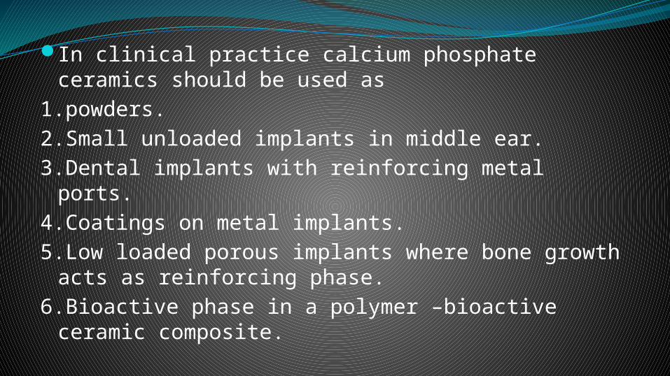
In clinical practice calcium phosphate ceramics should be used as
1.powders.2.Small unloaded implants in middle ear.3.Dental implants with reinforcing metal ports.4.Coatings on metal implants.5.Low loaded porous implants where bone growth
acts as reinforcing phase.6.Bioactive phase in a polymer –bioactive ceramic
composite.

Bonding mechanism:a cellular bone matrix from differentiated osteoblasts appear at the surface ,producing a narrow amorphous electron dense band only 3-5µm wide. Between this area and the cells
collagen bundles are seen.As the site matures the bonding zone shrinks to
a depth of only 0.05-0.2µm and thus normal bone attached through a thin epitaxial layer to the bulk implant.

Resorption / biodegradation of calcium phosphate ceramics is caused by
1.Physiochemical dissolution .2.Physical disintegration.3.Biological factors.All calcium phosphate ceramics biodegrade
to varying degrees in the following order: α-TCP > β –TCP > HA.

Rate of biodegradation increase as :1.Surface area increase.2.Crystallinity decrease.3.Crystal perfection decrease.4.Crystal and grain size decrease.5.Ionic substitution of CO3,Mg2+ in HA take placeFactors which decrease rate of biodegradation:1.F – substitution in HA .2.Mg2+ substitution in β-TCP.3.Decreased β-TCP/HA ratio in biphasic calcium
phosphate.

Composite ceramicsCertain restrictions in the use of bioceramics
due to 1.Uncertain life lifetime under complex stress
status.2.Slow crack growth.3.Cyclic fatigue.Two creative approaches to these mechanical
limitations:-use of bioactive ceramics as 1.coatings 2.composites.

Composites and coatings involve all 3 types of biomaterials-nearly inert,resorbable,bioactive.
Goal : 1.To increase the flexural strength and strain to failure.
2.decrease the elastic modulus.Most bioceramics are much stiffer than bone and
many exhibit poor fracture toughness.One approach to achieve properties analougous to
bone is to stiffen a compliant biocompatible synthetic polymer such as PE,with a higher modulus ceramic second phase –like HA powder.

The mechanical properties of PE-HA composite are close to/superior to those of bone.
Calcium phosphate –collagen composite: -collagen promotes mineral deposition by
providing binding sites for matrix proteins.-types 1 ,3 collagen have been combined with
HA,TCP, and autologous bone marrow to form a graft material devoid of structural support but augments fracture healing.

PE-HA composite used as a sleeve for the stem of a total hip implant.
Coatings Commonly used material for coating
are :1.carbon.
2.HA.Three types of carbon are used in biomedical
devices:1.LTI variety of pyrolytic carbon.2.Glassy (vitreous) carbon.3.Ultralow –temperature isotropic (ULTI) form
of vapor deposited carbon.

HA used as a coating on porous metal surfaces for fixation of orthopaedic prostheses.
This method combines type 2 & 3 methods of fixation.(biological & bioactive).
The plasma spray coating of HA is generally preffered, which substantially increase early stage interfacial bond strength of implants.

Calcium sulfate(POP)In a crystalline structure dscribed as
alphahemihydrate acts primarily as osteoconductive bone void filler.
Uses -1.filling of cysts,bone cavities,benign bone lesions and segmental defects.
2.expansion of grafts used for spinal fusion.
3.filling of bone graft harvest sites.

POLYMER BASED substitutesPolymers for graft substitutes include :-natural/synthetic.-Biodegradable/nonbiodegradable.Nonbiodegradable natural and synthetic polymers
are composites of polymer and ceramic.Biodegradable natural and synthetic materials
include -polyglycolic acid(PGA)-poly(lactic-co-glycolic )acid.

Miscellaneous bone graft substitutesCORALLINE HYDROXYAPATITE:-CHIROFF first observed that corals from marine
invertebrates have skeleton with a structure similar to both cortical and cancellous bone with interconnecting porosity.
-processed by a hydrothermal exchange method that converts the coral calcium phosphate to crystalline HA with pore diameters 200µm -500µm and in a structure very similar to that of human trabecular bone.