Boehmite and Gibbsite Nanoplates for the Synthesis of ...
14
Boehmite and Gibbsite Nanoplates for the Synthesis of Advanced Alumina Products Xin Zhang,* ,† Patricia L. Huestis, ‡ Carolyn I. Pearce, † Jian Zhi Hu, † Katharine Page, § Lawrence M. Anovitz, § Alexandr B. Aleksandrov, ∥ Micah P. Prange, † Sebastien Kerisit, † Mark E. Bowden, † Wenwen Cui, † Zheming Wang, † Nicholas R. Jaegers, † Trent R. Graham, ⊥ Mateusz Dembowski, † Hsiu-Wen Wang, § Jue Liu, § Alpha T. N’Diaye, % Markus Bleuel, & David F. R. Mildner, & Thomas M. Orlando, ∥ Greg A. Kimmel, † Jay A. La Verne, ‡ Sue B. Clark, †,# and Kevin M. Rosso* ,† † Pacific Northwest National Laboratory, Richland, Washington 99354, United States ‡ Radiation Laboratory and Department of Physics, University of Notre Dame, Notre Dame, Indiana 46556, United States § Oak Ridge National Laboratory, Oak Ridge, Tennessee 37830, United States ∥ School of Chemistry and Biochemistry, Georgia Institute of Technology, Atlanta, Georgia 30332, United States ⊥ The Voiland School of Chemical and Biological Engineering and # Department of Chemistry, Washington State University, Pullman, Washington 45177, United States % Advanced Light Source, Lawrence Berkeley National Laboratory, Berkeley, California 94720, United States & National Institute of Standards and Technology, Gaithersburg, Maryland 20899, United States * S Supporting Information ABSTRACT: Boehmite (γ-AlOOH) and gibbsite (α-Al- (OH) 3 ) are important archetype (oxy)hydroxides of alumi- num in nature that also play diverse roles across a plethora of industrial applications. Developing the ability to understand and predict the properties and characteristics of these materials, on the basis of their natural growth or synthesis pathways, is an important fundamental science enterprise with wide-ranging impacts. The present study describes bulk and surface characteristics of these novel materials in compre- hensive detail, using a collectively sophisticated set of experimental capabilities, including a range of conventional laboratory solids analyses and national user facility analyses such as synchrotron X-ray absorption and scattering spectroscopies as well as small-angle neutron scattering. Their thermal stability is investigated using in situ temperature-dependent Raman spectroscopy. These pure and effectively defect-free materials are ideal for synthesis of advanced alumina products. KEYWORDS: gibbsite, boehmite, aluminum oxides, nanoplates, material synthesis, thermal decomposition, neutron scattering, temperature-dependent Raman ■ INTRODUCTION Aluminum (oxyhydr)oxide nanomaterials are widespread in nature and industry, yet development of structurally and chemically well-defined model phases for fundamental and applied research needs is still lacking. In particular, the minerals boehmite (aluminum oxyhydroxide, γ-AlOOH) and gibbsite (aluminum hydroxide, α-Al(OH) 3 ) are abundant natural ores of aluminum as well as being important raw materials in industrial applications as adsorbents, 1−3 fire retardants, 4 coatings, 5 catalysts, 6,7 polishing agents, fillers, and fuel cells. 8 They are important precursors for the synthesis of different alumina products, such as γ-/δ-Al 2 O 3 , χ-Al 2 O 3 , 9−11 and α-Al 2 O 3 , 9−11 which are widely used in specialized industries including filler, catalysis, glass, ceramics, purification, paint, coating, and metallurgy. 12,13 Boehmite and gibbsite are also used in sensitive or specialized applications. For example, gibbsite is used as a substrate for treatment of stomach diseases and also serve as a vaccine adjuvant, 14,15 and boehmite is used as a host material for the light-emitting diodes (LEDs). 16 However, in support of this diversity of research and development areas is an underdeveloped capability in precision synthesis of representative model phases of specifically tailored particle size and shape at the nanoscale. Gibbsite is monoclinic (P2 1 /c space group) with a tabular pseudohexagonal habit Received: November 2, 2018 Accepted: November 26, 2018 Published: November 26, 2018 Article www.acsanm.org Cite This: ACS Appl. Nano Mater. 2018, 1, 7115-7128 © 2018 American Chemical Society 7115 DOI: 10.1021/acsanm.8b01969 ACS Appl. Nano Mater. 2018, 1, 7115−7128 Downloaded via NATL INST OF STANDARDS & TECHNOLOGY on August 23, 2019 at 15:50:01 (UTC). See https://pubs.acs.org/sharingguidelines for options on how to legitimately share published articles.
Transcript of Boehmite and Gibbsite Nanoplates for the Synthesis of ...
Boehmite and Gibbsite Nanoplates for the Synthesis of AdvancedAlumina ProductsXin Zhang,*,† Patricia L. Huestis,‡ Carolyn I. Pearce,† Jian Zhi Hu,† Katharine Page,§
Lawrence M. Anovitz,§ Alexandr B. Aleksandrov,∥ Micah P. Prange,† Sebastien Kerisit,†
Mark E. Bowden,† Wenwen Cui,† Zheming Wang,† Nicholas R. Jaegers,† Trent R. Graham,⊥
Mateusz Dembowski,† Hsiu-Wen Wang,§ Jue Liu,§ Alpha T. N’Diaye,% Markus Bleuel,&
David F. R. Mildner,& Thomas M. Orlando,∥ Greg A. Kimmel,† Jay A. La Verne,‡ Sue B. Clark,†,#
and Kevin M. Rosso*,†
†Pacific Northwest National Laboratory, Richland, Washington 99354, United States‡Radiation Laboratory and Department of Physics, University of Notre Dame, Notre Dame, Indiana 46556, United States§Oak Ridge National Laboratory, Oak Ridge, Tennessee 37830, United States∥School of Chemistry and Biochemistry, Georgia Institute of Technology, Atlanta, Georgia 30332, United States⊥The Voiland School of Chemical and Biological Engineering and #Department of Chemistry, Washington State University,Pullman, Washington 45177, United States%Advanced Light Source, Lawrence Berkeley National Laboratory, Berkeley, California 94720, United States&National Institute of Standards and Technology, Gaithersburg, Maryland 20899, United States
*S Supporting Information
ABSTRACT: Boehmite (γ-AlOOH) and gibbsite (α-Al-(OH)3) are important archetype (oxy)hydroxides of alumi-num in nature that also play diverse roles across a plethora ofindustrial applications. Developing the ability to understandand predict the properties and characteristics of thesematerials, on the basis of their natural growth or synthesispathways, is an important fundamental science enterprise withwide-ranging impacts. The present study describes bulk andsurface characteristics of these novel materials in compre-hensive detail, using a collectively sophisticated set ofexperimental capabilities, including a range of conventionallaboratory solids analyses and national user facility analyses such as synchrotron X-ray absorption and scattering spectroscopiesas well as small-angle neutron scattering. Their thermal stability is investigated using in situ temperature-dependent Ramanspectroscopy. These pure and effectively defect-free materials are ideal for synthesis of advanced alumina products.
KEYWORDS: gibbsite, boehmite, aluminum oxides, nanoplates, material synthesis, thermal decomposition, neutron scattering,temperature-dependent Raman
■ INTRODUCTION
Aluminum (oxyhydr)oxide nanomaterials are widespread innature and industry, yet development of structurally andchemically well-defined model phases for fundamental andapplied research needs is still lacking. In particular, theminerals boehmite (aluminum oxyhydroxide, γ-AlOOH) andgibbsite (aluminum hydroxide, α-Al(OH)3) are abundantnatural ores of aluminum as well as being important rawmaterials in industrial applications as adsorbents,1−3 fireretardants,4 coatings,5 catalysts,6,7 polishing agents, fillers,and fuel cells.8 They are important precursors for the synthesisof different alumina products, such as γ-/δ-Al2O3, χ-Al2O3,
9−11
and α-Al2O3,9−11 which are widely used in specialized
industries including filler, catalysis, glass, ceramics, purification,
paint, coating, and metallurgy.12,13 Boehmite and gibbsite arealso used in sensitive or specialized applications. For example,gibbsite is used as a substrate for treatment of stomach diseasesand also serve as a vaccine adjuvant,14,15 and boehmite is usedas a host material for the light-emitting diodes (LEDs).16
However, in support of this diversity of research anddevelopment areas is an underdeveloped capability in precisionsynthesis of representative model phases of specifically tailoredparticle size and shape at the nanoscale. Gibbsite is monoclinic(P21/c space group) with a tabular pseudohexagonal habit
Received: November 2, 2018Accepted: November 26, 2018Published: November 26, 2018
Article
www.acsanm.orgCite This: ACS Appl. Nano Mater. 2018, 1, 7115−7128
© 2018 American Chemical Society 7115 DOI: 10.1021/acsanm.8b01969ACS Appl. Nano Mater. 2018, 1, 7115−7128
Dow
nloa
ded
via
NA
TL
IN
ST O
F ST
AN
DA
RD
S &
TE
CH
NO
LO
GY
on
Aug
ust 2
3, 2
019
at 1
5:50
:01
(UT
C).
See
http
s://p
ubs.
acs.
org/
shar
ingg
uide
lines
for
opt
ions
on
how
to le
gitim
atel
y sh
are
publ
ishe
d ar
ticle
s.
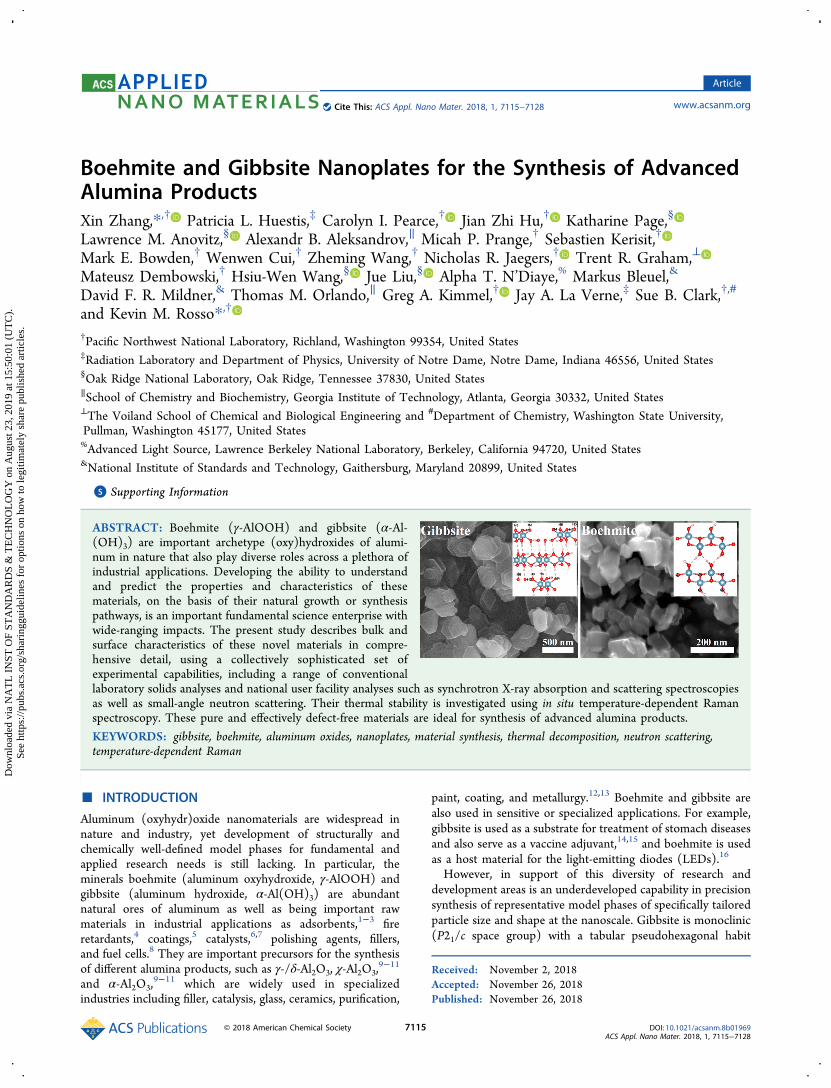
(Figure 1a). The morphology of lab-synthesized gibbsitenanoparticles was always visualized as hexagonal-shaped plates,which includes two dominant (001) basal surfaces with four(110) and two (100) edge surfaces.1 In contrast, boehmite isorthorhombic (Cmcm space group) with a tabular pseudohex-agonal or rhombic habit (Figure 1b). The morphology of lab-synthesized boehmite nanoparticles was always visualized aspseudohexagonal-shaped plates or rhombic-shaped plate,which includes two dominant (010) basal surfaces with two(100) and four (101) or with four (101) faces as edgesurfaces.2 Current methods for tailoring gibbsite and boehmiteparticles, however, rely on additive-assisted hydrothermalapproaches and typically lack phase purity, size, and shapecontrol within narrow distributions or produce materials ofunknown surface composition or structures because of anemphasis on bulk properties alone.17−19 The challengeprimarily relates to achieving structurally and chemicallyphase-pure materials from bulk interiors to the outermostsurface while also controlling size and shape. Part of theproblem stems from application of a limited set of character-ization tools that can incompletely describe both bulk andsurface properties.In recent previous work our team successfully laid out new
protocols for synthesizing structurally and chemically well-defined model gibbsite and boehmite materials, primarily onthe basis of bulk characterization.20,21 Our efforts have focusedon developing additive-free and novel morphology and size-controlled synthesis protocols that result in precision highquality materials with maximal yield.20,21
The purpose of the present work is to report on acharacterization campaign directed at achieving a comprehen-sive state-of-the-science understanding of the bulk and surfaceproperties of these materials. This study goes well beyondearlier work by comprehensively describing and comparing thestructures and properties of synthetic gibbsite and boehmitenanoparticles in an integrated fashion using a robust suite ofcharacterization tools. Here we feature gibbsite and boehmitenanoplates with average sizes of ∼280 and ∼35 nm,
respectively, characterizing their structure and properties indetail using X-ray photoelectron spectroscopy (XPS) forsurface composition and various morphology and structurecharacterization tools collectively spanning surface-to-bulkaspects, such as transmission electron microscopy (TEM),scanning electron microscopy (SEM), and atomic forcemicroscopy (AFM). Bulk properties were also assessed usingX-ray diffraction (XRD), nuclear magnetic resonance (NMR)spectroscopy, Fourier-transform infrared spectroscopy (FTIR),X-ray pair distribution function (PDF), and extended X-rayabsorption fine-structure spectroscopy (EXAFS) with datamodeling constrained by ab initio molecular dynamics (AIMD)simulations. Furthermore, we examined the thermal stability ofthese two materials using in situ temperature-programmedRaman spectroscopy and thermal gravimetric analysis/differ-ential thermal analysis (TGA/DTA). Finally, we begin toaddress the aggregation behavior of these two nanomaterialsusing Brunauer−Emmett−Teller (BET) measurements andsmall-angle neutron scattering (SANS).The results are presented in a logical flow from microscopic
morphology to bulk structure, bulk and surface chemicalproperties, and thermal stability and aggregation behavior. Theintegrated findings not only add to the knowledge databaseabout relationships between nucleation and growth pathwaysand nanophase property outcomes but also serve as animportant reference for the application of these materials forsynthesis of advanced alumina products.
■ SYNTHESIS, MEASUREMENT, AND DATAANALYSIS METHODS
Preparation of Hydrated Aluminum Hydroxide [Al(OH)3]Gel Precursors. Aluminum nitrate solution (0.25 M) was preparedby dissolving Al(NO3)3·9H2O (ACS reagent, ≥98%, Sigma-Aldrich)in deionized (DI) water, and then 1 M NaOH (≥98%, Sigma-Aldrich) solution was added to adjust the solution pH value to around5, or 10, to precipitate the Al(OH)3 gel precursors for the synthesis ofgibbsite and boehmite, respectively. Note: the pH of the 0.25 MAl(NO3)3 was around 2 due to the hydrolysis of Al3+ ions; the 1 MNaOH was used to adjust the solution pH to 5 or 10 to form the
Figure 1. Crystal structure of gibbsite (a) and boehmite (b). Blue, red, and white spheres represent aluminum, oxygen, and hydrogen atoms,respectively.
ACS Applied Nano Materials Article
DOI: 10.1021/acsanm.8b01969ACS Appl. Nano Mater. 2018, 1, 7115−7128
7116
Al(OH)3 gel. After stirring for 1 h, the gel-like precipitates werecollected by centrifuging using 8600 rpm and then were washed withDI water three times to remove the residue-soluble salts.Synthesis of Gibbsite Nanoplates.20 Al(OH)3 gel-like precip-
itates made from pH 5 as described above were dispersed into DIwater, and then the suspension was poured into a 20 mL Teflon liner;the pH value was then titrated to around 5 with 1 M NaOH. Note:the pH of the gel solution was slightly less than 5; 1 M NaOH wasalso used to adjust the solution pH to 5. The concentration of Al3+ inthe suspension was 0.5 M, and the volume of the suspension was 16mL, which is the 80% of the volume of the liner. Then the Teflon linerwas sealed into a Parr vessel and which was then heated to 80 °C in arotation oven (10 rpm) for 72 h. The resulting white precipitate wascollected by centrifuging using 8600 rpm and then washed with DIwater three times. Finally, the gibbsite solids were dried at 80 °C in anelectric oven overnight.Synthesis of Boehmite Nanoplates.21 Al(OH)3 gel-like
precipitates made from pH 10 as described above were dispersedinto DI water, and then the suspension was poured into a 20 mLTeflon liner; the pH was then titrated to around 10 with 1 M NaOH.The concentration of Al3+ in the suspension was 0.5 M, and thevolume of the suspension was 16 mL. Then the Teflon liner wassealed into a Parr vessel and was heated to 200 °C in a rotation oven(10 rpm) for 48 h. The resulting white precipitate was collected bycentrifuging using 8600 rpm and then washed with DI water threetimes. Finally, the boehmite solids were dried at 80 °C in an electricoven overnight.Scanning Electron Microscopy (SEM). The morphologies of
synthetic gibbsite and boehmite were characterized by SEM (FEI,Helios NanoLab 600i). Prior to imaging, using a sputter coater, ontoboth samples a carbon thin film layer around 5 nm thick wasdeposited to enhance the electrical conductivity electron beamimaging.Transmission Electron Microscopy (TEM). The morphologies
of synthetic gibbsite and boehmite were also characterized by TEM(FEI Titan TEM). To mount samples onto TEM grids, they were firstdispersed into DI water using a bath sonicator for ∼5 min. Thesesuspensions were then drop cast onto standard TEM grids (LaceyCarbon, 300 mesh, Copper, Ted Pella, Inc.) and then dried atambient conditions. All samples examined in the TEM used anacceleration voltage of 300 kV for optimal imaging results.Atomic Force Microscopy (AFM). The thickness and surface
roughness of the synthetic gibbsite nanoplates were characterized byAFM (Dimension Icon, Bruker) operating in contact mode withstandard silicon nitride tips (MLCT, Bruker). Typical imagingconditions used include a 1 Hz scan rate and a selected resolutionof 512 × 512 pixels. The sample was prepared by drop casting gibbsiteaqueous suspensions onto Si wafers (Nova Electronic Materials Ltd.)and then removing the residual suspension after 30 min using a high-purity N2 gas stream (99.9%). The sample was washed using DI waterthree times and then dried using the same high-purity N2. Prior tosample drop casting, the Si wafers were cleaned with DI water usingsonication twice and then sonicated in ethanol once. Finally, the Siwafers were plasma cleaned under Ar atmosphere for 30 min and thenwere treated by an ozone cleaner for another 30 min just prior to use.X-ray Diffraction. The crystal phases of the synthetic gibbsite and
boehmite were examined by XRD (Philips X’pert Multi-Purposediffractometer, PANAlytical) equipped with a Cu anode operated at40 mA and 50 kV. To mount these powder samples, they typicallywere lightly compressed using a clean glass microscope slide into atraditional well sample holder. To minimize preferred orientation forgibbsite, which given its prominent platelet form would tend to self-align under light compression, an additional pattern was recorded in acapillary sample using a microbeam diffractometer (Rigaku Rapid II)equipped with a rotating Cr anode. All XRD patterns were analyzedby whole-pattern fitting using Topas v5 (Bruker AXS), and the crystalstructures were obtained from the Inorganic Crystal StructureDatabase (Fachinformationzentrum Karlsruhe, Germany).Synchrotron X-ray Pair Distribution Function. Room temper-
ature synchrotron X-ray diffraction and total scattering data were
recorded for the synthetic gibbsite and boehmite in polyimidecapillaries, at the Advanced Photon Source (APS), Argonne NationalLaboratory, on beamline 11-ID-B. We used a rapid-acquisition pairdistribution function (RaPDF) method, with an X-ray energy of 86.7keV (λ = 0.1430 Å).22 We used a PerkinElmer amorphous Si two-dimensional image-plate detector (2048 × 2048 pixels and 200 × 200mm pixel size) for two-dimensional data collection, with a sample-to-detector distances of ∼950 and ∼180 mm for X-ray diffraction and forPDF data, respectively. These two-dimensional diffraction data wereconverted to one-dimensional form using the Fit2D software suite23
and using a CeO2 powder standard for calibration. The normalizedtotal scattering patterns, S(Q), were produced in the programPDFgetX224 by subtracting polyimide container scattering, utilizingthe appropriate sample composition, and applying standardcorrections for the area detector setup.22 PDF patterns, G(r), werecalculated via Fourier transformation of the total scattering data,utilizing a Q range of 0.1−26 Å−1.
Rietveld refinement of synchrotron diffraction data and localstructure PDF refinements were performed in Topas Academic v625,26
and calculations of partial PDFs from resulting models werecompleted in the PDFgui suite.27 The instrumental related dampening(dQ, instrumental fwhm of S(Q))27 and broadening (Qb)
28 for PDFfitting were refined by fitting standard Ni powder data between 1 and100 Å. The dQ and Qb were refined to be 0.041 and 0.017 Å−2. Thesetwo values were fixed during further structure refinements. A sincfunction (sin(Qr)/Qr) was convoluted to the calculated PDF toaccount for the termination effect due to the finite Qmax used forFourier transform.28 An empirical PDFgui-type delta1 (delt1/r) termwas used to model the correlated motion.27 A predetermined double-Gaussian function for a nanoplate with two characteristic lengths(thickness, l1, and width, l2) was used as a numerical approximationfor the characteristic particle shape to correct for the effects intrinsicto PDF modeling of small nanoparticles using bulk models.29−31
Synchrotron Al K-Edge Extended X-ray Absorption FineStructure (EXAFS) Spectroscopy. Al K-edge EXAFS spectra forgibbsite and boehmite were acquired at the Advanced Light Source(Berkeley, CA) at beamline 6.2.1.2. To mount samples, powder waslightly pressed into indium foil, which both secures the sample andminimizes charging during measurements, which was then attached tothe Cu metal sample holder using silver paint. A reference spectrumcollected on corundum (α-Al2O3) was used to calibrate the energyscale.32 The EXAFS signal was monitored at room temperature intotal electron yield (TEY) mode over the scan range from 1520 to1850 eV. EXAFS data were background corrected and analyzed usingthe Athena interface to the IFEFFIT program.33
Simulation of EXAFS Using Density Functional Theory.Calculations were performed using the pseudopotential plane-wavedensity functional theory (DFT) module (NWPW) of the NWChemcomputational chemistry package.34 The calculations made use of thegeneralized gradient approximation (GGA) exchange-correlationfunctional of Perdew, Burke, and Ernzerhof35,36 (PBE) with (gibbsite)and without (boehmite) the Grimme dispersion corrections.37
Softened Hamann pseudopotentials38 modified into a separableform suggested by Kleinman and Bylander39 were used for aluminum(10), oxygen (2), and hydrogen (0), with the number of coreelectrons shown in parentheses. The plane-wave cutoff energy was993 eV (73 Ry).
A constant-pressure geometry optimization (ionic positions, cellvolume, and cell shape are allowed to relax) was first performed forboth the gibbsite (P21/c space group) and boehmite (CmCcm spacegroup) unit cells with 4 × 8 × 4 and 12 × 3 × 12 k-point meshes,respectively, and a supercell was then created by scaling the optimizedunit cell 1 × 2 × 1 and 2 × 1 × 3 for gibbsite and boehmite,respectively. A 29-ps NVT (constant number of particles, constantvolume, and constant temperature) ab initio molecular dynamics(AIMD) simulation was then performed for each of the two supercellswith the Car−Parrinello approach.40−42 An integration time step of0.12 fs was used for the equations of motion with a fictitious mass of750 au for the electronic degrees of freedom. The temperature was setto 25 °C and was kept constant via Nose−Hoover thermostats with
ACS Applied Nano Materials Article
DOI: 10.1021/acsanm.8b01969ACS Appl. Nano Mater. 2018, 1, 7115−7128
7117
periods of 1200 au for both the ions and electrons. All hydrogenatoms were treated as deuterium atoms to allow for improvedadiabatic decoupling of the electron and ionic motion.After discarding the first 2 ps of each simulation, a configuration
was collected every 60 fs to generate a pool of 450 configuration fromeach AIMD simulation. For each configuration, a cluster with a radiusof 6 Å centered around one Al atom (randomly selected but the samefor each configuration) was generated to calculate all scattering pathswith effective distances less than the cluster radius for a Al K core holeof the central atom using FEFF9.43−45 For gibbsite, which has twosymmetrically distinct Al positions, the procedure was repeated foranother Al atom in the other crystallographic site, but the differencebetween the signals due to the two sites was negligible. The S0
2
parameter (0.922) calculated by FEFF9 was used in all calculations,and a value of 2 eV was used for ΔE0 for both materials. For eachmaterial, the EXAFS spectra of all configurations were averaged forcomparison with experiment. The Fourier transform was applied toaveraged EXAFS spectra using IFEFFIT46 in the range 1.5 < k < 8.3Å−1 with dk = 1 Å−1, weighted by k2, and truncated using a Hanningwindow.Nuclear Magnetic Resonance (NMR) Spectroscopy. Room
temperature single-pulse 27Al magic-angle spinning (MAS) NMRmeasurements were performed with a commercial 3.2 mm pencil-typeprobe and a Varian-Inova 850 MHz NMR spectrometer operating at amagnetic field of 19.975 T; corresponding 27Al Larmor frequencieswere 221.413 MHz. We used a single pulse sequence with a pulsewidth of 0.5 μs (corresponding to a solid π/4 pulse) and 27Al radio-frequency (rf) field strength of 83.3 kHz (i.e., 3.0 μs for liquid π/2calibrated by using 1 M Al(NO3)3 aqueous solution). Each spectrumwas acquired using an acquisition time of 20 ms and a recycle delaytime of 1 s, which was shown to be long enough to allow all aluminumspecies signals to be observed quantitatively.47 A 1 M Al(NO3)3aqueous solution (0 ppm) was used as a chemical shifts reference.Both hydrated (as-synthesized samples) and dehydrated 27Al MASNMR spectra were acquired with an accumulation number between20000 and 60000 scans to ensure observation of any traceundercoordinated alumina species. As-synthesized samples weredried at 80 °C overnight before analysis. Dehydrated versions of
these samples were prepared in a vacuum oven evacuated to about10−5 Torr and then heated to 50 °C for 12 h, before cooling andloading the samples into sealed 3.2 mm MAS rotors while inside anitrogen-filled glovebox to avoid exposure to air.
X-ray Photoelectron Spectroscopy (XPS). Survey and narrowscan XPS measurements were taken with a PHI VersaProbe II X-rayphotoelectron spectrometer located at the University of Notre Dame,using a monochromatic Al Kα X-ray source and a hemisphericalelectron energy analyzer. Survey scans were used to assess anyimpurities as well as relative composition, collected using a passenergy of 187.85 eV. More detailed elemental scans were made usinga pass energy of 23.5 eV. Samples were first affixed to an aluminumSEM stub using a conductive double stick carbon tab from Ted Pella,Inc. The stub with the material attached was then coated in a thinlayer of iridium (∼1.5 nm) to mitigate differential charging effects asthe instrumental charge correction methods were deemed insufficient.Survey scans were taken prior to coating the sample with iridium.Data analysis was completed using PHI MultiPak.
Fourier-Transform Infrared Spectroscopy (FTIR). FTIRmeasurements were performed in transmission mode using a BrukerVertex 70. Each measurement consisted of 256 scans taken from 400to 4000 cm−1 with a resolution of 4 cm−1. Samples for measurementswere prepared by mixing with KBr and pressed into a pellet prior toanalysis.
Raman Spectroscopy. Raman spectra were obtained using aBruker Senterra Raman microscope equipped with a Linkham variabletemperature vacuum translation stage at Georgia Tech. An excitationwavelength of 532 nm (20 mW) and a ×50 objective lens weretypically used, assuring no beam induced damage or heating occurred.About 5 mg of either boehmite or gibbsite powder was deposited on acopper plate, and 5 mg of water was added to make a slurry. Airdrying resulted in a dense flat layer that was placed on a heating platein the spectrometer and pumped to about 0.1 Torr. Spectra weretaken with a resolution of 9−15 cm−1 with ten scans and a scan timeof 10 s over the range of 70−3700 cm−1. Scans were made at aconstant temperature with a heating rate of 20 °C/s between scans.
Nitrogen Adsorption/Desorption Isotherms. Specific surfacearea, porosity, and size of the particles were obtained using a
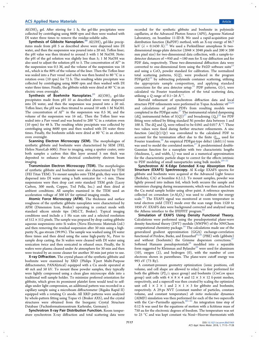
Figure 2. SEM and TEM images of gibbsite (a−c) and boehmite (d−f) nanoplates.
ACS Applied Nano Materials Article
DOI: 10.1021/acsanm.8b01969ACS Appl. Nano Mater. 2018, 1, 7115−7128
7118
Quantachrome Autosorb 1 with nitrogen gas as the adsorbate, withanalysis via the Brunauer−Emmett−Teller (BET) method. Measure-ments were performed on ∼0.25 g of material, which was firstoutgassed at a temperature of 105 °C for about 20 h. Data wereanalyzed using the included Autosorb analysis software.Thermal Gravimetric Analysis/Differential Thermal Anal-
ysis. TGA/DTA measurements were performed using a TGA/DSC-1from Mettler Toledo. Approximately 30 mg of the powders waspressed into a 100 μL aluminum crucible and heated from 25 to 600°C at a rate of 10 °C/min under a nitrogen flux of 50 mL/min.Background scans were collected on the empty crucible andsubtracted out using the STARe software.Small-Angle Neutron Scattering (SANS). Small-angle neutron
scattering experiments were performed to characterize the gibbsiteand boehmite grain sizes; methodological details have been reportedelsewhere48,49 and are therefore summarized briefly. Neutronscattering measurements were performed on the NGB30-SANS andBT5-USANS spectrometers at the National Institute of Standards andTechnology (NIST) Center for Neutron Research (NCNR)50−53 onaqueous dispersions. The wavelength was 2.38 Å with a wavelengthresolution Δλ/λ = 0.059. Data were collected over a Q range from 4.2× 10⁻⁵ to 2.7 × 10⁻³ Å−1, which corresponds to sizes from 2400 Å to∼15 μm. The horizontal Q resolution (full width at half-maximum,fwhm) was 2.5 × 10⁻⁵ Å−1.49 Standard titanium cells were used with a1 mm path length and two 1 mm thick quartz glass windows with thebeam incident along the surface normal. Approximately 1 wt % solidwas used. To keep the sample suspended during the measurement,these cells were placed in a sample tumbler54,55 and rotated along anaxis parallel to the beam at ∼10 rpm. The grain size distributions werecalculated using the total nonnegative least-squares approach coded inthe Irena plugin for IGOR.56 Each was run 10 times to estimateuncertainties, assuming, in each case, spherical grains. Details ofanalysis of the scattering data are provided in Ilavsky56 and Jemianand Anovitz and Cole.57
■ RESULTS AND DISCUSSION
Morphology Characterization. We begin with micro-scopic characterization to first build a visual impression of thephysical characteristics of the synthetic materials. SEM, TEM,and AFM were performed to characterize the size andmorphology of the gibbsite and boehmite nanoplates. Atomicscale structural details that provide context for particlemorphology are provided in the Introduction and Figure 1.As shown in Figure 2a−c and Figure S1, gibbsite nanoplatesare hexagonal-shaped particles with the average size around280 nm, the basal surface (001) with four (110) faces and two(100) faces on sides (diffraction pattern analysis in the Figure
2c); however, boehmite nanoplates are rhombic-shapedparticles with the average size around 35 nm (Figure 2d−f),the basal surface (010) with four (101) faces on sides(diffraction pattern analysis in the Figure 2f). As shown inFigure S1, the average thickness of gibbsite nanoplates was∼18 nm. The average thickness of boehmite nanoplates was∼6 nm, which was measured by TEM. Microscopicallyobserved particle dimensions are compared to detailed fits toXRD data below.
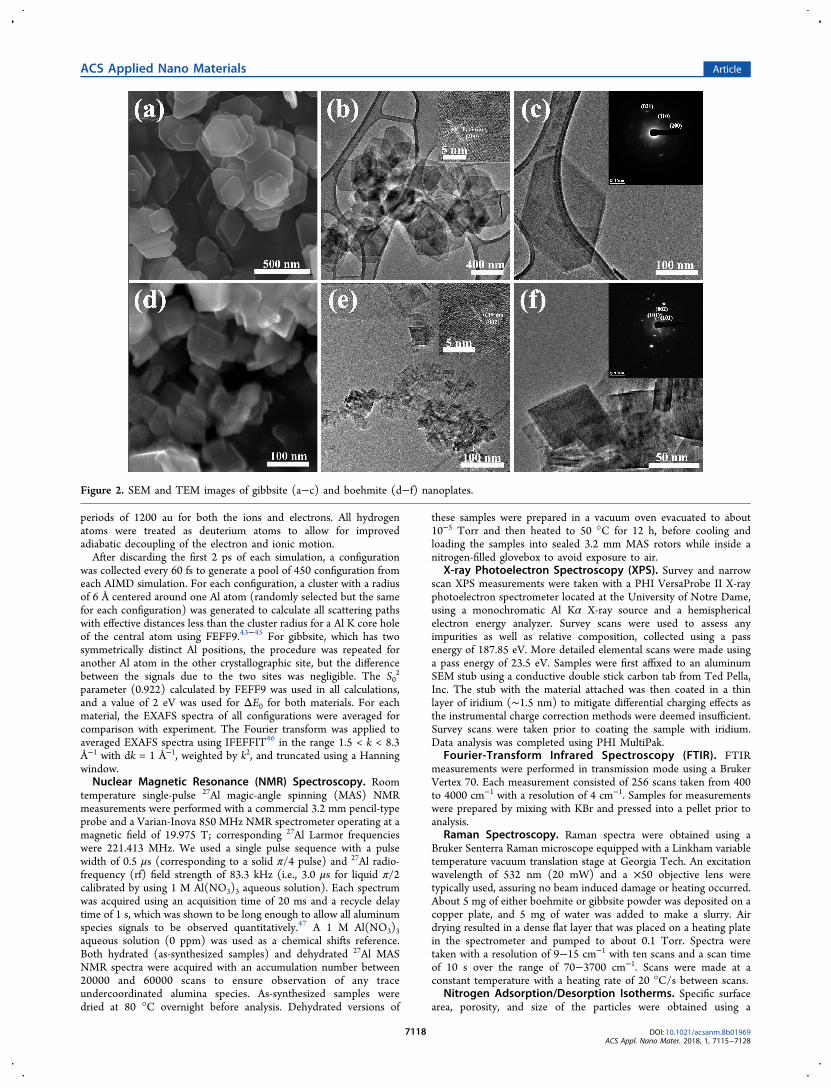
Structural Characterization. We now turn attention tothe bulk structure of the gibbsite and boehmite nanoplates,which was characterized in detail using conventional andsynchrotron-based XRD and data modeling. Figure 3a showscorresponding powder XRD patterns. The diffraction patternsare in good agreement with reference data for the respectivecompounds, although gibbsite shows strong preferredorientation of the (002) peak near 18° 2θ. This observationis consistent with the large thin plate morphology seen by SEMwith the surface of the plates being approximately parallel to(001). To eliminate the preferred orientation, a second patternwas collected on a microbeam XRD instrument using a sampleloaded into a glass capillary.Both patterns showed peak shapes, and in particular peak
widths, that did not vary smoothly with diffraction angle aswould be expected from isotropic size or strain effects. In thecase of boehmite, the pattern could be simulated well using amodel that incorporated anisotropic crystallite sizes. Figure 3bshows the results of Rietveld refinements for isotropic oranisotropic size broadening. In the isotropic case, the crystallitesize refined to 17 nm but the simulated pattern is too sharp forthe (020) peak while too broad for the (002) peak. Ananisotropic model58 provided a much better fit and resulted inan average crystallite size of 28 × 10 × 31 nm3 in the a, b, and cdirections, respectively, consistent with the SEM and TEMresults described above.The gibbsite pattern could not be satisfactorily modeled with
size broadening alone but required the additional incorpo-ration of microstrain broadening. Both broadening sourcesrequired anisotropic models, and for strain the phenomeno-logical model described by Stephens59 was employed. Ahexagonal model was chosen because it gave the greatestimprovement in fit for the fewest refined parameters. Figure S2shows the Rietveld fits for different combinations of size andstrain, using the capillary data where preferred orientation
Figure 3. XRD patterns of gibbsite (lower in a) and boehmite (upper in a) nanoplates and (b) detail of boehmite XRD pattern compared withsimulated patterns incorporating either isotropic or anisotropic crystallite size broadening.
ACS Applied Nano Materials Article
DOI: 10.1021/acsanm.8b01969ACS Appl. Nano Mater. 2018, 1, 7115−7128
7119

could be eliminated. Clearly the refinement that included bothsize and strain is best. The improvement is not simply theresult of adding more refineable parameters because there areconsistent trends of misfit with 2θ. If only strain is modeled,the low angle peaks are too sharp and the high angle peaks toobroad. The reverse is true for the size-only fit and arisesbecause of the different 2θ dependencies of size and strainbroadening. The average crystallite size obtained from the bestgibbsite fit was 34 × 50 × 9 nm3, and similar sizes wereobtained whether strain was considered or not.Results of Rietveld refinement of the synchrotron XRD data
for the gibbsite and boehmite nanoplates are shown in Figures4a and 4b, respectively. Lattice parameters, isotropic atomicdisplacement parameters (Beq) for Al (with all O isotropicatomic displacement Beqs held fixed to 0.8 Å2), and fractionalcoordinates of the atoms were refined in space group (SG)P121/c1 for gibbsite
60 and SG CmCm for boehmite.61 Tables 1and 2 list the refined parameters resulting from fitting. Resultsare consistent with previously reported gibbsite and boehmite
nanocrystalline structures, with goodness of fit values, Rwp, of6.16% and 8.31%, respectively. Both models required sizebroadening parameters; isotropic in the case of gibbsite andanisotropic (ellipsoidal) in the case of boehmite. In addition tothe isotropic size broadening, anisotropic strain broadening isagain required to model the X-ray diffraction pattern ofgibbsite.62
The isotropic crystallite dimension from the gibbsite datarefinement was 44(2) nm, which is considerably smaller thanthe 280 × 280 × 18 nm3 dimensions observed via microscopybut agree well with laboratory XRD fitting. By contrast,ellipsoidal particle dimensions determined by Rietveld analysisfor boehmite are 24.2(9) nm × 5.76(4) nm, which compareswell to the 35 nm × 6 nm dimensions found via microscopy.Crystallite sizes determined from diffraction are indicative ofaverage structural coherent grain size in particular crystallo-graphic directions (depending on the model) and thus oftenvary significantly from the shapes and dimensions observed viamicroscopy, small-angle scattering, and other morphologysensitive probes. The large discrepancy between microscopyand diffraction-based size estimates in the case of the gibbsitenanoplates indicates that the plate-shaped particles observableby SEM are each composed of smaller domains of coherentcrystallinity, approximately half the thickness of the plates andone-sixth their breadth. The regions between these domainsare likely to contribute to the remaining misfit in the Rietveldanalysis as well as being areas of higher reactivity. On the otherhand, the close agreement in diffraction and microscopy basedestimates in the case of the boehmite nanoplates indicateshighly crystalline (single or small number of grain) particles.
Figure 4. Results of Rietveld refinement of the (a) gibbsite and (b) boehmite nanoplates using X-ray diffraction data from 11-ID-B. First 80 Å ofPDF fits of 1 to 100 Å (c) gibbsite and (d) boehmite nanoplate X-ray PDF data, resulting from models with an anisotropic particle shape function.
Table 1. Crystal Structure of Gibbsite Nanoplates, Al(OH)3, Refined Using X-ray Diffraction Data from 11-ID-Ba
atom Wyck X y z occ Beq (Å2)
Al1 4e 0.1694(18) 0.0374(15) 0.0021(14) 1 1.20(7)*Al2 4e 0.333(2) 0.5104(15) 0.0004(15) 1 1.20(7)*O1 4e 0.072(2) 0.152(3) 0.3985(16) 1 0.8O2 4e 0.092(3) 0.133(3) 0.107(2) 1 0.8O3 4e 0.281(2) 0.712(2) 0.1096(17) 1 0.8O4 4e 0.396(3) 0.140(3) 0.393(2) 1 0.8O5 4e 0.407(2) 0.212(3) 0.1073(16) 1 0.8O6 4e 0.7496(19) 0.146(3) 0.0933(15) 1 0.8
aAl(OH)3 SG P121/c1, a = 8.6645(7) Å, b = 5.0594(4) Å, c = 12.5281(19) Å, β = 129.443(7)°, disotropic = 44(2) nm, Rp = 4.78%, RBragg = 3.12%,Rwp = 6.16%, GoF = 6.88. Refined values are given with estimated standard deviation from refinement in parentheses. Parameters with an asteriskwere constrained to be equivalent.
Table 2. Crystal Structure of Boehmite Nanoplates, AlOOH,Refined Using X-ray Diffraction Data from 11-ID-Ba
atom Wyck X y z occ Beq (Å2)
Al 4c 0 0.68048(23) 1 1 0.183(5)O1 4c 0 0.29276(38) 1 1 0.8O2 4c 0 0.07959(36) 1 1 0.8
aAlOOH SG CmCm, a = 2.86351(21) Å, b = 12.20722(18) Å, c =3.68868(29) Å, l1 = 24.2(9) nm, l2 = 5.76(4) nm, Rp = 6.37%, RBragg =3.36%, Rwp = 8.31%, GoF = 7.98. Refined values are given withestimated standard deviation from refinement in parentheses.
ACS Applied Nano Materials Article
DOI: 10.1021/acsanm.8b01969ACS Appl. Nano Mater. 2018, 1, 7115−7128
7120
To gain deeper insight into the coherent particledimensions, the materials were also characterized by X-rayPDF analysis. The first 80 Å of 100 Å fits to X-ray PDFs of thegibbsite, and boehmite nanoplates are given in Figure 4c,d.Similar models were used to those in Rietveld refinement,except both local structure data sets required anisotropicparticle shape models to fit the specific PDF intensity decay athigh r. It should also be noted that gibbsite fractionalcoordinates were held fixed to the values determined fromRietveld analysis (Table 1) to reduce the number of freeparameters for real-space fitting. The fit quality over the fullrange of the PDF is excellent in both cases, with resultinggoodness-of-fit, Rwp, of 18.62% for gibbsite and 14.18% forboehmite data sets. Refined model parameters from fits to thelocal structure are given in Tables S1 and S2. The results againsupport the observation of phase pure, high quality crystallinenanoplates. However, there are significantly higher atomicdisplacement values for Al and O sites in gibbsite nanoplatesrelative to those in boehmite nanoplates, another possibleindicator of structural disorder. It should be noted that in thissize regime (tens of nanometers) estimates of coherent particledimensions from analysis of Bragg data offer greater sensitivitythan analysis of X-ray PDF data. Nonetheless, anisotropic sizemodels are needed to fit the high-r PDF, and both samplesindicate a particle width at least 2 times larger than particle
thickness. These estimates vary again relative to the diffractionand microscopy determined dimensions.A closer look at the quality of local structure fits to the two
nanoplate data sets is given in Figure S3a for gibbsite and inFigure S3b for boehmite. The calculated partial PDFs,corresponding to Al−O, O−O, and Al−Al pair−pair distancesin each model, are shown for reference below that data and thefits. Results up to the first 10 Å in real space show that the localatomic structures are well-fit with the crystallographic modelsapplied. While the quality of the fits rules out significant localstructural distortions involving the X-ray sensitive elements inthe samples (Al and O), as well as significant amorphouscomponents and impurities, there are a few structural featuresat low r that suggest slight structural distortions in either bulkor surface structures relative to long-range crystalline models.In particular, the first sets of O−O correlations centered at∼2.4 Å in gibbsite, and ranging between approximately 2.5 and2.8 Å in boehmite, are not well captured. Local Al−Ocorrelations beyond the first neighbor (between approximately3.5 and 5 Å) are also slightly misfit. Several structuralmodifications were attempted in each case to capture thelocal distortions present in the Al−(O/OH)6 octahedral units,but it was evident from these attempts that an oxygen sensitiveprobe (such as neutron total scattering) may be needed todetermine the detailed nature of the slight distortions present.
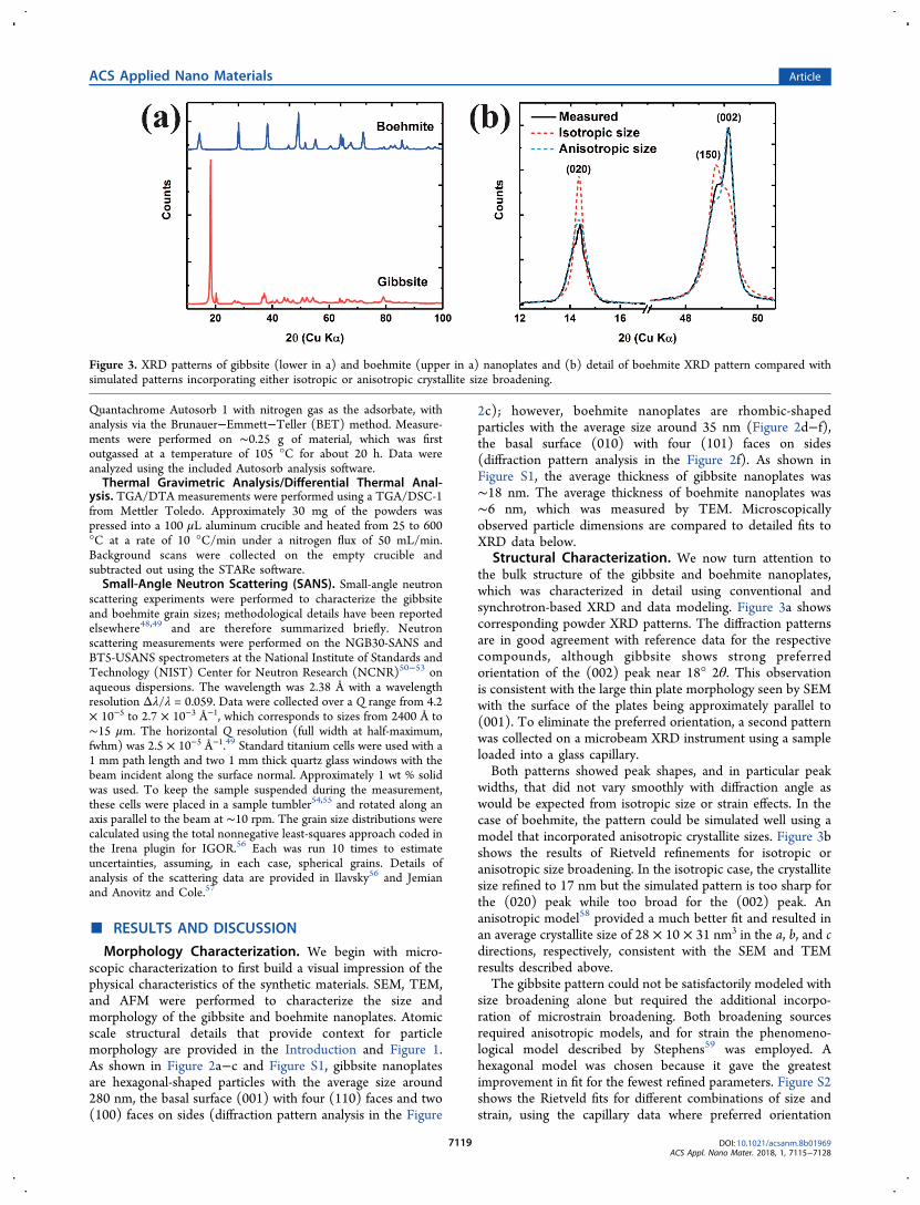
Figure 5. Comparison of Al K-edge experimental (solid lines) and calculated (dashed lines) EXAFS (left) and corresponding Fourier transformmagnitudes (right) at room temperature for gibbsite (blue) and boehmite (red).
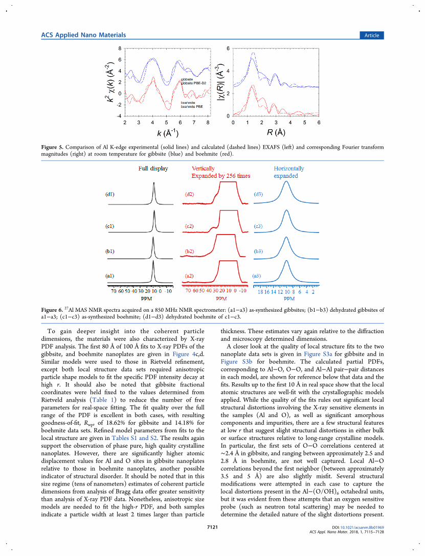
Figure 6. 27Al MAS NMR spectra acquired on a 850 MHz NMR spectrometer: (a1−a3) as-synthesized gibbsites; (b1−b3) dehydrated gibbsites ofa1−a3; (c1−c3) as-synthesized boehmite; (d1−d3) dehydrated boehmite of c1−c3.
ACS Applied Nano Materials Article
DOI: 10.1021/acsanm.8b01969ACS Appl. Nano Mater. 2018, 1, 7115−7128
7121
Neutron PDF studies would also allow determination of theH(D) atomic coordinates and H(D)-bearing local structure inthese samples. Such attempts are complicated by the need forsynthesizing deuterated gibbsite and boehmite nanocrystals butcould be pursued in the future.To gain a more localized perspective on the structures,
synchrotron X-ray absorption and scattering spectra werecollected. Interpretation of the experimental Al K-edge XANESspectra for these gibbsite and boehmite samples can be foundin our previous gibbsite20 and boehmite21 synthesis papers. AlK-edge EXAFS spectra of the gibbsite and boehmite samplesare shown in Figure 5 along with spectra calculated fromAIMD simulations, which provides a less ambiguous basis forEXAFS data interpretation than traditional shell-by-shellfitting. Good agreement was obtained between experimentaland calculated spectra (Figure 5) considering that only oneadjustable parameter was used for the calculated spectra (ΔE0,to adjust the position of the K-edge; see Methods section).Although slight differences in intensity can be seen in theFourier transforms between theory and experiment, thesimulations reproduced all the major peaks. The goodagreement indicates that the simulations reproduce well boththe structure and dynamics (i.e., thermal disorder) of the bulkmaterials. In addition, the good agreement indicates that thematerials used in the measurements were of good crystallinequality (i.e., negligible structural disorder), generally consistentwith the results of XRD and X-ray PDF analyses.Chemical Properties. Like XANES and EXAFS, NMR is
also a powerful tool for site-level structural characterization ofthese nanomaterials. 27Al MAS NMR was collected on thegibbsite and boehmite samples for its ability to quantifyproportions of Al coordination sites, including lowercoordinated Al sites that might exist at surfaces or grainboundaries, as well as assessing changes upon dehydration.Figure 6 summarizes the 27Al MAS NMR results obtained onthe gibbsite (a1−a3) and the boehmite (c1−c3) as well astheir dehydrated samples (b1−b3, and d1−d3), respectively.The use of high field of 850 MHz and moderate MAS spinningrate of 20 kHz allow the detection of tetrahedral (AlT) species(60−75 ppm) and pentahedral (AlP) species (∼25 to ∼45ppm) in addition to the expectedly abundant octahedral (AlO)species (with peak centered at about 10 ppm for gibbsites) (aand b series). A careful comparison reveals no changes on theAlO line shapes between the as-synthesized and the dehydratedgibbsites as evidenced in a3 and b3 in Figure 6 and Figure S4.The peak is asymmetric with a representative shoulder peak
centered at ∼11.8 ppm clearly observed, a typical feature forthe quadrupolar line shape associated with the octahedral siteof the gibbsites at high field of 850 MHz. After dehydration,the amount of low coordinated AlT and AlP sites is increased(Figure S4) likely due to the removal of the surface adsorbedH2O molecules that are weakly bound to the surface AlP andAlT sites. Dehydration allows more bare low coordinatedalumina sites to be detected, a result that is consistent with ourprior observations.47 The relative percentages of AlT and AlPfor gibbsites over the entire spectrum by peak integration areextremely low, i.e., 0.006% (AlT) and 0.2% (AlP) for the as-synthesized sample, 0.02% (AlT) and 0.5% (AlP) for thedehydrated gibbsites, indicating that the synthesized gibbsitesare very pure with generally negligible surface defects reflectedby AlP and AlT sites. It is also possible that this low density oflower coordinated Al sites is associated with minor bulk defectstied to the slight polycrystalline characteristic of the gibbsiteidentified by XRD and PDF above.In contrast to the gibbsite, there are no AlT sites detected by
MAS NMR on the boehmite samples even when it isdehydrated (Figure 6 and Figure S4). However, dehydrationintroduces observable line broadening on the AlO peak; the fullwidth at half-peak heights (FWHP) changes from 634 to 742Hz for the as-synthesized and the dehydrated samples,respectively. The peak for AlO is more symmetric comparedto the case of gibbsite, reflecting the increased AlO symmetry inboehmite. The amount of AlP is slightly increased (Figures S4-c2 and S4-d2) upon dehydration and the peak center for AlP isshifted upfield from about 36 to 31 ppm (Figure S4). Theexact reason for this upfield shift of AlP site is unknown butmay be due to the partial rearrangement of the alumina surfacedefect sites during the dehydration process. Like the case ofgibbsites, the relative amount of AlP is extremely small, i.e.,only about 0.2% for the as-synthesized and 0.4% for thedehydrated samples. This result clearly shows that thesynthesized boehmite sample is extremely pure with extremelysmall amount of defect AlP sites.FTIR was performed to complement the Al site-level
characteristics of these materials with spectral informationthat emphasizes the structural hydroxyl content. Figure S5adisplays the FTIR spectrum for gibbsite. In the hydroxylstretching region, three distinct peaks at 3468, 3528, and 3614cm−1 are present, along with a shoulder around 3395 cm−1.The low-frequency hydroxyl region shows a few peaks alongwith several shoulders. Other sources report more peaks inboth areas for synthetic gibbsite, though this is likely due to a
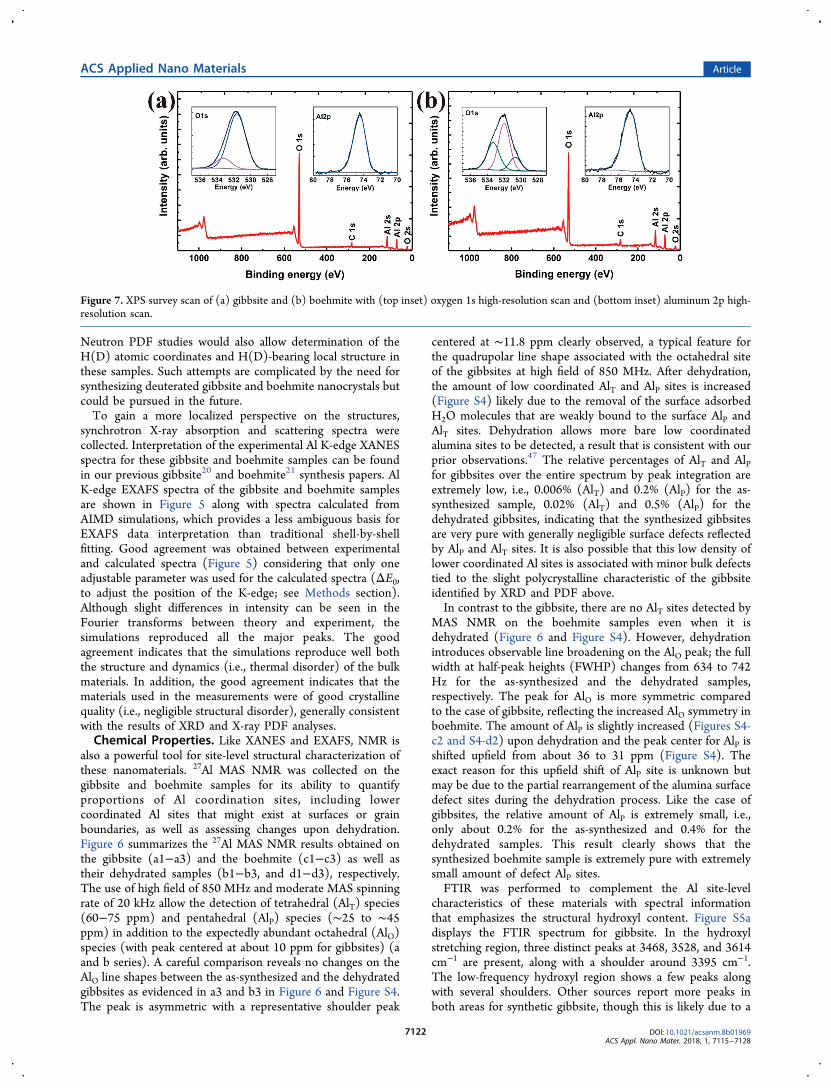
Figure 7. XPS survey scan of (a) gibbsite and (b) boehmite with (top inset) oxygen 1s high-resolution scan and (bottom inset) aluminum 2p high-resolution scan.
ACS Applied Nano Materials Article
DOI: 10.1021/acsanm.8b01969ACS Appl. Nano Mater. 2018, 1, 7115−7128
7122
difference in material size.63,64 In fact, the overall spectrum forgibbsite almost exactly matches that seen for a syntheticallyproduced gibbsite of a similar size.65
Figure S5b shows the FTIR spectrum for boehmite. In thehydroxyl stretching region, two distinct peaks at 3076 and3281 cm−1 are seen along with a small shoulder at 3525 cm−1.In the hydroxyl bending region, there are six distinct peakswith several shoulders. The shoulder at 3525 cm−1 is morepronounced in natural boehmite samples though lesspronounced in synthetic boehmite samples.66,67 A few extrapeaks in the hydroxyl bending region are present in naturalboehmite samples but do not seem to be present in syntheticsamples.63 The peak widths are what is expected for the size ofthe boehmite sample.XPS was used to specifically characterize the cleanliness and
composition of the nanomaterial surfaces. Survey XPS scans,with an information depth generally <5 nm, taken of bothgibbsite and boehmite revealed no detectable impurities otherthan the inevitable adventitious carbon, which was used tocharge correct the spectra. One can observe in the inset shownin Figure 7a the Al 2p peaks at 74.5 eV and the O 1s peaks at531.6 and 533.2 eV for gibbsite. The peak at 531.6 eV isattributed to the Al−O−H cluster found within the crystalstructure while the peak at 533.2 eV is attributed to adsorbedwater.68 The oxygen 1s peak for boehmite shows threecontributions: one at 530.7 eV, one at 532.0 eV, and one at533.3 eV (Figure 7b). The peak at 530.7 eV is attributed to theAl−O−Al structure and should be in a 1:1 ratio with the peakat 532.0 eV, which belongs to the structural hydroxyls.68 Theskewed ratio is likely due to an excess of adsorbed water. Thepeak at 533.3 eV is due to adsorbed water on the surface.
Thermal Stability. Because of the intrinsic importance ofthe water/hydroxyl content to the structural and chemicalquality of these materials, we performed detailed character-ization of temperature-dependent water loss and correspond-ing structural response as assessed by Raman spectroscopy.The mass loss curves and the heat flow curves are shown inFigure S6 for both gibbsite and boehmite. For gibbsite, ashallow endotherm corresponding to a mass loss of 0.8%appears at 100 °C and is likely due to the loss of adsorbedwater, an endotherm only found in synthetic gibbsite.66 Twomore endotherms occur at 265 and 310 °C and correspond tomass losses of 5.1% and 21.1%, respectively. The second ofthese endotherms can be attributed to the formation ofboehmite or a boehmite-like structure, which should give atheoretical mass loss of 23.1%.69 The final endotherm occurs at530 °C and corresponds to a mass loss of 34.6% that can beattributed to the dehydroxylation of the boehmite or aboehmite-like structure to alumina.63
The boehmite sample also shows an endotherm at 100 °Cwith a mass loss of 0.8% that can thus be attributed to the lossof adsorbed water. A shallow endotherm follows at 295 °Cwith a mass loss of 1.6% and is the start of the dehydroxylationof boehmite.66 The final endotherm occurs at 520 °C andcorresponds to a mass loss of 12.1%. Dehydroxylation toalumina is complete by 550 °C.In situ temperature-dependent Raman spectra are shown in
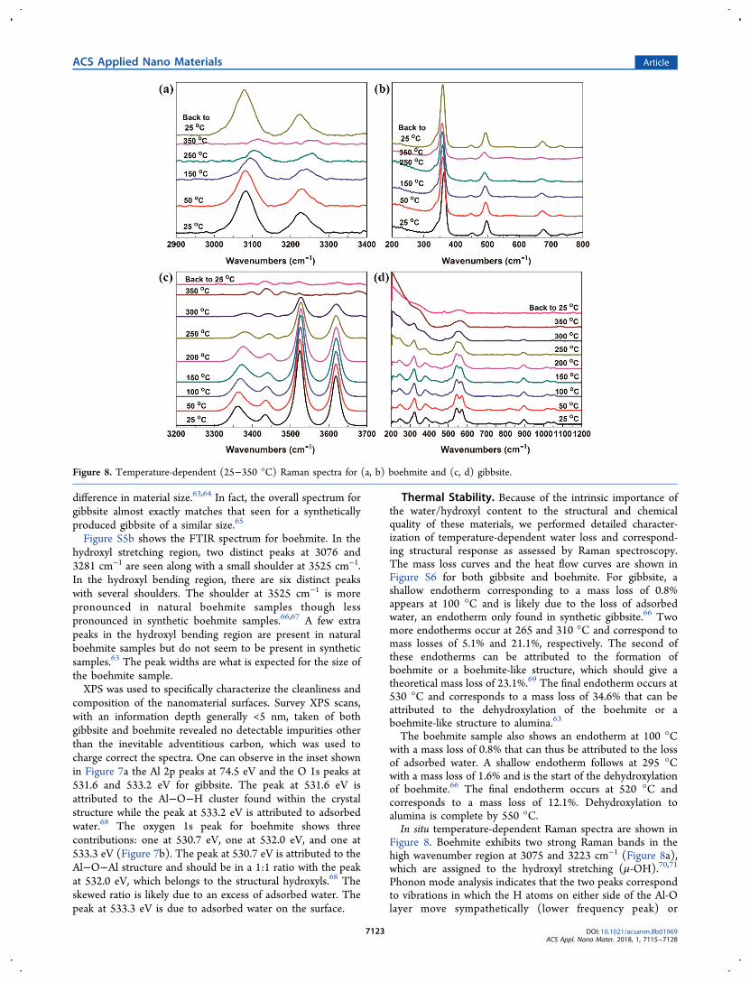
Figure 8. Boehmite exhibits two strong Raman bands in thehigh wavenumber region at 3075 and 3223 cm−1 (Figure 8a),which are assigned to the hydroxyl stretching (μ-OH).70,71
Phonon mode analysis indicates that the two peaks correspondto vibrations in which the H atoms on either side of the Al-Olayer move sympathetically (lower frequency peak) or
Figure 8. Temperature-dependent (25−350 °C) Raman spectra for (a, b) boehmite and (c, d) gibbsite.
ACS Applied Nano Materials Article
DOI: 10.1021/acsanm.8b01969ACS Appl. Nano Mater. 2018, 1, 7115−7128
7123

antisympathetically (higher frequency peak). Both bandsundergo a strong red-shift and intensity decreases withincreasing temperature. A measurement taken after coolingback to room temperature indicated these bands were notpermanently altered by being heated to 350 °C. However, thefwhm of the bond at 3075 cm−1 changed from 52 to 56 cm−1,possibly indicating slightly increased disorder in the interlayerhydrogen arrangement upon heating. There are two bands inthe hydroxyl translation region seen at 739 and 672 cm−1
(Figure 8b).70 The band at 672 cm−1 decreases in intensity andexperiences a red-shift as the temperature is increased whilethe band at 739 cm−1 is weak and becomes difficult to discernwith increasing temperatures. Both the location and intensityof these bands are preserved when the sample is cooled back toroom temperature. Three distinct peaks appear in the Al−Ostretching region at 495, 449, and 360 cm−1, and all of themshow a slight decrease in intensity and a red-shift upon heating.The Raman spectra indicated that no major permanentchanges occur for boehmite up to 350 °C. This result is notunexpected as the TGA reveals only a 2% mass loss at 350 °Cand no major endotherms prior to that temperature.Gibbsite contains six crystallographically distinct OH groups
each having C1 site symmetry (cf. the Supporting Informa-tion). Three of them (OH1, OH2, and OH4 in the Figure 1a)parallel to the (001) face to form intralayer hydrogen bonds,and another three (OH3, OH5, and OH6) form interlayerhydrogen bonds.72 In theory, six OH stretching peaks shouldbe detected in the Raman spectrum.72 However, there are onlyfour peaks at 3619, 3524, 3434, and 3362 cm−1 observed in the
μ-OH region of gibbsite Raman spectrum (Figure 8c,d). TheO···O distances between interlayer OH groups are shorter thanthe one between intralayer OH groups. Shorter O···Odistances lead to stronger hydrogen bonding, which inducelower OH stretching frequencies. Peaks at 3619 and 3524 cm−1
are composed of OH1 and OH2/OH4, respectively, and peaksat 3434 and 3362 cm−1 are composed of OH6 and OH3/OH5,respectively.70,72 All μ-OH bonds experience a red-shift and adecrease in intensity as a function of temperature. The fwhm ofthe bond at 3619 and 3524 cm−1 changed from 25 to 27 cm−1,then to 30 and 24 cm−1 to 25 cm−1, then to 27 cm−1,respectively, when the temperature changed from 25 to 200°C, and then to 300 °C. This behavior indicates the formationof disordered structures/defects. However, there were noobvious changes for the peaks at 3434 and 3362 cm−1, whichpointed out the interlayer hydrogen bonds are more stablethan the intralayer hydrogen bonds. When the temperatureincreased to 350 °C, both peaks at 3619 and 3524 cm−1
disappeared, which agree well with the TGA data and showedthe transformation of the gibbsite to χ-alumina.11 In thehydroxyl bending region, two peaks were observed at 1052 and1024 cm−1. There was no shift and the intensity of the peak at1052 cm−1 decreased steadily until it disappeared at 300 °C.The peak at 1024 cm−1 displayed a red-shift with increasingtemperature and disappeared by 300 °C. The hydroxyldeformation region shows several peaks occurring at 896,822, 714, 571, 545, and 509 cm−1. The bands at 896 and 822cm−1 experience no shift and slowly decrease in intensity untildisappearing at 350 °C. The peak at 714 cm−1 sees a slight red-
Figure 9. 1D patterns of SANS for (a) gibbsite and (c) boehmite. Grain size distribution for (b) gibbsite and (d) boehmite calculated using thetotal non-negative least-squares approach in IRENA from small and ultrasmall-angle neutron scattering data. Grain size distributions are shown asboth linear (left axis) and logarithmic (right axis) concentrations.
ACS Applied Nano Materials Article
DOI: 10.1021/acsanm.8b01969ACS Appl. Nano Mater. 2018, 1, 7115−7128
7124
shift and is gone by 250 °C. The bands at 571 and 545 cm−1
appear to merge together with increasing temperature. In theAl-O stretching region, all peaks were observed at 433, 396,381, 324, 284, 261, and 249 cm−1 virtually disappear by 350°C.The investigation using temperature-dependent Raman
spectroscopy showed there is no phase transformation ofboehmite when heating up to 350 °C, but there are disorderedstructures/defects formed in boehmite nanoparticles afterheating. There are disordered structures/defects formed ingibbsite nanoparticles when heating to 200 °C and the phasetransformation of gibbsite to χ-alumina happens when heatingto 350 °C period. There is no evidence support the formationof boehmite during the heating.Surface Area and Aggregation Behavior. Finally, we
extended our characterization study into understanding thephysical behavior of these nanomaterials in aggregate, whichrelates back to their bespoke individual particle-level character-istics. We first analyzed the dry unconsolidated material usingBET. Isotherms for nitrogen adsorption/desorption ongibbsite and boehmite are presented in Figures S7a and S7b,respectively. The isotherms do not show major deviationswhich is indicative of relatively smooth particles. The specificsurface area as obtained using a multipoint BET methodologywas determined to be 43.9 m2/g for gibbsite and 46.8 m2/g forboehmite. This may be compared to the calculated geometricsurface areas of around 51 m2/g and 150 m2/g, respectively,which are based on the measured size and thickness from theSEM, TEM, and AFM images. The fact that the measuredsurface areas are smaller than the calculated geometric valuesreflects minor (gibbsite) and substantial (boehmite) aggrega-tion in these dried powders. SEM (Figure 2a,d), TEM (Figure2b,e), and AFM (Figures S1 and S8) images show that theaggregation of gibbsite and boehmite nanoplates oftenoccurred in an aligned fashion along the basal surface normaldirection. In particular, the gibbsite nanoplates frequentlydisplayed this mutually oriented aggregation (Figure 2a),suggesting the possibility of relatively strong interparticle vander Waals attraction, forces that are considered primarycontributors to crystallographically oriented nanoparticleattachment.73
We performed SANS to investigate the grain size andaggregation behavior of the gibbsite and boehmite nanoplatesdispersed in aqueous suspension. SANS results indicate thatthe size distribution of the gibbsite is somewhat broader thanthat for boehmite. As shown in Figure 9a, several different sizedistributions were observed. These appear to be largelyseparate rather than representing a broad polydispersedistribution. As with the boehmite, there were relatively fewlarger aggregates formed in this sample, although theirconcentration is about an order of magnitude larger (Figure9b). Peaks for radii near 100 Å likely represent the radius ofthe boehmite plates, suggesting a fairly narrow sizedistribution.
■ CONCLUSIONSStructurally and chemically precise gibbsite and boehmitenanoplates have been synthesized with controlled sizes of∼280 and ∼35 nm, respectively, as part of a larger synthesiscampaign that is developing flexible and additive-free protocolsfor systematically tunable aluminum (oxyhydr)oxide nanoma-terials. Using a comprehensive integrated set of analyticalcapabilities, this study demonstrates that these protocols yield
materials that are essentially ideal for fundamental research onthe chemical and physical properties of gibbsite and boehmitefor diverse applications. Major conclusions can be summarizedas follows. First, the nanoscale morphology is preciselycontrolled over a narrow size and shape distribution, withparticle characteristics readily relatable back to their nominalatomic-scale crystal structures. Second, the materials arecompositionally pure, both in the bulk and at their surfaces,to the limits associated with air contact. Third, they containminimal to no detectable structural defects. Finally, theirthermal stabilities are consistent with expectations for the purebulk phases, including expected phase transitions, such thatthere appears to be no special nanoscale physics in play inthese particles at these dimensions. These materials cantherefore be viewed as homogeneous single-phase materials atthe individual particle level, on average. It can thus now beclaimed that chemically and structurally ideal gibbsite andboehmite nanoparticles of controlled uniform size and shapecan now be readily produced for synthesis of advanced aluminaproducts, using additive-free synthesis protocols that motivatedthe current comprehensive characterization study.
■ ASSOCIATED CONTENT*S Supporting InformationThe Supporting Information is available free of charge on theACS Publications website at DOI: 10.1021/acsanm.8b01969.
XRD patterns, NMR spectra, FTIR spectra, TGA curves,BET, and AFM images of gibbsite and boehmite samples(PDF)
■ AUTHOR INFORMATIONCorresponding Authors*E-mail: [email protected] (K.M.R.).*E-mail: [email protected] (X.Z.).ORCIDXin Zhang: 0000-0003-2000-858XCarolyn I. Pearce: 0000-0003-3098-1615Jian Zhi Hu: 0000-0001-8879-747XKatharine Page: 0000-0002-9071-3383Lawrence M. Anovitz: 0000-0002-2609-8750Sebastien Kerisit: 0000-0002-7470-9181Nicholas R. Jaegers: 0000-0002-9930-7672Trent R. Graham: 0000-0001-8907-8004Hsiu-Wen Wang: 0000-0002-2802-4122Jue Liu: 0000-0002-4453-910XGreg A. Kimmel: 0000-0003-4447-2440Kevin M. Rosso: 0000-0002-8474-7720NotesThe authors declare no competing financial interest.
■ ACKNOWLEDGMENTSThis work was supported by IDREAM (Interfacial Dynamicsin Radioactive Environments and Materials), an EnergyFrontier Research Center funded by the U.S. Department ofEnergy (DOE), Office of Science, Basic Energy Sciences(BES). Work performed at Argonne and use of the AdvancedPhoton Source were supported by the U.S. Department ofEnergy, Office of Science, Office of Basic Energy Sciences,under Contract DE-AC02-06CH11357. This research usedresources of the Advanced Light Source, which is a DOE
ACS Applied Nano Materials Article
DOI: 10.1021/acsanm.8b01969ACS Appl. Nano Mater. 2018, 1, 7115−7128
7125
Office of Science User Facility under Contract DE-AC02-05CH11231. The authors acknowledge the Center forSustainable Energy at Notre Dame (cSEND) MaterialsCharacterization Facilities for the use of the PHI VersaProbleII X-ray photoelectron spectrometer and the Mettler ToledoTGA/DSC-1. The authors thank Prof. Ian Carmichael formaking available the facilities of the Notre Dame RadiationLaboratory, which is supported by DOE BES through GrantDE-FC02-04ER15533. We acknowledge the support of theNational Institute of Standards and Technology, Center forNeutron Research, U.S. Department of Commerce inproviding the research neutron facilities used in this work.Access to NGB30-SANS and BT5-USANS was provided bythe Center for High Resolution Neutron Scattering, apartnership between the National Institute of Standards andTechnology and the National Science Foundation underAgreement DMR-1508249. A portion of this research wasperformed using EMSL, a national scientific user facilitysponsored by the DOE Office of Biological and EnvironmentalResearch and located at PNNL. PNNL is a multiprogramnational laboratory operated for DOE by Battelle MemorialInstitute under Contract DE-AC05-76RL0-1830.
■ REFERENCES(1) Granados-Correa, F.; Jimenez-Becerril, J. Chromium (VI)adsorption on boehmite. J. Hazard. Mater. 2009, 162, 1178−84.(2) Li, P.; Zheng, S.; Qing, P.; Chen, Y.; Tian, L.; Zheng, X.; Zhang,Y. The vanadate adsorption on a mesoporous boehmite and itscleaner production application of chromate. Green Chem. 2014, 16,4214−4222.(3) Zhang, H.; Li, P.; Wang, Z.; Zhang, X.; Zheng, S.; Zhang, Y. InSitu Synthesis of γ-AlOOH and Synchronous Adsorption Separationof V(V) from Highly Concentrated Cr(VI) Multiplex Complexsolutions. ACS Sustainable Chem. Eng. 2017, 5, 6674−6681.(4) Zhang, J.; Ji, Q.; Zhang, P.; Xia, Y.; Kong, Q. Thermal stabilityand flame-retardancy mechanism of poly(ethylene terephthalate)/boehmite nanocomposites. Polym. Degrad. Stab. 2010, 95, 1211−1218.(5) Esposito Corcione, C.; Manno, R.; Frigione, M. Sunlight-curableboehmite/siloxane-modified methacrylic based nanocomposites asinsulating coatings for stone substrates. Prog. Org. Coat. 2016, 95,107−119.(6) Liu, H.; Deng, J.; Li, W. Synthesis of Nickel NanoparticlesSupported on Boehmite for Selective Hydrogenation of p-Nitro-phenol and p-Chloronitrobenzene. Catal. Lett. 2010, 137, 261−266.(7) Mirzaee, M.; Bahramian, B.; Gholizadeh, J.; Feizi, A.; Gholami,R. Acetylacetonate complexes of vanadium and molybdenumsupported on functionalized boehmite nano-particles for the catalyticepoxidation of alkenes. Chem. Eng. J. 2017, 308, 160−168.(8) Oliveira, P. N.; Catarino, M.; Muller, C. M. O.; Brandao, L.;Pacheco Tanaka, D. A.; Bertolino, J. R.; Mendes, A. M.; Pires, A. T. N.Preparation and characterization of crosslinked PVAL membranesloaded with boehmite nanoparticles for fuel cell applications. J. Appl.Polym. Sci. 2014, 131, 40148.(9) Liu, S.; Chen, C.; Liu, Q.; Zhuo, Y.; Yuan, D.; Dai, Z.; Bao, J.Two-dimensional porous γ-AlOOH and γ-Al2O3 nanosheets: hydro-thermal synthesis, formation mechanism and catalytic performance.RSC Adv. 2015, 5, 71728−71734.(10) Zuo, Z.; Huang, W.; Han, P.; Gao, Z.; Li, Z. Theoretical studieson the reaction mechanisms of AlOOH- and γ-Al2O3-catalysedmethanol dehydration in the gas and liquid phases. Appl. Catal., A2011, 408, 130−136.(11) Louaer, S.; Wang, Y.; Guo, L. Fast synthesis and size control ofgibbsite nanoplatelets, their pseudomorphic dehydroxylation, andefficient dye adsorption. ACS Appl. Mater. Interfaces 2013, 5, 9648−9655.
(12) Trueba, M.; Trasatti, S. P. γ-Alumina as a Support for Catalysts:A Review of Fundamental Aspects. Eur. J. Inorg. Chem. 2005, 2005,3393−3403.(13) Liu, Q.; Wang, A.; Wang, X.; Gao, P.; Wang, X.; Zhang, T.Synthesis, characterization and catalytic applications of mesoporous γ-alumina from boehmite sol.Microporous Mesoporous Mater. 2008, 111,323−333.(14) Shi, Y.; HogenEsch, H.; Regnier, F. E.; Hem, S. L.Detoxification of endotoxin by aluminum hydroxide adjuvant. Vaccine2001, 19, 1747−1752.(15) Ulanova, M.; Tarkowski, A.; Hahn-Zoric, M.; Hanson, L. A.The Common vaccine adjuvant aluminum hydroxide up-regulatesaccessory properties of human monocytes via an interleukin-4-dependent mechanism. Infect. Immun. 2001, 69, 1151−1159.(16) Bai, X.; Caputo, G.; Hao, Z.; Freitas, V. T.; Zhang, J.; Longo, R.L.; Malta, O. L.; Ferreira, R. A.; Pinna, N. Efficient and tuneablephotoluminescent boehmite hybrid nanoplates lacking metal activatorcentres for single-phase white LEDs. Nat. Commun. 2014, 5, 5702.(17) Cai, W.; Yu, J.; Gu, S.; Jaroniec, M. Facile HydrothermalSynthesis of Hierarchical Boehmite: Sulfate-Mediated Transformationfrom Nanoflakes to Hollow Microspheres. Cryst. Growth Des. 2010,10, 3977−3982.(18) Fujii, T.; Kawasaki, S.-i.; Suzuki, A.; Adschiri, T. High-SpeedMorphology Control of Boehmite Nanoparticles by SupercriticalHydrothermal Treatment with Carboxylic Acids. Cryst. Growth Des.2016, 16, 1996−2001.(19) Liu, Y.; Ma, D.; Blackley, R. A.; Zhou, W.; Han, X.; Bao, X.Synthesis and characterization of gibbsite nanostructures. J. Phys.Chem. C 2008, 112, 4124−4128.(20) Zhang, X.; Zhang, X.; Graham, T. R.; Pearce, C. I.; Mehdi, B.L.; N’Diaye, A. T.; Kerisit, S. N.; Browning, N. D.; Clark, S. B.; Rosso,K. M. Fast Synthesis of Gibbsite Nanoplates and ProcessOptimization using Box-Behnken Experimental Design. Cryst. GrowthDes. 2017, 17, 6801−6808.(21) Zhang, X.; Cui, W.; Page, K. L.; Pearce, C. I.; Bowden, M. E.;Graham, T. R.; Shen, Z.; Li, P.; Wang, Z.; Kerisit, S. N.; N’Diaye, A.T.; Clark, S. B.; Rosso, K. M. Size and Morphology ControlledSynthesis of Boehmite Nanoplates and Crystal Growth Mechanisms.Cryst. Growth Des. 2018, 18, 3596−3606.(22) Chupas, P. J.; Qiu, X. Y.; Hanson, J. C.; Lee, P. L.; Grey, C. P.;Billinge, S. J. L. Rapid-acquisition pair distribution function (RA-PDF) analysis. J. Appl. Crystallogr. 2003, 36, 1342−1347.(23) Hammersley, A.; Svensson, S.; Hanfland, M.; Fitch, A.;Hausermann, D. Two-dimensional detector software: from realdetector to idealised image or two-theta scan. High Pressure Res.1996, 14, 235−248.(24) Qiu, X.; Thompson, J. W.; Billinge, S. J. L. PDFgetX2: a GUI-driven program to obtain the pair distribution function from X-raypowder diffraction data. J. Appl. Crystallogr. 2004, 37, 678−678.(25) Coelho, A. A. TOPAS and TOPAS-Academic: an optimizationprogram integrating computer algebra and crystallographic objectswritten in C++. J. Appl. Crystallogr. 2018, 51, 210−218.(26) Coelho, A. A.; Chater, P. A.; Kern, A. Fast synthesis andrefinement of the atomic pair distribution function. J. Appl. Crystallogr.2015, 48, 869−875.(27) Farrow, C. L.; Juhas, P.; Liu, J. W.; Bryndin, D.; Bozin, E. S.;Bloch, J.; Proffen, T.; Billinge, S. J. PDFfit2 and PDFgui: computerprograms for studying nanostructure in crystals. J. Phys.: Condens.Matter 2007, 19, 335219.(28) Chung, J. S.; Thorpe, M. Local atomic structure ofsemiconductor alloys using pair distribution functions. Phys. Rev. B:Condens. Matter Mater. Phys. 1997, 55, 1545−1553.(29) Farrow, C. L.; Billinge, S. J. Relationship between the atomicpair distribution function and small-angle scattering: implications formodeling of nanoparticles. Acta Crystallogr., Sect. A: Found. Crystallogr.2009, 65, 232−239.(30) Olds, D.; Wang, H.-W.; Page, K. DShaper: an approach forhandling missing low-Qdata in pair distribution function analysis ofnanostructured systems. J. Appl. Crystallogr. 2015, 48, 1651−1659.
ACS Applied Nano Materials Article
DOI: 10.1021/acsanm.8b01969ACS Appl. Nano Mater. 2018, 1, 7115−7128
7126
(31) Liu, J.; Olds, D.; Peng, R.; Yu, L.; Foo, G. S.; Qian, S.; Keum, J.;Guiton, B. S.; Wu, Z.; Page, K. Quantitative Analysis of theMorphology of {101} and {001} Faceted Anatase TiO2 Nanocrystalsand Its Implication on Photocatalytic Activity. Chem. Mater. 2017, 29,5591−5604.(32) Ildefonse, P.; Cabaret, D.; Sainctavit, P.; Calas, G.; Flank, A.M.; Lagarde, P. Aluminum x-ray absorption near edge structure inmodel compounds and Earth’s surface minerals. Phys. Chem. Miner.1998, 25, 112−121.(33) Ravel, B.; Newville, M. ATHENA, ARTEMIS, HEPHAESTUS:data analysis for X-ray absorption spectroscopy using IFEFFIT. J.Synchrotron Radiat. 2005, 12, 537−541.(34) Valiev, M.; Bylaska, E. J.; Govind, N.; Straatsma, T. P.; VanDam, H. J. J.; Wang, D.; Nieplocha, J.; Apra, E.; Windus, T. L.; deJong, W. A. NWChem: A comprehensive and scalable open-sourcesolution for large scale molecular simulations. Comput. Phys. Commun.2010, 181, 1477−1489.(35) Perdew, J. P.; Burke, K.; Ernzerhof, M. Generalized gradientapproximation made simple. Phys. Rev. Lett. 1996, 77, 3865−3868.(36) Perdew, J. P.; Burke, K.; Ernzerhof, M. Generalized gradientapproximation made simple. Phys. Rev. Lett. 1997, 78, 1396.(37) Grimme, S. Semiempirical GGA-type density functionalconstructed with a long-range dispersion correction. J. Comput.Chem. 2006, 27, 1787−1799.(38) Hamann, D. R. Generalized norm-conserving pseudopotentials.Phys. Rev. B: Condens. Matter Mater. Phys. 1989, 40, 2980−2987.(39) Kleinman, L.; Bylander, D. M. Efficacious form for modelpseudopotentials. Phys. Rev. Lett. 1982, 48, 1425−1428.(40) Blochl, P. E.; Parrinello, M. Adiabaticity in first-principlesmolecular dynamics. Phys. Rev. B: Condens. Matter Mater. Phys. 1992,45, 9413−9416.(41) Car, R.; Parrinello, M. Unified approach for moleculardynamics and density-functional theory. Phys. Rev. Lett. 1985, 55,2471−2474.(42) Laasonen, K.; Sprik, M.; Parrinello, M.; Car, R. “Ab initio”liquid water. J. Chem. Phys. 1993, 99, 9080−9089.(43) Rehr, J. J.; Albers, R. C. Theoretical approaches to x-rayabsorption fine structure. Rev. Mod. Phys. 2000, 72, 621.(44) Rehr, J. J.; Kas, J. J.; Prange, M. P.; Sorini, A. P.; Takimoto, Y.;Vila, F. D. Ab initio theory and calculations of X-ray spectra. C. R.Phys. 2009, 10, 548−559.(45) Rehr, J. J.; Kas, J. J.; Vila, F. D.; Prange, M. P.; Jorissen, K.Parameter-free calculations of x-ray spectra with FEFF9. Phys. Chem.Chem. Phys. 2010, 12, 5503−5513.(46) Newville, M. IFEFFIT: Interactive XAFS analysis and FEFFfitting. J. Synchrotron Radiat. 2001, 8, 322−324.(47) Hu, J. Z.; Zhang, X.; Jaegers, N. R.; Wan, C.; Graham, T. R.;Hu, M.; Pearce, C. I.; Felmy, A. R.; Clark, S. B.; Rosso, K. M.Transitions in Al Coordination during Gibbsite Crystallization UsingHigh-Field 27Al and 23Na MAS NMR Spectroscopy. J. Phys. Chem. C2017, 121, 27555−27562.(48) Anovitz, L. M.; Lynn, G. W.; Cole, D. R.; Rother, G.; Allard, L.F.; Hamilton, W. A.; Porcar, L.; Kim, M.-H. A new approach toquantification of metamorphism using ultra-small and small angleneutron scattering. Geochim. Cosmochim. Acta 2009, 73, 7303−7324.(49) Anovitz, L. M.; Freiburg, J. T.; Wasbrough, M.; Mildner, D. F.R.; Littrell, K. C.; Pipich, V.; Ilavsky, J. The effects of burial diagenesison multiscale porosity in the St. Peter Sandstone: An imaging, small-angle, and ultra-small-angle neutron scattering analysis. Mar. Pet. Geol.2018, 92, 352−371.(50) Barker, J. G.; Glinka, C. J.; Moyer, J. J.; Kim, M. H.; Drews, A.R.; Agamalian, M. Design and performance of a thermal-neutrondouble-crystal diffractometer for USANS at NIST. J. Appl. Crystallogr.2005, 38, 1004−1011.(51) Glinka, C. J.; Barker, J. G.; Hammouda, B.; Krueger, S.; Moyer,J. J.; Orts, W. J. The 30 m Small-Angle Neutron ScatteringInstruments at the National Institute of Standards and Technology.J. Appl. Crystallogr. 1998, 31, 430−445.
(52) Mildner, D. F. R.; Hammouda, B.; Kline, S. R. A refractivefocusing lens system for small-angle neutron scattering. J. Appl.Crystallogr. 2005, 38, 979−987.(53) Kline, S. R. Reduction and analysis of SANS and USANS datausing IGOR Pro. J. Appl. Crystallogr. 2006, 39, 895−900.(54) Olsson, A.; Hellsing, M. S.; Rennie, A. R. A holder to rotatesample cells to avoid sedimentation in small-angle neutron scatteringand ultra small-angle neutron scattering experiments. Meas. Sci.Technol. 2013, 24, 105901.(55) Leao, J. B.; Murphy, R. P.; Wagner, N. J.; Bleuel, M. Dynamicinfrared sample controlled (DISCO) temperature for the tumbler cellsfor ultra small angle neutron scattering (USANS). J. Neutron Res.2017, 19, 23−26.(56) Ilavsky, J.; Jemian, P. R. Irena: tool suite for modeling andanalysis of small-angle scattering. J. Appl. Crystallogr. 2009, 42, 347−353.(57) Anovitz, L. M.; Cole, D. R. Characterization and Analysis ofPorosity and Pore Structures. Rev. Mineral. Geochem. 2015, 80, 61−164.(58) Ectors, D.; Goetz-Neunhoeffer, F.; Neubauer, J. A generalizedgeometric approach to anisotropic peak broadening due to domainmorphology. J. Appl. Crystallogr. 2015, 48, 189−194.(59) Dinnebier, R. E.; Von Dreele, R.; Stephens, P. W.; Jelonek, S.;Sieler, J. Structure of sodium para-hydroxybenzoate, NaO2C−C6H4OH by powder diffraction-application of a phenomenologicalmodel of anisotropic peak width. J. Appl. Crystallogr. 1999, 32, 761−769.(60) Megaw, H. The crystal structure of Hydrargillite Al(OH)3. Z.Kristallogr. - Cryst. Mater. 1934, 87, 185−204.(61) Bokhimi, X.; Toledo-Antonio, J. A.; Guzman-Castillo, M. L.;Hernandez-Beltran, F. Relationship between Crystallite Size and BondLengths in Boehmite. J. Solid State Chem. 2001, 159, 32−40.(62) Stephens, P. W. Phenomenological model of anisotropic peakbroadening in powder diffraction. J. Appl. Crystallogr. 1999, 32, 281−289.(63) Kloprogge, J. T.; Ruan, H. D.; Frost, R. L. Thermaldecomposition of bauxite minerals: infrared emission spectroscopyof gibbsite, boehmite and diaspore. J. Mater. Sci. 2002, 37, 1121−1129.(64) Frost, R. L.; Kloprogge, J. T.; Russell, S. C.; Szetu, J. L.Vibrational Spectroscopy and Dehydroxylation of Aluminum (Oxo)-hydroxides: Gibbsite. Appl. Spectrosc. 1999, 53, 423−434.(65) Phambu, N.; Humbert, B.; Burneau, A. Relation between theInfrared Spectra and the Lateral Specific Surface Areas of GibbsiteSamples. Langmuir 2000, 16, 6200−6207.(66) Frost, R. L.; Kloprogge, J. T.; Russell, S. C.; Szetu, J.Dehydroxylation of Aluminum (Oxo)hydroxides Using InfraredEmission Spectroscopy. Part II: Boehmite. Appl. Spectrosc. 1999, 53,572−582.(67) Tettenhorst, R.; Hofmann, D. A. Crystal chemistry ofboehmite. Clays Clay Miner. 1980, 28, 373−380.(68) Kloprogge, J. T.; Duong, L. V.; Wood, B. J.; Frost, R. L. XPSstudy of the major minerals in bauxite: Gibbsite, bayerite and(pseudo-)boehmite. J. Colloid Interface Sci. 2006, 296, 572−576.(69) Denigres Filho, R. W. N.; Rocha, G. d. A.; Montes, C. R.;Vieira-Coelho, A. C. Synthesis and Characterization of BoehmitesObtained from Gibbsite in Presence of Different Environments.Mater. Res. 2016, 19, 659−668.(70) Ruan, H. D.; Frost, R. L.; Kloprogge, J. T. Comparison ofRaman spectra in characterizing gibbsite, bayerite, diaspore andboehmite. J. Raman Spectrosc. 2001, 32, 745−750.(71) Noel, Y.; Demichelis, R.; Pascale, F.; Ugliengo, P.; Orlando, R.;Dovesi, R. Ab initio quantum mechanical study of γ-AlOOHboehmite: structure and vibrational spectrum. Phys. Chem. Miner.2009, 36, 47−59.(72) Wang, S.; Johnston, C. T. Assignment of the structural OHstretching bands of gibbsite. Am. Mineral. 2000, 85, 739−744.(73) Zhang, X.; He, Y.; Sushko, M. L.; Liu, J.; Luo, L.; De Yoreo, J.J.; Mao, S. X.; Wang, C.; Rosso, K. M. Direction-specific van der
ACS Applied Nano Materials Article
DOI: 10.1021/acsanm.8b01969ACS Appl. Nano Mater. 2018, 1, 7115−7128
7127

Waals attraction between rutile TiO2 nanocrystals. Science 2017, 356,434−437.
ACS Applied Nano Materials Article
DOI: 10.1021/acsanm.8b01969ACS Appl. Nano Mater. 2018, 1, 7115−7128
7128
![Precipitation of spherical boehmite from concentrated ...nation step in metallurgical alumina production [23]. The precipitation of gibbsite or boehmite does not generate waste water](https://static.fdocuments.in/doc/165x107/5eb48fae5c1eda1ed720f825/precipitation-of-spherical-boehmite-from-concentrated-nation-step-in-metallurgical.jpg)