BMC Structural Biology BioMed Central - Brussels - Belgium
18
BioMed Central Page 1 of 18 (page number not for citation purposes) BMC Structural Biology Open Access Research article Molecular models of human P-glycoprotein in two different catalytic states Jean-Paul Becker 1 , Grégoire Depret 1 , Françoise Van Bambeke 2 , Paul M Tulkens 2 and Martine Prévost* 1 Address: 1 Structure et Fonction des Membranes Biologiques, Université Libre de Bruxelles, Boulevard du Triomphe CP 206/2, B-1050 Brussels, Belgium and 2 Unité de Pharmacologie cellulaire et moléculaire, Université catholique de Louvain, Avenue E. Mounier 73, B-1200 Brussels, Belgium Email: Jean-Paul Becker - [email protected]; Grégoire Depret - [email protected]; Françoise Van Bambeke - [email protected]; Paul M Tulkens - [email protected]; Martine Prévost* - [email protected] * Corresponding author Abstract Background: P-glycoprotein belongs to the family of ATP-binding cassette proteins which hydrolyze ATP to catalyse the translocation of their substrates through membranes. This protein extrudes a large range of components out of cells, especially therapeutic agents causing a phenomenon known as multidrug resistance. Because of its clinical interest, its activity and transport function have been largely characterized by various biochemical studies. In the absence of a high-resolution structure of P-glycoprotein, homology modeling is a useful tool to help interpretation of experimental data and potentially guide experimental studies. Results: We present here three-dimensional models of two different catalytic states of P- glycoprotein that were developed based on the crystal structures of two bacterial multidrug transporters. Our models are supported by a large body of biochemical data. Measured inter- residue distances correlate well with distances derived from cross-linking data. The nucleotide-free model features a large cavity detected in the protein core into which ligands of different size were successfully docked. The locations of docked ligands compare favorably with those suggested by drug binding site mutants. Conclusion: Our models can interpret the effects of several mutants in the nucleotide-binding domains (NBDs), within the transmembrane domains (TMDs) or at the NBD:TMD interface. The docking results suggest that the protein has multiple binding sites in agreement with experimental evidence. The nucleotide-bound models are exploited to propose different pathways of signal transmission upon ATP binding/hydrolysis which could lead to the elaboration of conformational changes needed for substrate translocation. We identified a cluster of aromatic residues located at the interface between the NBD and the TMD in opposite halves of the molecule which may contribute to this signal transmission. Our models may characterize different steps in the catalytic cycle and may be important tools to understand the structure-function relationship of P-glycoprotein. Published: 22 January 2009 BMC Structural Biology 2009, 9:3 doi:10.1186/1472-6807-9-3 Received: 31 July 2008 Accepted: 22 January 2009 This article is available from: http://www.biomedcentral.com/1472-6807/9/3 © 2009 Becker et al; licensee BioMed Central Ltd. This is an Open Access article distributed under the terms of the Creative Commons Attribution License (http://creativecommons.org/licenses/by/2.0 ), which permits unrestricted use, distribution, and reproduction in any medium, provided the original work is properly cited.
Transcript of BMC Structural Biology BioMed Central - Brussels - Belgium
BioMed CentralBMC Structural Biology
ss
Open AcceResearch articleMolecular models of human P-glycoprotein in two different catalytic statesJean-Paul Becker1, Grégoire Depret1, Françoise Van Bambeke2, Paul M Tulkens2 and Martine Prévost*1Address: 1Structure et Fonction des Membranes Biologiques, Université Libre de Bruxelles, Boulevard du Triomphe CP 206/2, B-1050 Brussels, Belgium and 2Unité de Pharmacologie cellulaire et moléculaire, Université catholique de Louvain, Avenue E. Mounier 73, B-1200 Brussels, Belgium
Email: Jean-Paul Becker - [email protected]; Grégoire Depret - [email protected]; Françoise Van Bambeke - [email protected]; Paul M Tulkens - [email protected]; Martine Prévost* - [email protected]
* Corresponding author
AbstractBackground: P-glycoprotein belongs to the family of ATP-binding cassette proteins whichhydrolyze ATP to catalyse the translocation of their substrates through membranes. This proteinextrudes a large range of components out of cells, especially therapeutic agents causing aphenomenon known as multidrug resistance. Because of its clinical interest, its activity andtransport function have been largely characterized by various biochemical studies. In the absenceof a high-resolution structure of P-glycoprotein, homology modeling is a useful tool to helpinterpretation of experimental data and potentially guide experimental studies.
Results: We present here three-dimensional models of two different catalytic states of P-glycoprotein that were developed based on the crystal structures of two bacterial multidrugtransporters. Our models are supported by a large body of biochemical data. Measured inter-residue distances correlate well with distances derived from cross-linking data. The nucleotide-freemodel features a large cavity detected in the protein core into which ligands of different size weresuccessfully docked. The locations of docked ligands compare favorably with those suggested bydrug binding site mutants.
Conclusion: Our models can interpret the effects of several mutants in the nucleotide-bindingdomains (NBDs), within the transmembrane domains (TMDs) or at the NBD:TMD interface. Thedocking results suggest that the protein has multiple binding sites in agreement with experimentalevidence. The nucleotide-bound models are exploited to propose different pathways of signaltransmission upon ATP binding/hydrolysis which could lead to the elaboration of conformationalchanges needed for substrate translocation. We identified a cluster of aromatic residues located atthe interface between the NBD and the TMD in opposite halves of the molecule which maycontribute to this signal transmission.
Our models may characterize different steps in the catalytic cycle and may be important tools to understand the structure-function relationship of P-glycoprotein.
Published: 22 January 2009
BMC Structural Biology 2009, 9:3 doi:10.1186/1472-6807-9-3
Received: 31 July 2008Accepted: 22 January 2009
This article is available from: http://www.biomedcentral.com/1472-6807/9/3
© 2009 Becker et al; licensee BioMed Central Ltd. This is an Open Access article distributed under the terms of the Creative Commons Attribution License (http://creativecommons.org/licenses/by/2.0), which permits unrestricted use, distribution, and reproduction in any medium, provided the original work is properly cited.
Page 1 of 18(page number not for citation purposes)
BMC Structural Biology 2009, 9:3 http://www.biomedcentral.com/1472-6807/9/3
BackgroundATP-binding cassette (ABC) proteins form a large proteinfamily in living organisms that carry various substratesacross cell membranes. There are 48 ABC transporters inhumans and mutations in many have been linked togenetic disorders[1,2].
The P-glycoprotein (P-gp), product of the mdr1 gene inhumans, is one of these ABC transporters that extrudes alarge range of structurally diverse compounds out of cells,a feature that has been described as poly-specificity[3,4].It is the most extensively studied ABC transporter and canbe used to understand the function of this important classof proteins. The overexpression of P-gp is associated witha multidrug resistance phenotype in various forms of can-cer[5], which is a major barrier to the successful treatmentof these diseases.
The vast majority of ABC transporters require energy inthe form of ATP to translocate their ligands across the cellmembrane. They require a minimum of four domains:two transmembrane domains (TMDs) form the ligandbinding site and two nucleotide-binding domains(NBDs) bind and hydrolyze ATP to proceed to the sub-strate translocation. Two subtypes of ABC transporters canbe defined on the basis of the direction of the transportreaction: ABC importers only present in prokaryotes andABC exporters. Like many eukaryotic ABC transporters P-gp comprises all four domains in a single polypeptidechain. It is composed of 1280 residues, organized as twohomologous halves (which are 43% identical in humanP-gp) each with 6 transmembrane (TM) segments and acytosolic NBD. Several helices of the TM domains havebeen proposed to accommodate the binding site(s) andconsequently are believed to form the pathway throughwhich the substrates cross the membrane. On the otherhand the NBDs couple the energy associated with ATPbinding and hydrolysis to drug transport[6]. The NBDsare better conserved than the TMDs and share severalcommon motifs including the Walker A and B motifs thatare found in other ATPases and the ABC signature that isunique to the family.
The P-gp mediated-transport mechanism has been exten-sively studied but remains controversial. Briefly it isbelieved[4,7] that the transport cycle is initiated by sub-strate binding in the TM domains of P-gp, which increasesthe ATP affinity for the protein. After binding and/orhydrolysis of a first ATP molecule in one of the NBDs sev-eral restructurings occur in the TM domains of the protein.This conformational change allows the release of the drugto the extracellular medium. P-gp returns to its originalconfiguration possibly after hydrolysis of a second ATPmolecule. The protein is then reset for another cycle. Twoalternative models for the transport cycle of P-gp[7] are
currently proposed and the most significant difference isin the nature of the mechanism that drives the drug froma high affinity site to a low affinity site in the TMDs. In onemodel[4] the formation of a closed NBD dimer provokesconformational changes that are transmitted to the drugbinding site. Two sequential ATP hydrolysis events resetthe P-gp molecule. Other models[8] require either one[9]or two[8] hydrolysis reactions to supply the efflux of thedrug and the resetting of the protein for another cycle.
The considerable body of biochemical data obtained onABC transporters has been more recently complementedby X-ray structures. At present several complete structuresare available. All are of bacterial origin: five are ABCimporters (BtuCD, ModBC-A, HI1470/1, MalFG/K,MetNI) [10-16] and two are drug exporters (SAV1866 andMsbA) [17-19]. In ABC importers the four domains existas separate units whereas bacterial exporters are half trans-porters with one TMD and one NBD per polypeptidewhich function as a homo or hetero-dimer. These struc-tures as well as genomic analysis and TM predictions[20]suggest that the domain organization of the importers isnot shared by that of the exporters and that the TMDtopology is also different.
Together biochemical data and structure determinationcan help in understanding the mechanism of the ABCexporters. However crystallization of membrane proteinsto obtain direct information on three-dimensional (3D)structure is still a difficult task and no atomic-level struc-ture of P-gp is available so far. Only structural informationhas been obtained at low resolution by electron micros-copy (EM)[21,22]. In the absence of a high resolutionstructure for P-gp use of molecular modeling has been rec-ommended in order to generate 3D models to sustaininterpretation of experimental data[23] and potentiallyguide experimental studies. Several 3D models of thenucleotide-free protein were first published and most ofthem were either completely or remotely based onretracted former MsbA crystal structures[24]. Only twostudies clearly stated that models constructed exclusivelyon MsbA dimeric structures were incompatible with cross-linking data on P-gp[25,26]. To unravel this problem, inone of these studies, the helices in the TMDs initiallymodeled from MsbA monomer structure were largelyreshuffled using additional restrained potentials, definedfrom cross linking data, in molecular dynamics simula-tions[26]. However the construction process of this modellacked experimental data to properly describe theNBD:TMD interfaces and the relative position of the TMhelices in each P-gp half.
The publication of the structures of SAV1866[17,18],which has a topology akin to that of P-gp prompted new3D models of the nucleotide-bound conformation of P-gp
Page 2 of 18(page number not for citation purposes)
BMC Structural Biology 2009, 9:3 http://www.biomedcentral.com/1472-6807/9/3
to be constructed [27-29]. These structures have beendetermined in presence of ADP and AMP-PNP. They arevery similar and feature a drug extrusion chamber open tothe extracellular side. New crystallographic studies of ABCexporters trapped in different conformational states areneeded to reveal changes along the catalytic cycle, f.i.upon substrate translocation and ATP hydrolysis. RevisedMsbA structures trapped in different conformations havebeen very recently made available: two nucleotide-boundstructures and two in absence of nucleotide[19]. The twonucleotide-bound structures possess an outward-facingconformation very similar to SAV1866 structures. Thenucleotide-free structures determined at a quite low reso-lution feature two strongly different inward (intracellu-lar)-facing conformations.
We present here four 3D models of P-gp describing twodifferent states along the catalytic cycle using the recent X-ray structures of bacterial ABC exporters as templates. Themodels elaborated by comparative modeling are con-fronted to a large number of available experimental data.Interactions between conserved residues describing path-ways from the ATP binding site towards the TMDs areexamined to propose potential ways of transmitting a sig-nal upon ATP binding or hydrolysis. Likewise conservedresidues are pinpointed in the regions which could possi-bly be involved in the hinge binding motion suggested bythe different crystallographic structures allowing the tran-sition between the different conformational states. Theinteraction of compounds with P-gp is clearly a complexprocess. One of the nucleotide-free 3D models is used todock several ligands in the central cavity harbored by theTM domains. The geometry of interaction of the ligands isexamined and compared with the experimental data.
MethodsSelection of templatesA PSI-BLAST search[30] identified two bacterial ABCexporters homologous to human P-gp. One is SAV1866from S. aureus whose structures were solved in complex toADP[17] (PDB code 2HYD) and to AMP-PNP[18] (PDBcode 2ONJ). The other is MsbA, a lipid flippase, which isavailable in two nucleotide-bound conformations (MsbAfrom S. typhimurium; PDB codes 3B60 and 3B5Z) and intwo nucleotide-free conformations (MsbA from E. coli orV. cholerae; PDB codes 3B5W and 3B5X)[19]. The nucle-otide-free structures include only the Cα positions.
Sequence alignmentThe sequences of human P-gp, S. aureus SAV1866, V. chol-erae MsbA, S. typhimurium MsbA and E. coli MsbA can beaccessed at the Universal Protein Ressource (UniProt).The percentage of sequence identity between P-gp andSAV1866 or MsbA ranges from 27% to 32% (sequence
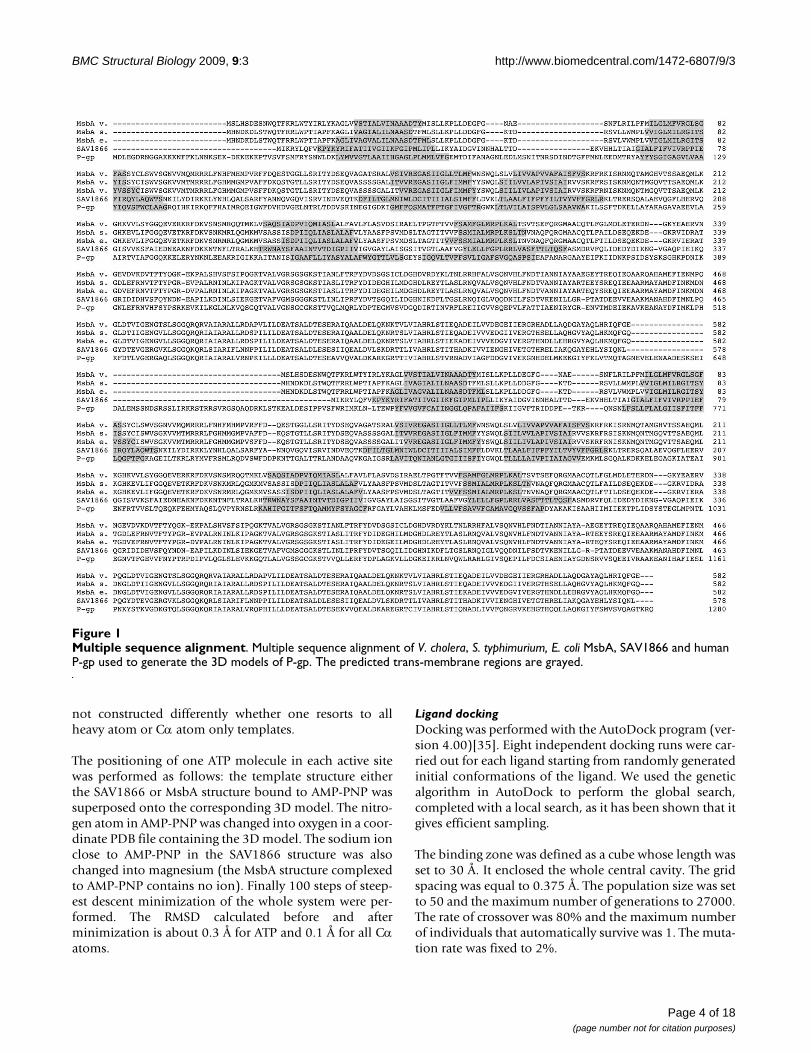
similarity is between 47% and 53%). Such percentages ofidentity make these templates potential candidates toelaborate 3D models of P-gp by comparative modeling.The similarity however varies noticeably for the cytosolicand transmembrane domains. The NBDs have sequenceidentity of about 50% and the TMDs have sequence iden-tity ranging between 15% and 23% (sequence similaritybetween 30% and 43%). A multiple-sequence alignmentof these protein sequences was performed using Clus-talW[31] (see Figure 1).
Trans-membrane domain predictionTrans-membrane domain predictions were performed onthe P-gp sequence using HMMTOP[32]. The results ofthese predictions which are reported on the ClustalWalignment (Figure 1) show a good correspondence withthe trans-membrane segments of SAV1866 and MsbAdetermined with PDBTM[33].
Model buildingThe multiple-sequence alignment was used as a basis toconstruct the models. It underwent only little modifica-tions within two short sequence regions: one at the C-ter-minus of TM1 and the second at the residue pairsAsp498–Glu499 in the N-terminal half and Glu1143–Glu1144 in the C-terminal half. These changes were moti-vated by the sequence alignment generated from the struc-tural superposition of the crystallographic structures ofSAV1866 and S. typhimurium MsbA, in presence of AMP-PNP.
Four models describing P-gp in two different catalyticstates were built using MODELLER 9v1[34]. Two modelsof P-gp in presence of nucleotides were constructed uponthe structure of SAV1866 (PDB code: 2HYD) and thestructure of S. typhimurium MsbA (PDB code: 3B60)respectively. Two models of P-gp in absence of nucleotidewere built upon either the structure of V. cholerae MsbA(PDB code: 3B5X) or that of E. coli MsbA (PDB code:3B5W). Each run produced ten different conformationsthat were optimized with the variable target functionmethod employing methods of conjugate gradients andmolecular dynamics with simulated annealing. Only thefive models with the lowest potential energy were kept forfurther analysis. Measured distances were calculated oneach model and averaged.
MODELLER builds models by satisfying different types ofspatial restraints which include homology-derivedrestraints, stereochemical restraints obtained fromCHARMM22 force field and statistical preferences fordihedral angles and non bonded distances obtained froma representative set of protein structures. Models are thus
Page 3 of 18(page number not for citation purposes)
BMC Structural Biology 2009, 9:3 http://www.biomedcentral.com/1472-6807/9/3
not constructed differently whether one resorts to allheavy atom or Cα atom only templates.
The positioning of one ATP molecule in each active sitewas performed as follows: the template structure eitherthe SAV1866 or MsbA structure bound to AMP-PNP wassuperposed onto the corresponding 3D model. The nitro-gen atom in AMP-PNP was changed into oxygen in a coor-dinate PDB file containing the 3D model. The sodium ionclose to AMP-PNP in the SAV1866 structure was alsochanged into magnesium (the MsbA structure complexedto AMP-PNP contains no ion). Finally 100 steps of steep-est descent minimization of the whole system were per-formed. The RMSD calculated before and afterminimization is about 0.3 Å for ATP and 0.1 Å for all Cαatoms.
Ligand dockingDocking was performed with the AutoDock program (ver-sion 4.00)[35]. Eight independent docking runs were car-ried out for each ligand starting from randomly generatedinitial conformations of the ligand. We used the geneticalgorithm in AutoDock to perform the global search,completed with a local search, as it has been shown that itgives efficient sampling.
The binding zone was defined as a cube whose length wasset to 30 Å. It enclosed the whole central cavity. The gridspacing was equal to 0.375 Å. The population size was setto 50 and the maximum number of generations to 27000.The rate of crossover was 80% and the maximum numberof individuals that automatically survive was 1. The muta-tion rate was fixed to 2%.
Multiple sequence alignmentFigure 1Multiple sequence alignment. Multiple sequence alignment of V. cholera, S. typhimurium, E. coli MsbA, SAV1866 and human P-gp used to generate the 3D models of P-gp. The predicted trans-membrane regions are grayed.
Page 4 of 18(page number not for citation purposes)
BMC Structural Biology 2009, 9:3 http://www.biomedcentral.com/1472-6807/9/3
The initial 3D structures of the ligands were generatedwith the CORINA program[36]. The computedGasteiger[37] and Kollman united-atom atomic partialcharges[38] were ascribed for the ligands and the proteinrespectively.
ResultsModeling of P-gp conformational statesIn each model three P-gp fragments were not modeledbecause they lacked template. These three regions are: thefirst thirty N-terminal amino-acids, the first extracellularloop (ECL1) and the sixty amino-acids of the linker whichconnects the N-terminal to the C-terminal halves of P-gp.The alignment of various mammalian homologues ofhuman P-gp reveals that the amino-acid sequences ofthese three regions are not chiefly conserved which sug-gests that they are not essential for the function of P-gp.This has been experimentally demonstrated for two ofthese three fragments. Deletions of the glycosylation siteslocated in ECL1 have shown that this region is not essen-tial for the activity of P-gp as a multidrug exporter[39].ECL1 was modeled as two helices in a model derived froman EM study[21]. However, because of the low resolutionof the EM structure and of the weak helical propensity ofthis fragment observed in a secondary structure predic-tion, we decided not to model this loop. As for the linkerregion, mutation or deletion experiments have proventhat only structural flexibility is required to ensure ATPhydrolysis and drug transport[40].
Four models of P-gp (Figure 2) were built. Two modelsdescribe a nucleotide-bound state. One of the two modelswas built using as a template the structure of MsbA S. typh-imurium complexed to AMP-PNP (PDB code: 3B60). Incontrast to ATP, AMP-PNP can not be hydrolyzed thusMsbA has been trapped in a conformation that describesa state resulting from ATP binding. The other model wasconstructed using the SAV1866 structure determined inpresence of ADP (PDB code: 2HYD). A more recent struc-ture of SAV1866 in complex with AMP-PNP (PDB code:2ONJ) shows no significant conformational change withthe ADP-bound structure suggesting that this structurealso mimics a post ATP-binding state. We used the ADP-bound structure of SAV1866 as a template because of itsbetter resolution.
The other two models describe P-gp in different nucle-otide-free conformations. One model was built using as atemplate the Vibrio cholera MsbA nucleotide-free structure(PDB code: 3B5W) and the other using the E. Coli nucle-otide-free structure (PDB code: 3B5X).
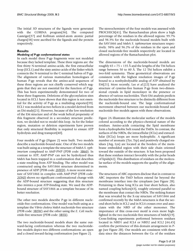
The two nucleotide-bound models share the same out-ward-facing conformation whereas the two nucleotide-free models depict two different conformations: an openand a closed inward-facing conformation (see Figure 2).
The stereochemistry of the four models was assessed withPROCHECK[41]. The Ramachandran plots show a highpercentage of the residues in the allowed regions: 99.7%and 98.3% for the nucleotide-bound models built withthe SAV1866 and MsbA S. typhimurium structures respec-tively. 98% and 96.2% of the residues in the open andclosed nucleotide-free models respectively are located inallowed regions of the Ramachandran plot.
The dimensions of the nucleotide-bound models areroughly 65 × 75 × 135 Å and the lengths of the TM helicesrange between 50 to 80 Å. The 12 TM helices exhibit atwo-fold symmetry. These geometrical observations areconsistent with the highest resolution images of P-gpbound to a nonhydrolyzable analog of ATP obtained byEM[21]. More recently, Lee et al.[22] have analyzed thestructure of cysteine-free human P-gp from two-dimen-sional crystals in lipid monolayer in the presence orabsence of various nucleotides. Their EM analysis predictsthe nucleotide-free structure to be distinctly different fromthe nucleotide-bound one. The large conformationalmovement observed between our nucleotide-bound andnucleotide-free models agrees with these observations.
Figure 2A illustrates the molecular surface of the modelscolored according to the physico-chemical nature of theresidues. Amino-acids contacting the lipid membraneform a hydrophobic belt round the TMDs. In contrast, thesurfaces of the NBDs, the intracellular (ICLs) and extracel-lular (ECLs) loops are more hydrophilic. A significantnumber of aromatic (Tyr, Trp) and positively charged res-idues (Arg, Lys) are located at the borders of the mem-brane embedded region with their side chain orientedtoward the outside of the structure. It has been proposedthat these residues interact favorably with the polar headsof lipids[42]. This distribution of residues on the molecu-lar surface of the models supports the quality of the align-ments.
The structures of ABC exporters disclose that in contrast toABC importers the TMD helices extend far beyond themembrane interface into the cytoplasm (see Figure 2B).Pertaining to these long ICLs are four short helices, alsonamed coupling helices[43], roughly oriented parallel tothe membrane that contact the NBDs. The striking featurethat was revealed by the X-ray structure of SAV1866 andconfirmed recently by the MsbA structures is that the sec-ond short helix in ICL2 and in ICL4 crosses over and asso-ciates with the NBD of the other monomer. Theimportance of this cross-over motif is even more high-lighted in the two nucleotide-free structures of MsbA[19].Cross-linking experiments performed between residueslocated in NBD1 and ICL4 or NBD2 and ICL2 [44,45]confirm the pertinence of this domain arrangement in P-gp (see Figure 2B). Our models are consistent with thesedata since the distances between the Cα of the residues
Page 5 of 18(page number not for citation purposes)
BMC Structural Biology 2009, 9:3 http://www.biomedcentral.com/1472-6807/9/3
Page 6 of 18(page number not for citation purposes)
A. Molecular surface representation of the 3D modelsFigure 2A. Molecular surface representation of the 3D models. (a) Nucleotide-bound model built with the SAV1866 template. (b) Nucleotide-bound model built with the S. typhimurium MsbA template. (c) Closed and (d) Open nucleotide-free models built with the V. cholerae MsbA and the E. coli MsbA templates respectively. The surface of hydrophobic residues (Ala, Leu, Val, Ile, Pro, Phe and Met) are colored in yellow. Other residues are colored in blue. The two red lines indicate the position of the lipid polar heads in the cellular membrane. B. View of a nucleotide-bound conformation of P-gp. The N-terminal half is highlighted: the three extracellular loops (ECL) are colored in pink (ECL1 is truncated (see text)), the two long intracellular loops are colored in yellow with the small coupling helices in pale green. The 6 trans-membrane helices are colored in blue (TM1), red (TM2), gray (TM3), orange (TM4), cyan (TM5) and green (TM6) and the nucleotide binding domain (NBD) is colored in magenta. The intracellular segment of TM1 and TM6 are depicted in light blue. All these segments are labeled from the N-terminus to the C-terminus.
BMC Structural Biology 2009, 9:3 http://www.biomedcentral.com/1472-6807/9/3
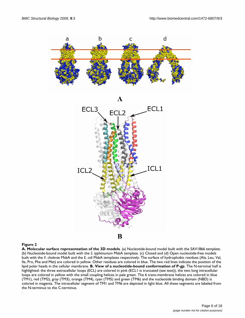
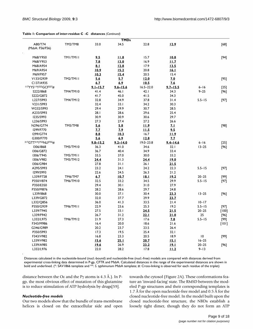
involved in the cysteine mutagenesis range from 8.5 Å to11.5 Å (see Table 1). Furthermore, the relative arrange-ment of ICL4 or ICL2 relative to the NBDs in our modelsagrees with several cross-linking data in CFTR[46], whichshares the same topology and exhibits 36% of sequencesimilarity with P-gp. In CFTR, Phe508 of NBD1 can cross-link to several residues in ICL4 and Cys276 in ICL2 formsa cross-link with residues of the C-terminal NBD (seeTable 1). The aligned sequences of the Q-loop regions andof these ICL portions feature about 50% similaritybetween P-gp and CFTR.
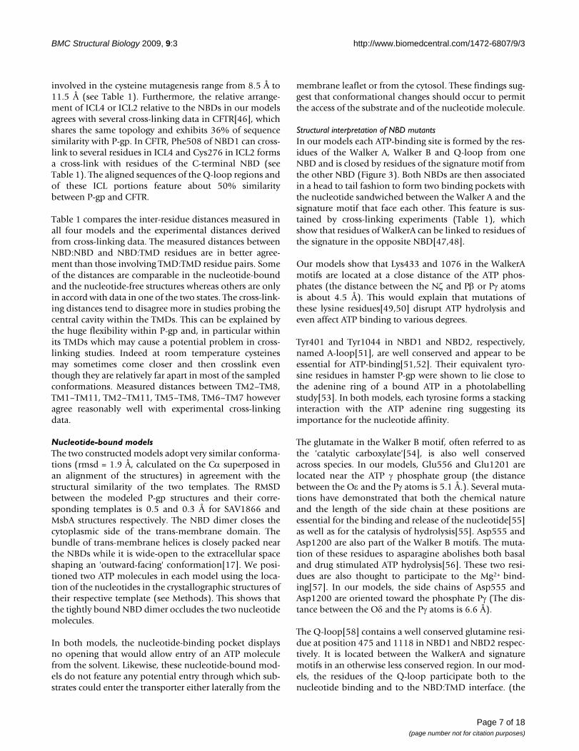
Table 1 compares the inter-residue distances measured inall four models and the experimental distances derivedfrom cross-linking data. The measured distances betweenNBD:NBD and NBD:TMD residues are in better agree-ment than those involving TMD:TMD residue pairs. Someof the distances are comparable in the nucleotide-boundand the nucleotide-free structures whereas others are onlyin accord with data in one of the two states. The cross-link-ing distances tend to disagree more in studies probing thecentral cavity within the TMDs. This can be explained bythe huge flexibility within P-gp and, in particular withinits TMDs which may cause a potential problem in cross-linking studies. Indeed at room temperature cysteinesmay sometimes come closer and then crosslink eventhough they are relatively far apart in most of the sampledconformations. Measured distances between TM2–TM8,TM1–TM11, TM2–TM11, TM5–TM8, TM6–TM7 howeveragree reasonably well with experimental cross-linkingdata.
Nucleotide-bound modelsThe two constructed models adopt very similar conforma-tions (rmsd = 1.9 Å, calculated on the Cα superposed inan alignment of the structures) in agreement with thestructural similarity of the two templates. The RMSDbetween the modeled P-gp structures and their corre-sponding templates is 0.5 and 0.3 Å for SAV1866 andMsbA structures respectively. The NBD dimer closes thecytoplasmic side of the trans-membrane domain. Thebundle of trans-membrane helices is closely packed nearthe NBDs while it is wide-open to the extracellular spaceshaping an 'outward-facing' conformation[17]. We posi-tioned two ATP molecules in each model using the loca-tion of the nucleotides in the crystallographic structures oftheir respective template (see Methods). This shows thatthe tightly bound NBD dimer occludes the two nucleotidemolecules.
In both models, the nucleotide-binding pocket displaysno opening that would allow entry of an ATP moleculefrom the solvent. Likewise, these nucleotide-bound mod-els do not feature any potential entry through which sub-strates could enter the transporter either laterally from the
membrane leaflet or from the cytosol. These findings sug-gest that conformational changes should occur to permitthe access of the substrate and of the nucleotide molecule.
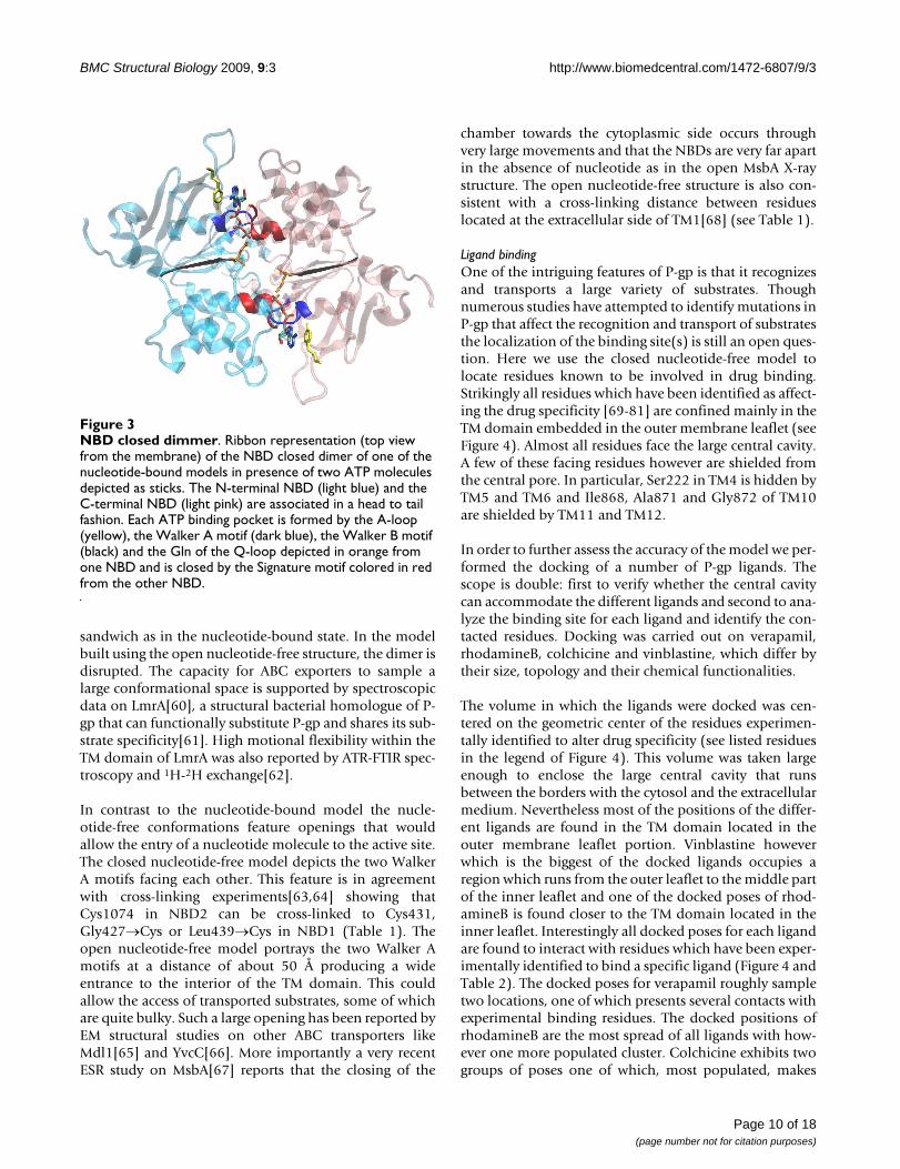
Structural interpretation of NBD mutantsIn our models each ATP-binding site is formed by the res-idues of the Walker A, Walker B and Q-loop from oneNBD and is closed by residues of the signature motif fromthe other NBD (Figure 3). Both NBDs are then associatedin a head to tail fashion to form two binding pockets withthe nucleotide sandwiched between the Walker A and thesignature motif that face each other. This feature is sus-tained by cross-linking experiments (Table 1), whichshow that residues of WalkerA can be linked to residues ofthe signature in the opposite NBD[47,48].
Our models show that Lys433 and 1076 in the WalkerAmotifs are located at a close distance of the ATP phos-phates (the distance between the Nζ and Pβ or Pγ atomsis about 4.5 Å). This would explain that mutations ofthese lysine residues[49,50] disrupt ATP hydrolysis andeven affect ATP binding to various degrees.
Tyr401 and Tyr1044 in NBD1 and NBD2, respectively,named A-loop[51], are well conserved and appear to beessential for ATP-binding[51,52]. Their equivalent tyro-sine residues in hamster P-gp were shown to lie close tothe adenine ring of a bound ATP in a photolabellingstudy[53]. In both models, each tyrosine forms a stackinginteraction with the ATP adenine ring suggesting itsimportance for the nucleotide affinity.
The glutamate in the Walker B motif, often referred to asthe 'catalytic carboxylate'[54], is also well conservedacross species. In our models, Glu556 and Glu1201 arelocated near the ATP γ phosphate group (the distancebetween the Oε and the Pγ atoms is 5.1 Å.). Several muta-tions have demonstrated that both the chemical natureand the length of the side chain at these positions areessential for the binding and release of the nucleotide[55]as well as for the catalysis of hydrolysis[55]. Asp555 andAsp1200 are also part of the Walker B motifs. The muta-tion of these residues to asparagine abolishes both basaland drug stimulated ATP hydrolysis[56]. These two resi-dues are also thought to participate to the Mg2+ bind-ing[57]. In our models, the side chains of Asp555 andAsp1200 are oriented toward the phosphate Pγ (The dis-tance between the Oδ and the Pγ atoms is 6.6 Å).
The Q-loop[58] contains a well conserved glutamine resi-due at position 475 and 1118 in NBD1 and NBD2 respec-tively. It is located between the WalkerA and signaturemotifs in an otherwise less conserved region. In our mod-els, the residues of the Q-loop participate both to thenucleotide binding and to the NBD:TMD interface. (the
Page 7 of 18(page number not for citation purposes)
BMC Structural Biology 2009, 9:3 http://www.biomedcentral.com/1472-6807/9/3
Table 1: Comparison of inter-residue C -C distances
Residues Region Cα-Cα distances (Å) Exp. cross-linking (Å) Ref.
Nucl.-bound* Nucl.-bound** Closed-nucl.-free Open-nucl.-free
NBDsS1072/L531 WalkerA/Signature 9.9 10.5 41.0 65.6 5.5–15 [47]S1072/S532 6.8 7.5 37.7 63.2G1073/L531 7.6 8.3 37.2 68.1G1073/S532 5.4 6.3 33.9 65.8G1073/G533 7.9 8.9 30.5 65.2C1074/L531 10.8 10.8 34.9 65.4G1075/L531 10.4 10.6 37.5 66.7S429/L1176 10.0 10.6 40.1 66.5G430/L1176 7.6 8.4 36.3 68.7G431/L1176 10.9 10.9 34.1 66.6G432/L1176 10.5 10.7 36.9 68.1C431/L1176 10.9 10.9 34.1 66.6 [48]L531/C1074 10.8 10.8 34.9 65.4G427/C1074 WalkerA/WalkerA 31.0 30.9 13.5 56.8 [63]L439/C1074 36.6 35.7 19.4 64.6C431/C1074 33.4 33.1 7.9 54.0 [64]
NBD:TMDL443/S909 8.6 9.8 14.2 9.2 6–16 [44]S474/R905 8.9 9.0 8.4 11.4
A266/F1086 8.6 10.0 13.8 9.7 5.5–15 [45]
Y490/V907(CFTR: F508/L1065)
7.9 9.1 12.5 9.2 [46]
Y490/L910(CFTR: F508/F1068)
7.2 6.9 8.7 8.0
Y490/T911(CFTR: F508/G1069)
9.5 8.2 11.5 9.5
Y490/F916(CFTR: F508/F1074)
9.3 10.2 10.6 10.3
E493/T911(CFTR: V510/G1069)
10.6 8.6 8.3 10.0
V478/T906(CFTR: W496/T1064)
6.5 7.4 8.2 9.6
F480/V907(CFTR: M498/L1065)
10.8 10.8 11.7 11.7
R547/T911(CFTR: K564/G1069)
11.0 9.9 15.4 13.0
G269/Q1107(CFTR: C276/Q1280)
8.4 10.4 13.5 11.8
G269/A1111(CFTR: C276/K1284)
8.5 10.1 13.4 11.2
Page 8 of 18(page number not for citation purposes)
BMC Structural Biology 2009, 9:3 http://www.biomedcentral.com/1472-6807/9/3
distance between the Oε and the Pγ atoms is 4.3 Å.). In P-gp, the most obvious effect of mutation of this glutamineis to reduce stimulation of ATP hydrolysis by drugs[59].
Nucleotide-free modelsOur two models show that the bundle of trans-membranehelices is closed on the extracellular side and open
towards the cytosol (Figure 2A). These conformations fea-ture an 'inward-facing' state. The RMSD between the mod-eled P-gp structures and their corresponding templates is1.7 Å for the open nucleotide-free model and 0.5 Å for theclosed nucleotide-free model. In the model built upon theclosed nucleotide-free structure, the NBDs establish aloosely tight dimer, though they do not form an ATP
TMDsA80/T74
(MsbA: F56/F56)TM2/TM8 33.0 34.5 22.8 12.9 [68]
M68/Y950 TM1/TM11 9.5 11.8 15.7 10.8 [94]M68/Y953 7.8 13.0 16.9 11.7M68/A954 8.1 12.8 17.9 13.5M69/A954 10.9 15.2 20.8 16.1M69/F957 10.3 15.4 20.5 15.4
V133/G939 TM2/TM11 5.6 5.7 12.0 7.0 [95]C137/A935 6.7 6.9 10.5 7.6
117YYS119/555GCF557# 9.1–15.7 9.6–15.6 16.5–22.0 9.7–15.5 6–16 [25]S222/I868 TM4/TM10 41.4 46.1 42.1 34.3 9–25 [96]S222/G872 41.7 45.0 41.5 34.3L227/S993 TM4/TM12 32.8 34.9 37.8 31.4 5.5–15 [97]V231/S993 32.4 33.1 34.2 30.3W232/S993 29.4 29.9 30.7 28.5A233/S993 28.1 28.6 29.6 25.4I235/S993 30.9 30.9 30.6 29.7L236/S993 27.3 27.4 27.2 26.6
N296/G774 TM5/TM8 5.6 5.8 11.9 7.1 [98]I299/F770 7.7 7.9 11.5 9.5I299/G774 8.8 10.3 16.7 11.9G300/F770 5.6 6.9 12.8 7.7
317GTT319/753NLF755# 9.8–15.2 9.2–14.0 19.0–23.8 9.4–14.6 6–16 [25]I306/I868 TM5/TM10 36.3 41.0 34.6 32.1 13–25 [96]I306/G872 36.7 40.4 34.9 33.4I306/T945 TM5/TM11 33.5 37.0 30.0 33.2I306/V982 TM5/TM12 24.4 31.3 24.4 19.0I306/G984 27.8 31.1 26.1 21.5A295/S993 23.2 24.1 24.2 22.3 5.5–15 [97]I299/S993 22.6 24.5 26.3 21.2L339/F728 TM6/TM7 6.7 10.7 18.1 19.2 20–25 [99]P350/V874 TM6/TM10 32.5 33.2 34.5 29.9 5.5–15 [97]P350/E350 29.4 30.1 31.0 27.9P350/M876 28.2 28.6 29.7 24.8L339/I868 30.0 37.1 30.4 23.3 13–25 [96]L339/G872 32.0 37.7 29.9 23.7L332/Q856 36.0 41.3 33.5 21.4 10–17P350/G939 TM6/TM11 22.9 23.6 25.3 19.2 5.5–15 [97]L339/T945 27.5 33.1 24.5 21.5 20–25 [100]L339/F942 26.7 31.2 22.1 21.0 25 [96]L332/L975 TM6/TM12 21.9 27.3 17.6 7.8 5.5–15 [99]F343/M986 16.4 20.0 18.6 21.6 [101]G346/G989 20.2 23.7 23.5 26.4P350/S993 17.3 19.5 25.4 32.1F343/V982 16.8 23.3 20.5 18.9 10 [99]L339/V982 15.6 25.1 20.7 15.1 16–25L339/A985 19.6 26.9 23.2 19.1 20–25 [96]L332/L976 22.0 28.2 17.8 11.2 9–13
Distances calculated in the nucleotide-bound (nucl.-bound) and nucleotide-free (nucl.-free) models are compared with distances derived from experimental cross-linking data determined in P-gp, CFTR and MsbA. Calculated distances in the range of the experimental distances are shown in bold and underlined. (*: SAV1866 template and **: S. typhimurium MsbA template; #: Cross-linking is observed for each residue of the triplet)
Table 1: Comparison of inter-residue C -C distances (Continued)
Page 9 of 18(page number not for citation purposes)
BMC Structural Biology 2009, 9:3 http://www.biomedcentral.com/1472-6807/9/3
sandwich as in the nucleotide-bound state. In the modelbuilt using the open nucleotide-free structure, the dimer isdisrupted. The capacity for ABC exporters to sample alarge conformational space is supported by spectroscopicdata on LmrA[60], a structural bacterial homologue of P-gp that can functionally substitute P-gp and shares its sub-strate specificity[61]. High motional flexibility within theTM domain of LmrA was also reported by ATR-FTIR spec-troscopy and 1H-2H exchange[62].
In contrast to the nucleotide-bound model the nucle-otide-free conformations feature openings that wouldallow the entry of a nucleotide molecule to the active site.The closed nucleotide-free model depicts the two WalkerA motifs facing each other. This feature is in agreementwith cross-linking experiments[63,64] showing thatCys1074 in NBD2 can be cross-linked to Cys431,Gly427→Cys or Leu439→Cys in NBD1 (Table 1). Theopen nucleotide-free model portrays the two Walker Amotifs at a distance of about 50 Å producing a wideentrance to the interior of the TM domain. This couldallow the access of transported substrates, some of whichare quite bulky. Such a large opening has been reported byEM structural studies on other ABC transporters likeMdl1[65] and YvcC[66]. More importantly a very recentESR study on MsbA[67] reports that the closing of the
chamber towards the cytoplasmic side occurs throughvery large movements and that the NBDs are very far apartin the absence of nucleotide as in the open MsbA X-raystructure. The open nucleotide-free structure is also con-sistent with a cross-linking distance between residueslocated at the extracellular side of TM1[68] (see Table 1).
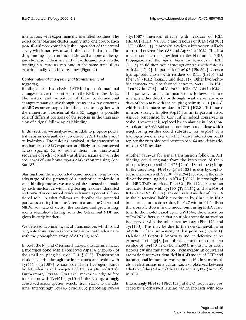
Ligand bindingOne of the intriguing features of P-gp is that it recognizesand transports a large variety of substrates. Thoughnumerous studies have attempted to identify mutations inP-gp that affect the recognition and transport of substratesthe localization of the binding site(s) is still an open ques-tion. Here we use the closed nucleotide-free model tolocate residues known to be involved in drug binding.Strikingly all residues which have been identified as affect-ing the drug specificity [69-81] are confined mainly in theTM domain embedded in the outer membrane leaflet (seeFigure 4). Almost all residues face the large central cavity.A few of these facing residues however are shielded fromthe central pore. In particular, Ser222 in TM4 is hidden byTM5 and TM6 and Ile868, Ala871 and Gly872 of TM10are shielded by TM11 and TM12.
In order to further assess the accuracy of the model we per-formed the docking of a number of P-gp ligands. Thescope is double: first to verify whether the central cavitycan accommodate the different ligands and second to ana-lyze the binding site for each ligand and identify the con-tacted residues. Docking was carried out on verapamil,rhodamineB, colchicine and vinblastine, which differ bytheir size, topology and their chemical functionalities.
The volume in which the ligands were docked was cen-tered on the geometric center of the residues experimen-tally identified to alter drug specificity (see listed residuesin the legend of Figure 4). This volume was taken largeenough to enclose the large central cavity that runsbetween the borders with the cytosol and the extracellularmedium. Nevertheless most of the positions of the differ-ent ligands are found in the TM domain located in theouter membrane leaflet portion. Vinblastine howeverwhich is the biggest of the docked ligands occupies aregion which runs from the outer leaflet to the middle partof the inner leaflet and one of the docked poses of rhod-amineB is found closer to the TM domain located in theinner leaflet. Interestingly all docked poses for each ligandare found to interact with residues which have been exper-imentally identified to bind a specific ligand (Figure 4 andTable 2). The docked poses for verapamil roughly sampletwo locations, one of which presents several contacts withexperimental binding residues. The docked positions ofrhodamineB are the most spread of all ligands with how-ever one more populated cluster. Colchicine exhibits twogroups of poses one of which, most populated, makes
NBD closed dimmerFigure 3NBD closed dimmer. Ribbon representation (top view from the membrane) of the NBD closed dimer of one of the nucleotide-bound models in presence of two ATP molecules depicted as sticks. The N-terminal NBD (light blue) and the C-terminal NBD (light pink) are associated in a head to tail fashion. Each ATP binding pocket is formed by the A-loop (yellow), the Walker A motif (dark blue), the Walker B motif (black) and the Gln of the Q-loop depicted in orange from one NBD and is closed by the Signature motif colored in red from the other NBD.
Page 10 of 18(page number not for citation purposes)
BMC Structural Biology 2009, 9:3 http://www.biomedcentral.com/1472-6807/9/3
interactions with experimentally identified residues. Theposes of vinblastine cluster mainly into one group. Eachpose fills almost completely the upper part of the centralcavity which narrows towards the extracellular side. Thedrug binding site in our model shows that none of the lig-ands because of their size and of the distance between thebinding site residues can bind at the same time all itsexperimentally identified residues (Figure 4).
Conformational changes: signal transmission and triggeringBinding and/or hydrolysis of ATP induce conformationalchanges that are transmitted from the NBDs to the TMDs.The nature and amplitude of these conformationalchanges remain elusive though the recent X-ray structuresof ABC exporters trapped in different states together withthe numerous biochemical data[82] suggest a possiblerole of different portions of the protein in the transmis-sion of a signal following ATP binding.
In this section, we analyze our models to propose poten-tial transmission pathways produced by ATP binding and/or hydrolysis. The residues involved in the transmissionmechanism of ABC exporters are likely to be conservedacross species. So to isolate them, the amino-acidsequence of each P-gp half was aligned separately with thesequences of 200 homologous ABC exporters using Con-Surf[83].
Starting from the nucleotide-bound models, so as to takeadvantage of the presence of a nucleotide molecule ineach binding pocket, we analyzed the interactions madeby each nucleotide with neighboring residues identifiedby ConSurf as conserved residues having a potential func-tional role. In what follows we describe the potentialpathways starting from the N-terminal and the C-terminalNBDs. For sake of clarity, the residues and protein frag-ments identified starting from the C-terminal NDB aregiven in curly brackets.
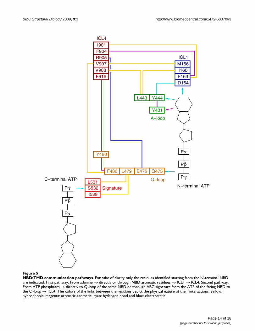
We detected two main ways of transmission, which couldoriginate from residues interacting either with adenine orwith the γ phosphate group of ATP (Figure 5).
In both the N- and C-terminal halves, the adenine makesa hydrogen bond with a conserved Asp164 {Asp805} ofthe small coupling helix of ICL1 {ICL3}. Transmissioncould also arise through the interactions of adenine withTyr444 {Tyr1087} whose side chain hydrogen bondsboth to adenine and to Asp164 of ICL1 {Asp805 of ICL3}.Furthermore, Tyr444 {Tyr1087} makes an edge-to-faceinteraction with Tyr401 {Tyr1044}, the A-loop, stronglyconserved across species, which, itself, stacks to the ade-nine. Interestingly Leu443 {Phe1086} preceding Tyr444
{Tyr1087} interacts directly with residues of ICL1(Ile160) {ICL3 (Val801)} and residues of ICL4 (Val 908){ICL2 (Ile265)}. Moreover, a cation-π interaction is likelyto occur between Phe1086 and Arg262 of ICL2. This lastinteraction has no equivalent in the N-terminal NBD.Propagation of the signal from the residues in ICL1{ICL3} could then occur through contacts with residuesof ICL4 {ICL2}. In particular Phe163 {Phe804} forms ahydrophobic cluster with residues of ICL4 (Ile901 andPhe904) {ICL2 (Leu258 and Ile261)}. Other hydropho-bic contacts are also formed between Met156 in ICL1{Leu797 in ICL3} and Val907 in ICL4 {Val264 in ICL2}.This pathway can be summarized as follows: adenineinteracts either directly or through nearby aromatic resi-dues of the NBDs with the coupling helix in ICL1 {ICL3}which itself contacts residues in ICL4 {ICL2}. This trans-mission strongly implies Asp164 as an important actor.Asp164 pinpointed by ConSurf is indeed conserved inMsbA. However it is replaced by an alanine in SAV1866.A look at the SAV1866 structures does not disclose whichneighboring residue could substitute for Asp164 as ahydrogen bond maker or which other interaction couldreplace the ones observed between Asp164 and either ade-nine or NBD residues.
Another pathway for signal transmission following ATPbinding could originate from the interaction of the γphosphate group with Gln475 {Gln1118} of the Q-loop.In the same loop, Phe480 {Phe1123} makes hydropho-bic interactions with Val907 {Val264} located in the mid-dle of the coupling helix in ICL4 {ICL2}. Interestingly, atthe NBD:TMD interface, Phe480 {Phe1123} shapes anaromatic cluster with Tyr490 {Tyr1133} and Phe916 ofICL4 {Phe267 of ICL2}. The equivalent residue of Phe916in the N-terminal half is substituted by Glu273 in ICL2but another aromatic residue, Phe267 within ICL2 fills inthe aromatic cluster in the model built using MsbA struc-ture. In the model based upon SAV1866, the orientationof Phe267 differs, such that no triple aromatic interactionis observed with the other two residues (Phe1123 andTyr1133). This may be due to the non-conservation inSAV1866 of the aromaticity at that position (Figure 1).Deletion of Tyr490 is known to induce defective or noexpression of P-gp[84] and the deletion of the equivalentresidue of Tyr490 in CFTR, Phe508, is the major cysticfibrosis causing mutation[85]. Remarkably an equivalentaromatic cluster was identified in a 3D model of CFTR andits functional importance was reported[46]. In some mod-els an electrostatic interaction was also observed betweenGlu476 of the Q-loop {Glu1119} and Arg905 {Arg262}in ICL4.
Interestingly Phe480 {Phe1123} of the Q-loop is also pre-ceded by a conserved leucine, which interacts with resi-
Page 11 of 18(page number not for citation purposes)
BMC Structural Biology 2009, 9:3 http://www.biomedcentral.com/1472-6807/9/3
dues of the ABC signature: Leu531 and Ile539 {Leu1176and Ile1184} which close the ATP binding pocket of theother NBD.
The second pathway can be summarized as follows: ATPphosphates interact with residues of the Q-loop whichitself contacts residues of the coupling helix in ICL4{ICL2}. In a more complex way the signal to ICL4 {ICL2}in one NBD could also originate from the phosphates ofthe facing NBD through residues of the ABC signaturewhich itself contacts the Q-loop.
Both proposed pathways emphasize the importance ofICL4 {ICL2} and to a lesser extent ICL1 {ICL3} in thetransmission of a signal from the ATP binding pocket. TheICL4 {ICL2} links the TM segments (either TM4–TM5 or
TM10–TM11) which mediate the hinge binding motionallowing the transition from the inward to the outwardconformations as suggested by the MsbA structures.Remarkably it was shown that CFTR mutants located inthe coupling helix in ICL4 displayed a decreased channelopen probability suggesting the importance of dynamiccontacts at these sites for the conformational change tooccur in this protein[46,86,87]. Also mutations in ICL1 ofCFTR were reported to impede transition to the open stateof the protein[88].
Comparison of the MsbA nucleotide-free and nucleotide-bound structures suggests that ABC exporters undergolarge conformational changes. These structures pinpointto a large motion of the portion formed by TM4/ICL2/TM5 {or TM10/ICL4/TM11} around a hinge defined by
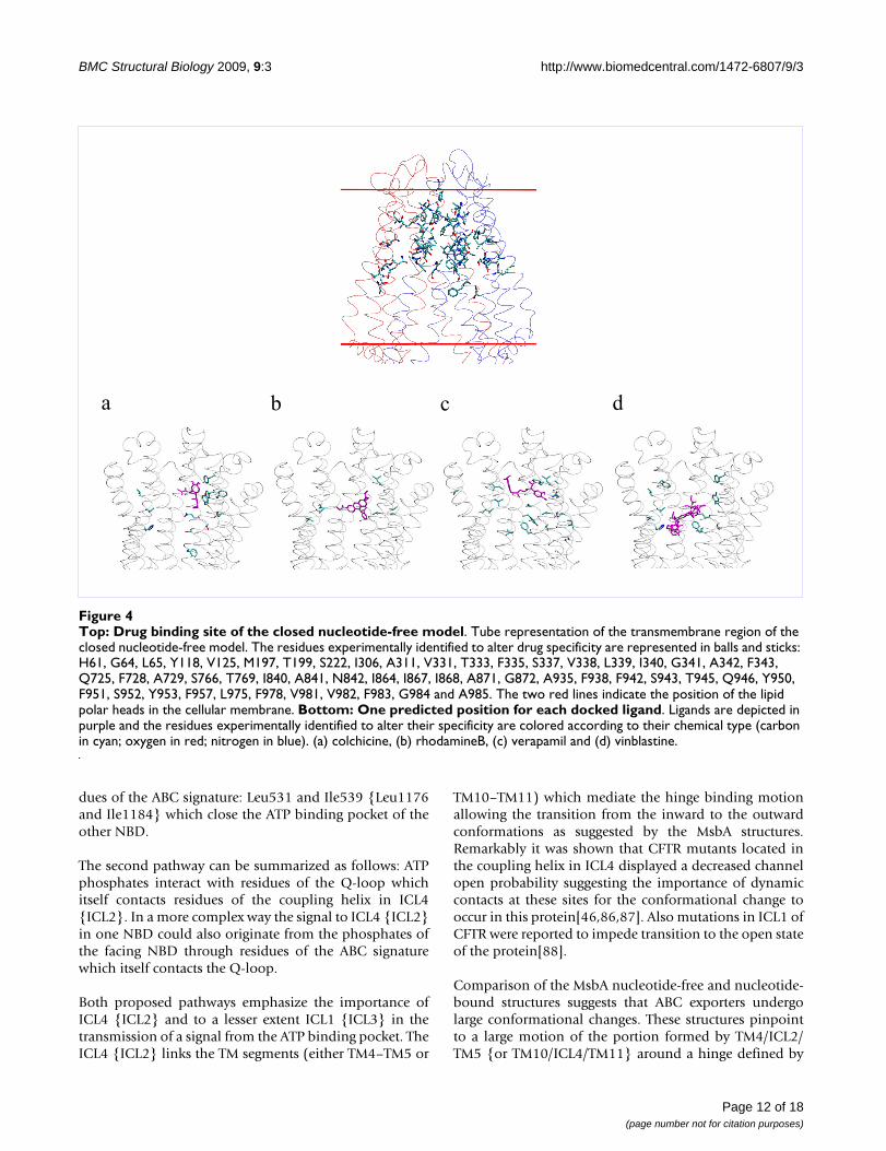
Top: Drug binding site of the closed nucleotide-free modelFigure 4Top: Drug binding site of the closed nucleotide-free model. Tube representation of the transmembrane region of the closed nucleotide-free model. The residues experimentally identified to alter drug specificity are represented in balls and sticks: H61, G64, L65, Y118, V125, M197, T199, S222, I306, A311, V331, T333, F335, S337, V338, L339, I340, G341, A342, F343, Q725, F728, A729, S766, T769, I840, A841, N842, I864, I867, I868, A871, G872, A935, F938, F942, S943, T945, Q946, Y950, F951, S952, Y953, F957, L975, F978, V981, V982, F983, G984 and A985. The two red lines indicate the position of the lipid polar heads in the cellular membrane. Bottom: One predicted position for each docked ligand. Ligands are depicted in purple and the residues experimentally identified to alter their specificity are colored according to their chemical type (carbon in cyan; oxygen in red; nitrogen in blue). (a) colchicine, (b) rhodamineB, (c) verapamil and (d) vinblastine.
a b c d
Page 12 of 18(page number not for citation purposes)
BMC Structural Biology 2009, 9:3 http://www.biomedcentral.com/1472-6807/9/3
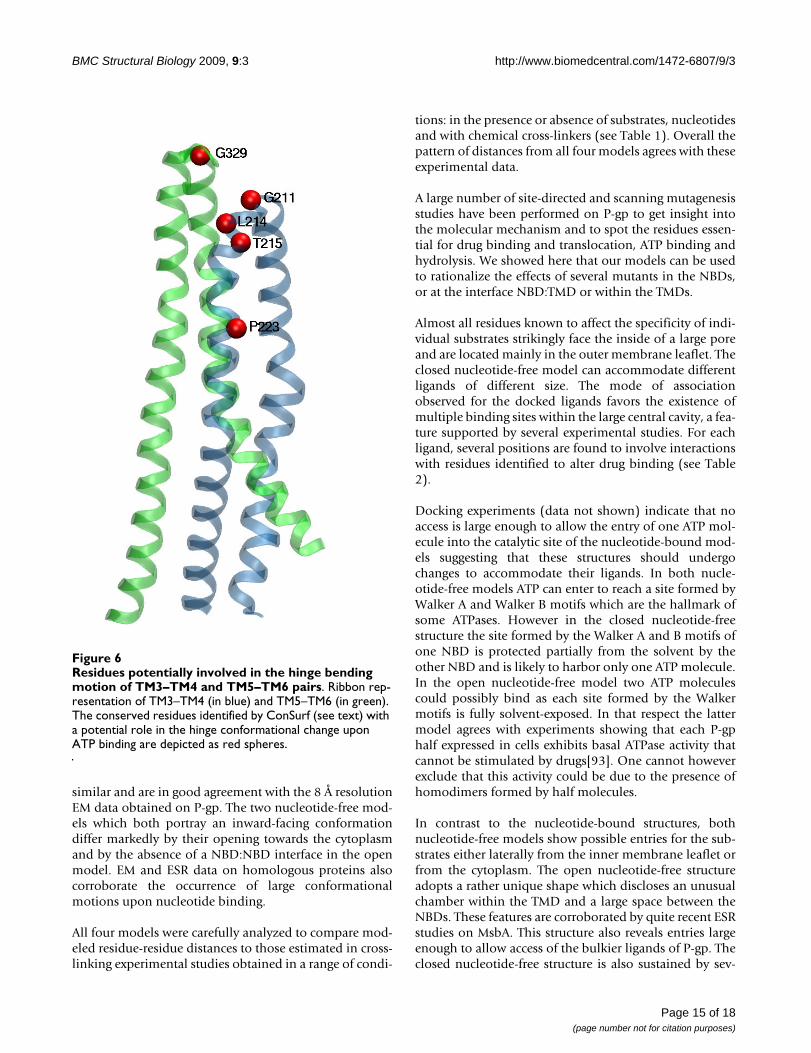
ECL2/ECL3 {or ECL5/ECL6}[19]. We looked at the con-served residues identified by ConSurf in these potentialhinge regions. The most conserved residues are Leu214,Thr215 and Leu216 {Leu 857, Thr858 and Leu859}located at the N-terminal side of TM4 {TM10} and thusclose to the extracellular loop between TM3 and TM4{TM9 and TM10} (Figure 6). Interestingly it has been pro-posed that threonine or serine residues can cause localalterations which may result in significant conformationalchanges across transmembrane helices and which mayplay a role in transmembrane signaling[89]. Pro223{Pro866} located further down TM4 {TM10} has alsobeen identified as a conserved residue with a potentialrole. Despite their disruptive nature, proline residues arestatistically well represented in transmembrane helices.Proline was reported to play an important role in produc-ing conformational changes essential for receptor signal-ing and channel gating[90,91].
CStudies[70,92] demonstrated that several residues in TM6in particular those located at the two extremes show largechanges in accessibility to covalent modification by male-
imide reagents suggesting that TM6 undergoes significantconformational changes upon the catalytic cycle. Ournucleotide-free and -bound models can readily explainmost of these site-directed labeling data. However, insteadof pointing to a large TM6 motion, our models featurestructural changes in TM6 neighborhood caused by thehinge bending motion of TM4–TM5 pair which swingsaway from TM6 and by the shift of TM7.
Discussion and ConclusionThe lack of a high resolution structure for P-gp and therecently determined X-ray structures of several ABCexporters trapped in different conditions prompted us toconstruct several 3D models of P-gp featuring differentstates along its catalytic cycle. Sequence identity and sim-ilarities, though varying markedly across the protein, aswell as experimental data support that SAV1866 andMsbA share the same architecture as P-gp.
We present here two nucleotide-bound and two nucle-otide-free models of P-gp based on four different templatestructures. The two nucleotide-bound models whichdepict an outward-facing conformation of P-gp are very
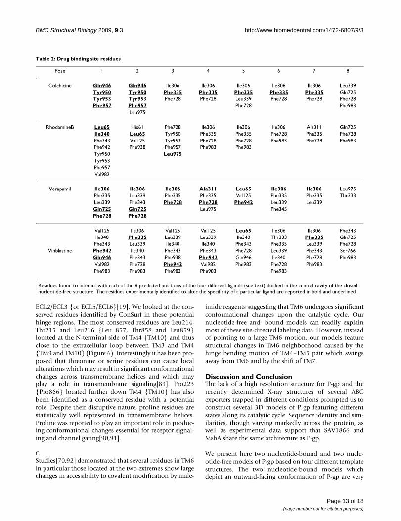
Table 2: Drug binding site residues
Pose 1 2 3 4 5 6 7 8
Colchicine Gln946 Gln946 Ile306 Ile306 Ile306 Ile306 Ile306 Leu339Tyr950 Tyr950 Phe335 Phe335 Phe335 Phe335 Phe335 Gln725Tyr953 Tyr953 Phe728 Phe728 Leu339 Phe728 Phe728 Phe728Phe957 Phe957 Phe728 Phe983
Leu975
RhodamineB Leu65 His61 Phe728 Ile306 Ile306 Ile306 Ala311 Gln725Ile340 Leu65 Tyr950 Phe335 Phe335 Phe728 Phe335 Phe728Phe343 Val125 Tyr953 Phe728 Phe728 Phe983 Phe728 Phe983Phe942 Phe938 Phe957 Phe983 Phe983Tyr950 Leu975Tyr953Phe957Val982
Verapamil Ile306 Ile306 Ile306 Ala311 Leu65 Ile306 Ile306 Leu975Phe335 Leu339 Phe335 Phe335 Val125 Phe335 Phe335 Thr333Leu339 Phe343 Phe728 Phe728 Phe942 Leu339 Leu339Gln725 Gln725 Leu975 Phe345Phe728 Phe728
Val125 Ile306 Val125 Val125 Leu65 Ile306 Ile306 Phe343Ile340 Phe335 Leu339 Leu339 Ile340 Thr333 Phe335 Gln725Phe343 Leu339 Ile340 Ile340 Phe343 Phe335 Leu339 Phe728
Vinblastine Phe942 Ile340 Phe343 Phe343 Phe728 Leu339 Phe343 Ser766Gln946 Phe343 Phe938 Phe942 Gln946 Ile340 Phe728 Phe983Val982 Phe728 Phe942 Val982 Phe983 Phe728 Phe983Phe983 Phe983 Phe983 Phe983 Phe983
Residues found to interact with each of the 8 predicted positions of the four different ligands (see text) docked in the central cavity of the closed nucleotide-free structure. The residues experimentally identified to alter the specificity of a particular ligand are reported in bold and underlined.
Page 13 of 18(page number not for citation purposes)
BMC Structural Biology 2009, 9:3 http://www.biomedcentral.com/1472-6807/9/3
Page 14 of 18(page number not for citation purposes)
NBD:TMD communication pathwaysFigure 5NBD:TMD communication pathways. For sake of clarity only the residues identified starting from the N-terminal NBD are indicated. First pathway: From adenine → directly or through NBD aromatic residues → ICL1 → ICL4. Second pathway: From ATP phosphates → directly to Q-loop of the same NBD or through ABC signature from the ATP of the facing NBD to the Q-loop → ICL4. The colors of the links between the residues depict the physical nature of their interactions: yellow: hydrophobic, magenta: aromatic-aromatic, cyan: hydrogen bond and blue: electrostatic.
P
P
α
β
γ
P
P
α
β
γ
ICL1
ICL4
L531
I901F904R905V907V908F916
M156I160F163D164
Signature
A−loop
Y401
S532I539
Q−loop
L443 Y444
C−terminal ATP
N−terminal ATP
P
P
Q475E476L479F480
Y490
BMC Structural Biology 2009, 9:3 http://www.biomedcentral.com/1472-6807/9/3
similar and are in good agreement with the 8 Å resolutionEM data obtained on P-gp. The two nucleotide-free mod-els which both portray an inward-facing conformationdiffer markedly by their opening towards the cytoplasmand by the absence of a NBD:NBD interface in the openmodel. EM and ESR data on homologous proteins alsocorroborate the occurrence of large conformationalmotions upon nucleotide binding.
All four models were carefully analyzed to compare mod-eled residue-residue distances to those estimated in cross-linking experimental studies obtained in a range of condi-
tions: in the presence or absence of substrates, nucleotidesand with chemical cross-linkers (see Table 1). Overall thepattern of distances from all four models agrees with theseexperimental data.
A large number of site-directed and scanning mutagenesisstudies have been performed on P-gp to get insight intothe molecular mechanism and to spot the residues essen-tial for drug binding and translocation, ATP binding andhydrolysis. We showed here that our models can be usedto rationalize the effects of several mutants in the NBDs,or at the interface NBD:TMD or within the TMDs.
Almost all residues known to affect the specificity of indi-vidual substrates strikingly face the inside of a large poreand are located mainly in the outer membrane leaflet. Theclosed nucleotide-free model can accommodate differentligands of different size. The mode of associationobserved for the docked ligands favors the existence ofmultiple binding sites within the large central cavity, a fea-ture supported by several experimental studies. For eachligand, several positions are found to involve interactionswith residues identified to alter drug binding (see Table2).
Docking experiments (data not shown) indicate that noaccess is large enough to allow the entry of one ATP mol-ecule into the catalytic site of the nucleotide-bound mod-els suggesting that these structures should undergochanges to accommodate their ligands. In both nucle-otide-free models ATP can enter to reach a site formed byWalker A and Walker B motifs which are the hallmark ofsome ATPases. However in the closed nucleotide-freestructure the site formed by the Walker A and B motifs ofone NBD is protected partially from the solvent by theother NBD and is likely to harbor only one ATP molecule.In the open nucleotide-free model two ATP moleculescould possibly bind as each site formed by the Walkermotifs is fully solvent-exposed. In that respect the lattermodel agrees with experiments showing that each P-gphalf expressed in cells exhibits basal ATPase activity thatcannot be stimulated by drugs[93]. One cannot howeverexclude that this activity could be due to the presence ofhomodimers formed by half molecules.
In contrast to the nucleotide-bound structures, bothnucleotide-free models show possible entries for the sub-strates either laterally from the inner membrane leaflet orfrom the cytoplasm. The open nucleotide-free structureadopts a rather unique shape which discloses an unusualchamber within the TMD and a large space between theNBDs. These features are corroborated by quite recent ESRstudies on MsbA. This structure also reveals entries largeenough to allow access of the bulkier ligands of P-gp. Theclosed nucleotide-free structure is also sustained by sev-
Residues potentially involved in the hinge bending motion of TM3–TM4 and TM5–TM6 pairsFigure 6Residues potentially involved in the hinge bending motion of TM3–TM4 and TM5–TM6 pairs. Ribbon rep-resentation of TM3–TM4 (in blue) and TM5–TM6 (in green). The conserved residues identified by ConSurf (see text) with a potential role in the hinge conformational change upon ATP binding are depicted as red spheres.
Page 15 of 18(page number not for citation purposes)
BMC Structural Biology 2009, 9:3 http://www.biomedcentral.com/1472-6807/9/3
eral experimental data. It is the only structure to agreewith the Walker A-Walker A cross-linking distance. Thisclosed nucleotide-free structure could possibly eitheroccur sequentially along the catalytic cycle after ligandsenter the open nucleotide-free structure or coexist with theopen structure as suggested by the high mobility noted byATR-FTIR and NMR for LmrA[60,62]. The outward facingconformation of the two nucleotide-bound models fea-tures a central pore open to the extracellular medium thatwould allow substrates to escape.
The determination of 3D models at different stages of thecatalytic cycle can help in proposing a role, in particular,of the residues essential for the transmission of a signalproducing conformational changes or responsible for theconformational changes themselves. We indeed identifiedtwo potential pathways formed by a chain of interactingresidues which could be involved in the propagation of asignal upon ATP binding from the catalytic site through-out to the TMDs. One highlighted a pathway describingcontacts between adenine either directly or through aro-matic residues with the coupling helix in ICL1 {or ICL3}which itself interacts with residues of ICL4 {or ICL2}. Theother pathway depicts a chain of interactions startingfrom the ATP phosphate groups to ICL4 {or ICL2} bymeans of the Q-loop. One of the interaction sites involvesa cluster of aromatic residues including residues of the Q-loop and of ICL4 or ICL2. Interestingly an equivalent clus-ter of aromatic residues was detected in CFTR and its roleon the channel gating was revealed. Mutagenesis of onlyone glutamine of the Q-loop has been shown to inhibitthe function suggesting that this mutant also affects theATP binding/or hydrolysis in the neighboring NBD [59].In that respect the second pathway may explain this obser-vation as it incorporates residues which have been sug-gested to play a role either in the NBD:TMDcommunication (residues of the Q-loop) or in the NBDdimerization (signature motif). It should also be men-tioned that several pathways either in series or in parallelcould occur to transmit the signal from the active site tothe TMD.
Our models are thus first approximation models and mayconstitute a useful starting point for the understanding ofthe complete structural picture of P-gp at the differentstages of the catalytic cycle. They may guide further inves-tigations of the role of residues at the NBD:NBD andNBD:TMD interfaces.
Authors' contributionsGD constructed the P-gp model based on the Sav1866template and participated in the elaboration of a coherentcollection of cross-linking and mutagenesis data. J-PBconstructed the other three models. J-PB and MP per-formed the structural analysis and the interpretation of
the mutagenesis data using all models of P-gp. FVB andPMT participated in the design of the study. J-PB and MPwrote the manuscript. All authors read and approved thefinal manuscript.
AcknowledgementsJ-PB is a post-doctoral fellow supported by the Belgian Fonds de la Recherche Scientifique (FNRS). MP and FVB are Maîtres de Recherches of the FNRS. This work was supported by the Belgian Fonds pour la Recherche Scientifique Médi-cale (FRSM 3.4583.08).
References1. Dean M, Hamon Y, Chimini G: The human ATP-binding cassette
(ABC) transporter superfamily. J Lipid Res 2001,42(7):1007-1017.
2. Gottesman MM, Ambudkar SV: Overview: ABC transporters andhuman disease. J Bioenerg Biomembr 2001, 33(6):453-458.
3. Bambeke FV, Balzi E, Tulkens PM: Antibiotic efflux pumps. Bio-chem Pharmacol 2000, 60(4):457-470.
4. Higgins CF, Linton KJ: The ATP switch model for ABC trans-porters. Nat Struct Mol Biol 2004, 11(10):918-926.
5. Leonard GD, Fojo T, Bates SE: The role of ABC transporters inclinical practice. Oncologist 2003, 8(5):411-424.
6. Ambudkar SV, Dey S, Hrycyna CA, Ramachandra M, Pastan I, Gottes-man MM: Biochemical, cellular, and pharmacological aspectsof the multidrug transporter. Annu Rev Pharmacol Toxicol 1999,39:361-398.
7. Sauna ZE, Kim I, Ambudkar SV: Genomics and the mechanism ofP-glycoprotein (ABCB1). J Bioenerg Biomembr 2007, 39(5–6):481-487.
8. Sauna ZE, Ambudkar SV: Evidence for a requirement for ATPhydrolysis at two distinct steps during a single turnover ofthe catalytic cycle of human P-glycoprotein. Proc Natl Acad SciUSA 2000, 97(6):2515-2520.
9. Senior AE, al-Shawi MK, Urbatsch IL: The catalytic cycle of P-glyc-oprotein. FEBS Lett 1995, 377(3):285-289.
10. Locher KP, Lee AT, Rees DC: The E. coli BtuCD structure: aframework for ABC transporter architecture and mecha-nism. Science 2002, 296(5570):1091-1098.
11. Hvorup RN, Goetz BA, Niederer M, Hollenstein K, Perozo E, LocherKP: Asymmetry in the structure of the ABC transporter-binding protein complex BtuCD-BtuF. Science 2007,317(5843):1387-1390.
12. Hollenstein K, Frei DC, Locher KP: Structure of an ABC trans-porter in complex with its binding protein. Nature 2007,446(7132):213-216.
13. Gerber S, Comellas-Bigler M, Goetz BA, Locher KP: StructuralBasis of Trans-Inhibition in a Molybdate/tungstate ABCTransporter. Science 2008, 321(5886):246-250.
14. Pinkett HW, Lee AT, Lum P, Locher KP, Rees DC: An inward-fac-ing conformation of a putative metal-chelate-type ABCtransporter. Science 2007, 315(5810):373-377.
15. Oldham ML, Khare D, Quiocho FA, Davidson AL, Chen J: Crystalstructure of a catalytic intermediate of the maltose trans-porter. Nature 2007, 450(7169):515-521.
16. Kadaba NS, Kaiser JT, Johnson E, Lee A, Rees DC: The high-affinityE. coli methionine ABC transporter: structure and allostericregulation. Science 2008, 321(5886):250-253.
17. Dawson RJP, Locher KP: Structure of a bacterial multidrugABC transporter. Nature 2006, 443(7108):180-185.
18. Dawson RJP, Locher KP: Structure of the multidrug ABC trans-porter Sav1866 from Staphylococcus aureus in complex withAMP-PNP. FEBS Lett 2007, 581(5):935-938.
19. Ward A, Reyes CL, Yu J, Roth CB, Chang G: Flexibility in the ABCtransporter MsbA: Alternating access with a twist. Proc NatlAcad Sci USA 2007, 104(48):19005-19010.
20. Davidson AL, Dassa E, Orelle C, Chen J: Structure, function, andevolution of bacterial ATP-binding cassette systems. Micro-biol Mol Biol Rev 2008, 72(2):317-364.
21. Rosenberg MF, Callaghan R, Modok S, Higgins CF, Ford RC: Three-dimensional structure of P-glycoprotein: the transmem-brane regions adopt an asymmetric configuration in thenucleotide-bound state. J Biol Chem 2005, 280(4):2857-2862.
Page 16 of 18(page number not for citation purposes)
http://www.ncbi.nlm.nih.gov/entrez/query.fcgi?cmd=Retrieve&db=PubMed&dopt=Abstract&list_uids=8549739
BMC Structural Biology 2009, 9:3 http://www.biomedcentral.com/1472-6807/9/3
22. Lee J, Urbatsch IL, Senior AE, Wilkens S: Nucleotide-inducedStructural Changes in P-glycoprotein Observed by ElectronMicroscopy. J Biol Chem 2008, 283(9):5769-5779.
23. Lawson J, O'Mara ML, Kerr ID: Struture-based interpretation ofthe mutagenesis database for the nucleotide bindingdomains of P-glycoprotein. Biochim Biophys Acta 2007,1778(2):376-391.
24. Chang G, Roth CB, Reyes CL, Pornillos O, Chen Y, Chen AP:Retraction. Science 2006, 314(5807):1875.
25. Stenham DR, Campbell JD, Sansom MSP, Higgins CF, Kerr ID, LintonKJ: An atomic detail model for the human ATP binding cas-sette transporter P-glycoprotein derived from disulfidecross-linking and homology modeling. FASEB J 2003,17(15):2287-2289.
26. Vandevuer S, Bambeke FV, Tulkens PM, Prévost M: Predicting thethree-dimensional structure of human P-glycoprotein inabsence of ATP by computational techniques embodyingcrosslinking data: insight into the mechanism of ligandmigration and binding sites. Proteins 2006, 63(3):466-478.
27. O'Mara ML, Tieleman DP: P-glycoprotein models of the apo andATP-bound states based on homology with Sav1866 andMalK. FEBS Lett 2007, 581(22):4217-4222.
28. Ravna AW, Sylte I, Sager G: Molecular model of the outward fac-ing state of the human P-glycoprotein (ABCB1) and compar-ison to a model of the human MRP5 (ABCC5). Theor Biol MedModel 2007, 4:33.
29. Globisch C, Pajeva IK, Wiese M: Identification of Putative Bind-ing Sites of P-glycoprotein Based on its Homology Model.ChemMedChem 2008, 3(2):280-295.
30. Altschul SF, Madden TL, Schäffer AA, Zhang J, Zhang Z, Miller W, Lip-man DJ: Gapped BLAST and PSI-BLAST: a new generation ofprotein database search programs. Nucleic Acids Res 1997,25(17):3389-3402.
31. Chenna R, Sugawara H, Koike T, Lopez R, Gibson TJ, Higgins DG,Thompson JD: Multiple sequence alignment with the Clustalseries of programs. Nucleic Acids Res 2003, 31(13):3497-3500.
32. Tusnády GE, Simon I: The HMMTOP transmembrane topologyprediction server. Bioinformatics 2001, 17(9):849-850.
33. Tusnády GE, Dosztányi Z, Simon I: Transmembrane proteins inthe Protein Data Bank: identification and classification. Bioin-formatics 2004, 20(17):2964-2972.
34. Sali A, Blundell T: Comparative protein modelling by satisfac-tion of spatial restraints. J Mol Biol 1993, 234:779-815.
35. Morris GM, Goodsell DS, Halliday RS, Huey R, Hart WE, Belew RK,Olson AJ: Automated Docking Using a Lamarckian GeneticAlgorithm and Empirical Binding Free Energy Function. JComputational Chemistry 1998, 19:1639-1662.
36. Sadowski J, Gasteiger J: From Atoms and Bonds to 3-Dimen-sional Atomic Coordinates – Automatic Model Builders.Chemical Reviews 1993, 93:2567-2581.
37. Gasteiger J, Marsili M: Iterative partial equalization of orbitalelectronegativity-a rapid access to atomic charges. Tetrahe-dron 1980, 36(22):3219-3228.
38. Weiner SJ, Kollman PA, Case DA, Singh UC, Ghio C, Alagona G, Pro-feta S, Weine P: A new force field for molecular mechanicalsimulation of nucleic acids and proteins. J Am Chem Soc 1984,106(3):765-784.
39. Schinkel AH, Kemp S, Dollé M, Rudenko G, Wagenaar E: N-glyco-sylation and deletion mutants of the human MDR1 P-glyco-protein. J Biol Chem 1993, 268(10):7474-7481.
40. Hrycyna CA, Airan LE, Germann UA, Ambudkar SV, Pastan I, Gottes-man MM: Structural flexibility of the linker region of humanP-glycoprotein permits ATP hydrolysis and drug transport.Biochemistry 1998, 37(39):13660-13673.
41. Laskowski RA, MacArthur MW, Moss DS, Thornton JM: PRO-CHECK: a program to check the stereochemical quality ofprotein structures. J Appl Cryst 1993, 26:283-291.
42. Killian JA, von Heijne G: How proteins adapt to a membrane-water interface. TIBS 2000, 25:429-434.
43. Dawson RJP, Hollenstein K, Locher KP: Uptake or extrusion:crystal structures of full ABC transporters suggest a com-mon mechanism. Mol Microbiol 2007, 65(2):250-257.
44. Zolnerciks JK, Wooding C, Linton KJ: Evidence for a Sav1866-likearchitecture for the human multidrug transporter P-glyco-protein. FASEB J 2007, 21(14):3937-3948.
45. Loo TW, Bartlett MC, Clarke DM: Processing mutations disruptinteractions between the nucleotide binding and transmem-brane domains of P-glycoprotein and the cystic fibrosistransmembrane conductance regulator (CFTR). J Biol Chem2008, 283(42):28190-28197.
46. Serohijos AWR, Hegedus T, Aleksandrov AA, He L, Cui L, DokholyanNV, Riordan JR: Phenylalanine-508 mediates a cytoplasmic-membrane domain contact in the CFTR 3D structure crucialto assembly and channel function. Proc Natl Acad Sci USA 2008,105(9):3256-3261.
47. Loo TW, Bartlett MC, Clarke DM: The "LSGGQ" motif in eachnucleotide-binding domain of human P-glycoprotein is adja-cent to the opposing walker A sequence. J Biol Chem 2002,277(44):41303-41306.
48. Loo TW, Bartlett MC, Clarke DM: Drug binding in human P-glyc-oprotein causes conformational changes in both nucleotide-binding domains. J Biol Chem 2003, 278(3):1575-1578.
49. Lerner-Marmarosh N, Gimi K, Urbatsch IL, Gros P, Senior AE: Largescale purification of detergent-soluble P-glycoprotein fromPichia pastoris cells and characterization of nucleotide bind-ing properties of wild-type Walker A, and Walker B mutantproteins. J Biol Chem 1999, 274(49):34711-34718.
50. Szakács G, Ozvegy C, Bakos E, Sarkadi B, Váradi A: Transition-stateformation in ATPase-negative mutants of human MDR1 pro-tein. Biochem Biophys Res Commun 2000, 276(3):1314-1319.
51. Kim I, Peng X, Sauna ZE, FitzGerald PC, Xia D, Müller M, NandigamaK, Ambudkar SV: The conserved tyrosine residues 401 and1044 in ATP sites of human P-glycoprotein are critical forATP binding and hydrolysis: evidence for a conserved sub-domain the A-loop in the ATP-binding cassette. Biochemistry2006, 45(24):7605-7616.
52. Carrier I, Urbatsch IL, Senior AE, Gros P: Mutational analysis ofconserved aromatic residues in the A-loop of the ABC trans-porter ABCB1A (mouse Mdr3). FEBS Lett 2007,581(2):301-308.
53. Sankaran B, Bhagat S, Senior AE: Photoaffinity labelling of P-glyc-oprotein catalytic sites. FEBS Lett 1997, 417(1):119-122.
54. Tombline G, Bartholomew LA, Tyndall GA, Gimi K, Urbatsch IL, Sen-ior AE: Properties of P-glycoprotein with mutations in the"catalytic carboxylate" glutamate residues. J Biol Chem 2004,279(45):46518-46526.
55. Tombline G, Bartholomew LA, Urbatsch IL, Senior AE: Combinedmutation of catalytic glutamate residues in the two nucle-otide binding domains of P-glycoprotein generates a confor-mation that binds ATP and ADP tightly. J Biol Chem 2004,279(30):31212-31220.
56. Urbatsch IL, Julien M, Carrier I, Rousseau ME, Cayrol R, Gros P:Mutational analysis of conserved carboxylate residues in thenucleotide binding sites of P-glycoprotein. Biochemistry 2000,39(46):14138-14149.
57. Hrycyna CA, Ramachandra M, Germann UA, Cheng PW, Pastan I,Gottesman MM: Both ATP sites of human P-glycoprotein areessential but not symmetric. Biochemistry 1999,38(42):13887-13899.
58. Kerr ID: Structure and association of ATP-binding cassettetransporter nucleotide-binding domains. Biochim Biophys Acta2002, 1561(1):47-64.
59. Urbatsch IL, Gimi K, Wilke-Mounts S, Senior AE: Investigation ofthe role of glutamine-471 and glutamine-1114 in the two cat-alytic sites of P-glycoprotein. Biochemistry 2000,39(39):11921-11927.
60. Siarheyeva A, Lopez JJ, Lehner I, Hellmich UA, van Veen HW, GlaubitzC: Probing the molecular dynamics of the ABC multidrugtransporter LmrA by deuterium solid-state nuclear mag-netic resonance. Biochemistry 2007, 46(11):3075-3083.
61. van Veen HW, Venema K, Bolhuis H, Oussenko I, Kox J, Poolman B,Driessen AJM, konings WN: Multidrug resistance mediated by abacterial homolog of thehuman multidrug transporterMDR1. Proc Natl Acad Sci USA 1996, 93:10668-10672.
62. Grimard V, Vigano C, Margolles A, Wattiez R, van Veen HW, KoningsWN, Ruysschaert JM, Goormaghtigh E: Structure and dynamicsof the membrane-embedded domain of LmrA investigatedby coupling polarized ATR-FTIR spectroscopy and (1)H/(2)Hexchange. Biochemistry 2001, 40(39):11876-11886.
63. Loo TW, Clarke DM: Drug-stimulated ATPase activity ofhuman P-glycoprotein is blocked by disulfide cross-linking
Page 17 of 18(page number not for citation purposes)
http://www.ncbi.nlm.nih.gov/entrez/query.fcgi?cmd=Retrieve&db=PubMed&dopt=Abstract&list_uids=9254694
http://www.ncbi.nlm.nih.gov/entrez/query.fcgi?cmd=Retrieve&db=PubMed&dopt=Abstract&list_uids=9254694
http://www.ncbi.nlm.nih.gov/entrez/query.fcgi?cmd=Retrieve&db=PubMed&dopt=Abstract&list_uids=8254673
http://www.ncbi.nlm.nih.gov/entrez/query.fcgi?cmd=Retrieve&db=PubMed&dopt=Abstract&list_uids=8254673
http://www.ncbi.nlm.nih.gov/entrez/query.fcgi?cmd=Retrieve&db=PubMed&dopt=Abstract&list_uids=8096511
http://www.ncbi.nlm.nih.gov/entrez/query.fcgi?cmd=Retrieve&db=PubMed&dopt=Abstract&list_uids=8096511
http://www.ncbi.nlm.nih.gov/entrez/query.fcgi?cmd=Retrieve&db=PubMed&dopt=Abstract&list_uids=8096511
http://www.ncbi.nlm.nih.gov/entrez/query.fcgi?cmd=Retrieve&db=PubMed&dopt=Abstract&list_uids=9753453
http://www.ncbi.nlm.nih.gov/entrez/query.fcgi?cmd=Retrieve&db=PubMed&dopt=Abstract&list_uids=9753453
http://www.ncbi.nlm.nih.gov/entrez/query.fcgi?cmd=Retrieve&db=PubMed&dopt=Abstract&list_uids=9395088
http://www.ncbi.nlm.nih.gov/entrez/query.fcgi?cmd=Retrieve&db=PubMed&dopt=Abstract&list_uids=9395088
http://www.ncbi.nlm.nih.gov/entrez/query.fcgi?cmd=Retrieve&db=PubMed&dopt=Abstract&list_uids=8855237
http://www.ncbi.nlm.nih.gov/entrez/query.fcgi?cmd=Retrieve&db=PubMed&dopt=Abstract&list_uids=8855237
BMC Structural Biology 2009, 9:3 http://www.biomedcentral.com/1472-6807/9/3
between the nucleotide-binding sites. J Biol Chem 2000,275(26):19435-19438.
64. Urbatsch IL, Gimi K, Wilke-Mounts S, Lerner-Marmarosh N, Rous-seau ME, Gros P, Senior AE: Cysteines 431 and 1074 are respon-sible for inhibitory disulfide cross-linking between the twonucleotide-binding sites in human P-glycoprotein. J Biol Chem2001, 276(29):26980-26987.
65. Hofacker M, Gompf S, Zutz A, Presenti C, Haase W, Does C van der,Model K, Tampé R: Structural and functional fingerprint of themitochondrial ATP-binding cassette transporter Mdl1 fromSaccharomyces cerevisiae. J Biol Chem 2007, 282(6):3951-3961.
66. Chami M, Steinfels E, Orelle C, Jault J, Pietro AD, Rigaud J, Marco S:Three-dimensional structure by cryo-electron microscopy ofYvcC an homodimeric ATP-binding cassette transporterfrom Bacillus subtilis. J Mol Biol 2002, 315(5):1075-1085.
67. Borbat PP, Surendhran K, Bortolus M, Zou P, Freed JH, MchaourabHS: Conformational Motion of the ABC Transporter MsbAInduced by ATP Hydrolysis. PLoS Biol 2007, 5(10):e271.
68. Buchaklian AH, Funk AL, Klug CS: Resting state conformation ofthe MsbA homodimer as studied by site-directed spin labe-ling. Biochemistry 2004, 43(26):8600-8606.
69. Shilling RA, Venter H, Velamakanni S, Bapna A, Woebking B, Shahi S,van Veen HW: New light on multidrug binding by an ATP-binding-cassette transporter. Trends Pharmacol Sci 2006,27(4):195-203.
70. Rothnie A, Storm J, Campbell J, Linton KJ, Kerr ID, Callaghan R: Thetopography of transmembrane segment six is altered duringthe catalytic cycle of P-glycoprotein. J Biol Chem 2004,279(33):34913-34921.
71. Pleban K, Kopp S, Csaszar E, Peer M, Hrebicek T, Rizzi A, Ecker GF,Chiba P: P-glycoprotein substrate binding domains arelocated at the transmembrane domain/transmembranedomain interfaces: a combined photoaffinity labeling-proteinhomology modeling approach. Mol Pharmacol 2005,67(2):365-374.
72. Loo TW, Clarke DM: Identification of residues in the drug-binding site of human P-glycoprotein using a thiol-reactivesubstrate. J Biol Chem 1997, 272(51):31945-31948.
73. Loo TW, Clarke DM: Identification of residues in the drug-binding domain of human P-glycoprotein. Analysis of trans-membrane segment 11 by cysteine-scanning mutagenesisand inhibition by dibromobimane. J Biol Chem 1999,274(50):35388-35392.
74. Loo TW, Clarke DM: Identification of residues within the drug-binding domain of the human multidrug resistance P-glyco-protein by cysteine-scanning mutagenesis and reaction withdibromobimane. J Biol Chem 2000, 275(50):39272-39278.
75. Loo TW, Clarke DM: Defining the drug-binding site in thehuman multidrug resistance P-glycoprotein using a meth-anethiosulfonate analog of verapamil, MTS-verapamil. J BiolChem 2001, 276(18):14972-14979.
76. Loo TW, Clarke DM: Location of the rhodamine-binding site inthe human multidrug resistance P-glycoprotein. J Biol Chem2002, 277(46):44332-44338.
77. Loo TW, Bartlett MC, Clarke DM: Permanent activation of thehuman P-glycoprotein by covalent modification of a residuein the drug-binding site. J Biol Chem 2003, 278(23):20449-20452.
78. Loo TW, Bartlett MC, Clarke DM: The drug-binding pocket ofthe human multidrug resistance P-glycoprotein is accessibleto the aqueous medium. Biochemistry 2004, 43(38):12081-12089.
79. Loo TW, Bartlett MC, Clarke DM: Transmembrane segment 1of human P-glycoprotein contributes to the drug-bindingpocket. Biochem J 2006, 396(3):537-545.
80. Loo TW, Bartlett MC, Clarke DM: Transmembrane segment 7of human P-glycoprotein forms part of the drug-bindingpocket. Biochem J 2006, 399(2):351-359.
81. Loo TW, Bartlett MC, Clarke DM: Suppressor mutations in thetransmembrane segments of P-glycoprotein promote matu-ration of processing mutants and disrupt a subset of drug-binding sites. J Biol Chem 2007, 282(44):32043-32052.
82. Linton KJ: Structure and function of ABC transporters. Physi-ology (Bethesda) 2007, 22:122-130.
83. Landau M, Mayrose I, Rosenberg Y, Glaser F, Martz E, Pupko T, Ben-Tal N: ConSurf 2005: the projection of evolutionary conser-vation scores of residues on protein structures. Nucleic AcidsRes 2005:W299-W302.
84. Loo TW, Bartlett MC, Clarke DM: Introduction of the mostcommon cystic fibrosis mutation (Delta F508) into human P-glycoprotein disrupts packing of the transmembrane seg-ments. J Biol Chem 2002, 277(31):27585-27588.
85. Cheng SH, Gregory RJ, Marshall J, Paul S, Souza DW, White GA,O'Riordan CR, Smith AE: Defective intracellular transport andprocessing of CFTR is the molecular basis of most cysticfibrosis. Cell 1990, 63(4):827-834.
86. Seibert FS, Linsdell P, Loo TW, Hanrahan JW, Clarke DM, Riordan JR:Disease-associated mutations in the fourth cytoplasmic loopof cystic fibrosis transmembrane conductance regulatorcompromise biosynthetic processing and chloride channelactivity. J Biol Chem 1996, 271(25):15139-15145.
87. Cotten JF, Ostedgaard LS, Carson MR, Welsh MJ: Effect of cysticfibrosis-associated mutations in the fourth intracellular loopof cystic fibrosis transmembrane conductance regulator. JBiol Chem 1996, 271(35):21279-21284.
88. Seibert FS, Jia Y, Mathews CJ, Hanrahan JW, Riordan JR, Loo TW,Clarke DM: Disease-associated mutations in cytoplasmicloops 1 and 2 of cystic fibrosis transmembrane conductanceregulator impede processing or opening of the channel. Bio-chemistry 1997, 36(39):11966-11974.
89. Ballesteros JA, Deupi X, Olivella M, Haaksma EE, Pardo L: Serineand threonine residues bend alpha-helices in the chi(1) = g(-) conformation. Biophys J 2000, 79(5):2754-2760.
90. Bright JN, Shrivastava IH, Cordes FS, Sansom MSP: Conformationaldynamics of helix S6 from Shaker potassium channel: simu-lation studies. Biopolymers 2002, 64(6):303-313.
91. Tieleman DP, Shrivastava IH, Ulmschneider MR, Sansom MS: Pro-line-induced hinges in transmembrane helices: possible rolesin ion channel gating. Proteins 2001, 44(2):63-72.
92. Storm J, Modok S, O'Mara ML, Tieleman DP, Kerr ID, Callaghan R:Cytosolic region of TM6 in P-glycoprotein: topographicalanalysis and functional perturbation by site directed labeling.Biochemistry 2008, 47(12):3615-3624.
93. Loo TW, Clarke DM: Reconstitution of drug-stimulatedATPase activity following co-expression of each half ofhuman P-glycoprotein as separate polypeptides. J Biol Chem1994, 269(10):7750-7755.
94. Loo TW, Bartlett MC, Clarke DM: ATP hydrolysis promotesinteractions between the extracellular ends of transmem-brane segments 1 and 11 of human multidrug resistance P-glycoprotein. Biochemistry 2005, 44(30):10250-10258.
95. Loo TW, Bartlett MC, Clarke DM: Val133 and Cys137 in trans-membrane segment 2 are close to Arg935 and Gly939 intransmembrane segment 11 of human P-glycoprotein. J BiolChem 2004, 279(18):18232-18238.
96. Loo TW, Clarke DM: Vanadate trapping of nucleotide at theATP-binding sites of human multidrug resistance P-glyco-protein exposes different residues to the drug-binding site.Proc Natl Acad Sci USA 2002, 99(6):3511-3516.
97. Loo TW, Clarke DM: The packing of the transmembrane seg-ments of human multidrug resistance P-glycoprotein isrevealed by disulfide cross-linking analysis. J Biol Chem 2000,275(8):5253-5256.
98. Loo TW, Bartlett MC, Clarke DM: Disulfide cross-linking analysisshows that transmembrane segments 5 and 8 of human P-glycoprotein are close together on the cytoplasmic side ofthe membrane. J Biol Chem 2004, 279(9):7692-7697.
99. Loo TW, Bartlett MC, Clarke DM: Nucleotide binding, ATPhydrolysis, and mutation of the catalytic carboxylates ofhuman P-glycoprotein cause distinct conformationalchanges in the transmembrane segments. Biochemistry 2007,46(32):9328-9336.
100. Loo TW, Clarke DM: Determining the dimensions of the drug-binding domain of human P-glycoprotein using thiol cross-linking compounds as molecular rulers. J Biol Chem 2001,276(40):36877-36880.
101. Loo TW, Clarke DM: Drug-stimulated ATPase activity ofhuman P-glycoprotein requires movement between trans-membrane segments 6 and 12. J Biol Chem 1997,272(34):20986-20989.
Page 18 of 18(page number not for citation purposes)
http://www.ncbi.nlm.nih.gov/entrez/query.fcgi?cmd=Retrieve&db=PubMed&dopt=Abstract&list_uids=9405384
http://www.ncbi.nlm.nih.gov/entrez/query.fcgi?cmd=Retrieve&db=PubMed&dopt=Abstract&list_uids=9405384
http://www.ncbi.nlm.nih.gov/entrez/query.fcgi?cmd=Retrieve&db=PubMed&dopt=Abstract&list_uids=9405384
http://www.ncbi.nlm.nih.gov/entrez/query.fcgi?cmd=Retrieve&db=PubMed&dopt=Abstract&list_uids=1699669
http://www.ncbi.nlm.nih.gov/entrez/query.fcgi?cmd=Retrieve&db=PubMed&dopt=Abstract&list_uids=1699669
http://www.ncbi.nlm.nih.gov/entrez/query.fcgi?cmd=Retrieve&db=PubMed&dopt=Abstract&list_uids=1699669
http://www.ncbi.nlm.nih.gov/entrez/query.fcgi?cmd=Retrieve&db=PubMed&dopt=Abstract&list_uids=8662892
http://www.ncbi.nlm.nih.gov/entrez/query.fcgi?cmd=Retrieve&db=PubMed&dopt=Abstract&list_uids=8662892
http://www.ncbi.nlm.nih.gov/entrez/query.fcgi?cmd=Retrieve&db=PubMed&dopt=Abstract&list_uids=8662892
http://www.ncbi.nlm.nih.gov/entrez/query.fcgi?cmd=Retrieve&db=PubMed&dopt=Abstract&list_uids=8702904
http://www.ncbi.nlm.nih.gov/entrez/query.fcgi?cmd=Retrieve&db=PubMed&dopt=Abstract&list_uids=8702904
http://www.ncbi.nlm.nih.gov/entrez/query.fcgi?cmd=Retrieve&db=PubMed&dopt=Abstract&list_uids=8702904
http://www.ncbi.nlm.nih.gov/entrez/query.fcgi?cmd=Retrieve&db=PubMed&dopt=Abstract&list_uids=9305991
http://www.ncbi.nlm.nih.gov/entrez/query.fcgi?cmd=Retrieve&db=PubMed&dopt=Abstract&list_uids=9305991
http://www.ncbi.nlm.nih.gov/entrez/query.fcgi?cmd=Retrieve&db=PubMed&dopt=Abstract&list_uids=9305991
http://www.ncbi.nlm.nih.gov/entrez/query.fcgi?cmd=Retrieve&db=PubMed&dopt=Abstract&list_uids=7907331
http://www.ncbi.nlm.nih.gov/entrez/query.fcgi?cmd=Retrieve&db=PubMed&dopt=Abstract&list_uids=7907331
http://www.ncbi.nlm.nih.gov/entrez/query.fcgi?cmd=Retrieve&db=PubMed&dopt=Abstract&list_uids=7907331
http://www.ncbi.nlm.nih.gov/entrez/query.fcgi?cmd=Retrieve&db=PubMed&dopt=Abstract&list_uids=9261097