BMC Genomics BioMed Central - weizmann.ac.il genomics.pdf · BioMed Central Page 1 of 16 (page ......
16
BioMed Central Page 1 of 16 (page number not for citation purposes) BMC Genomics Open Access Research article The evolving doublecortin (DCX) superfamily Orly Reiner* 1 , Frédéric M Coquelle 1,5 , Bastian Peter 2 , Talia Levy 1 , Anna Kaplan 1 , Tamar Sapir 1 , Irit Orr 3 , Naama Barkai 1 , Gregor Eichele 4 and Sven Bergmann 1,2 Address: 1 Department of Molecular Genetics, Weizmann Institute of Science, Rehovot, Israel, 2 Department of Medical Genetics, University of Lausanne, Switzerland, 3 Department of Biological Services, Weizmann Institute of Science, Rehovot, Israel, 4 Max-Planck Institute, Hannover, Germany and 5 CNRS – UMR 6026, Université de Rennes 1, Equipe SDM, Campus de Beaulieu – Bât. 13, 35042 Rennes cedex, France Email: Orly Reiner* - [email protected]; Frédéric M Coquelle - [email protected]; Bastian Peter - [email protected]; Talia Levy - [email protected]; Anna Kaplan - [email protected]; Tamar Sapir - [email protected]; Irit Orr - [email protected]; Naama Barkai - [email protected]; Gregor Eichele - [email protected]; Sven Bergmann - [email protected] * Corresponding author Abstract Background: Doublecortin (DCX) domains serve as protein-interaction platforms. Mutations in members of this protein superfamily are linked to several genetic diseases. Mutations in the human DCX gene result in abnormal neuronal migration, epilepsy, and mental retardation; mutations in RP1 are associated with a form of inherited blindness, and DCDC2 has been associated with dyslectic reading disabilities. Results: The DCX-repeat gene family is composed of eleven paralogs in human and in mouse. Its evolution was followed across vertebrates, invertebrates, and was traced to unicellular organisms, thus enabling following evolutionary additions and losses of genes or domains. The N-terminal and C-terminal DCX domains have undergone sub-specialization and divergence. Developmental in situ hybridization data for nine genes was generated. In addition, a novel co-expression analysis for most human and mouse DCX superfamily-genes was performed using high-throughput expression data extracted from Unigene. We performed an in-depth study of a complete gene superfamily using several complimentary methods. Conclusion: This study reveals the existence and conservation of multiple members of the DCX superfamily in different species. Sequence analysis combined with expression analysis is likely to be a useful tool to predict correlations between human disease and mouse models. The sub- specialization of some members due to restricted expression patterns and sequence divergence may explain the successful addition of genes to this family throughout evolution. Background Mutations in the X-linked gene doublecortin (DCX) result in lissencephaly or subcortical band heterotopia (SBH) [1,2] (see review in [3]). Lissencephaly is a severe brain malformation characterized by absent (agyria) or decreased (pachygyria) convolutions accompanied by thickening of the cortex [4]. SBH is a related disorder in which there are bilateral bands of gray matter interposed Published: 26 July 2006 BMC Genomics 2006, 7:188 doi:10.1186/1471-2164-7-188 Received: 26 April 2006 Accepted: 26 July 2006 This article is available from: http://www.biomedcentral.com/1471-2164/7/188 © 2006 Reiner et al; licensee BioMed Central Ltd. This is an Open Access article distributed under the terms of the Creative Commons Attribution License (http://creativecommons.org/licenses/by/2.0 ), which permits unrestricted use, distribution, and reproduction in any medium, provided the original work is properly cited.
Transcript of BMC Genomics BioMed Central - weizmann.ac.il genomics.pdf · BioMed Central Page 1 of 16 (page ......

BioMed CentralBMC Genomics
ss
Open AcceResearch articleThe evolving doublecortin (DCX) superfamilyOrly Reiner*1, Frédéric M Coquelle1,5, Bastian Peter2, Talia Levy1, Anna Kaplan1, Tamar Sapir1, Irit Orr3, Naama Barkai1, Gregor Eichele4 and Sven Bergmann1,2Address: 1Department of Molecular Genetics, Weizmann Institute of Science, Rehovot, Israel, 2Department of Medical Genetics, University of Lausanne, Switzerland, 3Department of Biological Services, Weizmann Institute of Science, Rehovot, Israel, 4Max-Planck Institute, Hannover, Germany and 5CNRS – UMR 6026, Université de Rennes 1, Equipe SDM, Campus de Beaulieu – Bât. 13, 35042 Rennes cedex, France
Email: Orly Reiner* - [email protected]; Frédéric M Coquelle - [email protected]; Bastian Peter - [email protected]; Talia Levy - [email protected]; Anna Kaplan - [email protected]; Tamar Sapir - [email protected]; Irit Orr - [email protected]; Naama Barkai - [email protected]; Gregor Eichele - [email protected]; Sven Bergmann - [email protected]
* Corresponding author
AbstractBackground: Doublecortin (DCX) domains serve as protein-interaction platforms. Mutations inmembers of this protein superfamily are linked to several genetic diseases. Mutations in the humanDCX gene result in abnormal neuronal migration, epilepsy, and mental retardation; mutations in RP1are associated with a form of inherited blindness, and DCDC2 has been associated with dyslecticreading disabilities.
Results: The DCX-repeat gene family is composed of eleven paralogs in human and in mouse. Itsevolution was followed across vertebrates, invertebrates, and was traced to unicellular organisms,thus enabling following evolutionary additions and losses of genes or domains. The N-terminal andC-terminal DCX domains have undergone sub-specialization and divergence. Developmental in situhybridization data for nine genes was generated. In addition, a novel co-expression analysis for mosthuman and mouse DCX superfamily-genes was performed using high-throughput expression dataextracted from Unigene. We performed an in-depth study of a complete gene superfamily usingseveral complimentary methods.
Conclusion: This study reveals the existence and conservation of multiple members of the DCXsuperfamily in different species. Sequence analysis combined with expression analysis is likely to bea useful tool to predict correlations between human disease and mouse models. The sub-specialization of some members due to restricted expression patterns and sequence divergencemay explain the successful addition of genes to this family throughout evolution.
BackgroundMutations in the X-linked gene doublecortin (DCX) resultin lissencephaly or subcortical band heterotopia (SBH)[1,2] (see review in [3]). Lissencephaly is a severe brain
malformation characterized by absent (agyria) ordecreased (pachygyria) convolutions accompanied bythickening of the cortex [4]. SBH is a related disorder inwhich there are bilateral bands of gray matter interposed
Published: 26 July 2006
BMC Genomics 2006, 7:188 doi:10.1186/1471-2164-7-188
Received: 26 April 2006Accepted: 26 July 2006
This article is available from: http://www.biomedcentral.com/1471-2164/7/188
© 2006 Reiner et al; licensee BioMed Central Ltd.This is an Open Access article distributed under the terms of the Creative Commons Attribution License (http://creativecommons.org/licenses/by/2.0), which permits unrestricted use, distribution, and reproduction in any medium, provided the original work is properly cited.
Page 1 of 16(page number not for citation purposes)
BMC Genomics 2006, 7:188 http://www.biomedcentral.com/1471-2164/7/188
in the white matter between the cortex and the lateral ven-tricles [5]. In general, lissencephaly and SBH are neuronalmigration disorders. SBH is very common among femaleswith mutations in DCX [1,2]. DCX [6-9] and its closelyrelated gene product DCLK (doublecortin-like kinase)[10,11] are classical microtubule (MT) associated proteins(MAPs), and contain two evolutionary conserved tubulinbinding repeat sequences. We have initially defined theevolutionary conservation based on a limited number ofproteins [12], following the expansion in availablesequences, but the recent explosion of available sequencedata, strongly warrants revisiting of this issue of the evolu-tion of the DCX protein family. Interestingly, most mis-sense mutations that are found in lissencephaly patientscluster in the defined DCX (doublecortin) domain[12,13]. These tandem repeat protein motifs are 11 kDaand are located in the N-terminal part of the 30 kDa DCXprotein. The structure of the DCX domain markedly dif-fers from that of classical MAPs adopting a globular struc-ture with a ubiquitin-like fold [14]. Cryo-electronmicroscopy studies revealed that DCX binds to MTs at anovel site, located in between the protofilaments [15].This binding site can explain why MTs are stabilized byDCX. Two additional genes of this superfamily are impli-cated in human diseases. RP1 is the protein product ofretinitis pigmentosa -1 (RP1) [16,17]. Mutations in thisgene result in progressive blindness, not only in humans,but also in a mouse model [18]. DCDC2 has been impli-cated in dyslexia [19,20]. Reduction of Dcdc2 in the devel-oping cortex using in utero electroporation inhibitedneuronal migration [19].
The notion that DCX domain proteins may function in aredundant way during cortical development was raisedwhen Dcx null mice exhibited a very mild cortical pheno-type [21], whereas acute knockdown of Dcx using RNAiresulted in a doublecortex phenotype [22]. Recent experi-mental evidence indicated that DCLK was redundant withDCX. Dclk -/- mice also exhibited a rather mild corticalphenotype, but Dcx/Dclk double mutant mice displayedsevere cortical defects [23,24]. Recently, an additionalmember of this protein family, doublecortin kinase 2 (orDCLK2), has been described, and found to posses MTbinding activities [25]. Nevertheless, Dcx and Dclk are buttwo genes among eleven DCX domain proteins in themouse genome [26]. Thus, the quest for how DCXdomain-containing proteins exert their multiple func-tions during cortical development is still open [27]. Ourprevious study has demonstrated common and uniqueactivities in regard to protein interactions, MAP activity,and the subcellular localization of nine of the mouse DCXdomain superfamily members [26]. Two unifying func-tional aspects were detected; all proteins affected MTpolymerization, and all interacted with the JNK scaffoldproteins, JIP1 and JIP2. Thus, the DCX superfamily of pro-
teins is likely to mediate signal transduction pathways.Proteins with a tandem DCX domain stabilized MTs intransfected cells. Some of the transfected proteins exhib-ited localization to actin-rich subcellular structures, and inaddition most of the DCX domain proteins exhibited anuclear localization. A distinct set of proteins interactedwith the scaffold protein neurabin 2, which binds to PP1[28] as well as to actin [29]. Positive interactions wereobserved with DCX, DCLK, and DCLK2, closely relatedfamily members, but not with other DCX-domain con-taining gene products.
In the current study, we studied the evolution of the DCXsuperfamily from unicellular organisms to humans. It waspossible to demonstrate emergence and disappearance ofspecific genes or individual domains. The N-terminal andC-terminal DCX domains have undergone sub-specializa-tion and divergence. In addition, the expression pattern ofgene members was studied using database mining and insitu hybridization tools. The sub-specialization of somemembers due to restricted expression patterns andsequence divergence may explain the successful additionof genes to this family throughout evolution.
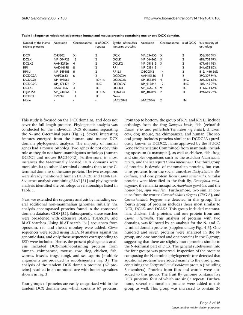
ResultsIdentification of proteins with a DCX domainHuman and mouse proteomes were searched forsequences similar to that of the human DCX domainyielding a total of 22 proteins containing one or two DCXrepeats (Table 1, the complete sequences used in thepresent study are found in supplementary Fig. 1).
Serine/threonine protein kinase domains were found inthree human/mouse proteins (DCLK, DCLK2, andDCLK3), and a ricin domain predicted to bind carbohy-drates was found in a human/mouse protein referred to asFLJ46154 [30]. The structure of the human FLJ46154 andDCDC2B proteins differed from other proteins with tan-dem repeats; they contained a repeat more similar to DCXC-terminal repeat, which appeared in the N-terminal partof this protein, and a second repeat more similar to DCXN-terminal repeat. In the mouse orthologs of these twoproteins, only one DCX domain was present. All themouse genes reside in chromosomal regions (Fig. 1b),which are synthenic to the human orthologs (supplemen-tary Fig. 2). This includes also the location of DCDC1 andBAC26042, however they are not true orthologs since thesequence similarity is very low (52%, among 46 out of 86amino acids) only in the DCX domain, and the phyloge-netic and evolutionary analysis, described below, indicatethat they are different. BAC26042 is also unique in itsclose physical proximity with FLJ46154, the distancebetween these two genes being only 2 kb, suggesting theymay share common regulatory elements.
Page 2 of 16(page number not for citation purposes)
BMC Genomics 2006, 7:188 http://www.biomedcentral.com/1471-2164/7/188
This study is focused on the DCX domains, and does notcover the full-length proteins. Phylogenetic analysis wasconducted for the individual DCX domains, separatingthe N- and C-terminal parts (Fig. 2). Several interestingfeatures emerged from the human and mouse DCXdomain phylogenetic analysis. The majority of humangenes had a mouse ortholog. Two genes do not obey thisrule as they do not have unambiguous orthologs (humanDCDC1 and mouse BAC26042). Furthermore, in mostinstances the N-terminally located DCX domains weremore similar to other N-terminal domains than to the C-terminal domains of the same protein. The two exceptionswere already mentioned; human DCDC2B and FLJ46154.Sequence analysis combining BLAT [31] and phylogeneticanalysis identified the orthologous relationships listed inTable 1.
Next, we extended the sequence analysis by including sev-eral additional non-mammalian genomes. Initially, theanalysis encompassed proteins found in the conserveddomain database CDD [32]. Subsequently, these searcheswere broadened with extensive BLAST, TBLASTN, andBLAT searches. Using BLAT search [31] sequences fromopossum, rat, and rhesus monkey were added. Cionasequences were added using TBLATN analysis against thegenomic data, and only those sequences corresponding toESTs were included. Hence, the present phylogenetic anal-ysis included DCX-motif-containing proteins fromhuman, chimpanzee, mouse, cow, dog, chicken, fish,worms, insects, frogs, fungi, and sea squirts (multiplealignments are provided in supplementary Fig. 3). Theanalysis of the tandem DCX domain proteins (67 pro-teins) resulted in an unrooted tree with bootstrap valuesshown in Fig. 3.
Four groups of proteins are easily categorized within thetandem DCX domain tree, which contains 67 proteins.
From top to bottom, the group of RP1 and RP1L1 includeorthologs from the frog Xenopus laevis, fish (zebrafishDanio rerio, and pufferfish Tetraodon nigrovidis), chicken,cow, dog, mouse, rat, chimpanzee, and human. The sec-ond group includes proteins similar to DCDC2A (previ-ously known as DCDC2, name approved by the HUGOGene Nomenclature Committee) from mammals, includ-ing opossum (a marsupial), as well as chicken, fish, frog,and simpler organisms such as the ascidian Halocynthiaroretzi, and the sea squirt Ciona intestinalis. The third groupof proteins is devoid of mammalian proteins, but con-tains proteins from the social amoebae Dictyostelium dis-coideum, and one protein from Ciona intestinalis. Similarproteins were identified in the fruit fly, Drosophila mela-nogaster, the malaria mosquito, Anopheles gambiae, and thehoney bee, Apis mellifera. Furthermore, two similar pro-teins from the worms Caenorhabditis elegans (ZYG-8), andCaenorhabditis briggsae are detected in this group. Thefourth group of proteins includes those most similar toDCX, DCLK, and DCLK2. This group included mamma-lian, chicken, fish proteins, and one protein from andCiona intestinalis. This analysis of proteins with twodomains, was followed by an analysis for the N- and C-terminal domain proteins (supplementary Figs. 4-5). Onehundred and seven proteins were analyzed in the N-group, and one hundred and one proteins in the C-group,suggesting that there are slightly more proteins similar tothe N-terminal part of DCX. The general subdivision intothe four groups was preserved. Inspection of the proteinscomposing the N-terminal phylogenetic tree detected thatadditional proteins were added mainly to the third groupcontaining the Dictyostelium discoideum protein (including8 members). Proteins from flies and worms were alsoadded to this group. The fruit fly genome contains fiveDCX proteins, four of which are single repeats. Further-more, several mammalian proteins were added to thisgroup as well. This group was increased to contain 26
Table 1: Sequence relationships between human and mouse proteins containing one or two DCX domains.
Symbol of the Homo sapiens proteins
Accession Chromosome # of DCX repeats
Symbol of the Mus musculus proteins
Accession Chromosome # of DCX % similarity of orthologs
DCX O43602 X 2 DCX NP_034155 X 2 358/360 99%DCLK NP_004725 13 2 DCLK NP_064362 3 2 681/702 97%DCLK2 AAH32726 4 2 DCLK2 NP_081815 3 2 679/691 98%RP1 AAD44198 8 2 RP1 NP_035413 1 2 544/675 80%RP1L1 NP_849188 8 2 RP1L1 Q8CGM2 14 2 812/1440 56%DCDC2A AAF23612 6 2 DCDC2A AAH45136 13 2 290/307 94%DCDC2B XP_497666 1 1C+1N DCDC2B XP_357395 4 1NC 207/303 68%DCDC2C XP_371476 2 1NC DCDC2C XP_917846 12 1NC 107/145 73%DCLK3 BAB21856 3 1C DCLK3 NP_766516 9 1C 411/633 64%FLJ46154 NP_940864 11 1C+1N FLJ46154 XP_489892 2 1C 494/649 76%DCDC1 P59894 11 1C NoneNone - - - BAC26042 BAC26042 2 1N
Page 3 of 16(page number not for citation purposes)

BMC Genomics 2006, 7:188 http://www.biomedcentral.com/1471-2164/7/188
Page 4 of 16(page number not for citation purposes)
A. Schematic representation of human and mouse proteins containing DCX domainsFigure 1A. Schematic representation of human and mouse proteins containing DCX domains. DCX domains more similar to the N-ter-minal repeat of DCX were labeled in green, whereas those more similar to the C-terminal repeat were labeled in purple. Pro-tein kinase domains are marked in yellow. The ricin domain is marked in brown. Human proteins are positioned on the top, the mouse proteins are shown below. B. Schematic presentation of the chromosomal location of the human and mouse DCX domain genes using UCSC site [51].

BMC Genomics 2006, 7:188 http://www.biomedcentral.com/1471-2164/7/188
Page 5 of 16(page number not for citation purposes)
Maximum Likelihood (ML) phylogenetic tree including DCX domain proteins from human and mouse, bootstrap values are indicatedFigure 2Maximum Likelihood (ML) phylogenetic tree including DCX domain proteins from human and mouse, bootstrap values are indicated.

BMC Genomics 2006, 7:188 http://www.biomedcentral.com/1471-2164/7/188
Page 6 of 16(page number not for citation purposes)
ML tree of the tandem DCX domain proteins from different speciesFigure 3ML tree of the tandem DCX domain proteins from different species. Bootstrap values are indicated.
BMC Genomics 2006, 7:188 http://www.biomedcentral.com/1471-2164/7/188
members in the N-group and 19 members in the C-group.This group included a protein from the unicellular organ-ism Plasmodium falciparum, the malaria parasite.
Inspection of the proteins composing the C-terminal phy-logenetic tree detected a group containing all the DCLK3proteins. It should be noted that this group as a whole isquite distinct from DCX, DCLK, and DCLK2. Proteins inthis group contain a single DCX domain from mammals(human, chimpanzee, cow, rat, and opossum), but alsofrom fruit flies, honeybees, and malaria mosquitoes. Anexception is the ciona protein demarking this group(Sca_10), which has a tandem repeat. One of the groupscontain both DCDC2A and DCDC2B proteins, and yet anadditional group contains several more DCDC2B pro-teins, suggesting probably less evolutionary conservedsequences in the C-terminal domains of this subset of pro-teins.
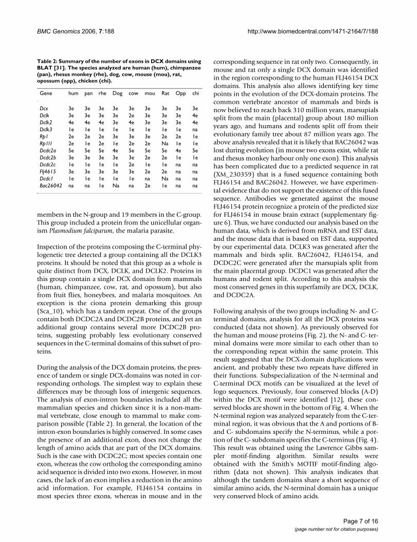
During the analysis of the DCX domain proteins, the pres-ence of tandem or single DCX-domains was noted in cor-responding orthologs. The simplest way to explain thesedifferences may be through loss of intergenic sequences.The analysis of exon-intron boundaries included all themammalian species and chicken since it is a non-mam-mal vertebrate, close enough to mammal to make com-parison possible (Table 2). In general, the location of theintron-exon boundaries is highly conserved. In some casesthe presence of an additional exon, does not change thelength of amino acids that are part of the DCX domains.Such is the case with DCDC2C; most species contain oneexon, whereas the cow ortholog the corresponding aminoacid sequence is divided into two exons. However, in mostcases, the lack of an exon implies a reduction in the aminoacid information. For example, FLJ46154 contains inmost species three exons, whereas in mouse and in the
corresponding sequence in rat only two. Consequently, inmouse and rat only a single DCX domain was identifiedin the region corresponding to the human FLJ46154 DCXdomains. This analysis also allows identifying key timepoints in the evolution of the DCX-domain proteins. Thecommon vertebrate ancestor of mammals and birds isnow believed to reach back 310 million years, marsupialssplit from the main (placental) group about 180 millionyears ago, and humans and rodents split off from theirevolutionary family tree about 87 million years ago. Theabove analysis revealed that it is likely that BAC26042 waslost during evolution (in mouse two exons exist, while ratand rhesus monkey harbour only one exon). This analysishas been complicated due to a predicted sequence in rat(XM_230359) that is a fused sequence containing bothFLJ46154 and BAC26042. However, we have experimen-tal evidence that do not support the existence of this fusedsequence. Antibodies we generated against the mouseFLJ46154 protein recognize a protein of the predicted sizefor FLJ46154 in mouse brain extract (supplementary fig-ure 6). Thus, we have conducted our analysis based on thehuman data, which is derived from mRNA and EST data,and the mouse data that is based on EST data, supportedby our experimental data. DCLK3 was generated after themammals and birds split. BAC26042, FLJ46154, andDCDC2C were generated after the marsupials split fromthe main placental group. DCDC1 was generated after thehumans and rodent split. According to this analysis themost conserved genes in this superfamily are DCX, DCLK,and DCDC2A.
Following analysis of the two groups including N- and C-terminal domains, analysis for all the DCX proteins wasconducted (data not shown). As previously observed forthe human and mouse proteins (Fig. 2), the N- and C- ter-minal domains were more similar to each other than tothe corresponding repeat within the same protein. Thisresult suggested that the DCX-domain duplications wereancient, and probably these two repeats have differed intheir functions. Subspecialization of the N-terminal andC-terminal DCX motifs can be visualized at the level oflogo sequences. Previously, four conserved blocks (A-D)within the DCX motif were identified [12], these con-served blocks are shown in the bottom of Fig. 4. When theN-terminal region was analyzed separately from the C-ter-minal region, it was obvious that the A and portions of B-and C- subdomains specify the N-terminus, while a por-tion of the C- subdomain specifies the C-terminus (Fig. 4).This result was obtained using the Lawrence Gibbs sam-pler motif-finding algorithm. Similar results wereobtained with the Smith's MOTIF motif-finding algo-rithm (data not shown). This analysis indicates thatalthough the tandem domains share a short sequence ofsimilar amino acids, the N-terminal domain has a uniquevery conserved block of amino acids.
Table 2: Summary of the number of exons in DCX domains using BLAT [31]. The species analyzed are human (hum), chimpanzee (pan), rhesus monkey (rhe), dog, cow, mouse (mou), rat, opossum (opp), chicken (chi).
Gene hum pan rhe Dog cow mou Rat Opp chi
Dcx 3e 3e 3e 3e 3e 3e 3e 3e 3eDclk 3e 3e 3e 3e 2e 3e 3e 3e 4eDclk2 4e 4e 4e 3e 4e 3e 3e 3e 4eDclk3 1e 1e 1e 1e 1e 1e 1e 1e naRp1 2e 2e 2e 3e 3e 3e 2e 2e 1eRp1l1 2e 1e 2e 1e 2e 2e Na 1e 1eDcdc2a 5e 5e 5e 4e 5e 5e 5e 4e 5eDcdc2b 3e 3e 3e 3e 3e 2e 2e 1e 1eDcdc2c 1e 1e 1e 1e 2e 1e 1e na naFlj4615 3e 3e 3e 3e 3e 2e 2e na naDcdc1 1e 1e 1e 1e 1e na Na na naBac26042 na na 1e Na na 2e 1e na na
Page 7 of 16(page number not for citation purposes)

BMC Genomics 2006, 7:188 http://www.biomedcentral.com/1471-2164/7/188
Expression analysis by in situ hybridizationTaken into consideration the similarities among the dif-ferent DCX-domain paralogs, and their common func-tions in relation to signal transduction and microtubuleregulation [26], it is important to establish when andwhere these genes are expressed. This will help in deline-ating their potential function. For example, the distinctionwhether a specific gene is expressed in proliferating,migrating, or differentiating cells is critical when trying tofigure out gene function. Additionally, coexpression in aparticular tissue may indicate that paralogs could cooper-ate or be redundant.
Our analysis was carried out by in situ hybridization atE14.5, a stage at which many differentiated cell types char-acteristic of an adult organism have formed yet at sametime such mid-gestation embryonic tissues still containprogenitor cells. This analysis was performed with thegoal to generate an expression profile "snapshot". Withthe exception of the ubiquitously expressed Dcdc2B (Fig.5D), expression patterns of genes encoding DCX-repeat-
containing proteins are to a greater or lesser extentregional. Dcx, Dclk and Dclk2 are expressed in the centraland peripheral nervous system including the brain, spinalcord, cranial and dorsal root ganglia and in the parasym-pathetic ganglia (Fig. 5A–C). A high power view (Fig. 5E–H) shows that in the developing neocortex Dcx and Dclktranscripts are much more abundant in the preplate, butindividual cells expressing the Dcx and Dclk genes can bedetected in the ventricular zone. Both Dclk2 and Dcdc2Bare expressed in the developing neocortex, largely uni-form and at low levels, but more pronounced in the ven-tricular zone than Dcx and Dclk. Outside the nervoussystem, prominent sites of Dcx and Dclk expression are theskeletal muscles, tongue muscles and individual cells ofthe olfactory epithelium (Fig. 5A,B). The latter tissue alsoexpresses Dclk2 (Fig. 5C).
BAC26042, FLJ46154 and Dcdc2A exhibit highly regionalexpression patterns, which in the brain appear to be simi-lar for BAC26042 and FLJ46154 (Fig. 5I–K). Fig. 5I and 5Jshow sagittal sections through the forebrain with
Sequence logos of the N-terminal and C-terminal DCX motifsFigure 4Sequence logos of the N-terminal and C-terminal DCX motifs. Multiple alignments of the motifs from the DCX motifs are shown as sequence logos. The height of each amino acid represents bits of information and is proportional to its conservation at that position (y-axis), after the sequences have been weighted and frequencies adjusted by the expected amino acid fre-quency. Below the logos is the numbering of amino acids within the internal A-D subdomains. This SeqLogo represents the Lawrence Gibbs sampler motif-finding algorithm.
Page 8 of 16(page number not for citation purposes)

BMC Genomics 2006, 7:188 http://www.biomedcentral.com/1471-2164/7/188
Page 9 of 16(page number not for citation purposes)
In situ hybridization patterns of genes containing the DCX protein domainFigure 5In situ hybridization patterns of genes containing the DCX protein domain. Blue stain denotes expression of the gene whose name is indicated in each panel. For details see Text. (A-D) Sagittal sections of whole E14.5 embryos. (E-H) Expression in fron-tal cortex. Note a few cells expressing in the ventricular zone. (I-K) Striking localized expression of BAC26042 (I), FLJ46154 (J), and Dcdc2A (K) in the E14.5 brain. In (I) "t" becomes "th" for thalamus. (L-Q) Expression patterns in the E14.5 eye. Abbrevia-tions: cerebellum (cb), choroids plexus (cp), cortex (cx), dorsal root ganglia (drg), hypothalamus (h), inner neuroblastic layer (inl), midbrain (mb), muscles (m), olfactory epithelium (oe), outer neuroblastic layer (onl), pons (p), preplate (pp), septum (s), spinal cord (sc), thalamus (th), thymus (ty). tongue (t), ventricular zone (VZ).

BMC Genomics 2006, 7:188 http://www.biomedcentral.com/1471-2164/7/188
Page 10 of 16(page number not for citation purposes)
A) Clustered Unigene [33] gene-tissue expression dataFigure 6A) Clustered Unigene [33] gene-tissue expression data. B) Gene-gene correlations based on Unigene expression data.

BMC Genomics 2006, 7:188 http://www.biomedcentral.com/1471-2164/7/188
BAC26042 and FLJ46154 transcripts present in the sep-tum, various cell groups of the ventral thalamus, and inthe posterior hypothalamus. Other sites of expression area group of neurons at the base of the olfactory bulb (Fig.5I,J), the pretectal area, the facial nucleus, and scatteredneurons in the ventral and dorsal parts of the spinal cord(data not shown). Dcdc2A expression in the CNS isrestricted to a group of scattered neurons in the lateralmost part of the developing cerebellum (Fig. 5K).BAC26042 and Dcdc2A are expressed in the choroid plexi(Fig. 5I,K).
The majority of DCX-repeat encoding genes are expressedin the developing retina. Three types of patterns emerge:Dcx, Dclk, Dclk2 transcripts are strongly expressed in thepostmitotic inner neuroblastic layer (Fig. 5L–N), whereasBAC26042 and FLJ46154 are also expressed in this layer,but in a more restricted fashion near and at its surface (Fig.5P,Q). Finally Rp1l1 transcripts are found in the outerneuroblastic layer that contains proliferating cells (Fig.5O). Radially arranged Dcx, Dclk or Dclk2-expressing cellsare detected in the outer neuroblastic layer which is remi-niscent of the situation seen in the ventricular zone of theneocortex (Fig. 5E–G).
In addition, lung and kidney express Dcx, Dclk andDcdc2A. Dclk2 transcripts are also found in the developingovary and weak expression is also seen throughout thekidney (data not shown).
Our analysis included most of the 11 genes listed in Table1, the exceptions being Dclk3, and Dcdc2C for which wecould not yet identify suitable templates. Rp1 was alsoexamined but it is not expressed at E14.5, except expres-sion noted in some midline cells of the spinal cord (datanot shown). To summarize our studies, we found that tis-sues destined to respond to electrical stimuli – central andperipheral nervous systems and skeletal muscles – repre-sent the most striking sites of expression of DCX-repeatencoding genes. Outside these tissues, expression ismostly low and usually not regional, the exceptions beingkidney and lung.
Expression analysis in human and mouseThe relevance of functional genomics approaches usingmouse models for studying human diseases obviouslydepends on the similarity of gene expression in the twospecies. Thus, we compared the expression of the humanmembers of the DCX gene superfamily investigated in thisstudy with their mouse orthologs. For this purpose, weused the Unigene database of expression data website. Tis-sue-dependent expression profiles for both human andmurine DCX repeat-containing proteins were generatedfrom the EST count provided by UNIGENE [33]. Since themouse-human comparison was a key feature, the analysis
was limited to tissues with a high total number of ESTcounts that were common to both organisms. We ana-lyzed data for ten different human genes, and eight mousegenes. For two human genes there were no correspondingexpression data in mouse: DCDC2B, which has a mouseortholog that is not listed in UNIGENE, and DCDC1,which does not have a mouse ortholog. The clusteredexpression data resulting from this analysis is shown Fig.6A and a gene-gene correlation based on this informationis shown in Fig. 6B.
We tested the significance of the correlation by randompermuation analysis. The correlations were re-calculated1000 times after rescambling for each gene independentlyall tissues at random. We found that all high correlation(>0.5) were significant (p < 0.01). Two clusters revealingvery high correlation were observed. The largest groupincluded human RP1 and RP1L1, and their murineorthologs. In addition, DCDC1, which so far had beenreported to be expressed mainly in testis, and embryonicbrain [34], was included in this group. This group is char-acterized by high levels of expression in the eye, which iscommon amongst most DCX proteins, and has beennoted in our in situ analysis. In addition to expression inthe eye, these genes are expressed at lower levels only in afew other tissues. In this group there is no clear distinctionin the gene-gene correlation in the expression in mouseand human. The correlation between the different mem-bers of this group is >0.9 in all cases. Both the human andthe mouse FLJ46154 are related to this group, however thecorrelation between the human and mouse FLJ46154 islow (0.3). The protein-products of these two genes havealso diverged, with a loss of a DCX domain in the mouseprotein. Thus, it may be possible that there has been lessconservation in the regulatory regions of these genes aswell.
The second group exhibiting high gene-gene correlationsincludes the murine genes Dcx, Dclk, and Dclk2, and theirhuman orthologs. Human DCLK2 exhibited somewhatlower correlations with its mouse ortholog (0.4) than theother genes in this group. This may stem from its generaloverall lower expression levels (Fig. 6A). Our in situ dataalso indicated a high similarity in the co-expression ofDcx, Dclk, and Dclk2. Furthermore, our functional analysis[26] indicated that this group shares more properties andonly they interact with the scaffold protein neurabin 2. Athird group of genes with lower levels of correlationinclude DCDC2A, DCLK3, Dcdc2A, and Dclk3. In thisgroup the correlation between the correspondingorthologs does not exceed 0.5. It should be noted thatthere are some additional high correlations between dif-ferent genes, for example; DCLK3 and Flj46154, orFLJ46154 with DCX, DCLK, and Dcx.
Page 11 of 16(page number not for citation purposes)

BMC Genomics 2006, 7:188 http://www.biomedcentral.com/1471-2164/7/188
DiscussionProtein architectureNumber of doublecortin repeatsThe DCX domain family of proteins consists of multiplemembers in the animal kingdom from the unicellularorganisms Plasmodium and Dictyostelium (in the non-aggregated part of its life cycle) to human. Although, Plas-modium and Dictyostelium do not have a nervous system,motility is one of their important characteristics. The shiftfrom a single to a tandem DCX repeat (or vice versa) is arather simple mode to modifying protein activity frombinding to tubulin and assisting MT polymerization (onerepeat) to enabling it to promote MT bundling (tworepeats). We have demonstrated experimental evidencefor this view for DCX [12], and for eight additional mem-bers of the mouse DCX domain proteins [26]. The dupli-cation of a single DCX motif was an ancient event becauseof the existence of a tandem repeat already in Dictyostel-ium, and since the similarity within the N-terminaldomains is generally higher than to the C-terminaldomains of the same protein. We were able to documentthe loss of a domain, by the loss of an exon ("domaindeath"). Interestingly, the group of proteins encoding asingle repeat is expanded in nematodes. These singlerepeat proteins are usually similar to the same extent tothe groups of the N- and C- terminal repeats. Our previousanalysis [12], structural analysis of DCX [14,15,35], andour current analysis reveals differences between the N-ter-minal and the C-terminal domains. Our analysis detecteda short stretch of conserved amino acids common to bothN-terminal and C-terminal repeats, however thesequences diverged and the N-terminal domain has aunique block of conserved amino acids. Close inspectionof the phylogenetic trees revealed higher evolutionaryconservation among proteins in the N-terminal groupthan those in the C-terminal group. Our analysis detectedthat both the N-terminal and C-terminal domains of DCXpromote assembly of MTs [12], however the N-terminalportion has higher activity than the C-terminal one. Inaddition, other proteins containing a single or tandemDCX domain were capable of promoting MT assembly[26]. Among the studied mouse proteins FLJ46154 exhib-ited the highest activity in promoting MT assembly [26].This protein contains a single DCX domain, which is moresimilar to the C-terminal domain of DCX. Future studiesdirected at the structural analysis of the different DCXdomains combined with the sequence analysis conductedhere may provide the basis for elucidating these func-tional variations. Another study indicated that the N-ter-minal domain of DCX binds only to assembled MTs,whereas the C-terminal domain binds both to assembledMTs and to unpolymerized tubulin [14]. Therefore, theconserved amino acid block common to both repeats maybe important in directing the binding to assembled MTs.
Additional domainsA significant portion of the DCX-domain containing pro-teins has a kinase domain attached to them. This groupincludes DCLK, and DCLK2, in which a tandem DCXrepeat is detected, and DCLK3, which contains a singlerepeat more similar to the C-terminal one. Orthologs ofthese proteins are also found also in invertebrates includ-ing nematodes. The C. elegans ortholog ZYG-8 has beenstudied extensively [36]. In nematodes, ZYG-8 was foundto be important for assembly of astral microtubules. Themutant ZYG-8 phenotype was observed with several dif-ferent alleles including mutations in the DCX domain,and the kinase domain, therefore suggesting a role forkinase activity in regulating MT assembly. So far, theendogenous substrates of these kinases, besides DCLKwhich undergoes autophosphorylation [37], areunknown.
Addition (or reduction) of the number of domains is notalways conserved in evolution. In close relationship withDCLK3, which contains a kinase domain, a branchincluding proteins with a different domain is found. Thisbranch includes the Drosophila melanogaster protein(EMAL_DROME), which contains an additional HELPdomain, and WD repeats, which can be detected in theclosely related Anopheles gambiae, and Apis mellifera pro-teins. The HELP (Hydrophobic ELP) motif is found inEMAP and EMAP-like proteins (ELPs), and has beenfound to mediate binding to MTs in vitro [38,39]. There-fore, the combinatorial possibilities of modifying orchanging functional activities will depend upon specificamino acid substitutions within the DCX domains, andaddition of other functional domains.
Additional ways to add on functionsOn the level of the whole organism, one of the best waysto modify protein activity is to vary the expression pattern.Indeed, even in the case of proteins with similar sequenceand similar expression pattern such as in the case of DCX,DCLK, and DCLK2 some notable differences in expres-sion pattern were noted. For example, the expression ofDCLK2 in the ventricular zone of the cerebral cortex ismuch more obvious than that of DCLK, suggesting thatthe former may have a pronounced role in regulation ofproliferation of neurogenic progenitors. In fact, theinvolvement of DCLK in regulation of mitosis has beendemonstrated recently [40]. DCLK regulated the forma-tion of bipolar mitotic spindles and the proper transitionfrom prometaphase to metaphase during mitosis inHEK293 cells and neuronal progenitors. In cultured corti-cal neural progenitors, DCLK RNAi disrupted the structureof mitotic spindles and the progression of M phase, caus-ing an increase of cell-cycle exit index and an ectopic com-mitment to a neuronal fate [40]. As mentioned above, theC. elegans ortholog of DCLK, ZYG-8 regulates assembly of
Page 12 of 16(page number not for citation purposes)

BMC Genomics 2006, 7:188 http://www.biomedcentral.com/1471-2164/7/188
astral microtubules [36]. Taken together, these findingssuggest that regulation of the MT-based mitotic spindlemay represent a conserved function among DCX-domainproteins.
Two genes FLJ46154, and BAC26042, exhibited very sim-ilar expression patterns in the mouse. In the mousegenome, the distance between these two genes is only 2kb, suggesting that they may share common regulatorysequences. Perhaps due to such a highly similar expres-sion pattern, the loss of BAC26042 in other mammalianspecies is not surprising. Partial copies of this gene (con-taining only one exon instead of two) exist in rat and inthe rhesus monkey. A proposed unified nomenclature forthe doublecortin superfamily based on correspondencewith the mouse gene nomenclature committee is pre-sented in supplementary figure 7.
Expression patterns and phylogenetic correlationsOne of the most exciting outcomes of the present analysisis that in some cases we observed a high correlationbetween orthologs and paralogs, and their expression pro-files. This was most striking in case of the human andmouse RP1, and RP1L1. A possible hypothesis, based onthese results is that a high correlation in the expression ofthe human and mouse genes may be indicative of a highprobability that a mouse mutant would model the humandisease (or vice versa). If we review this hypothesis withinthe limited genetic data available, we see that this hypoth-esis is supported. Rp1-/- mice closely reflect very well theprogressive blindness observed in human patients [16-18,41,42]. Furthermore, in these mice the JNK pathwaywas affected [43], and our functional analysis describe apossible connection between RP1 and the JNK pathway[26]. An unexpected member to this group is DCDC1. Thegene-to-gene function correlations are also very high incase of the human and mouse DCX, DCLK, and DCLK2genes. They are found on adjacent branches in the phylo-genetic analysis. These genes are highly expressed in thecentral nervous system not only in human and mouse, butalso in additional animals as chicken (DCX, DCLK [44],and DCLK2 (unigene data for Dcx Gga.2608, Dclk2Gga.16742), cow (dclk and dclk2; Bt.34533, Bt.55548),rat (dcx Rn.121471, dclk Rn.155540, dclk2 Rn.23327),and dog (dcx Cfa.19843, dclk Cfa.9988). Our analysisdetected expression of some of these genes in several addi-tional tissues such as muscle, heart, and kidney, siteswhich have not been previously described. Although Dcx-/- mice have minor neuronal abnormalities [21,45], theydo not adequately recapitulate the human disease. This isdue to gene redundancy because the double mutant miceDcx-/- and Dclk-/- exhibited perinatal lethality accompa-nied with multiple brain abnormalities [23,24]. However,using in utero electroporation in the rat and mouseembryo, reduction in the expression of Dcx resulted in
apparent inhibition of migration of neurons in the cere-bral cortex [22,46]. In the rat, it was possible to observeheterotropic positioning of neurons in postnatal ratbrains similar to SBH in human [22]. In contrast to thecases discussed, there are several genes for which theorthologs exhibit low gene-gene correlation; the lowestare DCLK3 and FLJ46154 (0.3), DCLK2 (0.4), andDCDC2A (0.5). Among these genes variability insequence analysis was noted as well. With future expan-sion of available information in databases, this analysiswill be extended to include many more tissues, additiondevelopmental stages, and additional species. We suggestthat this analysis may be useful in predicting possiblefunctional redundancy. Furthermore, it may provide use-ful clues in which tissues the mouse gene expression ismost similar to its human ortholog, thus suggesting thepossible relevance of a corresponding mouse model for ahuman disease.
ConclusionIn summary, the DCX-domain family of proteins hasproven to be a very successful and prolific protein familyinvolved in signal transduction and cytoskeletal regula-tion. Modifications in regulatory sequences allowingunique expression patterns, addition of new domains,and modifying the amino acid sequence of the DCXdomains themselves explains – at least in part- why thissuperfamily is thriving.
MethodsDatabase homology search and phylogenetic analysisOur database similarity search initiated with an NCBIblast search using the sequence of mouse doublecortinprotein (NP_034155.2). In addition, all proteins with theDCX domain were retrieved from the CDD, (ConservedDomain DB). In a similar fashion, DCX proteins from theEBI InterPro db, which were not detected in CDD wereextracted and added. In order not to miss relatedorthologs, BLAT analysis using UCSC BLAT site [31] wasconducted initiating with the human and mouse genes.Definition of orthologs was based on positioning in syn-tenic chromosomal localizations. In addition, an exhaus-tive TBLASTN search was conducted to detect DCX-relatedtranscripts in the EST database of Ciona intestinalis (seasquirt) [47]. The corresponding cDNAs were mapped totheir corresponding genomic localizations.
For our analysis, three datasets were generated, the firstdataset for the proteins with 2 DCX motifs (includingboth the N-term and C-term repeats) composed of 57 pro-teins from human, mouse, chicken, bovine, ciona, frog,fish, nematodes, chimp and flies.
The second dataset included the proteins with a singleDCX motif, which exhibited higher similarity to the
Page 13 of 16(page number not for citation purposes)

BMC Genomics 2006, 7:188 http://www.biomedcentral.com/1471-2164/7/188
mouse doublecortin C-terminal DCX motif, and the C-ter-minal ones of those with two DCX motifs. The singlerepeats exhibited which exhibited identical similarity toboth the N- and C-terminal repeats of mouse DCX wereincluded in both N-term and C-term groups. The compar-ison was based on two-way BLAST analysis usingblast2seq. This dataset integrated 77 proteins fromhuman, mouse, chicken, ciona, dog, bovine, frog, fish, seasquirt, nematode, ascidian, fruit fly and fungus.
The third dataset was composed of the proteins with a sin-gle DCX motif, which was more similar to the mouse dou-blecortin N-terminal DCX motif, and the N-terminal onesof those with two DCX motifs. This dataset included 85proteins from the species mentioned above.
Multiple alignment program, Clustalw, was run on eachof the datasets and the regions corresponding to the DCXmotifs were extracted, resulting in three multiple align-ments (data sets are deposited in supplementary data 1).
The Phylip package ver. 3.6 was used to build the ML treeswith boot strapping values.
One hundred datasets were generated using the programSEQBOOT from the original data (multiple alignmentdone by clustalw) for each of the 3 groups of motifs (2-repeats, C-terminal repeats and N-terminal repeats). Thiswas followed by running the program ProML (proteinMaximum Likelihood) on each of the datasets in thegroup, using the JTT model. A consensus tree (from all the100 trees) was generated using the program CONSENSE.
The trees were drawn using NJPLOT program or TreeView[48]. All sequences of the three original datasets are avail-able in supplementary Fig. 1.
Separate datasets were generated for the human andmouse proteins, the first dataset for the proteins that havetwo DCX motifs (N-terminal and C-terminal) composedof 13 proteins. The second dataset for the proteins with asingle DCX motif more similar to the mouse doublecortinC-term DCX motif, and the C-terminal ones of those withtwo DCX motifs. This dataset included 20 proteins. Athird dataset included the proteins with a single DCXmotif more similar to the mouse doublecortin N-termDCX motif, and the N-terminal ones of those with twoDCX motifs. This dataset has 21 proteins. These datasetswere subject to all the programs as mentioned above, clus-talw, seqboot, proml, consense and njplot. CONSENSEand NJPLOT were used to draw the human-mouse phylo-genetic trees.
Multiple alignment was done by Block Maker web serverat the Fred Hutchinson Cancer Research Center in Seattle,
Washington, USA [49]. The Logoshown represents theLawrence Gibbs sampler motif-finding algorithm, similar(but not identical) results were obtained using Smith'sMOTIF motif-finding algorithm.
Extraction of public large-scale expression data and cluster analysisEST counts from different human and murine tissue sam-ples were extracted for 10 human and 8 mouse genes fromthe UNIGENE website [33]. We discarded all human sam-ples with less than 150,000 total EST counts and retainedonly the murine samples that could be matched to thesesamples. The murine 'late gestation' sample was matchedto the human 'embryo'. Expression value are given as"counts per million of total ESTs". We applied a scalingtransformation 'x → t·arcsinh(x/t)' to the raw expressionvalues x to scale down (logarithmically) values above thethreshold t = 10.
In order to identify similarly expressed genes the com-bined set of expression data was clustered using Pearsoncorrelations over all tissues as similarity measures and sin-gle linkage for the generation of the dendrograms. Simi-larly, the tissues were clustered based on similarexpression across the genes. Alternative clustering meth-ods were tested and yielded similar results. We used thestandard Matlab clustering algorithm.
In situ hybridizationThe antisense RNA templates for in situ hybridizationwere generated by in vitro transcription using PCR prod-ucts (using the appropriate combinations of T7, T3, andSP6 primers) from the corresponding genes, which werecloned in BSIIKS+ or pGEMT. The sizes of the probesranged between 0.6 – 3 kb. The sequences of the templatescan be requested from the author. In several cases morethan one probe per gene was tested. Hybridizations anddata collection were done as described [50].
Authors' contributionsOR, SB, GE planned the experiments, collected and ana-lyzed the data, and wrote the manuscript. FMC, PB, TL,AK, TS, NB, conducted experiments, analyzed the data,and participated in manuscript writing.
Page 14 of 16(page number not for citation purposes)

BMC Genomics 2006, 7:188 http://www.biomedcentral.com/1471-2164/7/188
Additional material AcknowledgementsWe thank Prof. Shmuel Pietrekovski for helpful discussions. The work has been supported in part by the Fritz Thyseen Stifung Foundation, the Israeli Science Foundation (grant no. 270/04), Foundation Jérôme Lejeune, Min-erva foundation with funding from the Federal German Ministry for Educa-tion and Research, the German – Israeli collaboration grant Gr-1905, a grant from the Paul Godfrey Research Foundation in Childrens' diseases, the Kekst center, the Forcheimer center, the Jewish communal fund Albert Einstein College of Medicine of Yeshiva University, and the David and Fela Shapell Family Center for Genetic Disorders Research. O.R. is an Incum-bent of the Berstein-Mason professorial chair of Neurochemistry. F. C. was supported by a Post-Doctoral Fellowship from the Association pour la Recherche sur le Cancer (Villejuif, France), and by the Sir Charles Clore Post-Doctoral Fellowship, the Weizmann Institute of Science (Rehovot, Israel).
References1. des Portes V, Pinard JM, Billuart P, Vinet MC, Koulakoff A, Carrie A,
Gelot A, Dupuis E, Motte J, Berwald-Netter Y, Catala M, Kahn A,Beldjord C, Chelly J: A novel CNS gene required for neuronalmigration and involved in X-linked subcortical laminar het-rotropia and lissencephaly syndrome. Cell 1998, 92:51-61.
2. Gleeson JG, Allen KM, Fox JW, Lamperti ED, Berkovic S, Scheffer I,Cooper EC, Dobyns WB, Minnerath SR, Ross ME, Walsh CA: dou-blecortin, a brain-specific gene mutated in human X-linkedlissencephaly and double cortex syndrome, encodes a puta-tive signaling protein. Cell 1998, 92:63-72.
3. Reiner O, Coquelle FM: Missense mutations resulting in type 1lissencephaly. Cell Mol Life Sci 2005, 62(4):425-434.
4. Dobyns WB, Reiner O, Carrozzo R, Ledbetter DH: Lissencephaly:a human brain malformation associated with deletion of theLIS1 gene located at chromosome 17p13. J Am med Ass 1993,270:2838-2842.
5. Barkovich AJ, Guerrini R, Battaglia G, Kalifa G, N'Guyen T, Parmeg-giani A, Santucci M, Giovanardi-Rossi P, Granata T, D'Incerti L: Bandheterotopia: correlation of outcome with magnetic reso-nance imaging parameters. Ann Neurol 1994, 36(4):609-617.
6. Horesh D, Sapir T, Francis F, Caspi M, Grayer Wolf S, Elbaum M,Chelly J, Reiner O: Doublecortin, a stabilizer of microtubules.Hum Mol Genet 1999, 8:1599-1610.
7. Gleeson JG, Lin PT, Flanagan LA, Walsh CA: Doublecortin is amicrotubule-associated protein and is expressed widely bymigrating neurons. Neuron 1999, 23(2):257-271.
8. Francis F, Koulakoff A, Boucher D, Chafey P, Schaar B, Vinet MC, Fri-ocourt G, McDonnell N, Reiner O, Kahn A, McConnell SK, Berwald-Netter Y, Denoulet P, Chelly J: Doublecortin is a developmen-tally regulated, microtubule-associated protein expressed inmigrating and differentiating neurons. Neuron 1999,23(2):247-256.
9. Yoshiura K, Noda Y, Kinoshita A, Niikawa N: Colocalization ofdoublecortin with the microtubules: An ex vivo colocaliza-tion study of mutant doublecortin. J Neurobiol 2000,43(2):132-139.
10. Burgess HA, Reiner O: Doublecortin-like Kinase, is a Microtu-bule-Associated Protein Kinase Expressed in Growth Cones.MCN 2000, 16:529-541.
11. Lin PT, Gleeson JG, Corbo JC, Flanagan L, Walsh CA: DCAMKL1encodes a protein kinase with homology to doublecortinthat regulates microtubule polymerization. J Neurosci 2000,20(24):9152-9161.
12. Sapir T, Horesh D, Caspi M, Atlas R, Burgess HA, Grayer Wolf S,Francis F, Chelly J, Elbaum M, Pietrokovski S, Reiner O: Doublecor-tin Mutations Cluster in Evolutionary Conserved FunctionalDomains. Hum Mol Genet 2000, 5:703-712.
13. Taylor KR, Holzer AK, Bazan JF, Walsh CA, Gleeson JG: Patientmutations in doublecortin define a repeated tubulin-bindingdomain. J Biol Chem 2000, 275(44):34442-34450.
14. Kim MH, Cierpicki T, Derewenda U, Krowarsch D, Feng Y, DevedjievY, Dauter Z, Walsh CA, Otlewski J, Bushweller JH, Derewenda ZS:The DCX-domain tandems of doublecortin and doublecor-tin-like kinase. Nat Struct Biol 2003, 10(5):324-333.
Additional File 1Supplementary Fig. 1: Detailed list and sequences of all the DCX proteins used in this study. The list contains the set of proteins with two domains. In addition, the N-terminal domains including proteins with single domains more similar to the N-terminal domain of DCX, and the C-ter-minal domains including proteins with single domains more similar to the C-terminal domain of DCX are presented in this set.Click here for file[http://www.biomedcentral.com/content/supplementary/1471-2164-7-188-S1.doc]
Additional File 2Supplementary Fig. 2: Synthenic positions of mouse and human DCX domain proteins.Click here for file[http://www.biomedcentral.com/content/supplementary/1471-2164-7-188-S2.doc]
Additional File 3Supplementary Fig. 3: CLUSTAL W (1.83) multiple sequence alignment of the DCX proteins with tandem domains, N-terminal domains and C-terminal domains.Click here for file[http://www.biomedcentral.com/content/supplementary/1471-2164-7-188-S3.doc]
Additional File 4Supplementary Fig. 4: ML tree of the N-terminal DCX domain proteins from different species. Bootstrap values are indicated.Click here for file[http://www.biomedcentral.com/content/supplementary/1471-2164-7-188-S4.jpeg]
Additional File 5Supplementary Fig. 5: ML tree of the C-terminal DCX domain proteins from different species. Bootstrap values are indicated.Click here for file[http://www.biomedcentral.com/content/supplementary/1471-2164-7-188-S5.jpeg]
Additional File 6Supplementary Fig. 6: Western blot analysis using anti-FLJ46154 anti-bodies. The specificity of the antibodies was verified using cells transfected with Flag-FLJ46154 (A) compared with the preimmune serum. (B) Anti-FLJ46154 antibodies recognize a protein of approximate size 78 kDa in mouse brain extract.Click here for file[http://www.biomedcentral.com/content/supplementary/1471-2164-7-188-S6.jpeg]
Additional File 7Supplementary Fig. 7: Proposed nomenclature for the doublecortin super-family.Click here for file[http://www.biomedcentral.com/content/supplementary/1471-2164-7-188-S7.doc]
Page 15 of 16(page number not for citation purposes)
http://www.ncbi.nlm.nih.gov/entrez/query.fcgi?cmd=Retrieve&db=PubMed&dopt=Abstract&list_uids=9489699
http://www.ncbi.nlm.nih.gov/entrez/query.fcgi?cmd=Retrieve&db=PubMed&dopt=Abstract&list_uids=9489699
http://www.ncbi.nlm.nih.gov/entrez/query.fcgi?cmd=Retrieve&db=PubMed&dopt=Abstract&list_uids=9489699
http://www.ncbi.nlm.nih.gov/entrez/query.fcgi?cmd=Retrieve&db=PubMed&dopt=Abstract&list_uids=9489700
http://www.ncbi.nlm.nih.gov/entrez/query.fcgi?cmd=Retrieve&db=PubMed&dopt=Abstract&list_uids=9489700
http://www.ncbi.nlm.nih.gov/entrez/query.fcgi?cmd=Retrieve&db=PubMed&dopt=Abstract&list_uids=9489700
http://www.ncbi.nlm.nih.gov/entrez/query.fcgi?cmd=Retrieve&db=PubMed&dopt=Abstract&list_uids=7524438
http://www.ncbi.nlm.nih.gov/entrez/query.fcgi?cmd=Retrieve&db=PubMed&dopt=Abstract&list_uids=7524438
BMC Genomics 2006, 7:188 http://www.biomedcentral.com/1471-2164/7/188
15. Moores CA, Perderiset M, Francis F, Chelly J, Houdusse A, MilliganRA: Mechanism of microtubule stabilization by doublecortin.Mol Cell 2004, 14(6):833-839.
16. Pierce EA, Quinn T, Meehan T, McGee TL, Berson EL, Dryja TP:Mutations in a gene encoding a new oxygen-regulated pho-toreceptor protein cause dominant retinitis pigmentosa. NatGenet 1999, 22(3):248-254.
17. Sullivan LS, Heckenlively JR, Bowne SJ, Zuo J, Hide WA, Gal A, Den-ton M, Inglehearn CF, Blanton SH, Daiger SP: Mutations in a novelretina-specific gene cause autosomal dominant retinitis pig-mentosa. Nat Genet 1999, 22(3):255-259.
18. Gao J, Cheon K, Nusinowitz S, Liu Q, Bei D, Atkins K, Azimi A, DaigerSP, Farber DB, Heckenlively JR, Pierce EA, Sullivan LS, Zuo J: Pro-gressive photoreceptor degeneration, outer segment dys-plasia, and rhodopsin mislocalization in mice with targeteddisruption of the retinitis pigmentosa-1 (Rp1) gene. Proc NatlAcad Sci USA 2002, 99(8):5698-5703.
19. Meng H, Smith SD, Hager K, Held M, Liu J, Olson RK, Pennington BF,Defries JC, Gelernter J, O'Reilly-Pol T, Somlo S, Skudlarski P, ShaywitzSE, Shaywitz BA, Marchione K, Wang Y, Paramasivam M, Loturco JJ,Page GP, Gruen JR: DCDC2 is associated with reading disabilityand modulates neuronal development in the brain. Proc NatlAcad Sci USA 2005.
20. Schumacher J, Anthoni H, Dahdouh F, Konig IR, Hillmer AM, Kluck N,Manthey M, Plume E, Warnke A, Remschmidt H, Hulsmann J, CichonS, Lindgren CM, Propping P, Zucchelli M, Ziegler A, Peyrard-Janvid M,Schulte-Korne G, Nothen MM, Kere J: Strong Genetic Evidenceof DCDC2 as a Susceptibility Gene for Dyslexia. Am J HumGenet 2006, 78(1):52-62.
21. Corbo JC, Deuel TA, Long JM, LaPorte P, Tsai E, Wynshaw-Boris A,Walsh CA: Doublecortin is required in mice for lamination ofthe hippocampus but not the neocortex. J Neurosci 2002,22(17):7548-7557.
22. Bai J, Ramos RL, Ackman JB, Thomas AM, Lee RV, LoTurco JJ: RNAireveals doublecortin is required for radial migration in ratneocortex. Nat Neurosci 2003, 6(12):1277-1283.
23. Koizumi H, Tanaka T, Gleeson JG: doublecortin-like kinase Func-tions with doublecortin to Mediate Fiber Tract Decussationand Neuronal Migration. Neuron 2006, 49(1):55-66.
24. Deuel TA, Liu JS, Corbo JC, Yoo SY, Rorke-Adams LB, Walsh CA:Genetic Interactions between Doublecortin and Doublecor-tin-like Kinase in Neuronal Migration and Axon Outgrowth.Neuron 2006, 49(1):41-53.
25. Edelman AM, Kim WY, Higgins D, Goldstein EG, Oberdoerster M,Sigurdson W: Doublecortin kinase-2, a novel doublecortin-related protein kinase associated with terminal segments ofaxons and dendrites. J Biol Chem 2005, 280(9):8531-8543.
26. Coquelle FM, Levy T, Bergmann S, Wolf SG, Bar-El D, Sapir T, BrodyY, Orr I, Barkai N, Eichele G, Reiner O: Common and divergentroles for members of the mouse DCX superfamily. Cell Cycle2006, 5(9):976-983.
27. Weimer JM, Anton ES: Doubling Up on Microtubule Stabilizers:Synergistic Functions of Doublecortin-like Kinase and Dou-blecortin in the Developing Cerebral Cortex. Neuron 2006,49(1):3-4.
28. MacMillan LB, Bass MA, Cheng N, Howard EF, Tamura M, Strack S,Wadzinski BE, Colbran RJ: Brain actin-associated protein phos-phatase 1 holoenzymes containing spinophilin, neurabin, andselected catalytic subunit isoforms. J Biol Chem 1999,274(50):35845-35854.
29. Satoh A, Nakanishi H, Obaishi H, Wada M, Takahashi K, Satoh K,Hirao K, Nishioka H, Hata Y, Mizoguchi A, Takai Y: Neurabin-II/spinophilin. An actin filament-binding protein with one pdzdomain localized at cadherin-based cell-cell adhesion sites. JBiol Chem 1998, 273(6):3470-3475.
30. Liu Y, Chirino AJ, Misulovin Z, Leteux C, Feizi T, Nussenzweig MC,Bjorkman PJ: Crystal structure of the cysteine-rich domain ofmannose receptor complexed with a sulfated carbohydrateligand. J Exp Med 2000, 191(7):1105-1116.
31. UCSC BLAT Site [http://genome.ucsc.edu/cgi-bin/hgBlat?command=start]
32. Marchler-Bauer A, Anderson JB, Cherukuri PF, DeWeese-Scott C,Geer LY, Gwadz M, He S, Hurwitz DI, Jackson JD, Ke Z, Lanczycki CJ,Liebert CA, Liu C, Lu F, Marchler GH, Mullokandov M, ShoemakerBA, Simonyan V, Song JS, Thiessen PA, Yamashita RA, Yin JJ, Zhang D,
Bryant SH: CDD: a Conserved Domain Database for proteinclassification. Nucleic Acids Res 2005, 33(Database):D192-196.
33. Unigene [http://www.ncbi.nlm.nih.gov/entrez/query.fcgi?CMD=search&DB=unigene]
34. Zeng L, Gu S, Li Y, Zhao E, Xu J, Ye X, Wu Q, Wang L, Xie Y, MaoY: Identification of a novel human doublecortin-domain-con-taining gene (DCDC1) expressed mainly in testis. J Hum Genet2003, 48(7):393-396.
35. Kim MH, Derewenda U, Devedjiev Y, Dauter Z, Derewenda ZS:Purification and crystallization of the N-terminal domainfrom the human doublecortin-like kinase. Acta Crystallogr D BiolCrystallogr 2003, 59(Pt 3):502-505.
36. Gonczy P, Bellanger JM, Kirkham M, Pozniakowski A, Baumer K, Phil-lips JB, Hyman AA: zyg-8, a gene required for spindle position-ing in C. elegans, encodes a doublecortin-related kinase thatpromotes microtubule assembly. Dev Cell 2001, 1(3):363-375.
37. Burgess HA, Reiner O: Cleavage of Doublecortin-like Kinase byCalpain Releases an Active Kinase Fragment from a Micro-tubule Anchorage Domain. J Biol Chem 2001,276(39):36397-36403.
38. Eichenmuller B, Ahrens DP, Li Q, Suprenant KA: Saturable bindingof the echinoderm microtubule-associated protein (EMAP)on microtubules, but not filamentous actin or vimentin fila-ments. Cell Motil Cytoskeleton 2001, 50(3):161-172.
39. Eichenmuller B, Everley P, Palange J, Lepley D, Suprenant KA: Thehuman EMAP-like protein-70 (ELP70) is a microtubuledestabilizer that localizes to the mitotic apparatus. J BiolChem 2002, 277(2):1301-1309.
40. Shu T, Tseng HC, Sapir T, Stern P, Zhou Y, Sanada K, Fischer A,Coquelle FM, Reiner O, Tsai LH: Doublecortin-like Kinase Con-trols Neurogenesis by Regulating Mitotic Spindles and MPhase Progression. Neuron 2006, 49(1):25-39.
41. Liu Q, Zhou J, Daiger SP, Farber DB, Heckenlively JR, Smith JE, Sulli-van LS, Zuo J, Milam AH, Pierce EA: Identification and subcellularlocalization of the RP1 protein in human and mouse pho-toreceptors. Invest Ophthalmol Vis Sci 2002, 43(1):22-32.
42. Liu Q, Zuo J, Pierce EA: The retinitis pigmentosa 1 protein is aphotoreceptor microtubule-associated protein. J Neurosci2004, 24(29):6427-6436.
43. Liu J, Huang Q, Higdon J, Liu W, Xie T, Yamashita T, Cheon K, ChengC, Zuo J: Distinct gene expression profiles and reduced JNKsignaling in retinitis pigmentosa caused by RP1 mutations.Hum Mol Genet 2005, 14(19):2945-2958.
44. Capes-Davis A, Tolhurst O, Dunn JM, Jeffrey PL: Expression of dou-blecortin (DCX) and doublecortin-like kinase (DCLK) withinthe developing chick brain. Dev Dyn 2005, 232(2):457-467.
45. Kappeler C, Saillour Y, Baudoin JP, Tuy FP, Alvarez C, Houbron C,Gaspar P, Hamard G, Chelly J, Metin C, Francis F: Branching andnucleokinesis defects in migrating interneurons derivedfrom doublecortin knockout mice. Hum Mol Genet 2006,15(9):1387-1400.
46. Ramos RL, Bai J, Loturco JJ: Heterotopia Formation in Rat butNot Mouse Neocortex after RNA Interference Knockdownof DCX. Cereb Cortex 2005.
47. EST database of Ciona intestinalis (sea squirt) [http://ghost.zool.kyoto-u.ac.jp/indexr1.html]
48. Page RD: TreeView: an application to display phylogenetictrees on personal computers. Comput Appl Biosci 1996,12(4):357-358.
49. Henikoff S, Henikoff JG, Alford WJ, Pietrokovski S: Automatedconstruction and graphical presentation of protein blocksfrom unaligned sequences. Gene 1995, 163:17-26.
50. Yaylaoglu MB, Titmus A, Visel A, Alvarez-Bolado G, Thaller C, EicheleG: Comprehensive expression atlas of fibroblast growth fac-tors and their receptors generated by a novel robotic in situhybridization platform. Dev Dyn 2005, 234(2):371-386.
51. UCSC site [http://genome.ucsc.edu]
Page 16 of 16(page number not for citation purposes)
http://www.ncbi.nlm.nih.gov/entrez/query.fcgi?cmd=Retrieve&db=PubMed&dopt=Abstract&list_uids=9452470
http://www.ncbi.nlm.nih.gov/entrez/query.fcgi?cmd=Retrieve&db=PubMed&dopt=Abstract&list_uids=9452470
http://www.ncbi.nlm.nih.gov/entrez/query.fcgi?cmd=Retrieve&db=PubMed&dopt=Abstract&list_uids=9452470
http://www.ncbi.nlm.nih.gov/entrez/query.fcgi?cmd=Retrieve&db=PubMed&dopt=Abstract&list_uids=8902363