BMC Cancer BioMed Central - Home - Springer Central Page 1 of 10 (page number not for citation...
10
BioMed Central Page 1 of 10 (page number not for citation purposes) BMC Cancer Open Access Research article The expression of HSP60 and HSP10 in large bowel carcinomas with lymph node metastase Francesco Cappello* 1 , Sabrina David 1 , Francesca Rappa 2 , Fabio Bucchieri 1 , Lorenzo Marasà 2 , Tommaso E Bartolotta 1 , Felicia Farina 1 and Giovanni Zummo 1 Address: 1 Sezione di Anatomia Umana, Dipartimento di Medicina Sperimentale, Università degli Studi di Palermo, Italy and 2 Reparto di Anatomia Patologica, Ospedale "Civico", Palermo, Italy Email: Francesco Cappello* - [email protected]; Sabrina David - [email protected]; Francesca Rappa - [email protected]; Fabio Bucchieri - [email protected]; Lorenzo Marasà - [email protected]; Tommaso E Bartolotta - [email protected]; Felicia Farina - [email protected]; Giovanni Zummo - [email protected] * Corresponding author Abstract Background: The involvement of Heat Shock Proteins (HSP) in cancer development and progression is a widely debated topic. The objective of the present study was to evaluate the presence and expression of HSP60 and HSP10 in a series of large bowel carcinomas and locoregional lymph nodes with and without metastases. Methods: 82 Astler and Coller's stage C2 colorectal cancers, of which 48 well-differentiated and 34 poorly- differentiated, were selected along with 661 lymph nodes, including 372 with metastases and 289 with reactive hyperplasia only, from the same tumours. Primitive tumours and both metastatic and reactive lymph nodes were studied; specifically, three different compartments of the lymph nodes, secondary follicle, paracortex and medullary sinus, were also analysed. An immunohistochemical research for HSP60 and HSP10 was performed and the semiquantitative results were analysed by statistical analysis to determine the correlation between HSPs expression and 1) tumour grading; 2) degree of inflammation; 3) number of lymph nodes involved; 4) lymph node compartment hyperplasia. Moreover, western blotting was performed on a smaller group of samples to confirm the immunohistochemical results. Results: Our data show that the expression of HSP60, in both primary tumour and lymph node metastasis, is correlated with the tumoral grade, while the HSP10 expression is not. Nevertheless, the levels of HSP10 are commonly higher than the levels of HSP60. In addition, statistical analyses do not show any correlation between the degree of inflammation and the immunopositivity for both HSP60 and HSP10. Moreover, we find a significant correlation between the presence of lymph node metastases and the positivity for both HSP60 and HSP10. In particular, metastatic lymph nodes show a higher percentage of cells positive for both HSP60 and HSP10 in the secondary follicles, and for HSP10 in the medullary sinuses, when compared with hyperplastic lymph nodes. Conclusion: HSP60 and HSP10 may have diagnostic and prognostic significance in the management of this tumour and their overexpression in tumoral cells may be functionally related to tumoral progression. We hypothesise that their expression in follicular and medullary cells of lymph nodes may be induced by formation of metastases. Further studies based on these observations could lead to a better understanding of the HSPs involvement in colorectal cancer progression, as well as other neoplasms. Published: 28 October 2005 BMC Cancer 2005, 5:139 doi:10.1186/1471-2407-5-139 Received: 31 March 2005 Accepted: 28 October 2005 This article is available from: http://www.biomedcentral.com/1471-2407/5/139 © 2005 Cappello et al; licensee BioMed Central Ltd. This is an Open Access article distributed under the terms of the Creative Commons Attribution License (http://creativecommons.org/licenses/by/2.0 ), which permits unrestricted use, distribution, and reproduction in any medium, provided the original work is properly cited.
Transcript of BMC Cancer BioMed Central - Home - Springer Central Page 1 of 10 (page number not for citation...

BioMed CentralBMC Cancer
ss
Open AcceResearch articleThe expression of HSP60 and HSP10 in large bowel carcinomas with lymph node metastaseFrancesco Cappello*1, Sabrina David1, Francesca Rappa2, Fabio Bucchieri1, Lorenzo Marasà2, Tommaso E Bartolotta1, Felicia Farina1 and Giovanni Zummo1Address: 1Sezione di Anatomia Umana, Dipartimento di Medicina Sperimentale, Università degli Studi di Palermo, Italy and 2Reparto di Anatomia Patologica, Ospedale "Civico", Palermo, Italy
Email: Francesco Cappello* - [email protected]; Sabrina David - [email protected]; Francesca Rappa - [email protected]; Fabio Bucchieri - [email protected]; Lorenzo Marasà - [email protected]; Tommaso E Bartolotta - [email protected]; Felicia Farina - [email protected]; Giovanni Zummo - [email protected]
* Corresponding author
AbstractBackground: The involvement of Heat Shock Proteins (HSP) in cancer development and progression is a widelydebated topic. The objective of the present study was to evaluate the presence and expression of HSP60 andHSP10 in a series of large bowel carcinomas and locoregional lymph nodes with and without metastases.
Methods: 82 Astler and Coller's stage C2 colorectal cancers, of which 48 well-differentiated and 34 poorly-differentiated, were selected along with 661 lymph nodes, including 372 with metastases and 289 with reactivehyperplasia only, from the same tumours. Primitive tumours and both metastatic and reactive lymph nodes werestudied; specifically, three different compartments of the lymph nodes, secondary follicle, paracortex andmedullary sinus, were also analysed. An immunohistochemical research for HSP60 and HSP10 was performed andthe semiquantitative results were analysed by statistical analysis to determine the correlation between HSPsexpression and 1) tumour grading; 2) degree of inflammation; 3) number of lymph nodes involved; 4) lymph nodecompartment hyperplasia. Moreover, western blotting was performed on a smaller group of samples to confirmthe immunohistochemical results.
Results: Our data show that the expression of HSP60, in both primary tumour and lymph node metastasis, iscorrelated with the tumoral grade, while the HSP10 expression is not. Nevertheless, the levels of HSP10 arecommonly higher than the levels of HSP60. In addition, statistical analyses do not show any correlation betweenthe degree of inflammation and the immunopositivity for both HSP60 and HSP10. Moreover, we find a significantcorrelation between the presence of lymph node metastases and the positivity for both HSP60 and HSP10. Inparticular, metastatic lymph nodes show a higher percentage of cells positive for both HSP60 and HSP10 in thesecondary follicles, and for HSP10 in the medullary sinuses, when compared with hyperplastic lymph nodes.
Conclusion: HSP60 and HSP10 may have diagnostic and prognostic significance in the management of thistumour and their overexpression in tumoral cells may be functionally related to tumoral progression. Wehypothesise that their expression in follicular and medullary cells of lymph nodes may be induced by formation ofmetastases. Further studies based on these observations could lead to a better understanding of the HSPsinvolvement in colorectal cancer progression, as well as other neoplasms.
Published: 28 October 2005
BMC Cancer 2005, 5:139 doi:10.1186/1471-2407-5-139
Received: 31 March 2005Accepted: 28 October 2005
This article is available from: http://www.biomedcentral.com/1471-2407/5/139
© 2005 Cappello et al; licensee BioMed Central Ltd. This is an Open Access article distributed under the terms of the Creative Commons Attribution License (http://creativecommons.org/licenses/by/2.0), which permits unrestricted use, distribution, and reproduction in any medium, provided the original work is properly cited.
Page 1 of 10(page number not for citation purposes)
BMC Cancer 2005, 5:139 http://www.biomedcentral.com/1471-2407/5/139
BackgroundHeat shock proteins (HSP) are a family of molecules thatare highly conserved during evolution and involved inmany cellular functions, such as protein folding. Conse-quently, their alteration may have multiple pathophysio-logic effects and the number of papers studying theirexpression in normal and pathologic conditions is con-stantly increasing [1-3]. In particular, the role of a numberof HSPs, such as HSP27, -70, -72 and -90, during carcino-genesis has already been widely investigated, in vivo and invitro, in many conditions, such as lung [4], breast [5],esophageal [6] and ovarian [7] cancer, as well as osteosa-rcoma [8], and lymphoblastic leukemia [9]. The dataobtained in these studies seems to suggest that this groupof HSPs may be useful as tools in the management ofprimitive neoplasms. Some articles have also suggested apossible relationship between HSP expression and lymphnode metastasis formation [10-15].
HSP60 and HSP10 are two chaperones that interact in atwo-step folding mechanism in the mitochondria ofprokaryotic and eukaryotic cells [16]. In addition, theseproteins may be involved in other cellular functions, suchas mediating specific tumour signals, but these roles arenot yet well understood [3]. In the last few years, ourresearch group has evaluated the presence and expressionof HSP60 and HSP10 in a series of carcinogenetic models,such as the "dysplasia-carcinoma" sequences of uterineexocervix [17,18], large bowel [18,19] and prostate [20].These data have highlighted that these chaperones areoverexpressed during the carcinogenetic steps; in particu-lar, they accumulate in the cytoplasm of dysplastic andneoplastic cells, and their levels of expression increases inthe sequence leading from dysplasia towards carcinoma.We have hypothesised that HSP60 and HSP10 might beconsidered as new diagnostic and prognostic tools forthese cancers [21,22], being involved in the molecularsteps of carcinogenesis, analogously to what has alreadybeen demonstrated with other tumours [23-28].
HSP10 was recently shown to be selectively expressed bymyelocyte and megakaryocyte precursors in normalhuman bone marrow [29]. This feature disappears duringlineage maturation, and it was hypothesised that HSP10might have another role during differentiation and/orproliferation of those normal cellular lineages apart fromthe co-chaperonin one, although the obtained resultscould not explain this selective expression.
In view of these factors, in the present work the presenceand expression of HSP60 and HSP10 were studied in aseries of advanced large bowel carcinomas (LBC) withlymph node metastases. In particular, we investigatedwhether their expression was dependent on the grade ofdifferentiation of the tumour and presence of regional
lymph node metastasis. Moreover, we analysed the signif-icance of the data to determine the correlation betweenHSP expression and both degree of inflammation andnumber of lymph nodes involved. Finally, three differentcompartments of each reactive lymph node, the secondaryfollicles (SF), the paracortex (PC) and the medullarysinuses (MS), were examined; lymph nodes with metas-tases (MLN) were compared with lymph nodes with reac-tive hyperplasia (HLN) only, to determine any differencesin HSP60 and HSP10 expression.
MethodsImmunohistochemistry82 LBC of Astler and Coller's stage C2, with locoregionalnode metastases, were collected. 48 well-differentiated(G1) carcinomas were compared with 34 poorly-differen-tiated (G3) ones. The specimens were formalin-fixed andparaffin-embedded. From each case, a 5 micra section ofboth tumour infiltrating the bowel wall (Ti) and lymphnode with metastasis (Ni) were obtained. 10 specimens ofnormal colonic mucosae were selected, from which 5micra sections were obtained as controls.
From all the tumours collected, 661 lymph nodes wereselected and divided in two groups; the first comprising of372 lymph nodes with metastases (MLN) and the secondof 289 lymph nodes with reactive hyperplasia (HLN)only. A 5 micra section was obtained from each sample.
An immunostaining for HSP60 (monoclonal, SIGMA,H4149, 1:400), and HSP10 (Polyclonal, StressGen, SPA-110, 1:400) was performed on the Ti, Ni, MLN and HLNsections, using an avidin-biotin complex kit (LSAB2,DAKO, Cat. No. K677). A non-immune serum was runconcurrently as negative control. 3-3'-diaminobenzidine(DAB chromogen solution, DAKO, Cat. No. K3467) wasused as develop chromogen. Nuclear counterstaining wasobtained using hematoxylin (DAKO, Cat. No. S2020).
Three independent observers (F.C., F.R. and L.M.) exam-ined the specimens and performed a semiquantitativeanalysis to evaluate the percentage of positive cells in 10HPF. The mean of the triplicate observation data was usedfor statistical analyses. Moreover, we semiquantified thedegree of inflammation (infiltrating lymphocytes) in eachtumoral specimen on a scale of 0–3 + (from absent tostrongly positive).
We analysed the significance of the data using the Student"t" test (P < 0.05). A one-way "analysis of variance"(ANOVA) was used to determine the correlation betweenHSP expression and 1) tumour grading; 2) degree ofinflammation; 3) number of lymph nodes involved; 4)lymph node compartment hyperplasia.
Page 2 of 10(page number not for citation purposes)
BMC Cancer 2005, 5:139 http://www.biomedcentral.com/1471-2407/5/139
Western blottingBiopsies from specimens of Ti were frozen in liquid nitro-gen for molecular biology. Specifically, 10 G1 and 10 G3specimens of both tumours and metastatic lymph nodeswere collected randomly, along with 4 biopsies of normalcolonic mucosae as controls.
20 µg of total tissue extracts in each lane and a proteinmarker (Kaleidoscope prestained standard, Bio-Rad, Cat.No 1610324) were separated by electrophoresis on dena-
turing 15% polyacrylamide slab gel (SDS-PAGE) andtransferred to a nitrocellulose membrane (Nitrocell Paper,Bio-Rad, Cat. No 1620115). After 1 hour at room temper-ature (RT) with a blocking buffer (5% low-fat dried milkin TBST: 50 mM Tris-HCl pH 7,5, 150 mM NaCl, 0,1 %Tween-20) under gentle shaking, the membrane was incu-bated with anti-HSP60 primary antibody (monoclonalmouse, SIGMA, H4149, 1:1000) overnight at 4°C. Afterwashings, the membrane was incubated with HRP-conju-gated secondary antibody (anti-mouse, Pierce, 1:10000,Cat. No 31432) for 1 hour at RT with shaking and the spe-cific binding was detected using a chemiluminescent sub-strate (SuperSignal West Pico ChemiluminescentSubstrate, Pierce, Cat. No 34080) for autoradiography.
The same membrane was stripped with a stripping buffer(Restore TM Western Blot Stripping Buffer, Pierce, Cat. No21059) and incubated with anti-HSP10 primary antibody
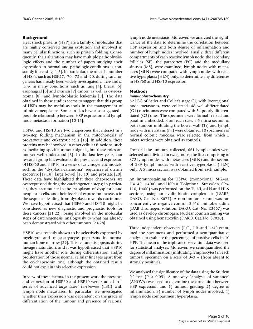
Graphics showing comparison between data obtained by quantitative analyses of HSP60 (a) and HSP10 (b) positive tumoral cells in G1 and G3 Ti and NiFigure 2Graphics showing comparison between data obtained by quantitative analyses of HSP60 (a) and HSP10 (b) positive tumoral cells in G1 and G3 Ti and Ni.
HSP60 positivity in carcinoma (arrow) but not in normal tis-sues (arrowhead) in specimens from both well differentiated (a) and poorly differentiated (b) tumours (Magnification: 10×)Figure 1HSP60 positivity in carcinoma (arrow) but not in normal tis-sues (arrowhead) in specimens from both well differentiated (a) and poorly differentiated (b) tumours (Magnification: 10×). A higher magnification (40×) shows that the positivity is diffuse into cytoplasm of tumoral cells of both G1 (c) and G3 (d) specimens; few positive interstitial (inflammatory) cells were scattered in the interposed stroma. HSP10 is also diffusely expressed by tumoral cells of both G1 (e) and G3 (f) LBC (Magnification: 10×). A higher magnification (40×) of both G1 (g) and G3 (h) shows that the positivity is mainly localised in the cytoplasm of neoplastic elements.
Page 3 of 10(page number not for citation purposes)

BMC Cancer 2005, 5:139 http://www.biomedcentral.com/1471-2407/5/139
(polyclonal rabbit, StressGen, SPA-110, 1:2000), follow-ing the procedures described above (secondary antibody:anti-rabbit, Pierce, 1:20000 Cat. No 31462).
ResultsHSP60 and HSP10 positivity in primitive versus metastatic tumours of different gradeHSP60 was present in 40 out of 48 (83.3%) Ti G1 (fig.1a,c) and in 32 out of 34 (94.1%) Ti G3 LBC (fig. 1b,d),while HSP10 was present in all examined specimens of Tiof both G1 (fig. 1e,g) and G3 (fig. 1f,h) LBC. In these spec-imens, both molecules were present in the cytoplasm ofneoplastic cells, and they were also rarely present in someinflammatory elements scattered in the stroma. In partic-ular, statistical analyses, we did not find any correlationbetween the degree of inflammation and the immunopo-sitivity for HSP60 (p > 0.05) and HSP10 (p > 0.1).
In addition, the percentage of HSP60 positive cells washigher in the G3 group (mean: 70%) compared with theG1 (mean: 35%) (fig. 2a), while a similar number ofHSP10 positive elements were present in both groups(mean: respectively 74% and 75%) (fig. 2b). Normal epi-thelium above the infiltrating neoplasms resulted com-monly negative or with few scattered positive elements
(fig. 1b), similarly to what observed in the biopsies of nor-mal colonic mucosae (data not shown). Statistical analy-ses showed that the difference between the number ofHSP60 positive cells in G1 and G3 LBC was significant (p< 0.0005), while statistic difference was not found inHSP10 positivity (p > 0.05).
Fig. 2c shows that 28 out of 48 Ni of G1 LBC (58%) werepositive for HSP60 (fig. 3a), compared to 26 out of 34 Niof G3 CRC (76%) (fig. 3b). Fig. 2d shows that all Ni ofboth G1 (fig. 3c) and G3 (fig. 3d) LBC were positive toHSP10. Statistical analyses showed that the differencebetween the number of HSP60 positive Ni in G1 and G3LBC was significant (p < 0.01), while statistic differencewas not found for HSP10 positivity (p > 0.05). BothHSP60 and HSP10 positivity was often co-localisedwithin vascular (fig. 3e) and nervous (fig. 3f) structuresinvaded by neoplastic tissue in both Ti and Ni.
The results of the immunoblotting analyses were compa-rable to the immunohistochemical data (fig. 4). Thequantity of HSP60 was higher in G3 specimens of both Tiand Ni, when compared to G1. HSP10 was present in asimilar amount in all examined specimens. Specimens ofnormal colonic tissue were commonly under the thresh-old of detectability for both HSP60 and HSP10.
HSP60 and HSP10 positivity in metastatic versus hyperplastic lymo nodesWe selected 661 lymph nodes from all the tumours(mean: 8.1; range: 5–12; S.D. 2.2) and divided in twogroups; the first comprising of 372 MLN and the second289 HLN. Firstly, we examined the presence of a statisticcorrelation between HSPs expression and number lymphnode involved by the disease. We found the presence of asignificant correlation between the presence of metastasesand the positivity for both HSP60 (p < 0.005) and HSP10(p < 0.001) in lymph nodes.
Western blot analyses for the research of HSP60 and HSP10 in tissue extracts of normal colonic mucosa (1), Ti G1 (2), Ti G3 (3), Ni G1 (4) and Ni G3 (5)Figure 4Western blot analyses for the research of HSP60 and HSP10 in tissue extracts of normal colonic mucosa (1), Ti G1 (2), Ti G3 (3), Ni G1 (4) and Ni G3 (5).
Infiltrated lymph nodes from G1 (a) and G3 (b) LBC show tumoral glands positive for HSP60Figure 3Infiltrated lymph nodes from G1 (a) and G3 (b) LBC show tumoral glands positive for HSP60. Metastases also show glands positive for HSP10 in both G1 (c) and G3 (d) carcino-mas (Magnification: 40×). HSP60 positivity shows vascular (e) and neural (f) invasion by cancer (Magnification: 10×).
Page 4 of 10(page number not for citation purposes)

BMC Cancer 2005, 5:139 http://www.biomedcentral.com/1471-2407/5/139
Subsequently, a semiquantitative analysis on the immu-nohistochemical observations in the lymph node com-partments of HLN was performed. Table 1 summarizesthese results. In SF cells of the HLN group, only 5% werepositive for HSP60 and 13% for HSP10. On the contrary,we found an increase in the number of HSP60 (28%) andHSP10 (35%) positive cells in SF of MLN (fig. 5a). We alsofound a great increase in the number of HSP10 positivecells in MS cells (38%) of MLN when compared to theHLN group (3%). There was no significant difference inthe number of HSP60 positive cells between both HLNand MLN groups in MS (fig. 5b), similarly to the numberof cells positive to both chaperones in PC of both HLNand MLN groups (fig. 5c). Statistical analyses showed asignificant difference between the number of HSP60 pos-itive cells in SF of the HLN and MLN groups (p < 0.0003).
Analogously, the number of HSP10 positive cells in bothSF (p < 0.001) and MS (p < 0.0001) presented a significantdifference. The positivity for both HSP60 (fig. 6a) andHSP10 (fig. 6b) in the cells of all reactive lymph nodecompartments was commonly localised in the cytoplasm.
DiscussionAlthough HSPs were first defined as proteins induced byenvironmental and pathophysiologic stress, they are alsoimplicated in protein-protein interactions, such as fold-ing, translocation, and prevention of inappropriate pro-tein aggregation. Recently, other functions concerningtheir pivotal roles during cancer development and pro-gression have been suggested (30).
In a study conducted on a series of esophageal squamouscell carcinomas, the overexpression of HSP70 was corre-lated with lymph node metastasis, and lymphatic vesselinvasion, and the authors suggested that HSP70 expres-sion might be used to assess the clinical outcome after sur-gery [12]. A reduced expression of HSP70 and HSP40 hasalso been associated with a lower histopathologic differ-entiation in a series of gastric carcinomas [31]. In addi-tion, Hwang et al. [13] have demonstrated that theexpression of HSP70 and HSP110 was increased in highly
metastatic colorectal cancer cell lines, but not in weakmetastatic cells, suggesting that the expression of theseHSPs is highly correlated with the advanced clinical stagesand positive lymph node involvement. In multivariateanalyses concerning the type, grade, stage of the tumour,invasion of lymphatics, blood vessels and nerves as well aslymph nodes, in 36 pancreatic adenocarcinomas, HSP70immunoexpression was found to be an independentprognostic factor [32]. Finally, in an immunohistochemi-cal study on 102 esophageal squamous cell carcinomaspecimens Kawanishi et al. [33] suggested that the expres-sion of HSP27 and HSP70 was frequently reduced andtherefore it should be considered an independent prog-nostic factor in this disease.
In their study on primary invasive ductal carcinomas ofthe breast with lymph node metastasis, Storm et al. [11]found that HSP27 might confer cytoprotection for meta-static cells, and they postulated that HSP27 overexpres-sion is associated with reduced disease-free survival inbreast carcinomas. Contrastingly, Tetu et al. [34] did notfind any predictive role for HSP27 in the outcome innode-positive breast carcinomas.
Piselli et al. [35] recently performed a cytofluorimetricanalysis on human pancreatic adenocarcinoma cells, bothgrown in vitro and collected ex vivo from primarytumours or lung metastases of tumour-engrafted SCIDmice; they were the first to demonstrate an HSP60 surfaceexpression on metastatic cells, but this expression was notcorrelated with metastasization. In a multivariate analysison a series of metastatic breast cancers, Schneider et al.[36] demonstrated that neither HSP27 nor HSP60 expres-sion was able to exclude axillary node invasion com-pletely. Finally, Ito et al. [37] studied the expression ofHSP27, -60, -70 and -90 in 24 squamous cell carcinomasof the tongue using immunohistochemistry, finding thatHSP immunoexpression might change during tumorigen-esis, but there was no correlation between HSP stainingand survival period, clinical stage, lymph node metastasis,histological grade or p53 immunostaining.
Table 1: Mean percentages and ranges of immunopositive cells
HLN PART MEAN RANGE MLN PART MEAN RANGE
SF 5% 0–8% SF 28% 7–41%HSP60 PC 2% 0–4% HSP60 PC 2% 0–4%
MS 1% 0–2% MS 3% 0–6%
SF 13% 4–22% SF 35% 18–58%HSP10 PC 5% 2–8% HSP10 PC 9% 4–19%
MS 3% 1–5% MS 38% 22–65%
Mean percentages and ranges of immunopositive cells in hyperplastic lymph nodes (HLN), left side, and metastatic lymph node (MLN), right side. SF: secondary follicles; PC: paracortex; MS: medullary sinuses. See text for more details.
Page 5 of 10(page number not for citation purposes)

BMC Cancer 2005, 5:139 http://www.biomedcentral.com/1471-2407/5/139
As far as we are aware, the present work is the first studyreporting the expression of HSP60 and HSP10 in a seriesof LBC with lymph node metastases, and the immunolo-calisation of these molecules in the different compart-ments of reactive lymph nodes.
The presence and expression of HSP60 and HSP10 insome carcinogenic models, in particular, both pre-tumoral (dysplastic) and neoplastic lesions of largebowel, as well as uterine exocervix and prostate gland hasbeen investigated previously [17-20]. These experimentsshowed that the level of these two strictly related mito-chondrial chaperonins increases from normal throughdysplastic towards neoplastic tissues. These proteinsresulted diffusely localised in the cytoplasms of dysplasticand neoplastic cells. Moreover, few scattered inflamma-tory elements were occasionally positive at stromal level.Considering this overexpression, it was hypothesised thatthese molecules might have different functions duringcancer development, apart from the mitochondrial regen-eration during normal cell proliferation. Nevertheless, theexact nature of this role is still not understood.
In the present paper, the expression of HSP60 in Ti and Niwas found to be dependent on the tumoral grade, whilethe expression of HSP10 was not. The number of HSP60positive tumoral cells in Ti of G3 LBC was higher than inG1, and the number of HSP60 positive Ni in G3 LBC washigher than in G1. Immunohistochemical results wereconfirmed by immunoblotting analysis. Therefore, wepostulate a prognostic significance of these data. HSP10was strongly positive in all examined specimens, andthese results may have a diagnostic utility. Interestingly, ahigher positivity for HSP60, but not for HSP10, was corre-lated with the presence of lymph node metastasis and thisdata may have a histopathologic value.
Although normal epithelia periodically regenerate cellularelements by mitosis of basal cells, the present work showsthat HSP60 and HSP10, in normal epithelia, are under theantibody detection threshold for immunohistochemicalanalyses, while neoplastic elements show a strong cyto-plasmic expression of these proteins. Many papers havealready shown the involvement of the HSP60/HSP10complex in preventing the activation of the apoptoticmachinery. Samali et al [38] were the first to demonstratethat pro-caspase-3 is present in the mitochondrial fractionof Jurkat T cells in a complex with the chaperon proteinsHSP60 and HSP10 and that the release of mitochondrialHSP may also accelerate caspase activation in the cyto-plasm of intact cells. These data are in accordance with thefindings of Xanthoudakis et al. [39] who showed thatATP-dependent 'foldase' activity of HSP60 may induce
High magnifications (100×) of immunostaining for HSP60 (a) and HSP10 (b) show cytoplasmic positivityFigure 6High magnifications (100×) of immunostaining for HSP60 (a) and HSP10 (b) show cytoplasmic positivity.
Diagrams showing the differences of the mean number of positive cells between HLN and MLN in the lymph node compartments: a) secondary follicles; b) paracortex; c) med-ullary sinusesFigure 5Diagrams showing the differences of the mean number of positive cells between HLN and MLN in the lymph node compartments: a) secondary follicles; b) paracortex; c) med-ullary sinuses.
Page 6 of 10(page number not for citation purposes)
BMC Cancer 2005, 5:139 http://www.biomedcentral.com/1471-2407/5/139
pro-caspase-3 maturation in Jurkat cells stimulated toundergo apoptosis by a Fas-independent pathway andthat this represents an important regulatory event inapoptotic cell death. Lin et al. [40] suggested that overex-pression of the combination of HSP60 and HSP10 and ofHSP60 or HSP10 individually may protect myocytesagainst apoptosis in an in vitro model of ischemia/reper-fusion injury. More recently they have demonstrated thatHSP10 overexpression may inactivate Raf, ERK, andp90Ribosomal kinase (p90RSK), suggesting that onlyHSP10 is involved in the complex mechanisms that pro-tect myocytes against simulated ischemia and reoxygena-tion induced death.
We could postulate that both HSP60 and HSP10 are up-regulated in cancer for extramitochondrial functions, i.e.in the block of the apoptotic machinery that usually takesplace during cancer development and progression.Although HSP60 and HSP10 should be functionally cor-related, HSP10 is present in a higher number of specimensand with a higher expression than HSP60. This result mayindicate a different function of HSP10 inside the cyto-plasm of tumoral cells. In a recent study, where the expres-sion of HSP10 and HSP60 was investigated in a series ofnormal human bone marrows, similar data were found[29]. Interestingly, a selective preference of HSP10 formyeloid and megakaryocytic precursors was discovered[29]. Therefore, other roles of HSP10 apart from the co-chaperonin one during bone marrow cell proliferationand differentiation of normal cells could be hypothesised.This is backed up by other studies on this co-chaperonin[41,42].
In the present study, the presence and localisation ofHSP60 and HSP10 in a series of human lymph nodes wereevaluated. Lymph node functioning is crucial for an indi-vidual's survival. Normal lymph nodes are very smallstructures, not always clinically detectable in humanbody. They react to antigens by uptaking and processingthem, eventually destroying them. Reacting lymph nodesenlarge due to immunologic stimulation that drives theirhyperplasia. Examining a histological section of hyper-plastic lymph node, different compartments can be distin-guished: 1) the follicles, where precursors of plasma cellsand memory B cells are formed; 2) paracortex, where spe-cific cellular response takes place, generating antigen-spe-cific T cells; 3) medullar sinuses, where the lymph carriedfrom afferent to efferent lymphatic vessels is cleared bymacrophages [43-45]. Commonly, hyperplasia involves alllymph node structures; as already demonstrated, in astudy on a wide series of HLN and MLN from axilla, themost common pattern of hyperplasia is the mixed type(66.5% of 996 HLN and 68.6% of 4711 MLN). In partic-ular, hyperplasia mainly involved both follicles and para-cortex area (22.2% of MLN and 19.2% of HLN), although
often sinuses were also found enlarged [46]. A fundamen-tal step during the examination of an enlarged lymphnode is the distinction between a reactive hyperplasia anda neoplasm. The latter may be of primary (lymphoma) orsecondary (metastasis) type. In an investigation on aseries of 1159 lymph nodes from breast cancer, microme-tastases (involving less that 25% of the lymph nodal vol-ume) have been shown often to be present also in verysmall nodes (less that 2 mm in diameter) [47]. Unfortu-nately, the detection of metastases in a lymph node is dif-ficult if they are small in dimension. Several studies haveshown that a number of histologically negative lymphnodes may present, at a retrospective analysis, a microme-tastasis revealed only immunohistochemically or withimmunofluorescence techniques; the presence ofmicrometastasis in the lymph node has great prognosticand therapeutic implications [48-53].
In this study, the number of SF cells positive for bothHSP60 and HSP10 increased significantly in MLN, whencompared to HLN. We postulate that this increase may berelated to formation of metastases. As a consequence,although cytokeratin staining is a much easier way todetect micrometastasis, the statistically significant incre-ment in the MLN group of the number of HSP60 andHSP10 positive elements in secondary follicles could alsobe considered diagnostic to predict the presence of lymphnode metastases. Therefore, a lymph node with germinalcentre presenting a strong staining for HSP60 and/orHSP10 without any apparent metastasis should be exam-ined in detail, since this observation may reflect anincreased likelihood of finding a micrometastasis.
Since MS of MLN showed a significantly higher amount ofHSP10, but not HSP60, positive cells, when compared tothe MS of HLN, we could assume that HSP10 in this siteis under unknown stimulation inducing its overexpres-sion, for functional roles, i.e. cell proliferation. The role ofHSP60 during proliferation and differentiation of eukary-otic cells has already been demonstrated [54-56]. Moreo-ver, HSP60 and HSP10, working together, could protectmitochondrial function and prevent apoptotic cell death[37,39] although some studies have shown that thesemolecules do not always act as a single functional unit invivo [41,57].
ConclusionMany papers have been focusing on the role of some HSPsto predict cancer progression [11-13,58]. We found partic-ularly interesting the paper of Storm [11], who firstshowed the expression of HSP27 in metastatic lymphnodes to confer cytoprotection for metastatic cells ofbreast carcinoma and its association with the reduced dis-ease-free survival. In addition, a selective expression ofHSP70 in the germinal centres of HLN has been demon-
Page 7 of 10(page number not for citation purposes)

BMC Cancer 2005, 5:139 http://www.biomedcentral.com/1471-2407/5/139
strated, although the meaning of this overexpression isnot understood [59].
Our researches showed an overexpression of HSP60 andHSP10 in LBC with lymph node metastases. Both mole-cules could be useful for histopathologic diagnosis of thisneoplasm, as well as to better assess the prognosis. Wecould also assume that both proteins are involved in LBCprogression, i.e. exercising an antiapoptotic effect. Wehypothesise that the increased expression of HSP60 andHSP10 in reactive lymph node cells is due to their role inproliferation of normal cells. Recently it has been shownthat HSP60 activates macrophages and T-cell, and thiscould also be a possible explanation for its overexpressionin lymph nodes with metastatic cancer upregulation [60].Based on these considerations, it could be postulated thatparacrine factors, such as cytokines, may induce an up-regulation of these molecules, but the exact role of theoverexpression remains to be confirmed.
Serial sectioning could be used to further study if any ofthe HLN population that present HSP60 and HSP10 stain-ing in the range of MLN contain occult metastases; itcould also be interesting to compare HLN from LBC spec-imens with non-neoplastic setting ones, i.e. resections ofactive inflammatory bowel disease, to confirm whetherthe former could be used as proper controls. Finally, theselective overexpression of HSP10 in MS of MLN couldsupport the hypothesis that this molecule, followingunknown biological stimulations, acts independentlyfrom HSP60.
In conclusion, our study could add new data to the classicclassification of G1 or G3, since both HSP60 and HSP10positivity may help to detect more aggressive tumors.Moreover, the comparison of the expression levels ofthese chaperones with other predictors of survival, asgrading, number of metastatic lymph nodes and degree oftumor infiltrating lymphocytes may be useful in coloncancer management.
AbbreviationsHSP: Heat Shock Proteins
LBC: Large Bowel Carcinomas
SF: Secondary Follicles
PC: Paracortex
MS: Medullary Sinuses
MLN: Lymph Nodes with Metastases
HLN: Reactive Hyperplasia
G1: Well-Differentiated Tumours
G3: Poorly-Differentiated Tumours
Ti: Specimens of Tumour Infiltrating the Bowel Wall
Ni: Specimens of Lymph Node with Metastasis
Competing interestsThe author(s) declare that they have no competing inter-ests.
Authors' contributionsFC designed the study, examined the immunohistochem-ical results, performed a semiquantitative analysis anddrafted the manuscript;
SD collected of specimens, carried out the immunohisto-chemistry and carried out the Western blotting analyses;
FR collected of specimens, examined the immunohisto-chemical results and performed a semiquantitative analy-sis;
FB participated in the design of the study and in the draftof the manuscript, performed the statistical analysis andexamined the Western blotting results ;
LM participated in the collection of specimens, examinedthe immunohistochemcal results and performed a semi-quantitative analysis;
FF participated in the and examination of the Westernblotting results and in the draft of the manuscript;
GZ coordinated the design and execution of the study.
All authors red and approved the final manuscript.
AcknowledgementsThis study was funded by MIUR ex-60% funds of Professor G. Zummo, Prof. F. Farina and Dr. F. Cappello.
References1. Wataba K, Saito T, Fukunaka K, Ashihara K, Nishimura M, Kudo R:
Over-expression of heat shock proteins in carcinogenicendometrium. Int J Cancer 2001, 91:448-456.
2. Rashmi R, Kumar S, Karunagaran D: Ectopic expression of HSP70confers resistance and silencing its expression sensitizeshuman colon cancer cells to curcumin-induced apoptosis.Carcinogenesis 2004, 25:179-187.
3. Mosser DD, Morimoto RI: Molecular chaperones and the stressof oncogenesis. Oncogene 2004, 23:2907-2918.
4. Zhong L, Peng X, Hidalgo GE, Doherty DE, Stromberg AJ, Hirschow-itz EA: Antibodies to HSP70 and HSP90 in serum in non-smallcell lung cancer patients. Cancer Detect Prev 2003, 27:285-290.
5. O'Neill PA, Shaaban AM, West CR, Dodson A, Jarvis C, Moore P,Davies MP, Sibson DR, Foster CS: Increased risk of malignantprogression in benign proliferating breast lesions defined by
Page 8 of 10(page number not for citation purposes)

BMC Cancer 2005, 5:139 http://www.biomedcentral.com/1471-2407/5/139
expression of heat shock protein 27. Br J Cancer 2004,90:182-188.
6. Lambot MA, Peny MO, Fayt I, Haot J, Noel JC: Overexpression of27-kDa heat shock protein relates to poor histological differ-entiation in human oesophageal squamous cell carcinoma.Histopathology 2000, 36:326-330.
7. Elpek GO, Karaveli S, Simsek T, Keles N, Aksoy NH: Expression ofheat-shock proteins HSP27, HSP70 and HSP90 in malignantepithelial tumour of the ovaries. APMIS 2003, 111:523-530.
8. Uozaki H, Ishida T, Kakiuchi C, Horiuchi H, Gotoh T, Iijima T, Ima-mura T, Machinami R: Expression of heat shock proteins in oste-osarcoma and its relationship to prognosis. Pathol Res Pract2000, 196:665-673.
9. Lauten M, Beger C, Gerdes K, Asgedom G, Kardinal C, Welte K,Schrappe M: Expression of heat-shock protein 90 in glucocor-ticoid-sensitive and -resistant childhood acute lymphoblasticleukaemia. Leukemia 2003, 17:1551-1556.
10. Ciocca DR, Clark GM, Tandon AK, Fuqua SA, Welch WJ, McGuireWL: Heat shock protein HSP70 in patients with axillarylymph node-negative breast cancer: prognostic implications.J Natl Cancer Inst 1993, 85:570-574.
11. Storm FK, Mahvi DM, Gilchrist KW: Heat shock protein 27 over-expression in breast cancer lymph node metastasis. Ann SurgOncol 1996, 3:570-573.
12. Noguchi T, Takeno S, Shibata T, Uchida Y, Yokoyama S, Muller W:Expression of heat shock protein 70 in grossly resectedesophageal squamous cell carcinoma. Ann Thorac Surg 2002,74:222-226.
13. Hwang TS, Han HS, Choi HK, Lee YJ, Kim YJ, Han MY, Park YM: Dif-ferential, stage-dependent expression of HSP70, HSP110and Bcl-2 in colorectal cancer. J Gastroenterol Hepatol 2003,18:690-700.
14. Noguchi T, Wada S, Takeno S, Moriyama H, Kimura Y, Uchida Y:Lymph node metastasis could be predicted by evaluation ofmacrophage infiltration and HSP70 expression in superficialcarcinoma of the esophagus. Oncol Rep 2003, 10:1161-1164.
15. Zuo DS, Dai J, Bo AH, Fan J, Xiao XY: Significance of expressionof heat shock protein90alpha in human gastric cancer. WorldJ Gastroenterol 2003, 9:2616-2618.
16. Richardson A, Schwager F, Landry SJ, Georgopoulos C: The impor-tance of a mobile loop in regulating chaperonin/ co-chaper-onin interaction: humans versus Escherichia coli. J Biol Chem2001, 276:4981-4987.
17. Cappello F, Bellafiore M, Palma A, Marciano V, Martorana G, BelfioreP, Martorana A, Farina F, Zummo G, Bucchieri F: Expression of 60-kD heat shock protein increases during carcinogenesis in theuterine exocervix. Pathobiology 2002, 70:83-88.
18. Cappello F, Bellafiore M, David S, Anzalone R, Zummo G: Ten kilo-dalton heat shock protein (HSP10) is overexpressed duringcarcinogenesis of large bowel and uterine exocervix. CancerLett 2003, 196:35-41.
19. Cappello F, Bellafiore M, Palma A, David S, Marciano V, Bartolotta T,Sciume C, Modica G, Farina F, Zummo G, Bucchieri F: 60KDa chap-eronin (HSP60) is over-expressed during colorectal carcino-genesis. Eur J Histochem 2003, 47:105-110.
20. Cappello F, Rappa F, David S, Anzalone R, Zummo G: Immunohis-tochemical evaluation of PCNA, p53, HSP60, HSP10 andMUC-2 presence and expression in prostate carcinogenesis.Anticancer Res 2003, 23:1325-1331.
21. Cappello F: HSP60 and HSP10 as diagnostic and prognostictools in the management of exocervical carcinoma. GynecolOncol 2003, 91:661.
22. Pomara G, Cappello F: RE: Heat shock proteins: their role inurological tumors. J Urol 2003, 170:927-928.
23. Hsu PL, Hsu SM: Abundance of heat shock proteins (HSP89,HSP60, and HSP27) in malignant cells of Hodgkin's disease.Cancer Res 1998, 58:5507-5513.
24. Schneider J, Jimenez E, Marenbach K, Romero H, Marx D, Meden H:Immunohistochemical detection of HSP60-expression inhuman ovarian cancer. Correlation with survival in a seriesof 247 patients. Anticancer Res 1999, 19:2141-2146.
25. Laad AD, Thomas ML, Fakih AR, Chiplunkar SV: Human gammadelta T cells recognize heat shock protein-60 on oral tumorcells. Int J Cancer 1999, 80:709-714.
26. Trieb K, Gerth R, Windhager R, Grohs JG, Holzer G, Berger P, KotzR: Serum antibodies against the heat shock protein 60 are
elevated in patients with osteosarcoma. Immunobiology 2000,201:368-376.
27. Lebret T, Watson RW, Molinie V, O'Neill A, Gabriel C, FitzpatrickJM, Botto H: Heat shock proteins HSP27, HSP60, HSP70, andHSP90: expression in bladder carcinoma. Cancer 2003,98:970-977.
28. Pignatelli D, Ferreira J, Soares P, Costa MJ, Magalhaes MC: Immuno-histochemical study of heat shock proteins 27, 60 and 70 inthe normal human adrenal and in adrenal tumors with sup-pressed ACTH production. Microsc Res Tech 2003, 61:315-323.
29. Cappello F, Tripodo C, Farina F, Franco V, Zummo G: HSP10 selec-tive preference for myeloid and megakaryocytic precursorsin normal human bone marrow. Eur J Histochem 2004,49:261-265.
30. Papp E, Nardai G, Soti C, Csermely P: Molecular chaperones,stress proteins and redox homeostasis. Biofactors 2003,17:249-257.
31. Isomoto H, Oka M, Yano Y, Kanazawa Y, Soda H, Terada R, YasutakeT, Nakayama T, Shikuwa S, Takeshima F, Udono H, Murata I, OhtsukaK, Kohno S: Expression of heat shock protein (HSP) 70 andHSP 40 in gastric cancer. Cancer Lett 2003, 198:219-228.
32. Sagol O, Tuna B, Coker A, Karademir S, Obuz F, Astarcioglu H, Kupe-lioglu A, Astarcioglu I, Topalak O: Immunohistochemical detec-tion of pS2 protein and heat shock protein-70 in pancreaticadenocarcinomas. Relationship with disease extent andpatient survival. Pathol Res Pract 2002, 198:77-84.
33. Kawanishi K, Shiozaki H, Doki Y, Sakita I, Inoue M, Yano M, TsujinakaT, Shamma A, Monden M: Prognostic significance of heat shockproteins 27 and 70 in patients with squamous cell carcinomaof the esophagus. Cancer 1999, 85:1649-1657.
34. Tetu B, Brisson J, Landry J, Huot J: Prognostic significance ofheat-shock protein-27 in node-positive breast carcinoma: animmunohistochemical study. Breast Cancer Res Treat 1995,36:93-97.
35. Piselli P, Vendetti S, Vismara D, Cicconi R, Poccia F, Colizzi V, DelpinoA: Different expression of CD44, ICAM-1, and HSP60 on pri-mary tumor and metastases of a human pancreatic carci-noma growing in scid mice. Anticancer Res 2000, 20:825-831.
36. Schneider J, Pollan M, Ruibal A, Jimenez E, Lucas AR, Nunez MI,Sanchez J, Tejerina A: Histologic grade and CD44 are independ-ent predictors of axillary lymph node invasion in early (T1)breast cancer. Tumour Biol 1999, 20:319-30.
37. Ito T, Kawabe R, Kurasono Y, Hara M, Kitamura H, Fujita K, KanisawaM: Expression of heat shock proteins in squamous cell carci-noma of the tongue: an immunohistochemical study. J OralPathol Med 1998, 27:18-22.
38. Samali A, Cai J, Zhivotovsky B, Jones DP, Orrenius S: Presence of apre-apoptotic complex of pro-caspase-3, HSP60 and HSP10in the mitochondrial fraction of jurkat cells. EMBO J 1999,18:2040-2048.
39. Xanthoudakis S, Roy S, Rasper D, Hennessey T, Aubin Y, Cassady R,Tawa P, Ruel R, Rosen A, Nicholson DW: HSP60 accelerates thematuration of pro-caspase-3 by upstream activator pro-teases during apoptosis. EMBO J 1999, 18:2049-2056.
40. Lin KM, Lin B, Lian IY, Mestril R, Scheffler IE, Dillmann WH: Com-bined and individual mitochondrial HSP60 and HSP10expression in cardiac myocytes protects mitochondrial func-tion and prevents apoptotic cell deaths induced by simulatedischemia-reoxygenation. Circulation 2001, 103:1787-1792.
41. Lin KM, Hollander JM, Kao VY, Lin B, Macpherson L, Dillmann WH:Myocyte protection by 10 kD heat shock protein (HSP10)involves the mobile loop and attenuation of the Ras GTP-asepathway. FASEB J 2004, 18:1004-1006.
42. Yang EC, Guo J, Diehl G, DeSouza L, Rodrigues MJ, Romaschin AD,Colgan TJ, Siu KW: Protein expression profiling of endometrialmalignancies reveals a new tumor marker: chaperonin 10. JProteome Res 2004, 3:636-643.
43. Hsu SM, Cossman J, Jaffe ES: Lymphocyte subsets in normalhuman lymphoid tissues. Am J Clin Pathol 1983, 80:21-30.
44. Dardick I, Dardick AM: Morphometry of normal human lym-phoid tissues. Nuclear parameters for comparative studiesof lymphoma. Arch Pathol Lab Med 1984, 108:190-196.
45. van den Oord JJ, de Wolf-Peeters C, Desmet VJ: The compositenodule. A structural and functional unit of the reactivehuman lymph node. Am J Pathol 1986, 122:83-91.
Page 9 of 10(page number not for citation purposes)
http://www.ncbi.nlm.nih.gov/entrez/query.fcgi?cmd=Retrieve&db=PubMed&dopt=Abstract&list_uids=8455204
http://www.ncbi.nlm.nih.gov/entrez/query.fcgi?cmd=Retrieve&db=PubMed&dopt=Abstract&list_uids=8455204
http://www.ncbi.nlm.nih.gov/entrez/query.fcgi?cmd=Retrieve&db=PubMed&dopt=Abstract&list_uids=8915490
http://www.ncbi.nlm.nih.gov/entrez/query.fcgi?cmd=Retrieve&db=PubMed&dopt=Abstract&list_uids=8915490
http://www.ncbi.nlm.nih.gov/entrez/query.fcgi?cmd=Retrieve&db=PubMed&dopt=Abstract&list_uids=9850087
http://www.ncbi.nlm.nih.gov/entrez/query.fcgi?cmd=Retrieve&db=PubMed&dopt=Abstract&list_uids=9850087
http://www.ncbi.nlm.nih.gov/entrez/query.fcgi?cmd=Retrieve&db=PubMed&dopt=Abstract&list_uids=7579511
http://www.ncbi.nlm.nih.gov/entrez/query.fcgi?cmd=Retrieve&db=PubMed&dopt=Abstract&list_uids=7579511
http://www.ncbi.nlm.nih.gov/entrez/query.fcgi?cmd=Retrieve&db=PubMed&dopt=Abstract&list_uids=7579511
http://www.ncbi.nlm.nih.gov/entrez/query.fcgi?cmd=Retrieve&db=PubMed&dopt=Abstract&list_uids=9466730
http://www.ncbi.nlm.nih.gov/entrez/query.fcgi?cmd=Retrieve&db=PubMed&dopt=Abstract&list_uids=9466730
http://www.ncbi.nlm.nih.gov/entrez/query.fcgi?cmd=Retrieve&db=PubMed&dopt=Abstract&list_uids=6190394
http://www.ncbi.nlm.nih.gov/entrez/query.fcgi?cmd=Retrieve&db=PubMed&dopt=Abstract&list_uids=6190394
http://www.ncbi.nlm.nih.gov/entrez/query.fcgi?cmd=Retrieve&db=PubMed&dopt=Abstract&list_uids=6546504
http://www.ncbi.nlm.nih.gov/entrez/query.fcgi?cmd=Retrieve&db=PubMed&dopt=Abstract&list_uids=6546504
http://www.ncbi.nlm.nih.gov/entrez/query.fcgi?cmd=Retrieve&db=PubMed&dopt=Abstract&list_uids=6546504
http://www.ncbi.nlm.nih.gov/entrez/query.fcgi?cmd=Retrieve&db=PubMed&dopt=Abstract&list_uids=2934988

BMC Cancer 2005, 5:139 http://www.biomedcentral.com/1471-2407/5/139
46. Cappello F, Bellafiore M, Palma A, Marciano V, Zummo G, Farina F,Bucchieri F: Study of axillary lymph node asymmetry in afemale population. J Anat 2001, 199:617-620.
47. Cappello F, Barresi E, Martorana A: Breast cancer and lymphnode metastasis: relation of lymph node dimensions andmetastasis. Pathologica 2000, 92:5-8.
48. Gonzalez Bosquet J, Keeney GL, Mariani A, Webb MJ, Cliby WA:Cytokeratin staining of resected lymph nodes may improvethe sensitivity of surgical staging for endometrial cancer.Gynecol Oncol 2003, 91:518-525.
49. Jiao X, Eslami A, Ioffe O, Kwong KF, Henry M, Zeng Q, Refaely Y, Bur-rows W, Gamliel Z, Krasna MJ: Immunohistochemistry analysisof micrometastasis in pretreatment lymph nodes frompatients with esophageal cancer. Ann Thorac Surg 2003,76:996-999.
50. Lara JF, Young SM, Velilla RE, Santoro EJ, Templeton SF: The rele-vance of occult axillary micrometastasis in ductal carcinomain situ: a clinicopathologic study with long-term follow-up.Cancer 2003, 98:2105-2113.
51. Munakata S, Aihara T, Morino H, Takatsuka Y: Application ofimmunofluorescence for intraoperative evaluation of senti-nel lymph nodes in patients with breast carcinoma. Cancer2003, 98:1562-1568.
52. Juretzka MM, Jensen KC, Longacre TA, Teng NN, Husain A: Detec-tion of pelvic lymph node micrometastasis in stage IA2-IB2cervical cancer by immunohistochemical analysis. GynecolOncol 2004, 93:107-111.
53. Lentz SE, Muderspach LI, Felix JC, Ye W, Groshen S, Amezcua CA:Identification of micrometastases in histologically negativelymph nodes of early-stage cervical cancer patients. ObstetGynecol 2004, 103:1204-1210.
54. Meinhardt A, Wilhelm B, Seitz J: Expression of mitochondrialmarker proteins during spermatogenesis. Hum Reprod Update1999, 5:108-119.
55. Bethke K, Staib F, Distler M, Schmitt U, Jonuleit H, Enk AH, Galle PR,Heike M: Different efficiency of heat shock proteins (HSP) toactivate human monocytes and dendritic cells: superiority ofHSP60. J Immunol 2002, 169:6141-6148.
56. Flohe SB, Bruggemann J, Lendemans S, Nikulina M, Meierhoff G, FloheS, Kolb H: Human heat shock protein 60 induces maturationof dendritic cells versus a Th1-promoting phenotype. J Immu-nol 2003, 170:2340-2348.
57. Dubaquie Y, Looser R, Funfschilling U, Jeno P, Rospert S: Identifica-tion of in vivo substrates of the yeast mitochondrial chaper-onins reveals overlapping but non-identical requirement forHSP60 and HSP10. EMBO J 1998, 17:5868-5876.
58. Cuezva JM, Chen G, Alonso AM, Isidoro A, Misek DE, Hanash SM,Beer DG: The bioenergetic signature of lung adenocarcino-mas is a molecular marker of cancer diagnosis and progno-sis. Carcinogenesis 2004, 25:1157-1163.
59. Leopardi O, Naughten W, Giannulis I, Mirra M, Frigo B: HSP70 isselectively over-expressed in the blast cells of the germinalcentres and paracortex in reactive lymph nodes. Histopathol-ogy 2001, 39:566-571.
60. Breloer M, More SH, Osterloh A, Stelter F, Jack RS, Bonin A: Macro-phages as main inducers of IFN-gamma in T cells followingadministration of human and mouse heat shock protein 60.Int Immunol 2002, 14:1247-1253.
Pre-publication historyThe pre-publication history for this paper can be accessedhere:
http://www.biomedcentral.com/1471-2407/5/139/pre-pub
Page 10 of 10(page number not for citation purposes)
http://www.ncbi.nlm.nih.gov/entrez/query.fcgi?cmd=Retrieve&db=PubMed&dopt=Abstract&list_uids=9774331
http://www.ncbi.nlm.nih.gov/entrez/query.fcgi?cmd=Retrieve&db=PubMed&dopt=Abstract&list_uids=9774331