BLS & ACLS in Twenty-Ten - Cardiac Resuscitation · PDF fileBLS & ACLS in 2011 An Introduction...
13
BLS & ACLS in 2011 An Introduction to the 2010 AHA CPR Guidelines for Healthcare Providers AnnMarie Fitzgerald Chase MSN, RN, CEN
Transcript of BLS & ACLS in Twenty-Ten - Cardiac Resuscitation · PDF fileBLS & ACLS in 2011 An Introduction...
BLS & ACLS in 2011
An Introduction to the 2010 AHA CPR Guidelines
for Healthcare Providers
AnnMarie Fitzgerald Chase MSN, RN, CEN
1
Feeling stressed, overworked, and in information overload? Well, you can relax
when it comes to the new 2010 American Heart Association CPR Guidelines, as
BCLS and ACLS just got streamlined and simplified. Although making your life
easier was not the driving force behind the changes, it seems likely that this will
be the case. The American Heart Association (AHA) based the new guidelines on
the latest science in the field of resuscitation in order to reduce disability and save more lives. So sit
back and relax as we go over what has changed for 2010.
“Approximately every 25 seconds, an American will have a coronary event, and approximately
every minute, someone will die of one.”1 While there are no reliable national statistics on
cardiopulmonary resuscitation,2 one focus of the ongoing AHA Get with the Guidelines–Resuscitation
initiative is to build this database.3 The most recent published studies demonstrated a less than 8
percent survival rate for out-of-hospital cardiac arrest4 and an average 17.6 percent survival-to-
discharge rate for inpatients regardless of the cardiac rhythm at arrest.5 This demonstrates the need for
healthcare providers to deliver more effective resuscitation care. This article is geared toward
healthcare providers practicing in the hospital setting and will review the key changes for adult patients,
which will be taught in BLS and ACLS classes in the near future. For a comprehensive list of all the
changes and the rationale behind them you can read the
2010 AHA Guidelines for Cardiopulmonary Resuscitation
and Emergency Cardiovascular Care Science by following
this link:
http://circ.ahajournals.org/content/vol122/18_suppl_3/.
The changes were officially released on October 18, 2010,
2
and are the result of an extensive literature review by international resuscitation experts.6 Over a three-
year period, these experts discussed and debated the results of thousands of resuscitation studies
before coming to the consensus statements we will review today. BLS and ACLS course instructors are in
the process of learning about the updates. Interim 2010 materials that supplement the 2005 materials
will be used until March 2011 when all instructors have completed updated training. Complete new
course materials based on the 2010 update will be rolled out in classes by AHA training centers after
instructors have received this updated training.7 This article is designed to be an introduction to the
changes and does not constitute training or replace an AHA class.
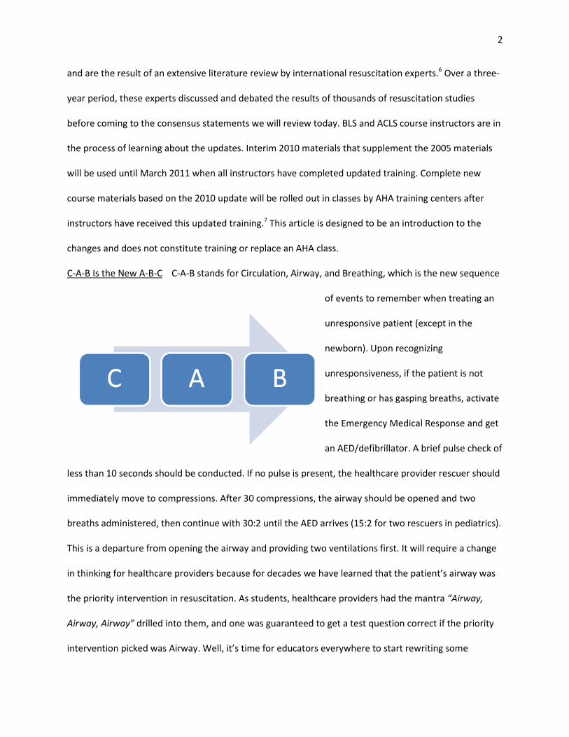
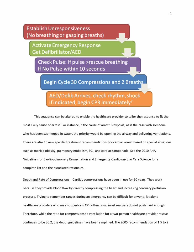
C-A-B Is the New A-B-C C-A-B stands for Circulation, Airway, and Breathing, which is the new sequence
of events to remember when treating an
unresponsive patient (except in the
newborn). Upon recognizing
unresponsiveness, if the patient is not
breathing or has gasping breaths, activate
the Emergency Medical Response and get
an AED/defibrillator. A brief pulse check of
less than 10 seconds should be conducted. If no pulse is present, the healthcare provider rescuer should
immediately move to compressions. After 30 compressions, the airway should be opened and two
breaths administered, then continue with 30:2 until the AED arrives (15:2 for two rescuers in pediatrics).
This is a departure from opening the airway and providing two ventilations first. It will require a change
in thinking for healthcare providers because for decades we have learned that the patient’s airway was
the priority intervention in resuscitation. As students, healthcare providers had the mantra “Airway,
Airway, Airway” drilled into them, and one was guaranteed to get a test question correct if the priority
intervention picked was Airway. Well, it’s time for educators everywhere to start rewriting some
3
questions, as one of the bigger changes for 2010 is a shift in priority to Compressions first. But why? It’s
not that the airway is not important; it is, but the act of opening the airway and ventilating the patient
delays the start of compressions. Adults are most likely to arrest with ventricular fibrillation (VF) or
pulseless ventricular tachycardia (VT) as the underlying cause. The treatment for these rhythms is
immediate CPR and early defibrillation. In fact, “the highest survival rates from cardiac arrest are
reported among patients of all ages who have a witnessed arrest with a rhythm of ventricular fibrillation
(VF) or pulseless ventricular tachycardia(VT).”5 On the other hand, animal studies have demonstrated
decreased survival rates when compressions where delayed or interrupted.6 The 2005 guidelines already
emphasized this need to minimize interruptions in compressions, and in 2010 this idea has been taken a
step further by doing compressions first. Starting with
compressions will only delay ventilations for about 18
seconds as the provider delivers 30 compressions prior to
opening the airway and ventilating. In most hospital
settings, cardiac arrests are responded to by a team,
usually with at least two healthcare providers present.
Frequently, compressions, calling for help, opening the airway, and providing ventilation breaths happen
almost simultaneously by the team while waiting for a defibrillator and additional help. The new 2010
guidelines recognize this and emphasize starting compressions first while the airway is being opened by
the second provider. As stated above, the most critical part of treatment for VF and pulseless VT is CPR
and early defibrillation, and the new C-A-B sequence is intended to better address this need. Finally, the
adage “look, listen, and feel” has been removed from BLS, as it was found to be time consuming and
inconsistently applied.8 Unresponsiveness and lack of breathing (or inadequate breathing) are now to be
assessed at the same time prior to the pulse check.
4
This sequence can be altered to enable the healthcare provider to tailor the response to fit the
most likely cause of arrest. For instance, if the cause of arrest is hypoxia, as is the case with someone
who has been submerged in water, the priority would be opening the airway and delivering ventilations.
There are also 15 new specific treatment recommendations for cardiac arrest based on special situations
such as morbid obesity, pulmonary embolism, PCI, and cardiac tamponade. See the 2010 AHA
Guidelines for Cardiopulmonary Resuscitation and Emergency Cardiovascular Care Science for a
complete list and the associated rationales.
Depth and Rate of Compressions Cardiac compressions have been in use for 50 years. They work
because theyprovide blood flow by directly compressing the heart and increasing coronary perfusion
pressure. Trying to remember ranges during an emergency can be difficult for anyone, let alone
healthcare providers who may not perform CPR often. Plus, most rescuers do not push hard enough.
Therefore, while the ratio for compressions to ventilation for a two-person healthcare provider rescue
continues to be 30:2, the depth guidelines have been simplified. The 2005 recommendation of 1.5 to 2
5
inches was changed in 2010 to at least 2 inches in adults. In infants and children, it is at least one-third
of the AP chest diameter or 1.5 inches in infants and 2 inches in children.6 This should eliminate the
confusion associated with a range of depths: just remember to push down at least 2 inches on the adult
patient.
The rate of compressions per minute has stayed the same at 100 a minute, but the emphasis is
now on at least 100 a minute, not around 100 a minute. In order to achieve return of spontaneous
circulation (ROSC) with good neurological function, effective compressions must be achieved to deliver
oxygen to the heart and brain. “In most studies, more compressions are associated with higher survival
rates, and fewer compressions are associated with lower survival rates.”6 The number of compressions
per minute actually delivered is dependent upon the rate per minute and the number of times CPR is
interrupted. The healthcare provider should still allow for adequate chest recoil and should continue to
minimize interruptions. Push hard and fast and use an AED as soon as it is available. Does your hospital
have AEDs in non-ACLS designated areas? The goal is to defibrillate the patient within three minutes of
collapse.7 If an AED is available, do all staff know where it is and how to use it? This early intervention
can save lives before the code team arrives. Collapse-to-defibrillation times in hospitals should be
measured; this may be one of the quality measures for sudden cardiac arrest monitored by the Joint
Commission in the near future.8 Follow this link
http://www.jointcommission.org/sudden_cardiac_arrest_initiatives/ for more information.
The precordial thump is back; sort of. For in-hospital, witnessed, monitored unstable VT (including
pulseless) healthcare providers may use the precordial thump when a defibrillator is not immediately
available, as it has been reported to convert ventricular tachycardias in a few studies. The thump should
never delay the arrival of the defibrillator or initiation of CPR and comes with complications such as
6
sternal fracture, stroke, osteomyelitis, and triggering malignant arrhythmias.6 It should not be used for
VF as it has not been demonstrated to result in ROSC for this arrhythmia.
Simplified ACLS Drugs and Algorithms BLS remains the bedrock of ACLS, but drugs and devices are de-
emphasized. Both the drugs and the algorithms have been streamlined and simplified. The algorithms
are circular and focus on quality CPR and defibrillation in two-minute cycles. Drugs, IV access, and
intubation are secondary. Atropine is no longer recommended for management of PEA and asystole and
is completely removed from the cardiac arrest algorithm. The evidence shows that routine use of
atropine is not effective in treating PEA and Asystole6, although it is still recommended for bradycardia
(see below). The medications in the cardiac arrest algorithm are limited to epinephrine (dose 1 mg,),
vasopressin (dose 40 units), and amiodarone (first dose 300 mg, second dose 150mg), and that’s it!
The bradycardia algorithm is simplified as well: For symptomatic bradycardia, atropine (0.5 mg
up to 3 mg total) and then consider transcutaneous pacing or dopamine or epinephrine infusion—
simple. Even the tachycardia algorithm is easier because adenosine is now recommended for “the initial
diagnosis and treatment of stable, undifferentiated regular, monomorphic wide-complex tachycardia.”6
It should still not be used for irregular wide complex tachycardia as it may result in VF.
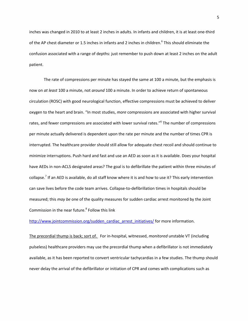
Ventilations & Oxygen Avoiding hyperventilation is key; hyperventilation increases gastric distension
and increases intrathoracic pressure, thereby decreasing venous return and with it, the chance of
survival. Tidal volume should be enough to make the chest rise and fall. When an advanced airway is
placed, ventilations should be given asynchronously every 6 to 8 seconds, and hyperventilation should
be avoided. Hyperoxia post arrest should be avoided as well as its harmful
effects have been documented6. During a code, pure oxygen is
administered, often resulting in post-arrest PaO2 levels (amount of oxygen
present in the blood) ranging from 80 mg to 500 mg, but the oxygen
7
saturation will show 100 percent. Therefore, it is considered reasonable, if equipment is available, to
monitor and maintain oxyhemoglobin saturation levels between 94 percent and 99 percent.6
Continuous Capnography Monitoring For patients who are intubated anytime during resuscitation,
continuous quantitative capnography is now recommended.6 End tidal capnography produces a
continuous waveform that monitors the amount of exhaled CO2. It can serve as a physiologic indicator of
cardiac output, because blood must circulate through the lungs in order for CO2 to be exhaled.
Therefore, continuous capnography can detect good CPR earlier than conventional methods and is also
the most reliable way to confirm endotracheal tube placement. It is also a noninvasive way to determine
whether effective compressions are being performed since rising CO2 levels can be an early indication of
ROSC and, conversely, falling Petco2 levels may indicate decreased cardiac output and re-arrest.
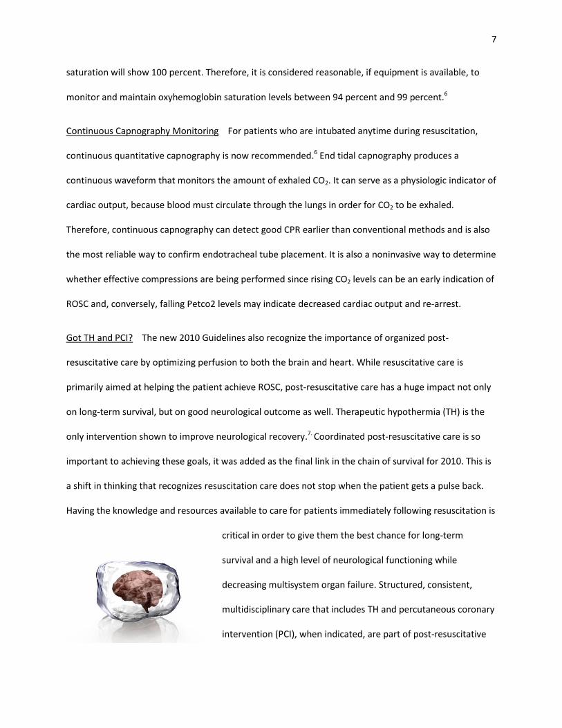
Got TH and PCI? The new 2010 Guidelines also recognize the importance of organized post-
resuscitative care by optimizing perfusion to both the brain and heart. While resuscitative care is
primarily aimed at helping the patient achieve ROSC, post-resuscitative care has a huge impact not only
on long-term survival, but on good neurological outcome as well. Therapeutic hypothermia (TH) is the
only intervention shown to improve neurological recovery.7. Coordinated post-resuscitative care is so
important to achieving these goals, it was added as the final link in the chain of survival for 2010. This is
a shift in thinking that recognizes resuscitation care does not stop when the patient gets a pulse back.
Having the knowledge and resources available to care for patients immediately following resuscitation is
critical in order to give them the best chance for long-term
survival and a high level of neurological functioning while
decreasing multisystem organ failure. Structured, consistent,
multidisciplinary care that includes TH and percutaneous coronary
intervention (PCI), when indicated, are part of post-resuscitative
8
care aimed at supporting both cardiac and neurological functioning. TH has
been extended to PEA and asystole arrest rhythms for patients who have a
ROSC, are comatose, and are able to maintain a blood pressure.
The goal for reperfusion therapy in STEMI patients is now three
minutes7 from when the patient arrives in the ED because immediate PCI
has been associated with favorable outcomes in adult patients resuscitated
after cardiac arrest. After arrest, PCI may be a reasonable intervention even
if the initial EKG does not show STEMI. The EKG may be misleading after cardiac arrest, so it is
reasonable to presume cardiac ischemia after an arrest. TH is considered safe when used in combination
with PCI.11 Bundled post-resuscitative care includes TH and PCI, when indicated, blood pressure
optimization, 12-lead EKG, oxygen saturation, and blood chemistry monitoring.7.It is now recommended
that if coordinated post-resuscitative care can’t be delivered at a hospital, the patient should be
transferred to a hospital that is able to provide post-arrest care.6 (Note: Therapeutic hypothermia for
use in post resuscitative care is included in the 2010 AHA Guidelines and is not an FDA-cleared
application).
More Training with Emphasis Teams
The AHA now recognizes what many healthcare providers have
known for some time: Two years in between trainings is too
long to maintain skills and mastery of BLS and ACLS. While
interval training is recommended, the ideal time for retraining
is unknown. One should also recognize that completing the
AHA class is only the first step in competence. Classes and
interval training should include performance assessment as well as written exams. It is suggested that
9
high-fidelity mannequins and simulation may be helpful in skill and competency attainment. Studies of
computerized simulations and case studies reveal that this learning modality can “teach decision making
skills and can facilitate learners’ understanding of broad core concepts.”12 Simulation can be an excellent
learning tool because it is interactive and can facilitate active learning, which “is more apt to stimulate
higher cognitive processes such as those associated with critical thinking.”13 ACLS classes should also
continue to include training in teamwork and leadership skills so that teams can work collaboratively to
really minimize interruptions in compressions. Training as a team can help to identify barriers to
effective ACLS delivery as well as identify learning needs of the team. Coordinated teamwork between
pre-hospital and hospital healthcare providers is also critical in order to effectively link the chain of
survival for cardiac arrest and stroke.
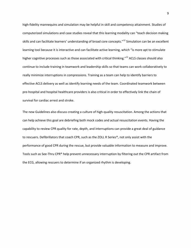
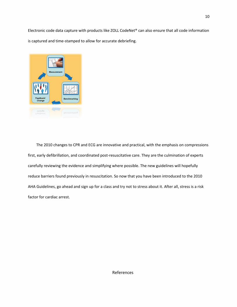
The new Guidelines also discuss creating a culture of high-quality resuscitation. Among the actions that
can help achieve this goal are debriefing both mock codes and actual resuscitation events. Having the
capability to review CPR quality for rate, depth, and interruptions can provide a great deal of guidance
to rescuers. Defibrillators that coach CPR, such as the ZOLL R Series®, not only assist with the
performance of good CPR during the rescue, but provide valuable information to measure and improve.
Tools such as See-Thru CPR® help prevent unnecessary interruption by filtering out the CPR artifact from
the ECG, allowing rescuers to determine if an organized rhythm is developing.
10
Electronic code data capture with products like ZOLL CodeNet® can also ensure that all code information
is captured and time-stamped to allow for accurate debriefing.
The 2010 changes to CPR and ECG are innovative and practical, with the emphasis on compressions
first, early defibrillation, and coordinated post-resuscitative care. They are the culmination of experts
carefully reviewing the evidence and simplifying where possible. The new guidelines will hopefully
reduce barriers found previously in resuscitation. So now that you have been introduced to the 2010
AHA Guidelines, go ahead and sign up for a class and try not to stress about it. After all, stress is a risk
factor for cardiac arrest.
References
11
1. Roger VL et al. (2010) Heart Disease and Stroke Statistics—2011 Update: A Report from the American Heart Association, Circulation 2010, Dec 15 (Epub ahead of print) p. e2. 2. AHA (2010) Cardiopulmonary Resuscitation (CPR) Statistics. Retrieved December 17, 2010 from: http://www.americanheart.org/presenter.jhtml?identifier=4483. 3. AHA Website (2010) Get With the Guidelines-Resuscitation. Retrieved December 17, 2010 from: http://www.heart.org/HEARTORG/HealthcareResearch/GetWithTheGuidelines-Resuscitation/Get-With-The-Guidelines-Resuscitation_UCM_314496_SubHomePage.jsp. 4. AHA (2010) CPR & Sudden Cardiac Arrest (SCA) Fact Sheet, as of April 26, 2010. Downloaded December 17, 2010 from: http://www.heart.org/HEARTORG/CPRAndECC/WhatisCPR/CPRFactsandStats/CPR-Statistics_UCM_307542_Article.jsp. 5. Cooper JA, Cooper JD, and Cooper, JM (2006) Cardiopulmonary Resuscitation: History, Current Practice and Future Direction. Circulation, December 19/26, p. 2839. 6. AHA (2010) Highlights of the 2010 American Heart Association Guidelines for CPR and ECC, p. 1. 7. AHA Website (2010) in American Heart Association Instructor Network, https://myportal.americanheart.org/eccportal/ecc/ecc;jsessionid=W4II3ORGZNTT0CQFCU1CF3Q?paf_dm=shared&DPSLogout=true&_requestid=70585.
8. The Joint Commission Website (2010) Sudden Cardiac Arrest Initiatives. Retrieved December 17, 2010 from: http://www.jointcommission.org/sudden_cardiac_arrest_initiatives/. 9. Hazinzski MF, Schexnayder S, Samson R (2010) Handbook of Emergency Cardiovascular Care for Healthcare Providers, BLS Adult HCP Algorithm. First American Heart Association Printing November 2010, p. 2–3. 10. AHA (2010) American Heart Association Guidelines for CPR and Emergency Cardiovascular Care Comparison Chart of Key Changes. Released Oct. 18, 2010. 11. Morrison LJ et al. (2010) Part 8: Advanced Life Support: 2010 International Consensus on Cardiopulmonary Resuscitation and Emergency Cardiovascular Care Science with Treatment Recommendations, Circulation, 122:S345–S421.
12. Bolan C (2003) Incorporating the experiential learning theory into the instructional design of online courses. Nurse Educator, 28(1)10–14.

12
13. Billings DM, Halstead JA (2009) Teaching in nursing: A guide for faculty, 6th ed. Saunders-Elsevier, St. Louis, MO. p. 156.