BLOOD COLLECTION TUBES - Catholic Medical Center...BD Vacutainer® Venous Blood Collection Tube...
27
Catholic Medical Center Laboratory 100 McGregor Street, Manchester, NH 03102 BLOOD COLLECTION TUBES LIGHT BLUE TOP TUBE – 2.7 ml draw and 1.8 ml draw Liquid 3.2% sodium citrate anticoagulant RED TOP TUBE – 4 ml draw Sprayed on clot activator GOLD TOP TUBE (SST) – 3.5 ml draw Sprayed on clot activator with gel for serum separation LIGHT GREEN TOP TUBE – 3 ml draw Lithium heparin anticoagulant with gel for plasma separation PURPLE TOP TUBE – 2 ml draw or 4ml draw or 6ml draw Sprayed on K2EDTA Anticoagulant The following tubes are available upon request through Catholic Medical Center’s Laboratory Customer Services: NAVY BLUE TOP TUBE – 6 ml draw (trace element) No additive NAVY BLUE TOP TUBE – 7 ml draw Na Heparin NAVY BLUE TOP TUBE – 6 ml draw K2EDTA anticoagulant GREEN TOP TUBE – 4 ml draw Sodium heparin anticoagulant GRAY TOP TUBE – 6 ml draw Powder sodium fluoride / potassium oxalate anticoagulant YELLOW TOP TUBE – 8.5 ml draw Liquid ACD solution A YELLOW TOP TUBE – 6 ml draw Liquid ACD solution B YELLOW TOP TUBE – 8.3 ml draw SPS (Sodium Polyanelthol Sulfonate) After specimen collection, all tubes - with the exception of the Light Blue Top tube (Sodium Citrate) - are to be gently inverted 8-10 times. This will ensure the proper distribution of additives with the specimen. The Light Blue Top Sodium Citrate tube should be gently inverted only 3-4 times to avoid activation of platelets which could skew test results. Gold and red top tubes should be allowed to clot for a minimum of 30 minutes and centrifuged within a maximum of 2 hours after collection. Serum for the red top tube must be removed as soon as possible from the cells to maintain the integrity of the specimen. Extra red top tubes that are collected in the emergency will only be viable for one hour on arrival in the lab.
Transcript of BLOOD COLLECTION TUBES - Catholic Medical Center...BD Vacutainer® Venous Blood Collection Tube...

Catholic Medical Center Laboratory 100 McGregor Street, Manchester, NH 03102
BLOOD COLLECTION TUBES
LIGHT BLUE TOP TUBE – 2.7 ml draw and 1.8 ml draw
Liquid 3.2% sodium citrate anticoagulant
RED TOP TUBE – 4 ml draw
Sprayed on clot activator
GOLD TOP TUBE (SST) – 3.5 ml draw
Sprayed on clot activator with gel for serum separation
LIGHT GREEN TOP TUBE – 3 ml draw
Lithium heparin anticoagulant with gel for plasma separation
PURPLE TOP TUBE – 2 ml draw or 4ml draw or 6ml draw
Sprayed on K2EDTA Anticoagulant
The following tubes are available upon request through Catholic Medical Center’s Laboratory
Customer Services:
NAVY BLUE TOP TUBE – 6 ml draw (trace element)
No additive
NAVY BLUE TOP TUBE – 7 ml draw
Na Heparin
NAVY BLUE TOP TUBE – 6 ml draw
K2EDTA anticoagulant
GREEN TOP TUBE – 4 ml draw
Sodium heparin anticoagulant
GRAY TOP TUBE – 6 ml draw
Powder sodium fluoride / potassium oxalate anticoagulant
YELLOW TOP TUBE – 8.5 ml draw
Liquid ACD solution A
YELLOW TOP TUBE – 6 ml draw
Liquid ACD solution B
YELLOW TOP TUBE – 8.3 ml draw
SPS (Sodium Polyanelthol Sulfonate)
After specimen collection, all tubes - with the exception of the Light Blue Top tube (Sodium
Citrate) - are to be gently inverted 8-10 times. This will ensure the proper distribution of
additives with the specimen. The Light Blue Top Sodium Citrate tube should be gently inverted
only 3-4 times to avoid activation of platelets which could skew test results.
Gold and red top tubes should be allowed to clot for a minimum of 30 minutes and centrifuged
within a maximum of 2 hours after collection. Serum for the red top tube must be removed as
soon as possible from the cells to maintain the integrity of the specimen. Extra red top tubes that
are collected in the emergency will only be viable for one hour on arrival in the lab.

BD Vacutainer® Venous Blood Collection
Tube Guide
BD Diagnostics Preanalytical Systems 1 Becton Drive Franklin Lakes, NJ 07417 USA
* Invert gently, do not shake ** The performance characteristics of these tubes have not been established for infectious disease testing in general; therefore, users must
validate the use of these tubes for their specific assay-instrument/reagent system combinations and specimen storage conditions. *** The performance characteristics of these tubes have not been established for immunohematology testing in general; therefore, users must validate the use of these tubes for their specific assay-instrument/reagent system combinations and specimen storage conditions.
BD Global Technical Services: [email protected] Customer Service: 1.888.237.2762www.bd.com/vacutainer
BD, BD Logo and all other trademarks are property of Becton, Dickinson and Company. © 2008 BD Printed in USA 08/08 VS5229-9
Note: BD Vacutainer® Tubes for pediatric and partial draw applications can be found on our website.
For the full array of BD Vacutainer® Blood Collection Tubes, visit www.bd.com/vacutainer.
Many are available in a variety of sizes and draw volumes (for pediatric applications). Refer to our website for full descriptions.
BD Vacutainer® Tubes with
BD Hemogard™ Closure
BD Vacutainer® Tubes with
Conventional Stopper Additive
Inversions at Blood
Collection* Laboratory UseYour Lab’s Draw Volume/Remarks
GoldRed/Gray
• Clot activator and gel for serum separation
5 For serum determinations in chemistry. May be used for routine blood donor screening and diagnostic testing of serum for infectious disease.** Tube inversions ensure mixing of clot activator with blood. Blood clotting time: 30 minutes.
Light Green
Green/Gray
• Lithium heparin and gel for plasma separation
8 For plasma determinations in chemistry. Tube inversions ensure mixing of anticoagulant (heparin) with blood to prevent clotting.
Red Red
• Silicone coated (glass)• Clot activator, Silicone
coated (plastic)
0 5
For serum determinations in chemistry. May be used for routine blood donor screening and diagnostic testing of serum for infectious disease.** Tube inversions ensure mixing of clot activator with blood. Blood clotting time: 60 minutes.
OrangeGray/Yellow
• Thrombin 8 For stat serum determinations in chemistry. Tube inversions ensure mixing of clot activator (thrombin) with blood to activate clotting.
Royal Blue
• Clot activator (plastic serum)
• K2EDTA (plastic)
8 8
For trace-element, toxicology, and nutritional-chemistry determinations. Special stopper formulation provides low levels of trace elements (see package insert). Tube inversions ensure mixing of either clot activator or anticoagulant (EDTA) with blood.
Green Green
• Sodium heparin• Lithium heparin
8 8
For plasma determinations in chemistry. Tube inversions ensure mixing of anticoagulant (heparin) with blood to prevent clotting.
Gray Gray
• Potassium oxalate/ sodium fluoride
• Sodium fluoride/Na2 EDTA• Sodium fluoride
(serum tube)
8
88
For glucose determinations. Oxalate and EDTA anticoagulants will give plasma samples. Sodium fluoride is the antiglycolytic agent. Tube inversions ensure proper mixing of additive with blood.
Tan
• K2EDTA (plastic) 8 For lead determinations. This tube is certified to contain less than .01 µg/mL(ppm) lead. Tube inversions prevent clotting.
Yellow
• Sodium polyanethol sulfonate (SPS)
• Acid citrate dextrose additives (ACD): Solution A - 22.0 g/L trisodium citrate, 8.0 g/L citric acid, 24.5 g/L dextrose Solution B - 13.2 g/L trisodium citrate, 4.8 g/L citric acid, 14.7 g/L dextrose
8 8 8
SPS for blood culture specimen collections in microbiology. ACD for use in blood bank studies, HLA phenotyping, and DNA and paternity testing.
Tube inversions ensure mixing of anticoagulant with blood to prevent clotting.
Lavender Lavender
• Liquid K3EDTA (glass)• Spray-coated K2EDTA
(plastic)
8 8
K2EDTA and K3EDTA for whole blood hematology determinations. K2EDTA may be used for routine immunohematology testing, and blood donor screening.*** Tube inversions ensure mixing of anticoagulant (EDTA) with blood to prevent clotting.
White
• K2EDTA with gel 8 For use in molecular diagnostic test methods (such as, but not limited to, polymerase chain reaction [PCR] and/or branched DNA [bDNA] amplification techniques.) Tube inversions ensure mixing of anticoagulant (EDTA) with blood to prevent clotting.
Pink Pink
• Spray-coated K2EDTA (plastic)
8 For whole blood hematology determinations. May be used for routine immunohematology testing and blood donor screening.*** Designed with special cross-match label for patient information required by the AABB. Tube inversions prevent clotting.
Light Blue
Light Blue
• Buffered sodium citrate 0.105 M (≈3.2%) glass 0.109 M (3.2%) plastic
• Citrate, theophylline, adenosine, dipyridamole (CTAD)
3-4
3-4
For coagulation determinations. CTAD for selected platelet function assays and routine coagulation determination. Tube inversions ensure mixing of anticoagulant (citrate) to prevent clotting.
Clear
ClearNew
Red/Light Gray
• None (plastic) 0 For use as a discard tube or secondary specimen tube.

Centrifuge at full speed
1100 – 1300g for 13 mm Plus Plastic tubes
1000 – 1300g for 16 mm Plus Plastic tubes
for 10 minutes in a swing-bucket unit or 15 minutes for a fixed-angle unit (balance tubes in centrifuge).
Gel barrier will form to separate plasma from red blood cells.
x 8
Ready for
Analysis
How to Prepare a Quality Sample
Spin
10Minutes
Invert
8-10Times
Using BD Vacutainer® PST™ Tubes
Centrifuge at1100 - 1300g
Gently invert 8 -10 times immediately after collection to mix lithium heparin anticoagulant with blood.
Insufficient mixing may lead to microclot and fibrin strand formation.
Use in laboratory for plasma determinations in chemistry.
BDPAS-PSTQualWC-7846.indd 1 3/6/06 12:27:10 PM

• Allow blood to clot for a minimum of 5 minutes in a vertical position.
• Observe a dense clot.
Spinas low as
3*
Minutes
Invert
5-6Times
Gently invert 5-6 times to mix clot activator with blood.
• Centrifuge at * 4000g for 3 minutes, 2000g for 4 minutes, or 1500–2000g for 10 minutes (balance tube in centrifuge), at 23-27°C.
• Barrier will form, separating serum specimen from clot.
• Transport spun tube to laboratory.
Clot
5Minutes
x 5-6
How to Prepare a Quality SampleBD Vacutainer® Rapid Serum Tube
BD DiagnosticsPreanalytical Systems1 Becton DriveFranklin Lakes, NJ 07417www.bd.com/vacutainer
Reference Number: 368774
Material: PET
Tube Size (mm): 13 x 100
Draw Volume (mL): 5.0
Closure Type/Color: BD Hemogard™/Orange
Label Type: Paper V-Notch™
Additive: Thrombin-based Clot Activator
Packaging Box/Case Quantities: 100/1000
Ordering Information
For more information please contact your local BD Sales Consultant
BD, BD Logo and all other trademarks are property of Becton, Dickinson and Company. © 2010 BD VS8876
BD Global Technical Services at 1.800.631.0174 BD Customer Service at 1.888.237.2762
Swing-headCentrifuge
2000g
4000g
1500–2000g
Swing-headCentrifuge
2000g
4000g
1500–2000g
Swing-headCentrifuge
2000g
4000g
1500–2000gSwing-head
Centrifuge
2000g
4000g
1500–2000g
Swing-headCentrifuge
2000g
4000g
1500–2000g
BD Diagnostics Preanalytical Systems 1 Becton Drive Franklin Lakes, NJ 07417 www.bd.com/vacutainer
BD, BD Logo, and all other trademarks are the property of Becton, Dickinson and Company. ©2006 BD. 03/06 VS7531
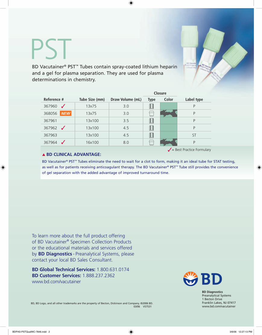
PST BD Vacutainer® PST™ Tubes contain spray-coated lithium heparin and a gel for plasma separation. They are used for plasma determinations in chemistry.
Closure
Reference # Tube Size (mm) Draw Volume (mL) Type Color Label type
367960 13x75 3.0 P
368056 13x75 3.0 P
367961 13x100 3.5 P
367962 13x100 4.5 P
367963 13x100 4.5 ST
367964 16x100 8.0 P
✓
✓
✓
NEW
To learn more about the full product offering of BD Vacutainer® Specimen Collection Products or the educational materials and services offered by BD Diagnostics - Preanalytical Systems, please contact your local BD Sales Consultant.
BD Global Technical Services: 1.800.631.0174BD Customer Services: 1.888.237.2362www.bd.com/vacutainer
▲ BD CLINICAL ADVANTAGE:
BD Vacutainer® PST™ Tubes eliminate the need to wait for a clot to form, making it an ideal tube for STAT testing,
as well as for patients receiving anticoagulant therapy. The BD Vacutainer® PST™ Tube still provides the convenience
of gel separation with the added advantage of improved turnaround time.
✓= Best Practice Formulary
BDPAS-PSTQualWC-7846.indd 2 3/6/06 12:27:13 PM

Maximum Fill*
Minimum FillIndicator
BD V
acut
aine
r® P
lus
Plas
tic
Citr
ate
Tub
e
Note: The quantity of blood drawn into evacuated tubes varies with altitude, ambient temperature, barometric pressure, tube age, venous pressure and filling technique.
Sufficient volume achieved if blood drawn falls above minimum fill indicator. For blood transfer, do not fill above illustrated dashed maximum line.
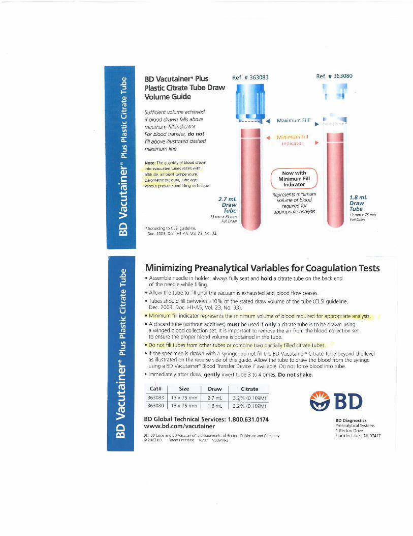
BD Vacutainer® Plus Plastic Citrate Tube Draw Volume Guide
* According to CLSI guideline, Dec. 2003, Doc. H1-A5, Vol. 23, No. 33.
2.7 mL Draw Tube
13 mm x 75 mm Full Draw
1.8 mL Draw Tube13 mm x 75 mmFull Draw
Ref. # 363083 Ref. # 363080
Represents minimum volume of blood
required for appropriate analysis
Now with Minimum Fill
Indicator

BD Diagnostics Preanalytical Systems 1 Becton Drive Franklin Lakes, NJ 07417BD, BD Logo and BD Vacutainer® are trademarks of Becton, Dickinson and Company.
© 2007 BD Patents Pending 10/07 VS5944-3
• Assemble needle in holder; always fully seat and hold a citrate tube on the back end of the needle while filling.
• Allow the tube to fill until the vacuum is exhausted and blood flow ceases.
• Tubes should fill between ±10% of the stated draw volume of the tube (CLSI guideline, Dec. 2003, Doc. H1-A5, Vol. 23, No. 33).
• Minimum fill indicator represents the minimum volume of blood required for appropriate analysis.
• A discard tube (without additives) must be used if only a citrate tube is to be drawn using a winged blood collection set. It is important to remove the air from the blood collection set to ensure the proper blood volume is obtained in the tube.
• Do not fill tubes from other tubes or combine two partially filled citrate tubes.
• If the specimen is drawn with a syringe, do not fill the BD Vacutainer® Citrate Tube beyond the level as illustrated on the reverse side of this guide. Allow the tube to draw the blood from the syringe using a BD Vacutainer® Blood Transfer Device if available. Do not force blood into tube.
• Immediately after draw, gently invert tube 3 to 4 times. Do not shake.
BD V
acut
aine
r® P
lus
Plas
tic
Citr
ate
Tub
e Minimizing Preanalytical Variables for Coagulation Tests
Cat# Size Draw Citrate
363083 13 x 75 mm 2.7 mL 3.2% (0.109M)
363080 13 x 75 mm 1.8 mL 3.2% (0.109M)
BD Global Technical Services: 1.800.631.0174www.bd.com/vacutainer


BD Vacutainer® Order of Draw for Multiple Tube CollectionsDesigned for Your Safety
Handle all biologic samples and blood collection “sharps” (lancets, needles, luer adapters and blood collection sets) according to the policies and procedures of your facility. Obtain appropriate medical attention in the event of any exposure to biologic samples (for example, through a puncture injury) since they may transmit viral hepatitis, HIV (AIDS), or other infectious diseases. Utilize any built-in used needle protector if the blood collection device provides one. BD does not recommend reshielding used needles, but the policies and procedures of your facility may differ and must always be followed. Discard any blood collection “sharps” in biohazard containers approved for their disposal.
Note: Always follow your facility’s protocol for order of draw
BD, BD Logo and all other trademarks are property of Becton, Dickinson and Company. ©2004 BD.Printed in USA 06/2004 VS5729-4
BD Diagnostics Preanalytical Systems 1 Becton Drive Franklin Lakes, NJ 07417
www.bd.com/vacutainer
BD Global Technical Services
1.800.631.0174
BD Customer Service
1.888.237.2762www.bd.com/vacutainer
= 1 inversion
BD Vacutainer® Blood Collection Tubes (glass or plastic)
or
or
or
• Blood Cultures - SPS
• Citrate Tube*
• BD Vacutainer® SST™
Gel Separator Tube• Serum Tube
(glass or plastic)
• Heparin Tube
• BD Vacutainer® PST™
Gel Separator Tube With Heparin
• EDTA Tube
• Fluoride (glucose) Tube
8 to 10 times
3 to 4 times
5 times
5 times (plastic) none (glass)
8 to 10 times
8 to 10 times
8 to 10 times
8 to 10 times
Closure Color Collection Tube Mix by Inverting
* When using a winged blood collection set for venipuncture and a coagulation (citrate) tube is the first specimen tube to be drawn, a discard tube should be drawn first. The discard tube must be used to fill the blood collection set tubing’s “dead space” with blood but the discard tube does not need to be completely filled. This important step will ensure maintenance of the proper blood-to-additive ratio of the blood specimen. The discard tube should be a nonad-ditive or coagulation tube.
Reflects change in NCCLS recommended Order of Draw (NCCLS H3-A5, Vol 23, No 32, 8.10.2)

PROPER SPECIMEN LABELING
Catholic Medical Center Laboratory 100 McGregor Street, Manchester, NH 03102
PATIENT SAFETY IS IN YOUR HANDS.
Label all specimens in the presence of the patient
List the full legal name , First and Last List another unique identifier i.e. Date of
Birth or Medical Record Number List the date and time of the collection List the collectors ID

PROPER SPECIMEN LABELING
Catholic Medical Center Laboratory 100 McGregor Street, Manchester, NH 03102
LABELING All specimens arriving in the laboratory must be properly labeled to support Patient Safety. Two unique identifiers for the patient must be on the specimen. The first and last name of the patient is mandatory. Either a date of birth or the medical record number is acceptable as the second identifier. The patient’s social security is not necessary. The date and time of collection, as well as the ID of the collector should be noted if not captured electronically. The labeling information must be confirmed with the patient when ever possible before labeling the specimen by asking the patient to recite the spelling of their name and date of birth. If the patient is unable to confirm by spelling the name etc, the name of a care giver that provides confirmation of the identity of the patient should be listed. Any discrepancies must be reconciled. Information presented on the order request and specimen labels must be in agreement. Additionally, this information should be concurrent to the information present on the patient identification band. GUIDELINES FOR LABELING:
• Immediately after collection, legibly label the specimen in the presence of the patient. • For positive patient identification ask the patient to spell their first and last name
along with reciting their date of birth. Compare with the preprinted label if available. If no label is available, hand label the tube with the patient’s full name, date of birth, date and time of collection, and the code/initials of the collector. This information along with the patient’s demographics should also appear on the requisition. Patient Identification Label For Specimens
NAME DOB Phleb Initials Date Time

PROPER SPECIMEN LABELING
Catholic Medical Center Laboratory 100 McGregor Street, Manchester, NH 03102
Labels whether electronically or manually generated should be:
• Placed vertically over the tube’s paper label with the name seated near the cap of the tube.
• Placed on straight, not wrapped around the tube or tube bottom, or overlapping the tube cap
• Free of wrinkles and tears • Free of stray marks in the bar-coded area • Placed on the tube in single thickness (only a single label placed on the tube)
SPECIMEN REJECTION DUE TO LABELING ERROR The laboratory strives for 100% compliance when it comes to specimen labeling and Patient Safety. Specimens are considered mislabeled when there is a mismatch between patient specific identifiers and information accompanying the specimen. When insufficient, inconsistent, or inaccurate identification exists, the laboratory will recommend that a new specimen be obtained if feasible, based on the outline of the following charts (Retrievable Specimens, Irretrievable or Precious Specimens). LABELING GUIDELINES: It is expected that lab specimens will be labeled at the point of collection in the presence of the patient. The labels are required to have 2 unique patient identifiers, Date and Time of collection and the collectors ID. Immediate notification to the provider is imperative for all discrepancies. **************************************************************** RETRIEVABLE SPECIMENS For Retrievable Samples to include but not limited to: urines, blood collections, Stool, Semen, Swabs, (except those obtained in the OR). Blood Bank specimens must match exactly and have all of the labeling criteria or they will be rejected. * For specimens mailed to the laboratory by the patient, such as fecal occult blood cards, the second identifier can be the address and phone number of the patient as the card template displays. The requisit ion information accompanying the stool cards should match the name, address and phone number as w ritten on the stool card. Type of Labeling Error QM Form/
Discard Form/ Precious Spec Form as appropriate
Specimen held/ Processed off line until clarification
Pathologist Consult
Rejected
Unlabeled specimen Y N N Y Last name only, no DOB or MR#
Y N N Y
PROPER SPECIMEN LABELING
Catholic Medical Center Laboratory 100 McGregor Street, Manchester, NH 03102
First name only with or without DOB or MR#
Y N N Y
Last name, first initial No DOB or MR#
Y N N Y
Last name, First Initial, DOB or MR#
Y N N Y
Last name, nickname DOB or MR#
Y Y Y To be determined
Last name change due to marital status. Must have documentation from the patient that the name has changed as of a date. Info must be captured as a chartable comment in soft.
Y Y N Y if not confirmed. NO If info is confirmed by the patient.
Lack of JR. SR. N N N N Full name, No DOB or MR#
Y Y Y To be determined
Specimen label does not match the name on the requisition
Y N N Y
The secondary label does not match the primary label
Y N N
Y
Name is misspelled Y Y Y To be determined
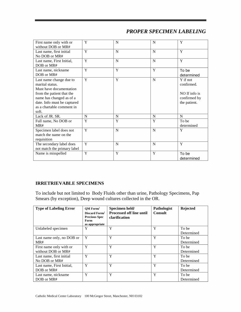
IRRETRIEVABLE SPECIMENS To include but not limited to Body Fluids other than urine, Pathology Specimens, Pap Smears (by exception), Deep wound cultures collected in the OR. Type of Labeling Error QM Form/
Discard Form/ Precious Spec Form as appropriate
Specimen held/ Processed off line until clarification
Pathologist Consult
Rejected
Unlabeled specimen Y Y Y To be Determined
Last name only, no DOB or MR#
Y Y Y To be Determined
First name only with or without DOB or MR#
Y Y Y To be Determined
Last name, first initial No DOB or MR#
Y Y Y To be Determined
Last name, First Initial, DOB or MR#
Y Y Y To be Determined
Last name, nickname DOB or MR#
Y Y Y To be Determined

PROPER SPECIMEN LABELING
Catholic Medical Center Laboratory 100 McGregor Street, Manchester, NH 03102
Last name change due to marital status
Y Y Y To be Determined
Lack of JR. SR. N N N N Full name, No DOB or MR#
Y Y Y To be determined
Specimen label does not match the name on the requisition
Y Y Y To be Determined
The secondary label does not match the primary label
Y Y Y
To be Determined
Name is miss spelled Y Y Y To be Determined
Decisions to discard the precious specimens will be made by the pathologist. Consult the pathologist “On Call”. For third shift, preserve the specimen until a pathologist can be reached in the early AM. Any documentation to accept the specimen for testing must be obtained in writing. This
documentation must be signed off by the physician who procured the specimen (Precious Specimen Form completed)
All discrepancies must be documented as a chartable comment in SCC order entry by a laboratory supervisor or manager.
The laboratory strives for 100% compliance when it comes to specimen labeling and Patient Safety. Authorized by Weldon Sanford MD Laboratory Medical Director __________________________ 4/21/2009ad; updated 05/12/09 as. updated 10/04/10 as

Benefi ts of Proper Secondary Label Alignment
Examples of misaligned secondary labels
Steps to properly alignsecondary labels
BD Vacutainer® Plus Plastic TubesHelp to improve your lab’s effi ciency. Misaligned secondary labeling means extra work and less effi ciency in the lab. To help minimize this
error, all BD Vacutainer® Plus Plastic Tubes have been restyled to include a visual guide for proper placement of secondary labels.
✔ Step 1Using the colored sidebar as your guide, position the secondary label on the tube, leaving the colored sidebar exposed.
✔ Step 2Smoothly wrap the secondary label around the tube, leaving a clear window to the sample.
Sample visibility Ability to know the tube type if the closure is removed
Easy to see fi ll indicator Consistent bar code location, creating instrument-readable labels
Draw volume
Tube type and additive concentration (if applicable)
BD lot number & expiration date
BD reference number
Color-coded sidebarColor-coded notch
Provides visual guide for proper placement of secondary labeling
Nominal fi ll indicator
BD sterile symbol
BD, BD Logo, and all other trademarks are the property of Becton, Dickinson and Company. ©2006 BD. VS7428-1www.TimeMed.comwww.TimeMed.com
BD DiagnosticsPreanalytical Systems1 Becton DriveFranklin Lakes, NJ 07417
For more information, please contact:BD Global Technical Services: 1.800.631.0174BD Customer Service: 1.888.237.2762www.bd.com/vacutainer
For more information, please contact:TimeMed Labeling Systems, Inc.144 Tower Drive • Burr Ridge, IL 60527Phone: 1.800.323.4840 • Fax: 1.800.548.5359
A Perfect Alignment for a Perfect Solution!

UNACCEPTABLE SPECIMENS
In the event a specimen is deemed unacceptable, the ordering provider will be made aware of the
status of the specimen and related testing as soon as possible.
The laboratory reserves the right to reject specimens that are:
Improperly labeled - including but not limited to, missing two unique patient identifiers,
incorrect spelling, or wrong date of birth.
Resulting from inappropriate patient preparation for the test.
Missing clinical information necessary for the interpretation of the test.
Missing collection date and time.
Submitted without an order or patient demographic information.
Submitted in an inappropriate container to perform the test.
Shipped or stored in unsuitable transport media.
Submitted in an expired container/tube.
Submitted in an inadequately filled tube.
Submitted in specimen collection containers were leaking.
Submitted in a way that may pose a safety hazard.
Hemolyzed or unexpectedly clotted.
Not maintained at optimum specimen storage /transport temperatures.
Received after expected delivery time to the lab was exceeded.
Compromised because the integrity of the specimen was not maintained.
Note:
For those specimens identified as irretrievable and suboptimal, testing can only occur when
permission is given by signature of the ordering provider and/or approved by the pathologist. A
Disclaimer will be appended to the test result stating the suboptimal condition.
The ordering provider/patient will be notified if recollection is desired.

Catholic Medical Center Laboratory 100 McGregor Street, Manchester, NH 03102
SPECIMEN PROCESSING
To maintain the integrity of blood specimens, clotted samples should be centrifuged within
two hours of collection but not before they have been allowed to properly clot,
approximately 30 minutes.
To operate the centrifuge, balance the samples (a water balance tube of identical size and fill
may be necessary). The separator tubes and red top tubes should be spun for 15 minutes at
approximately 3000 rpms. The cycle of the centrifuge should not be interrupted once it is
operational. Allow the centrifuge to come to a complete stop before opening the lid.
Respinning separator tubes may cause tainted testing results. CMC will supply and
maintain the fixed angle centrifuges for the processing of specimens that will be tested at
CMC.
Inpatient blood specimens may be sent to the lab as soon as they are collected and labeled.
The laboratory will take responsibility for processing the specimens using the automated
processor. The instrument (Tecan) will electronically receive the specimen in the lab
computer system, centrifuge, aliquot, and sort the specimens eliminating the risk of
mislabeling the prodigy specimens.
Specimens collected in the outpatient locations or during down time may have to be
separated after centrifuging to maintain the integrity of the testing sample. Red top tubes
should never be sent without centrifuging and separating the specimen. Separating is
accomplished by aspirating/removing the Serum/Plasma (liquid) component of the
centrifuged specimen with a plastic disposable dropper and placing the contents of the
dropper in a clean well-labeled transport tube with a secure cap. Avoid aspiration of the
specimen close to the cell layer. Place the specimen at the appropriate storage temperature.
Usually when freezing specimens, the serum/plasma is the component that is frozen and the
cells remain at refrigerated or room temperature and may even be immediately discarded.
Specimens with limited stability should be processed, separated and stored as required.
Specimens that are collected on ice should be chilled throughout the preparation process and
stored frozen as soon as possible. Specimens that are frozen should not be allowed go
through a freeze-thaw-freeze cycle.
Specimen such as urine should be kept refrigerated after collection to maintain the integrity
of the specimen.
Please note; Metal testing requires special processing and transport equipment. See specific
Metal Free Processing Procedure or call Customer support (663-8031) for assistance.

Catholic Medical Center Laboratory 100 McGregor Street, Manchester, NH 03102
SPECIMEN PACKAGING
SPECIMEN PACKAGING
All diagnostic specimens submitted to the lab will be packaged in at least a primary and a
secondary container. This precaution is to prevent the accidental exposure of potentially
infectious samples to the staff and the public. The primary container is the vessel that
contains the actual specimen, the collection tube, bottle, or jar etc. The secondary
container is the packaging the primary container is placed in. Most often specimen tubes
are placed in small zip lock plastic bags. Larger specimens may be placed in large plastic
bags or hard walled plastic containers.
There are two sections to the plastic specimen transport bags used as secondary
containers. The larger pouch is meant for the specimens and thinner pouch is used for the
transport of the paper requisitions. Specimens and paperwork should be packaged
separately in the same bag to prevent contamination in the event of a spill.
All lids to specimen jars, cups, jugs and the like are secured and tightened to prevent
leakage. The leakage may render the specimen unable to be tested. All specimens that
have been collected with a needle and syringe must have the needle removed and
replaced with a stopper to prevent leakage.
Light sensitive samples must be packaged to prevent the specimen’s exposure to any light
source. This can be accomplished by wrapping the primary container with aluminum foil.
Lab specimens are often susceptible to variations in temperature. Packaging specimens
to maintain the optimum temperature specific to the testing is critical to the accuracy of
the results.
Packaging for the specimen to travel in the pneumatic tube system requires additional
attention. Along with the primary and secondary containers additional padding is
required in the transport carrier. Please note the complete procedure is located on the
CMC Intranet / Administrative Manual/Computerized Tube System.
Single specimens may be placed in a zip lock bag and the requisition may be placed in the
attached pocket of the bag. Multiple blood specimens are preferably “racked” as opposed to
individual specimen bags. This saves time, cuts back biohazard waste, and lessens the
ergonomic impact of zipping and unzipping multiple specimen bags. Specimens should stay
in the upright position.

Catholic Medical Center Laboratory 100 McGregor Street, Manchester, NH 03102
SPECIMEN PACKAGING
Inpatient sweep collection may be racked in the following manner, then placed in the black
transport bag for delivery to the lab.
To Rack Specimens
Place specimens by patient in the slots of the rack, running the length of the rack. Place
multiple tubes of each patient behind the original tube.
Patient Rack
11 12
#1 #2 #3 #4 #5 #6 #7 #8 #9
= Blue top tube =Gold top separator tube = lavender tube =urine collection tube
OUTPATIENT PACKAGING
Completed requisitions are placed upside down in the order in which the specimens have
been collected. The rack is then placed in a large zip lock bag for transport. All additional
completed forms necessary to process the specimens, especially those specimens that will be
forwarded to another testing facility, should be attached to the original requisition.
Incomplete or missing forms could delay the turnaround time for resulting. These specimens
are refrigerated until ready for pick up unless otherwise stated in the specific test
requirements.
Beware! Exposing specimens to extreme weather conditions will compromise the integrity
of the testing specimens. If placing specimens in a pick-up box for the courier, the box must
be locked to insure the privacy of the patients and the security of the samples.
Cold packs should be added to the pick up boxes in extreme heat and bottles of hot water
may be placed in the box in extreme cold. It is best to place the box in the foyer of the
building to protect it from the extreme temperatures. Non blood specimens such as swabs
and urine tubes can be placed in the rack as well. Any collection vessel that is not
accommodated by the rack may be individually bagged and placed in the large zip bag for
transport.


Catholic Medical Center Laboratory 100 McGregor Street, Manchester, NH 03102
SPECIMEN STABILITY TIME SENSITIVE TESTS CHART
Test Name Specimen Stability from Collection Time Special Instructions
Ammonia 20 minutes on ice Must be collected on ice and performed within 20 minutes of collection.
Beta-Hydroxy 30 minutes RT or Refrig BNP whole blood 7 hrs Room Temp
or Refrig plasma 24 hrs - Room Temp or Refrig
B12 and Folate 8 hrs- Room Temp or Refrig After 8 hrs protect from light and refrigerate C-Diff 48 hr Refrigerated CBC 24 hr- Room Temp Clotted samples or those containing clots, fibrin strands,
or platelet clumps, grossly hemolyzed specimens, insufficient volume, and those drawn above an IV are not acceptable. Sample stability is 24 hours at room temperature.
Cold Agglutinin Room temperature specimen clots at 37 degrees Cryoglobulin 3 days at Room temperature D-Dimer 24 hr- Room Temp or Refrig Direct Bili 24 hr- Room Temp or Refrig Protect from Light Total Bilirubin 24 hr- Room Temp or Refrig Protect from Light ED Cardiac Panel-Biosite 4 hrs- Room Temp
ESR 24 hr- Room Temp Insufficient volume (Tube must be 2/3 full), specimens more then 24 hours post-collection, hemolysis, clotted specimens, and improperly labeled specimens.
Fibrinogen 4 hr- Room Temp Patient plasma should be tested within 4 hours (centrifuged or uncentrifuged) if stored in an unopened tube at Room Temp Short draw tubes, clotted and hemolyzed specimens are never acceptable.
HIT- Heparin Dependent Antibodies Screen- IgGAM
Separated plasma: freeze immediately 6 months frozen at -20
Plasma should be separated as soon as possible after collection, this test cannot be added on.
Herpes Viral Culture 4 days refrigerated, 6 months frozen
Viral Culture Medium(VCM) only
Homocysteine 1 hour- on ice separate plasma and perform testing immediately, refrigerated plasma stable for 7 days
Ionized Calcium Room temperature Specimen should never have been uncapped. Kleihauer Betke 2 hrs - Room Temp
24 hours on ice
Lactic Acid 1 hour- on ice Must be collected on ice and performed within 1 hr of collection

Catholic Medical Center Laboratory 100 McGregor Street, Manchester, NH 03102
Test Name Specimen Stability from Collection Time Special Instructions
LDH Specimen kept at Room Temp, serum/plasma must be separated from cells within 2 hrs of collection.
This test cannot be added- on.
Lupus Screen 90 days frozen, prepare platelet poor plasma, freeze immediately.
O&P Screen 24 hrs - Room Temp if not in preservative 2 weeks if in preservative
Osmolality-Serum 3 hours at Room Temperature up to 10 hours refrigerated
Osmolality-Urine 3 hours at room temperature up to 24 hours refrigerated 1 week frozen
PFA 4 hrs- Room Temp Patient samples are stable for up to 4 hours and must be stored at room temperature. Do not refrigerate or centrifuge sample prior to analysis.Do not use the pneumatic tube; hand deliver to the lab via transport personnel. Hemolyzed specimens not accepted.
PT and/or PT Mixing Study
24 hr- Room Temp Patient plasma should be tested within 24 hours (centrifuged or uncentrifuged) if stored in an unopened tube at Room Temp Short draw tubes, clotted and hemolyzed specimens are never acceptable. Frozen platelet poor plasma good for 2 weeks at minus 20 degrees, and good for 12 months at minus 70 degrees.
PTT and/or PTT Mixing Study
4 hr- Room Temp Patient plasma should be tested within 4 hours (centrifuged or uncentrifuged) if stored in an unopened tube at Room Temp Short draw tubes, clotted and hemolyzed specimens are never acceptable. Frozen platelet poor plasma good for 2 weeks at minus 20 degrees, and good for 12 months at minus 70 degrees.
Retic 24 hr- Room Temp Clotted samples or those containing clots, fibrin strands, or platelelt clumps, grossly hemolyzed sepcimens, insufficient volume, and those drawn above an IV are not acceptable. Sample stability is 24 hours at room temperature.
Stool culture 24 hr- Room Temp no preservative 96 hrs preservative
TEG 2 hrs- Room Temp Specimens are stable at room temperature for two (2) hours. They must not be refrigerated, nor centrifuged. Do not use the pneumatic tube.
Urinalysis Room Temp - 2 hours Refigerated 24 hours In preservative- 72 hours

Catholic Medical Center Laboratory 100 McGregor Street, Manchester, NH 03102
Test Name Specimen Stability from Collection Time Special Instructions
Urine Culture 24 hr- Refrig unpreserved; 48 hr preserved RT or Refrig
Urine Tox Screen- Bisosite
48 hrs Refrig
Viral culture 72 hours refrigetated Viral Culture Medium(VCM) only Rubeola Screen Varicella Zoster Screen
48 hrs- Refrig longer than 48 hrs if spec is frozen

Catholic Medical Center Laboratory 100 McGregor Street, Manchester, NH 03102
TRANSPORTING AND TRACKING SPECIMENS
TRANSPORTING SPECIMENS
Specimens may be brought directly to the lab from within the hospital. Manually
transporting the specimens must be done by placing the packaged specimens into the
black specimen transport container marked “diagnostic specimens”. This bag is
specifically designated for specimen transport use.
Laboratory staff pick up and transport surgical specimens to the pathology lab.
There are special ergonomic carts designated specifically to handle the various size
specimen containers. The cart carrier acts as the secondary transport container to prevent
exposure to staff and the public. The carrier also serves to protect patient information.
Pneumatic Tube: A variety of hospital units and the Notre Dame Pavilion are connected
to the main lab via a computerized pneumatic tube system. The tube system allows for
the speedy transport of specimens from critical units directly to the laboratory. The
specimens travel by special padded carrier to their designated location. Their arrival is
announced with an audible bell within minutes of the original departure. Please refer to
the “Computerized Pneumatic Tube Policy” located on the CMC intranet under
Administrative policies. This policy speaks to the general operation, limitations, training
and maintenance of the Computerized Tube System.
Outreach specimens are picked up by the courier and placed in temperature appropriate
coolers for transport. The coolers are kept in the controlled environment inside the
vehicle to avoid exposure to the extreme seasonal temperatures in the trunk. Specimens
are transported to the laboratory within the time required to maintain specimen integrity.
TRACKING SPECIMENS
It is the intent of Catholic Medical Center to ensure that all laboratory specimens
accepted into the facility reach the appropriate testing area and have a traceable path from
the specimen collection to receipt in the final testing location under safe, ergonomic, and
HIPAA compliant conditions.
The ultimate goal is to integrate the various department systems to allow test order
placement, tracking and receipt of these specimens electronically. The organization will
work to that essential end.

Catholic Medical Center Laboratory 100 McGregor Street, Manchester, NH 03102
TRANSPORTING AND TRACKING SPECIMENS
Current Tracking:
In-house specimens are managed by specific lab department pending lists, tracking
specimens that are collected but not received in the lab.
Specimens are electronically tracked from the Patient Service Centers to the lab to eliminate
lost specimens.
Tracking of Operating Room, Endoscopy and Radiology specimens is accomplished
manually by the lab registration staff via sign off sheet.
Courier tracking for clients is limited to tracking at the specimen level only.

![l>lf·· E ·B; -I,:,C-·-1·1V · cat. no.i bd lj.657 bd lj.6]5 bd 4630 bd 4·627 bd 4628 bd 4886 bd 4546 bd 4·545 bd 4544 bd 4542 bd lj,588 bd lj.593 bd 0102 bd 4636 bd 4632 bd](https://static.fdocuments.in/doc/165x107/5f7c69bb7d840d18665ab1e6/llf-e-b-ic-11v-cat-noi-bd-lj657-bd-lj65-bd-4630-bd-4627-bd-4628-bd.jpg)