Blood Chapter 14. Blood Connective tissue with a fluid matrix Three main functions:...
75
Blood Chapter 14
-
Upload
eric-reeves -
Category
Documents
-
view
218 -
download
2
Transcript of Blood Chapter 14. Blood Connective tissue with a fluid matrix Three main functions:...
BloodChapter 14

Blood
• Connective tissue with a fluid matrix
• Three main functions:– Transportation– Regulation– Protection

Physical Characteristics of Blood• Heavier, thicker, and 3-4 X more viscous
than water
• 38o C (100.4oF)
• pH : 7.35 – 7.45
• 8% of body weight
• 4-6 liters in an adult
• Varies with electrolyte concentration and amount of adipose tissue

8% of body weight
70 kg person
Total blood weight =
0.08 X 70kg = 5.6 kg
One kilogram of blood ≈ 1 liter
Total blood volume = 5.6 liters
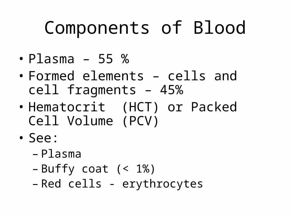
Components of Blood
• Plasma – 55 %• Formed elements – cells and cell
fragments – 45%• Hematocrit (HCT) or Packed Cell Volume
(PCV)• See:
– Plasma– Buffy coat (< 1%)– Red cells - erythrocytes


Formation of blood cells
Before birth blood is formed by the yolk sac, liver, spleen, thymus gland, lymph nodes and red bone marrow.
After birth only by red bone marrow
Stem cells are hemocytoblasts
Process is hematopoiesis
Colony stimulating factors


ErthrocytesBiconcave discs without nuclei
1/3 hemoglobin by volume
Hemoglobin – 4 peptide chains and an iron containing pigment called heme
The iron binds reversibly with oxygen
oxyhemoglobin deoxyhemoglobin
Cyanotic – skin and mucous membranes
appear blue


Also transports 23 % of CO2
bound to globin as carbaminohemoglobin
CO – carbon monoxide binds more tightly to hemoglobin than oxygen
Males = 5.4 million RBCs per cubic mm
Females and children = 4.8 million/ cubic mm
People at high altitudes = 8 million/cubic mm


RBCs live about 120 days
Iron is removed from heme, combines with transferrin and is taken to bone marrow
80% of the iron is stored in the liver in an iron-protein complex called ferritin


The rest of the heme is a greenish pigment called biliverdin which is broken down to a yellow-orange substance called bilirubin
Jaudice


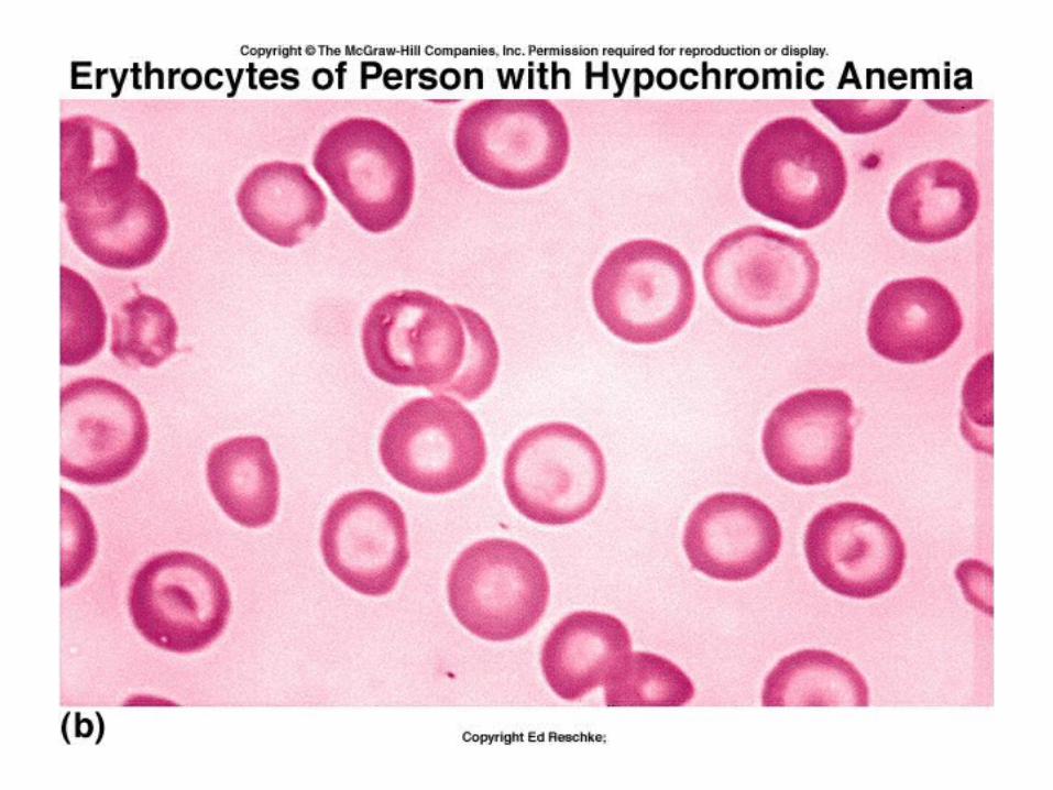
Anemia is the inability of the blood to carry sufficient oxygen to the body.
low #’s of RBCslack of hemoglobin

Pernicious anemia is a lack of RBCs due to lack of vitamin B12
Folic acid also needed for DNA synthesis
Iron deficiency anemia
Sickle cell anemia
Polycythemia is an excess of RBCs – blood gets too thick to flow

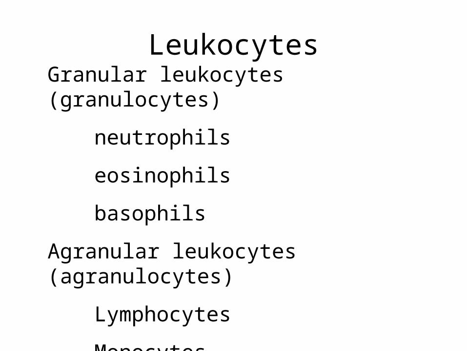
LeukocytesGranular leukocytes (granulocytes)
neutrophils
eosinophils
basophils
Agranular leukocytes (agranulocytes)
Lymphocytes
Monocytes
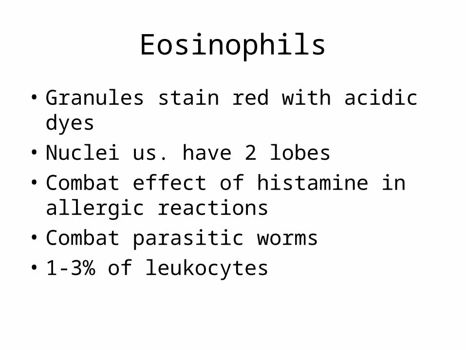
Eosinophils
• Granules stain red with acidic dyes
• Nuclei us. have 2 lobes
• Combat effect of histamine in allergic reactions
• Combat parasitic worms
• 1-3% of leukocytes


Basophils
•Have granules that stain blue with basic dyes
•Release heparin, and histamine
•Increase in allergic reactions that intensify the inflammatory response
•Less than 1% of leukocytes


Neutrophils
•Granules stain pale purple in a combination of acidic and basic dye
•Older cells have many lobed nuclei – gives the name polymorphonuclear leukocytes (PMNs)
•Younger neutrophils are called bands
•Phagocytes
•54 -62 % of the leukocytes


Lymphocytes•May be small or large
•Nuclei stain darkly, very little cytoplasm
•Only 2% of lymphocytes are in blood
•B lymphocytes make antibodies
•T lymphocytes attack invaders directly
•Long lived
•25-33% of leukocytes


Monocytes•Largest leukocyte
•Nuclei horseshoe-shaped
•Cytoplasm blue-gray and foamy
•When leave blood and enter tissues become transformed into macrophages
•3 - 9 % of leukocytes


Normal blood contains 5,000 -10,000/mm3
An increase in the number of wbcs is leukocytosis
A deficiency in wbcs is leukopenia
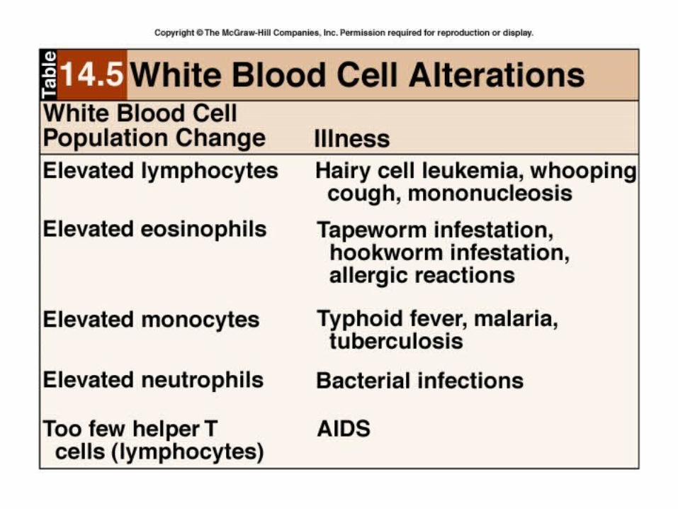
Differential white blood cell count is useful in diagnosis of disease
Major histocompatibility (MHC) antigens used in tissue typing


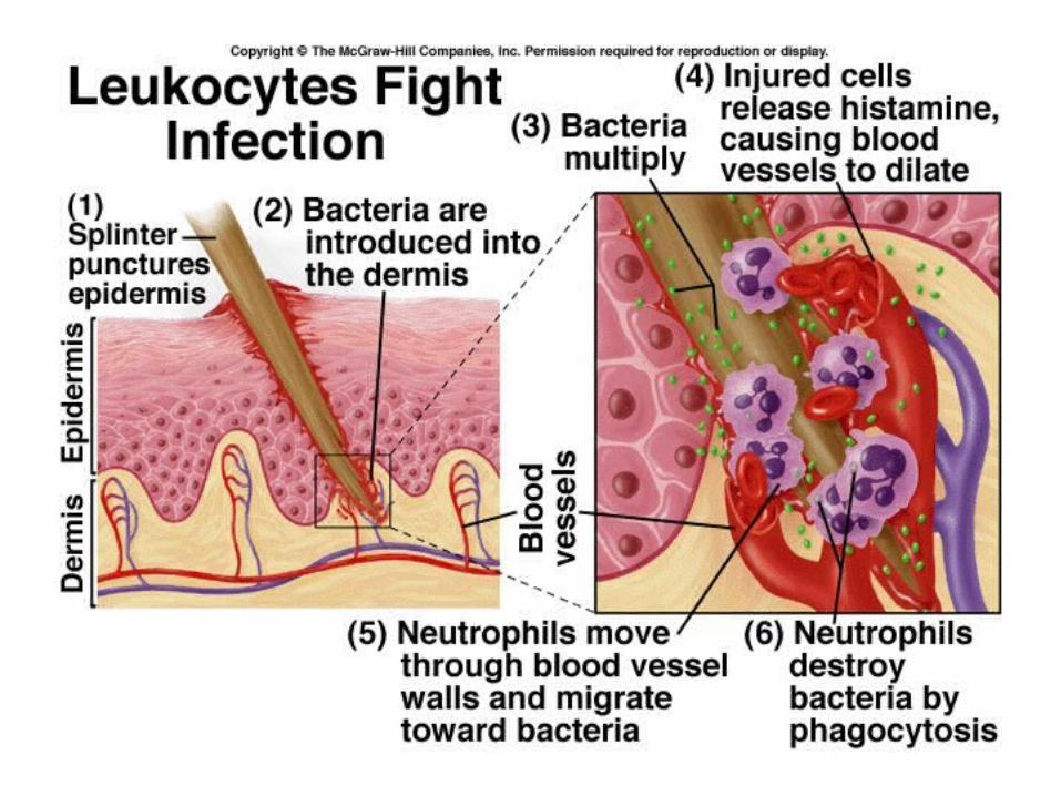
Function of leukocytes
Protect against infection
phagocytosis, antibodies, direct attack
Diapedesis
Damaged tissue releases chemicals that attract leukocytes – positive chemotaxis


Platelets, or thrombocytes, are cell fragments
•Formed from megakaryocytes
•130,000 – 360,000/mm3
•Involved in blood clotting
•Release serotonin which contracts smooth muscle in walls of blood vessels – reducing blood flow – and blood loss

Plasma
• 92 % water
• 8% solutes – ions, nutrients, enzymes, gases, wastes, hormones, but mostly proteins
•These proteins are mostly made by the liver


Serum
Plasma without the clotting factors
Still contains :
ions, nutrients, enzymes, gases, wastes, hormones, and some proteins – including antibodies

Hemostasis
• Means stoppage of bleeding
1. Vascular spasm
2. Platelet plug formation
3. Blood coagulation - clotting

Vasospasm
• Smooth muscle in vessel wall contracts
• Decreased diameter of vessel decreases blood flow
• Lasts several minutes to several hours

Platelet plug formation
1. Platelets contact damaged vessel –platelet adhesion
2. Platelets become activated, dump granules – platelet release reaction
3. Platelets become sticky and accumulate – platelet aggregation


Blood coagulation
• Clotting in an unbroken vessel – thrombosis
• Clot is a thrombus
• If it breaks free and travels in the blood stream it is an embolus
• If it lodges elsewhere in the body, it is an embolism

Blood clotting can be divided into 3 stages:
1. formation of prothrombinase
2.Conversion of prothrombin to thrombin
3.Conversion of soluble fibrinogen to insoluble fibrin

Stage 1 formation of prothrombinase (prothrombin activator) can be started one of two ways:
Extrinsic pathway (clotting mechanism):tissue factor leaks into the blood from
outside the vessels
Intrinsic pathway –more complex, slower, by roughened endothelium of exposure to foreign substances – Hageman factor


Stage 2 – Prothrombinase + calcium
Convert prothrombin to thrombin
Stage 3 – Thrombin + calcium
convert fibrinogen to fibrin
Threads of fibrin form a net and trap blood cells, forming a clot.
Clot retraction
Platelet-derived growth factor → repair


Hemophila
• Due to a deficiency of one of the clotting factors
• Hereditary trait
Role of vitamin K
• Normal clotting requires vitamin K, but it is not a clotting factor
• It is required for the synthesis of four of the clotting factors by the liver.
• Normally made by bacteria in large intestine
• Sometimes given before surgery
Fibrolytic system
• Plasminogen, an inactive enzyme, is incorporated into a clot
• Plasminogen can be activated to plasmin, (or fibrinolysin) which digests fibrin and inactivates other clotting factors

Thrombolytic agents
• Streptokinase
• Tissue plasminogen activator – t-PA

Anticoagulants
• Prostaglandins
• Heparin
• Warfarin or coumadin – antagonists to vitamin K
• Chelating agents tie up calcium
• Aspirin inhibits vasoconstriction and platelet aggregation


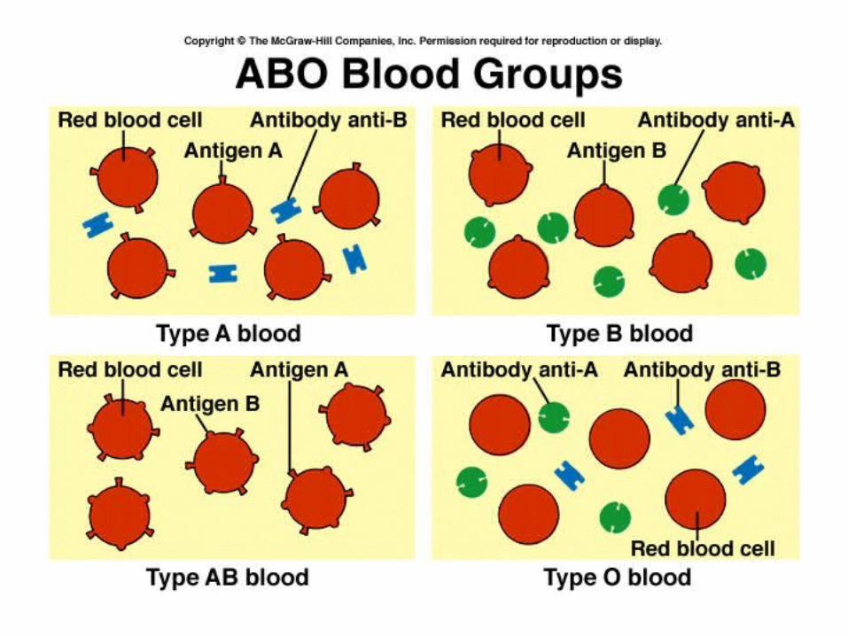

Blood Groups and Blood Types• Erythrocytes have surface antigens, called
isoantigens or agglutinogens• Most common are the ABO and Rh groups• A and B are isoantigens, O is absence of
antigen• Antibodies activate complement which
causes hemolysis• Typing outside the body uses
agglutination, NOT clotting !

Persons with type A blood make antibodies against B
Persons with type B blood make antibodies against A
Persons with type O blood make antibodies against A and B
Persons with type AB do not make antibodies.


Rh factor
• First discovered in the rhesus monkey
• Either have the antigen = positive
• Or don’t have the antigen = negative
• Do not automatically make antibodies – must first encounter the antigen
• Hemolytic disease of the newborn – or- erythroblastosis fetalis
• Give RhoGAM – anti Rh antibodies


Remember:
Rh factor is only a problem if the mother is negative and the father is positive.