Blood - anatomy-review-semester-1.weebly.com
14
Blood ✮ connective tissue ✮ forms mostly in red bone marrow Functions ● Transports vital substances ● Maintains stability of interstitial fluid ● Distributes heat 3 Types 1. red blood cells or Erythrocytes 2. white blood cells or Leukocytes 3. platelets (cell fragments) or Thrombocytes Volume ● varies with. . . ○ body size ○ changes in fluid concentration ○ changes in electrolyte concentration ○ amount of adipose tissue ● ≊ 5 liters/ 8% body weight Composition ● 45% formed elements + 55% plasma ● Hematocrit: measurement of cell volume to total volume Origin
Transcript of Blood - anatomy-review-semester-1.weebly.com

Blood connective tissue forms mostly in red bone marrow
Functions Transports vital substances Maintains stability of interstitial fluid Distributes heat
3 Types 1. red blood cells or Erythrocytes 2. white blood cells or Leukocytes 3. platelets (cell fragments) or Thrombocytes
Volume varies with. . .
body size changes in fluid concentration changes in electrolyte concentration amount of adipose tissue
≊ 5 liters/ 8% body weight Composition
45% formed elements + 55% plasma Hematocrit: measurement of cell volume to total volume
Origin

Erythrocytes
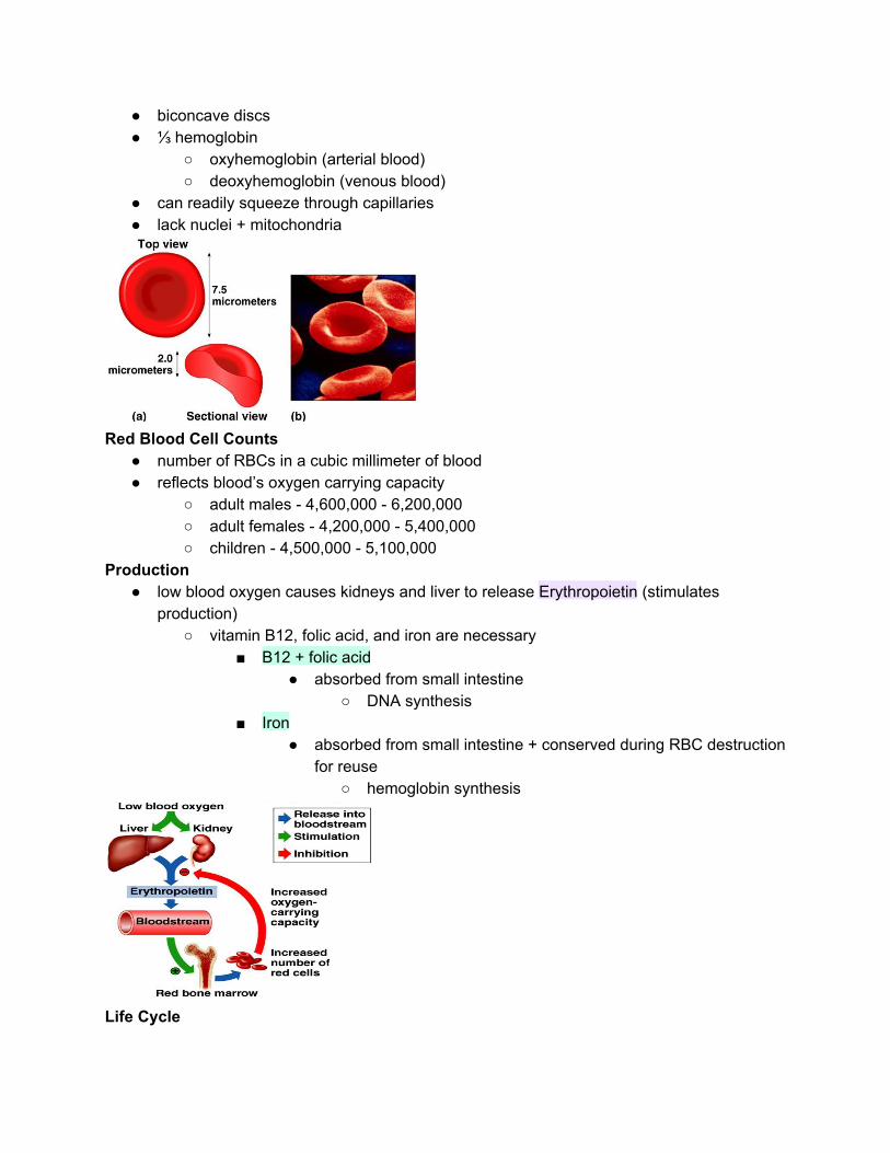
biconcave discs ⅓ hemoglobin
oxyhemoglobin (arterial blood) deoxyhemoglobin (venous blood)
can readily squeeze through capillaries lack nuclei + mitochondria
Red Blood Cell Counts
number of RBCs in a cubic millimeter of blood reflects blood’s oxygen carrying capacity
adult males 4,600,000 6,200,000 adult females 4,200,000 5,400,000 children 4,500,000 5,100,000
Production low blood oxygen causes kidneys and liver to release Erythropoietin (stimulates
production) vitamin B12, folic acid, and iron are necessary
B12 + folic acid absorbed from small intestine
DNA synthesis Iron
absorbed from small intestine + conserved during RBC destruction for reuse
hemoglobin synthesis
Life Cycle
1. circulate for ≊ 120 days 2. macrophages in spleen + liver destroys worn out RBCs 3. hemoglobin is broken down into heme + globin 4. iron from heme returns to red bone marrow 5. bilirubin + biliverdin excreted in bile
Types of Anemia
Type Cause Defect
Aplastic anemia Toxic chemicals, radiation Damaged bone marrow
Hemolytic anemia Toxic chemicals RBC destroyed
Iron deficiency anemia Dietary lack of iron Hemoglobin deficiency
Pernicious anemia Inability to absorb vitamin B12
Excess of immature cells
Sickle cell disease Defective gene RBC abnormally shaped
Thalassemia Defective gene Hemoglobin deficiency; RBC shortlived
Destruction 1. Squeezing through capillaries of active tissues damages RBC 2. Macrophages in the spleen +liver phagocytize damaged RBC 3. Hemoglobin → heme + globin 4. Heme → iron + biliverdin 5. Iron is made available for reuse in the synthesis of new hemoglobin or is stored in the
liver as ferritin 6. Some biliverdin → bilirubin 7. Biliverdin + bilirubin are excreted in bile as bile pigments 8. Globin → amino acids that are metabolized by macrophages/ released into the blood
Antigens and Antibodies
antigens a chemical that stimulates cells to produce antibodies antibodies a protein that reacts against a specific antigen
ABO Blood Group Based on the presence/absence of two major antigens on RBC membranes
antigen A or antigen B

Blood Types for Transfusion
Blood of Recipient Prefered Blood Type of Donor Permissible Blood Type of Donor (emergency)
A A A,O
B B B,O
AB AB AB,A,B,O
O O O
agglutination clumping of RBC in response to a reaction between an antibody + antigen occurs in wrong blood type transfusion/disorders; blood clumps together
Rh Blood Group Rh positive presence of antigen D and/or other Rh antigens on the RBC membranes Rh negative absence of antigen D and/or other Rh antigens on the RBC membranes

Leukocytes Functions
protects against disease hormones stimulate development
interleukins colonystimulating factors
2 Types 1. Granulocytes (cytoplasmic granules are present)
Neutrophils (segs, polymorphonuclear leukocyte, or bands (young neutrophils)) lightpurple granules in acidbase strain lobed nucleus 1st to arrive at infection phagocytic 54%64% of leukocytes elevated in bacterial infections

Eosinophils
deepred granules in acid stain bilobed nucleus defend against parasitic worm infestations 1%3% of leukocytes elevated in parasitic worm infestations/ allergic reactions
Basophils
deepblue granules in basic stain release histamine + heparin <1% of leukocytes similar to eosinophils in size + shape of nuclei
2. Agranulocytes (cytoplasmic granules are not present)
lymphocytes slightly larger that RBC large spherical nucleus surrounded by thin rim of cytoplasm T cells + B cells → important to immunity B cells produce antibodies 25%33% of leukocytes
Monocytes
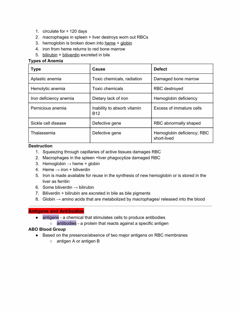
largest spherical, kidneyshaped, oval/lobed nuclei leave bloodstream to become macrophages 3%9% of leukocytes Phagocytize bacteria, dead cells, and other debris
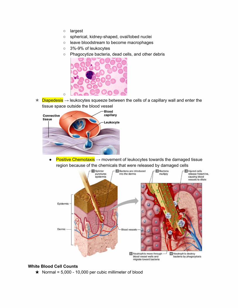
Diapedesis → leukocytes squeeze between the cells of a capillary wall and enter the
tissue space outside the blood vessel
Positive Chemotaxis → movement of leukocytes towards the damaged tissue
region because of the chemicals that were released by damaged cells
White Blood Cell Counts
Normal = 5,000 10,000 per cubic millimeter of blood

Leukopenia low WBC count (below 5,000) Typhoid fever, flu, measles, mumps, chicken pox, and AIDS
Leukocytosis high WBC count (above 10,000) acute infections, vigorous exercise, great loss of body fluids
Differential WBC count lists percentages of types of leukocytes may change in particular diseases
Thrombocytes cell fragments of megakaryocytes 130,000 360,000 per cubic millimeter of blood
Functions controls blood loss from broken vessels
Plasma
liquid portion of blood (straw colored)
55% blood 92% water

Gases and Nutrients
Gases Oxygen Carbon Dioxide
Nutrients amino acids simple sugars nucleotides lipids
Nonprotein Nitrogenous Substances

molecules containing nitrogen but are not proteins urea product of protein catabolism; about 50% of NPN substances uric acid product of nucleic acid catabolism amino acids product of protein catabolism creatine stores phosphate creatinine product of creatine metabolism BUN blood urea nitrogen; indicate health of kidney
**catabolism means breaking down molecules by metabolic pathways into smaller units producing energy Plasma Electrolytes
absorbed from the intestine/released as byproducts of cellular metabolism Sodium, potassium, calcium, magnesium, chloride, bicarbonate, phosphate, and
sulfate (sodium and chloride are most abundant)
Hemostasis stoppage of bleeding 1. Blood Vessel Spasm
triggered by pain receptors, platelet receptors, platelet release, or serotonin smooth muscle in vessel contracts
2. Platelet Plug Formation triggered by exposure of platelets to collagen
platelets adhere to rough surface to form plug
3. Blood Coagulation
Extrinsic clotting mechanism chemical outside of blood triggers blood coagulation
triggered by thromboplastin (not found in blood) triggered when blood contacts damaged tissue
Intrinsic clotting mechanism

chemical inside blood triggers blood coagulation triggered by Hageman factor (found inside blood) triggered when blood contacts foreign substance
Fate of blood Clots
After forming, a blood clot retracts and pulls the edges of a broken vessel together while squeezing the fluid serum from the clot
platederived growth factor stimulate smooth muscle cells and fibroblasts to repair damaged blood vessel walls
plasmin digest the blood clot

thrombus abnormal blood clot embolus blood clot moving through blood
Prevention of Coagulation as a clot forms, fibrin absorbs thrombin and prevents the clotting reaction from spreading antithrombin inactivates additional thrombin by binding to it and blocking its action on
fibrinogen heparin (an anticoagulant)
Green Boxes and Clinical Application

1. Leukemia Myeloid Leukemia
bone marrow produces too many immature granulocytes Leukemia cells crowd out other blood cells
anemia bleeding susceptible to infections
Lymphoid Leukemia lymphocytes are cancerous symptoms similar to myeloid leukemia
Treatments drugs marrow and umbilical cord transplants chemotherapy regimens