Block Gastro 2012 Lower & Accesory GI Tract
29
COLON
-
Upload
silvestri-purba -
Category
Documents
-
view
217 -
download
1
description
GIT3
Transcript of Block Gastro 2012 Lower & Accesory GI Tract

COLON
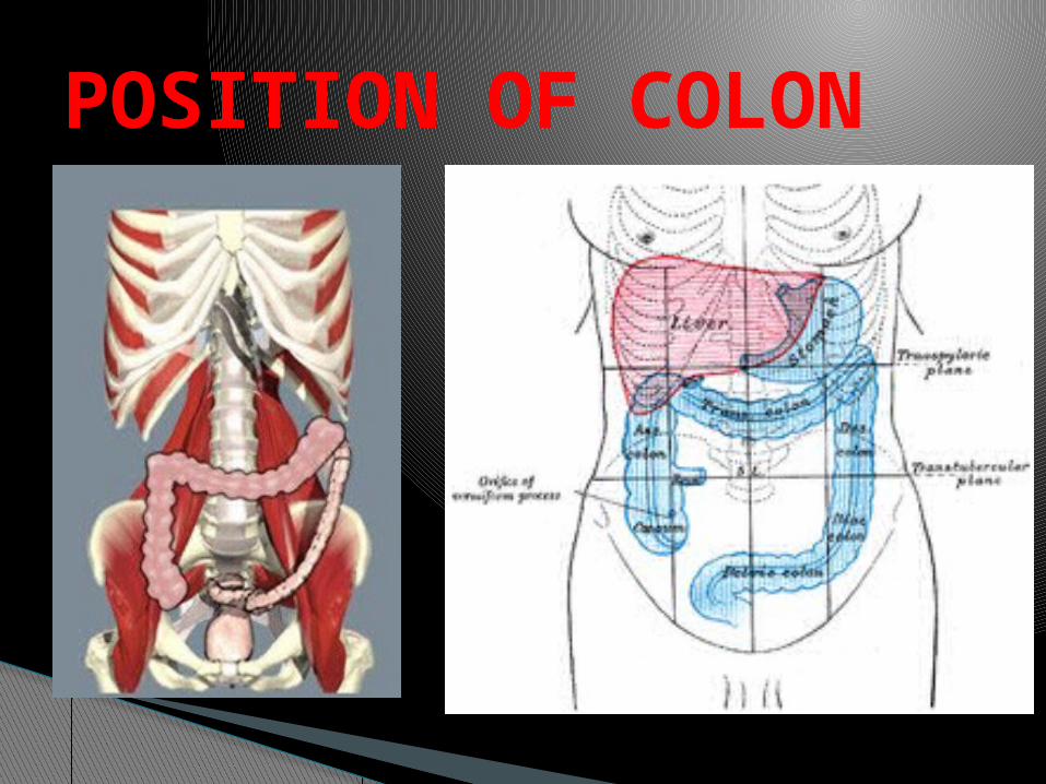
POSITION OF COLON

DIVISION OF COLON
80 cm•Cecum•Colon ascendens•Colon transversum•Colon descendens•Colon sigmoideum•Rectum•Canalis analis
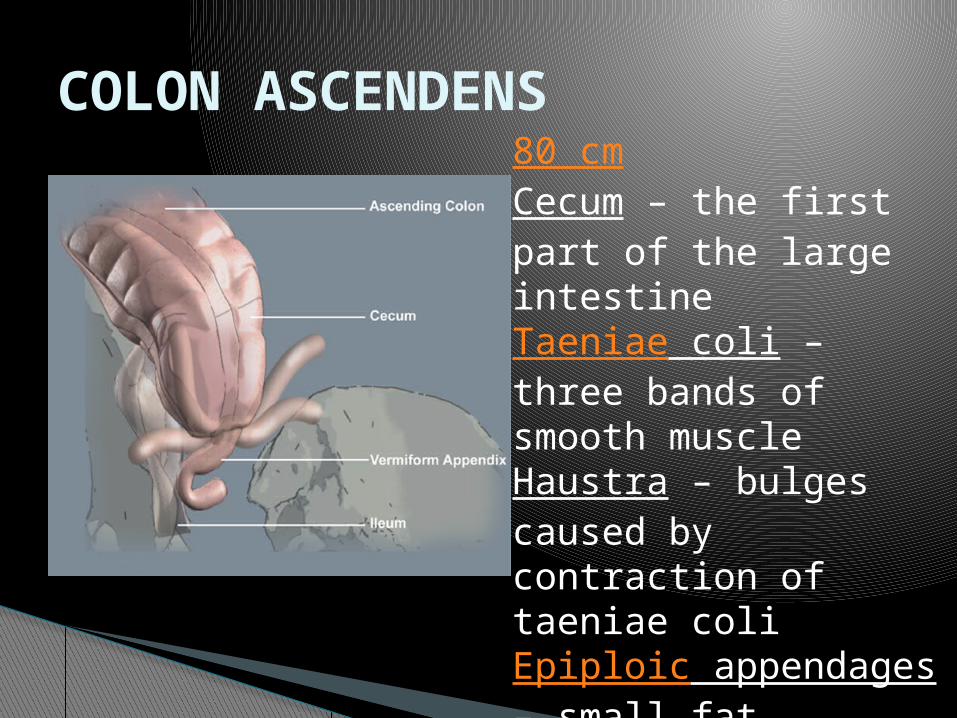
COLON ASCENDENS80 cmCecum – the first part of the large intestineTaeniae coli – three bands of smooth muscle Haustra – bulges caused by contraction of taeniae coli Epiploic appendages – small fat accumulations on the viscera

04/18/2023Wahyuni Atmodjo 6
Location: right lumbar hypochondriaca
Length 8 inches (15-20 cm)
Right colic flexure colon transversum
Secondarily peritoneal Taenia, Haustra, Plicae
semilunares Epiploic appendages
Ascending colon

04/18/2023Wahyuni Atmodjo 7
Hepatic flexure : ren dex, duod inf
Splenic flexure : ren sin, spleenLength 45 – 50cm Intraperitoneal Supported by transverse mesocolon and gastrocolic lig
Colon transversum

04/18/2023Wahyuni Atmodjo 8
Location : left lumbar Begin at splenic flexureLength : 25 cmDiameter smaller than ascending colon
Secondarily peritoneal
Colon descendens

04/18/2023Wahyuni Atmodjo 9
Location: pelvic Length : 25-40 cm Related to urinary bladder (male), uterus and vagina (female)
Continues to rectum at 3rd sacral level
Peritoneal Function: storage of feces
Colon Sigmoid

04/18/2023Wahyuni Atmodjo 10
Rectum ◦Retroperitoneal
Anal canal◦Retroperitoneal◦Inferior to pelvic diaphragm◦Anal triangle of perineum
Rectum and Anal canal

04/18/2023Wahyuni Atmodjo 11
Internal sphincter : ◦ circular muscular
layer under involuntary control.
External sphincter : ◦ striated muscle
under voluntary control.
Anal sphinchter

04/18/2023Wahyuni Atmodjo 12
Internal structure
o Anal columns
o Anal valves
o Pectinate line
o Anal verge
o Anal glands
o Anal sphincter
internal
o Anal sphincter
external

04/18/2023Wahyuni Atmodjo 13
◦A. mesenterica superior: jejenum, ileum, cecum, appendix
◦A. mesenterica inferior: colon, sigmoid, rectum
Blood supply of colon
A. rectalis sup a. mesenterica inf A. rectalis med a. iliaca interna A. rectalis inf a. pudenda interna

04/18/2023Wahyuni Atmodjo 14
A. colica sinistra Aa. sigmoidea A. rectalis superior A. rectosigmoidea A. marginalis
A. Mesenterica superior
A. pancreaticoduodenalis inferior
A. jejunalis A. ilealis A. colica media A. colica dextra A. ileocolica A. marginalis
A. Mesenterica inferior

Superior mesenteric ileocolic aright colic amiddle colic a
Inferior mesenteric left colic asigmoid arteries superior rectal a
ARTERY OF COLON

04/18/2023Wahyuni Atmodjo 16
Blood supply A. rectalis inferior A. rectalis media A. rectalis superior
Venous return◦v. rectalis inferior◦v. rectalis media ◦v. rectalis
superior
Vasculature

Simpatis
Lesser splanchnic nn
Parasimpatis
Plexus sacralis S2– S4

HEPAR,VESICA FELLEA, PANCREAS

POSITION OF THE LIVER
Located at right hypochondriaca epigastrica region Palpable: costal margin, lateral to rectus abdominis

STRUCTURE OF LIVER
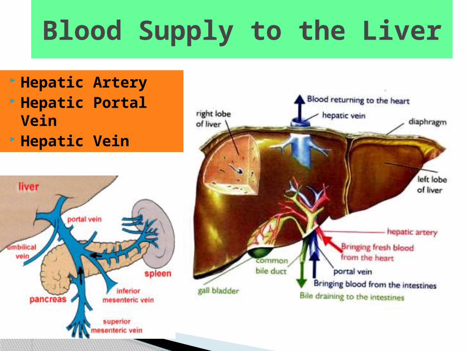
Blood Supply to the Liver
Hepatic Artery Hepatic Portal
Vein Hepatic Vein
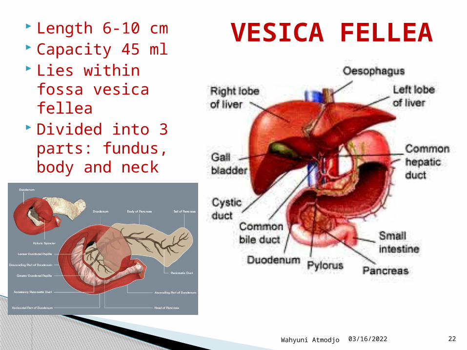
Length 6-10 cm Capacity 45 ml Lies within fossa
vesica fellea Divided into 3
parts: fundus, body and neck
VESICA FELLEA

Function of Pancreas
Endocrine Alpha cells: Glucagon Beta Cells: Insulin Delta Cells: Somatostatin
Exocrine gland 1 - 1.5l pancreatic juice / day Enzymatic component - digestive enzymes
(1) Proteases,(2) Lipolytic ,(3) Amylase

POSITION OF PANCREAS

04/18/2023Wahyuni Atmodjo 25
epigastricleft hypochondriaca
Head , Body, Tail Pancreatic ducts:
◦ Primary pancreatic duct duodenal papilla
◦ Accessory pancreatic accessory duodenal papilla about 2 cm above the primary duct
Structure of Pancreas

BLOOD SUPPLY OF PANCREAS

POSITION OF LIEN
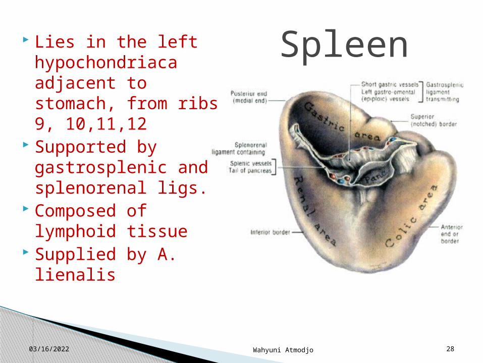
The spleen is approximately 11 cm in length. weighs between 150 -200 gr, lies in front of the 9th to the 12th thoracic ribs.
04/18/2023 28Wahyuni Atmodjo
Spleen Lies in the left hypochondriaca adjacent to stomach, from ribs 9, 10,11,12
Supported by gastrosplenic and splenorenal ligs.
Composed of lymphoid tissue
Supplied by A. lienalis

04/18/2023Wahyuni Atmodjo 29
Blood supply of lien