“Blind Spot Amsler Grid” to determine the Quality of ...€¦ · “Blind Spot Amsler Grid”...
4
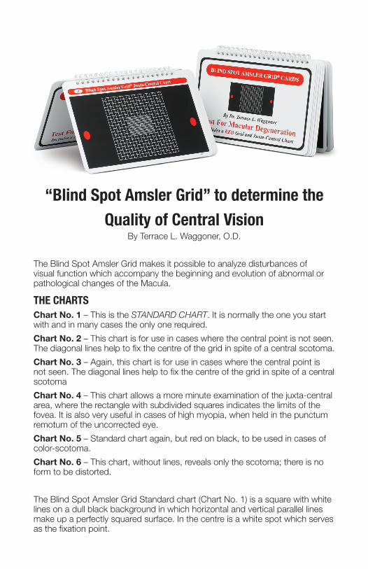
“Blind Spot Amsler Grid” to determine the Quality of Central Vision By Terrace L. Waggoner, O.D. The Blind Spot Amsler Grid makes it possible to analyze disturbances of visual function which accompany the beginning and evolution of abnormal or pathological changes of the Macula. THE CHARTS Chart No. 1 – This is the STANDARD CHART. It is normally the one you start with and in many cases the only one required. Chart No. 2 – This chart is for use in cases where the central point is not seen. The diagonal lines help to fix the centre of the grid in spite of a central scotoma. Chart No. 3 – Again, this chart is for use in cases where the central point is not seen. The diagonal lines help to fix the centre of the grid in spite of a central scotoma Chart No. 4 – This chart allows a more minute examination of the juxta-central area, where the rectangle with subdivided squares indicates the limits of the fovea. It is also very useful in cases of high myopia, when held in the punctum remotum of the uncorrected eye. Chart No. 5 – Standard chart again, but red on black, to be used in cases of color-scotoma. Chart No. 6 – This chart, without lines, reveals only the scotoma; there is no form to be distorted. The Blind Spot Amsler Grid Standard chart (Chart No. 1) is a square with white lines on a dull black background in which horizontal and vertical parallel lines make up a perfectly squared surface. In the centre is a white spot which serves as the fixation point.
Transcript of “Blind Spot Amsler Grid” to determine the Quality of ...€¦ · “Blind Spot Amsler Grid”...
“Blind Spot Amsler Grid” to determine the Quality of Central Vision
By Terrace L. Waggoner, O.D.
The Blind Spot Amsler Grid makes it possible to analyze disturbances of visual function which accompany the beginning and evolution of abnormal or pathological changes of the Macula.
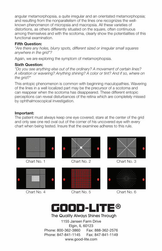
THE CHARTSChart No. 1 – This is the STANDARD CHART. It is normally the one you start with and in many cases the only one required.
Chart No. 2 – This chart is for use in cases where the central point is not seen. The diagonal lines help to fix the centre of the grid in spite of a central scotoma.
Chart No. 3 – Again, this chart is for use in cases where the central point is not seen. The diagonal lines help to fix the centre of the grid in spite of a central scotoma
Chart No. 4 – This chart allows a more minute examination of the juxta-central area, where the rectangle with subdivided squares indicates the limits of the fovea. It is also very useful in cases of high myopia, when held in the punctum remotum of the uncorrected eye.
Chart No. 5 – Standard chart again, but red on black, to be used in cases of color-scotoma.
Chart No. 6 – This chart, without lines, reveals only the scotoma; there is no form to be distorted.
The Blind Spot Amsler Grid Standard chart (Chart No. 1) is a square with white lines on a dull black background in which horizontal and vertical parallel lines make up a perfectly squared surface. In the centre is a white spot which serves as the fixation point.
angular metamorphopsia, a quite irregular and an orientated metamorphopsia; and resulting from the nonparallelism of the lines one recognizes the well-known phenomenon of micropsia and macropsia. All these varieties of distortions, as others differently situated on the square, often continuous among themselves and with the scotoma, clearly show the potentialities of this functional examination.
Fifth Question: "Are there any holes, blurry spots, different sized or irregular small squares anywhere in the grid"?
Again, we are exploring the symptom of metamorphopsia.
Sixth Question: "Do you see anything else out of the ordinary? A movement of certain lines? A vibration or wavering? Anything shining? A color or tint? And if so, where on the grid?"
This entopic phenomenon is common with beginning maculopathies. Wavering of the lines in a well localized part may be the precursor of a scotoma and can reappear when the scotoma has disappeared. These different entopic perceptions can reveal disturbances of the retina which are completely missed by ophthalmoscopical investigation.
Important: The patient must always keep one eye covered; stare at the center of the grid and only see one red oval out of the corner of his uncovered eye with every chart when being tested. Insure that the examinee adheres to this rule.
1155 Jansen Farm DriveElgin, IL 60123
Phone: 800-362-3860 Fax: 888-362-2576Phone: 847-841-1145 Fax: 847-841-1149
www.good-lite.com
Chart No. 1
Chart No. 4
Chart No. 2
Chart No. 5
Chart No. 3
Chart No. 6
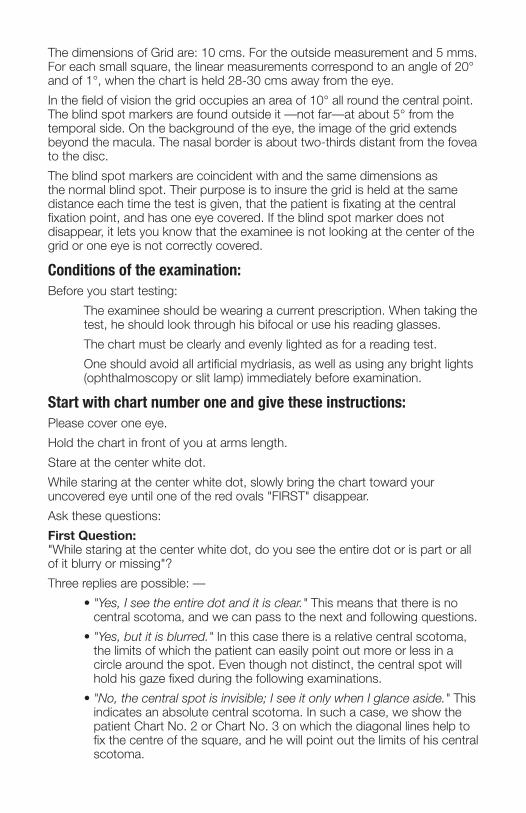
The dimensions of Grid are: 10 cms. For the outside measurement and 5 mms. For each small square, the linear measurements correspond to an angle of 20° and of 1°, when the chart is held 28-30 cms away from the eye.
In the field of vision the grid occupies an area of 10° all round the central point. The blind spot markers are found outside it —not far—at about 5° from the temporal side. On the background of the eye, the image of the grid extends beyond the macula. The nasal border is about two-thirds distant from the fovea to the disc.
The blind spot markers are coincident with and the same dimensions as the normal blind spot. Their purpose is to insure the grid is held at the same distance each time the test is given, that the patient is fixating at the central fixation point, and has one eye covered. If the blind spot marker does not disappear, it lets you know that the examinee is not looking at the center of the grid or one eye is not correctly covered.
Conditions of the examination:Before you start testing:
The examinee should be wearing a current prescription. When taking the test, he should look through his bifocal or use his reading glasses.
The chart must be clearly and evenly lighted as for a reading test.
One should avoid all artificial mydriasis, as well as using any bright lights (ophthalmoscopy or slit lamp) immediately before examination.
Start with chart number one and give these instructions:Please cover one eye.
Hold the chart in front of you at arms length.
Stare at the center white dot.
While staring at the center white dot, slowly bring the chart toward your uncovered eye until one of the red ovals "FIRST" disappear.
Ask these questions:
First Question: "While staring at the center white dot, do you see the entire dot or is part or all of it blurry or missing"?
Three replies are possible: —
•"Yes, I see the entire dot and it is clear." This means that there is no central scotoma, and we can pass to the next and following questions.
• "Yes, but it is blurred." In this case there is a relative central scotoma, the limits of which the patient can easily point out more or less in a circle around the spot. Even though not distinct, the central spot will hold his gaze fixed during the following examinations.
•"No, the central spot is invisible; I see it only when I glance aside." This indicates an absolute central scotoma. In such a case, we show the patient Chart No. 2 or Chart No. 3 on which the diagonal lines help to fix the centre of the square, and he will point out the limits of his central scotoma.
Second Question: "Is the center of the grid blurry, distorted, or are parts of it missing"?
This question deals with juxta - and paracentral scotoma, absolute or relative, of which the patient can easily point out the location, the outline and the form. Is the spot in question clearly defined? Is it completely blurred (absolute scotoma) or do white lines show through (relative scotoma)? Is the blurring everywhere equal, or does it show some parts more blurred and other parts less blurred?
Third Question: "Do you see the four corners and sides of the grid"?
Possible replies:
•"Yes, it is complete." Then we can go on to the next question.
•"No, one corner is cut off," or "one side," and the patient shows us the corner or the corners, or the sides which are missing.
Here we have to deal with a scotoma, which we call "exterior" because it invades the area in which we are interested from the outside. What we are about to find here is the arcuate scotoma of Bjerrum found in chronic glaucoma, which coming from the neighboring blind spot, covers in its curved course the superior or inferior temporal angle of our square, or the two at once. Or the patient may point out the loss of the other side of the square; this is the nasal restriction, also characteristic of chronic glaucoma and which accentuates its menace at least 10° from the-centre. In the case of a very advanced glaucoma, as also in pigmentary degeneration of the retina, the all-round encirclement of the focus point (annular scotoma) will be shown to the patient by the disappearance of each corner and each side of the square. The field left in the middle will be easily outlined and measured by the number of small squares left intact in each direction.
Another interesting exterior scotoma is the caecocentral scotoma of toxic amblyopia which is bilateral and temporal to the blind spot. This first becomes obvious on our square, often partially absolute (nucleus) and partially relative. In these cases, our Chart No. 5 with red squares on a black ground will be helpful.
Fourth Question: "Do the lines appear to be straight and continuous from top to bottom and side to side or are some distorted, wavy, blurred or broken"?
With this question, we are exploring the symptom of metamorphopsia.
The infinitely diverse replies which are made to this fourth question reveal that there are many different types of metamorphopsia. "The whole square is like an unstretched canvas," says one patient; "in this place all the lines seem to have been drawn by free hand and not ruled," says another; "the left side of the square seems swollen," says a third, and with a finger will indicate a regular convex line; "just above the white spot," says a fourth, "the lines are completely broken"; and a fifth, "there, the horizontal lines are waving much more than the vertical lines"; and a sixth, "these small squares here are bigger than those there," and so on.
One learns to recognize, by experience, a diffuse as well as a localized metamorphopsia, a gross and a minute metamorphopsia, a wavy and an
“Blind Spot Amsler Grid” to determine the Quality of Central Vision
By Terrace L. Waggoner, O.D.
The Blind Spot Amsler Grid makes it possible to analyze disturbances of visual function which accompany the beginning and evolution of abnormal or pathological changes of the Macula.
THE CHARTSChart No. 1 – This is the STANDARD CHART. It is normally the one you start with and in many cases the only one required.
Chart No. 2 – This chart is for use in cases where the central point is not seen. The diagonal lines help to fix the centre of the grid in spite of a central scotoma.
Chart No. 3 – Again, this chart is for use in cases where the central point is not seen. The diagonal lines help to fix the centre of the grid in spite of a central scotoma
Chart No. 4 – This chart allows a more minute examination of the juxta-central area, where the rectangle with subdivided squares indicates the limits of the fovea. It is also very useful in cases of high myopia, when held in the punctum remotum of the uncorrected eye.
Chart No. 5 – Standard chart again, but red on black, to be used in cases of color-scotoma.
Chart No. 6 – This chart, without lines, reveals only the scotoma; there is no form to be distorted.
The Blind Spot Amsler Grid Standard chart (Chart No. 1) is a square with white lines on a dull black background in which horizontal and vertical parallel lines make up a perfectly squared surface. In the centre is a white spot which serves as the fixation point.
angular metamorphopsia, a quite irregular and an orientated metamorphopsia; and resulting from the nonparallelism of the lines one recognizes the well-known phenomenon of micropsia and macropsia. All these varieties of distortions, as others differently situated on the square, often continuous among themselves and with the scotoma, clearly show the potentialities of this functional examination.
Fifth Question: "Are there any holes, blurry spots, different sized or irregular small squares anywhere in the grid"?
Again, we are exploring the symptom of metamorphopsia.
Sixth Question: "Do you see anything else out of the ordinary? A movement of certain lines? A vibration or wavering? Anything shining? A color or tint? And if so, where on the grid?"
This entopic phenomenon is common with beginning maculopathies. Wavering of the lines in a well localized part may be the precursor of a scotoma and can reappear when the scotoma has disappeared. These different entopic perceptions can reveal disturbances of the retina which are completely missed by ophthalmoscopical investigation.
Important: The patient must always keep one eye covered; stare at the center of the grid and only see one red oval out of the corner of his uncovered eye with every chart when being tested. Insure that the examinee adheres to this rule.
1155 Jansen Farm DriveElgin, IL 60123
Phone: 800-362-3860 Fax: 888-362-2576Phone: 847-841-1145 Fax: 847-841-1149
www.good-lite.com
Chart No. 1
Chart No. 4
Chart No. 2
Chart No. 5
Chart No. 3
Chart No. 6