Blends of Poly-(E-caprolactone) and Polysaccharides in ... by extrusion or injection molding at...
16
Blends of Poly-(E-caprolactone) and Polysaccharides in Tissue Engineering Applications Gianluca Ciardelli,* ,² Valeria Chiono, ² Giovanni Vozzi, ²,‡ Mariano Pracella, § Arti Ahluwalia, ‡ Niccoletta Barbani, ² Caterina Cristallini, ‡ and Paolo Giusti ²,§ Department of Chemical Engineering, Industrial Chemistry and Materials Science, University of Pisa, Via Diotisalvi 2, 56126 Pisa, Italy, Centro “E. Piaggio”, University of Pisa, Via Diotisalvi 2, 56126 Pisa, Italy, and C.N.R. Institute for Composite and Biomedical Materials, Via Diotisalvi 2, 56126 Pisa, Italy Received February 2, 2005 Bioartificial blends of poly-(-caprolactone) (PCL) with a polysaccharide (starch, S; dextran, D; or gellan, G) (PCL/S, PCL/D, PCL/G 90.9/9.1 wt ratio) were prepared by a solution-precipitation technique and widely characterized by differential scanning calorimetry analysis (DSC), Fourier transform infrared-attenuated total reflectance spectroscopy (FTIR-ATR), optical microscopy (OM), wide-angle X-ray diffraction analysis (WAXD), and thermogravimetry (TGA). DSC showed that the polysaccharide reduced the crystallinity of PCL and had a nucleation effect, which was also confirmed by OM analysis. Hoffman-Weeks analysis was performed on PCL and blend samples allowing calculation of their equilibrium melting temperatures (T m 0 ). WAXD showed that the crystalline unit cell type was the same for PCL and blends. FTIR-ATR did not evidence interactions between blend components. Thermal stability was affected by the type of polysaccharide. Microparticles (<125 μm) were produced from blends by cryogenical milling and characterized by scanning electron microscopy analysis (SEM). Selective laser sintering (SLS), a new rapid prototyping technology for scaffold fabrication, was applied to sinter blend microparticles according to a PC-designed two-dimensional geometry (strips and 2 × 2 mm 2 square-meshed grids). The optimal experimental conditions for sintering were established and laser beam parameters (beam speed, BS, and power, P) were found for each blend composition. Morphology of sintered objects was analyzed by SEM and found to be dependent on the morphology of the sintered powders. Sintered samples were analyzed by chemical imaging (CI), FTIR-ATR, DSC, and contact angle analysis. No evidence of the occurrence of degradation phenomena was found by FTIR-ATR for sintered samples, whereas DSC parameters of PCL and blends showed changes which could be attributed to some molecular weight decrease of PCL during sintering. CI of sintered samples showed that the polysaccharide phase was homogeneously dispersed within the PCL matrix, with the only exception being the PCL/D blend. The contact angle analysis showed that all samples were hydrophilic. Fibroblasts were then seeded on scaffolds to evaluate the rate and the extent of cell adhesion and the effect of the polysaccharides (S, D, G) on the bioactivity of the PCL-based blends. Introduction Tissue engineering has emerged as a promising approach to treat the loss or malfunction of a tissue or organ. Several tissue-engineering methods typically employ three-dimen- sional biodegradable polymer matrixes as temporary scaffolds to engineer new natural tissues from isolated cells. 1-6 The most important requirement for a biodegradable polymer to be used in medical applications is its biocom- patibility in a specific environment, together with the noncytotoxicity of its degradation products. 7 Aliphatic polyesters such as polylactide (PLA), polyglycolide (PGA), poly(lactide-co-glycolide) (PLGA), and poly--caprolactone (PCL) are among the few synthetic polymers that meet these requirements and have been used in the tissue engineering of cartilage, 8-11 bone, 12-14 tendon, 15 skin, 16 liver, 18 and heart valves. 19 However, cell affinity toward synthetic polymers is generally poor as a consequence of their low hydrophilicity and lack of surface cell recognition sites. 20,21 Cell adhesion on scaffolds is markedly influenced by the physical and chemical properties of the material surface layer, which influence the non-receptor-mediated and receptor- mediated attachment mechanisms. Non-receptor-mediated cell adhesion consists of a nonspecific cell-material interac- tion via weak chemical bonding (hydrogen bonding, elec- trostatic, polar, ionic interactions between molecules on cell membrane and functional chemical groups on the polymer substrate). These types of interactions cannot ensure the transmission of adequate signals from the extra-cellular matrix environment into cells. On the contrary, receptor- mediated cell adhesion is due to the extra cellular matrix (ECM) molecules, such as fibronectin, vitronectin, collagen, and laminin. Cells bind specific amino acid sequences of ECM molecules through integrin receptors. 22 * To whom correspondence should be addressed. E-mail: [email protected]. Fax: +39/050/511266. ² Department of Chemical Engineering, Industrial Chemistry and Materi- als Science, University of Pisa. ‡ Centro “E. Piaggio”, University of Pisa. § C.N.R. Institute for Composite and Biomedical Materials. 1961 Biomacromolecules 2005, 6, 1961-1976 10.1021/bm0500805 CCC: $30.25 © 2005 American Chemical Society Published on Web 04/27/2005
Transcript of Blends of Poly-(E-caprolactone) and Polysaccharides in ... by extrusion or injection molding at...
Blends of Poly-( E-caprolactone) and Polysaccharides in TissueEngineering Applications
Gianluca Ciardelli,*,† Valeria Chiono,† Giovanni Vozzi,†,‡ Mariano Pracella,§ Arti Ahluwalia,‡
Niccoletta Barbani,† Caterina Cristallini,‡ and Paolo Giusti†,§
Department of Chemical Engineering, Industrial Chemistry and Materials Science, University of Pisa,Via Diotisalvi 2, 56126 Pisa, Italy, Centro “E. Piaggio”, University of Pisa, Via Diotisalvi 2, 56126 Pisa,Italy, and C.N.R. Institute for Composite and Biomedical Materials, Via Diotisalvi 2, 56126 Pisa, Italy
Received February 2, 2005
Bioartificial blends of poly-(ε-caprolactone) (PCL) with a polysaccharide (starch, S; dextran, D; or gellan,G) (PCL/S, PCL/D, PCL/G 90.9/9.1 wt ratio) were prepared by a solution-precipitation technique andwidely characterized by differential scanning calorimetry analysis (DSC), Fourier transform infrared-attenuatedtotal reflectance spectroscopy (FTIR-ATR), optical microscopy (OM), wide-angle X-ray diffraction analysis(WAXD), and thermogravimetry (TGA). DSC showed that the polysaccharide reduced the crystallinity ofPCL and had a nucleation effect, which was also confirmed by OM analysis. Hoffman-Weeks analysiswas performed on PCL and blend samples allowing calculation of their equilibrium melting temperatures(Tm
0 ). WAXD showed that the crystalline unit cell type was the same for PCL and blends. FTIR-ATR didnot evidence interactions between blend components. Thermal stability was affected by the type ofpolysaccharide. Microparticles (<125 µm) were produced from blends by cryogenical milling andcharacterized by scanning electron microscopy analysis (SEM). Selective laser sintering (SLS), a new rapidprototyping technology for scaffold fabrication, was applied to sinter blend microparticles according to aPC-designed two-dimensional geometry (strips and 2× 2 mm2 square-meshed grids). The optimalexperimental conditions for sintering were established and laser beam parameters (beam speed, BS, andpower, P) were found for each blend composition. Morphology of sintered objects was analyzed by SEMand found to be dependent on the morphology of the sintered powders. Sintered samples were analyzed bychemical imaging (CI), FTIR-ATR, DSC, and contact angle analysis. No evidence of the occurrence ofdegradation phenomena was found by FTIR-ATR for sintered samples, whereas DSC parameters of PCLand blends showed changes which could be attributed to some molecular weight decrease of PCL duringsintering. CI of sintered samples showed that the polysaccharide phase was homogeneously dispersed withinthe PCL matrix, with the only exception being the PCL/D blend. The contact angle analysis showed that allsamples were hydrophilic. Fibroblasts were then seeded on scaffolds to evaluate the rate and the extent ofcell adhesion and the effect of the polysaccharides (S, D, G) on the bioactivity of the PCL-based blends.
Introduction
Tissue engineering has emerged as a promising approachto treat the loss or malfunction of a tissue or organ. Severaltissue-engineering methods typically employ three-dimen-sional biodegradable polymer matrixes as temporary scaffoldsto engineer new natural tissues from isolated cells.1-6
The most important requirement for a biodegradablepolymer to be used in medical applications is its biocom-patibility in a specific environment, together with thenoncytotoxicity of its degradation products.7 Aliphaticpolyesters such as polylactide (PLA), polyglycolide (PGA),poly(lactide-co-glycolide) (PLGA), and poly-ε-caprolactone(PCL) are among the few synthetic polymers that meet theserequirements and have been used in the tissue engineering
of cartilage,8-11 bone,12-14 tendon,15 skin,16 liver,18 and heartvalves.19 However, cell affinity toward synthetic polymersis generally poor as a consequence of their low hydrophilicityand lack of surface cell recognition sites.20,21
Cell adhesion on scaffolds is markedly influenced by thephysical and chemical properties of the material surface layer,which influence the non-receptor-mediated and receptor-mediated attachment mechanisms. Non-receptor-mediatedcell adhesion consists of a nonspecific cell-material interac-tion via weak chemical bonding (hydrogen bonding, elec-trostatic, polar, ionic interactions between molecules on cellmembrane and functional chemical groups on the polymersubstrate). These types of interactions cannot ensure thetransmission of adequate signals from the extra-cellularmatrix environment into cells. On the contrary, receptor-mediated cell adhesion is due to the extra cellular matrix(ECM) molecules, such as fibronectin, vitronectin, collagen,and laminin. Cells bind specific amino acid sequences ofECM molecules through integrin receptors.22
* To whom correspondence should be addressed. E-mail:[email protected]. Fax:+39/050/511266.
† Department of Chemical Engineering, Industrial Chemistry and Materi-als Science, University of Pisa.
‡ Centro “E. Piaggio”, University of Pisa.§ C.N.R. Institute for Composite and Biomedical Materials.
1961Biomacromolecules 2005,6, 1961-1976
10.1021/bm0500805 CCC: $30.25 © 2005 American Chemical SocietyPublished on Web 04/27/2005
Polysaccharides are interesting materials for tissue engi-neering applications since their carbohydrate moieties interactwith or are integral components of many cell adhesionmolecules and matrix glycoproteins.23
Bioartificial polymeric materials have been produced byblending synthetic and natural polymers in order to combinethe good mechanical characteristics, easy processability, andlow production and transformation costs of the former withthe specific tissue and cell compatibility of the latter.24-27
Moreover, blending synthetic and natural polymers allowsfor a control of the degradation rate of the system:28 as thedegradation kinetics of a bioartificial blend increase withincreasing the natural polymer amount, the blend compositioncan be adjusted to make the scaffold degradation rate matchwith the growth rate of the regenerating tissue.
Our research group has already realized and studiedvarious systems consisting of a biological (mainly fibrin,collagen, and hyaluronic acid) and a synthetic (polyurethanes,poly(vinyl alcohol) (PVA), and poly(acrylic acid) (PAA))polymer.29 Blends between collagen and PAA24 showedstrong interactions between the ionized functional groups ofthe two components, which increased the denaturationtemperature of collagen. Blends between hyaluronic acid orits ester derivatives with PVA were produced and used toobtain hydrogels by a freeze-thaw technique.30,31 Recentstudies have been carried out by our group also on blendsof poly(vinyl alcohol) (PVA) with starch, and PVA withchitosan or dextran for their potential use as dialysismembranes or delivery systems for human growth hormone,respectively.28
The combination between synthetic and natural polymershas also been widely investigated in the field of particulatecarriers with engineered surface properties for drug targeting.The synthetic polymers such as PCL, PLA, and PLGA arehydrophobic which leads to complement activation and liveraccumulation and they also lack surface recognition functionsfor specific mucoadhesion or receptor recognition. Thesedrawbacks can be overcome by producing polysaccharide-coated particles whose core is made of a synthetic polymer,resulting in a control of the specific interactions with targetcells and the nonspecific interactions with blood componentsand phagocytic cells.32-34
In this research, we studied PCL-based bioartificial blends,as PCL is a regulatory approved biodegradable and biocom-patible synthetic polymer with good mechanical properties,35-37
which may undergo enzymatic degradation through hydroly-sis of its ester bonds by lipase,38 cholesterol esterase,39,40andcarboxyl esterase.39 However, degradation kinetics of PCLare slow due to its hydrophobic and semicrystalline nature,which makes its resorption time longer than 2 years.9 Onthe other hand, PCL has a low glass transition temperatureof around -60 °C, which gives the polymer a rubberybehavior at room temperature and, as a consequence, a goodpermeability to low molecular weight drugs in deliverysystems for biomedical uses. The main applications of PCLin the tissue engineering field include tissue engineeredskin,41 drug delivery systems,42,43 axonal regeneration,44,45
and scaffolds for supporting fibroblasts and osteoblastsgrowth.46,47
In this work, binary blends were produced between PCLand one of three selected polysaccharides (starch, gellan, anddextran) with the aim of finding out among them which ismore suitable for the production of bioactive scaffolds withhigh biocompatibility. Our choice focused on starch, gellan,and dextran, as they are natural biodegradable hydrophilicpolymers, which display enzymatic degradation behavior anda good biocompatibility.
Starch is a biopolymer present as minute granules in theroots, seeds, and stems of a variety of plants, including corn,wheat, rice, barley, and potatoes. It is composed of twocomponents: amylose, which is a linear polymer consistingof D-glucose units joined byR-1,4 glycosidic bonds, andamylopectin, which is a highly branched polymer with amolecular weight that can reach tens of millions.48 Starch-based blends have been widely used in several biomedicalapplications as they offer the possibility of obtaining verydistinct structure/properties combinations, varying the syn-thetic component of the blend, processing methods, additives,and reinforcement materials.49-51 Resulting applications covera wide range of fields, such as tissue engineering scaffolds,49
bone cements,50 hydrogels for the controlled release ofdrugs,50 and bone substitutes in the orthopaedic field.51
Gellan is an anionic exocellular polysaccharide secretedfrom theSphingomonas elodeabacterium52 and composedof repeated tetrasaccharide units consisting of glucose,glucuronic acid, and rhamnose residues in a 2:1:1 ratio,joined in a linear chain ([f3)-â-D-glucose-(1f4)-â-D-glucuronic acid-(1f4)-â-D-glucose-(1f4)-R-L-rhamnose-(1f]n).53 Gellan, which is primarily used as a thickener orgelling agent in a wide range of food applications,54 hasrecently been investigated by our group as a suitablecandidate for reactive blending with gelatin, leading to theproduction of biocompatible scaffolds.55 Other applicationsof gellan in the biomedical field include capsules for drugdelivery systems.56
Dextran is a high molecular weight polymer ofD-glucoseand it is produced by different bacterial strains. In the fieldof biomaterials, dextran is mainly used as a plasma expander,whereas dextran derivatives find applications in bone repair.57
Blends between polylactide and dextran have recently beenstudied as promising materials for tissue engineering scaf-folds.20,21
In the literature, blends between PCL and starch have beenproduced by melt extrusion for biodegradable packagingapplications.58,59Some physicochemical characterizations ofPCL/starch blends have been carried out with the purposeof using them for biomedical applications. Demirgo¨z et al.developed a methodology for cross-linking commercialstarch-based thermoplastic blends.60 The method was basedon the reaction between the starch hydroxyl groups and tri-sodium tri-meta phosphate and led to materials with a lowerwater-up take ability, a slower degradation rate, improvedmechanical properties and modified surface properties whichcould affect interactions with proteins and cells in vivo. Manoet al.61 studied the thermal properties of thermoplastic starch/synthetic polymer blends (including PCL/starch blends)aimed at biomedical applications and found out that thevolatilisation of the plasticizer (glycerol) could occur during
1962 Biomacromolecules, Vol. 6, No. 4, 2005 Ciardelli et al.

processing by extrusion or injection molding at temperaturesabove 150°C. Degradation of starch could occur at someextent only for more extreme processing conditions, as starchweight loss was recorded in the 280-350 °C temperaturerange by thermogravimetric analysis.
However, the literature does not report specific studies onscaffolds made of blends between PCL and starch, gellan,or dextran.
Beside the choice of the materials for scaffold production,the novel content of the present paper also consists of theuse of selective laser sintering (SLS) as a technique forscaffold fabrication. Briefly, SLS is a new rapid prototypingtechnique, based on the use of the heating energy of a CO2
laser to sinter powder particles following a computer-designed geometry.62,63SLS creates objects from a computer-generated model: the model is sliced into a set of two-dimensional layers which are reproduced by laser sinteringof powder particles.
In this work, we sintered strips as a first trial to find outthe optimal conditions for sintering. Then, we obtained two-dimensional (2D) grid-shaped scaffolds.
The physicochemical properties of both blend powders andsintered samples were evaluated, and a comparison was madebetween the characteristics of pure PCL and those of blends,based on our previous work on PCL.62 A wide range ofcharacterization techniques were used with the aim ofobtaining specific information. Thermogravimetric analysis(TGA) assessed the thermal stability of blend samples, tobetter define the correct parameters for the laser sinteringprocessing. Differential scanning calorimetry (DSC) wasperformed both under isothermal and nonisothermal condi-tions to give insight into blend compatibility and thermalparameters. Furthermore, the nonisothermal analysis carriedout on sintered and unsintered samples allowed detection ofany difference in the melting and crystallization behaviorbetween the two kinds of samples due to the sinteringprocess. Optical microscopy analysis (OM) allowed observa-tion of morphological characteristics of blend samples. Wide-angle X-ray diffraction analysis (WAXD) was performed toevidence the effect of blend composition on the crystallinestructure. Fourier transform infrared-attenuated total reflec-tance spectroscopy (FTIR-ATR) was carried out with theaim of evidencing both the interactions between blendcomponents and any possible chemical variation occurredduring sintering. Chemical imaging (CI) is a powerfultechnique for the analysis of surface composition and surfacedistribution of phases. At last, cell adhesion tests wereperformed on sintered scaffolds to compare blend compat-ibility and bioactivity as a function of the polysaccharidetype.
Experimental Section
Materials. Polymers used were a commercial poly-(ε-caprolactone) (PCL) supplied by Polysciences, Inc. with anaverage molecular weight (Mw) of 45 000 and three biopoly-mers: soluble starch ACS for analysis (S; Carlo Erba),dextran T40 (D; Pharmacia Fine Chemicals), and gellan gum(G; Fluka). Soluble starch composition was experimentally
found to be 90 wt % amylose and 10 wt % amylopectin. Allother chemicals were analytical pure grade and used asdelivered, without further purification.
Preparations of Blend and PCL Microparticles. Threeblends containing 90.9% (wt/wt) PCL and 9.1% (wt/wt) S,G, or D were produced by a solution-precipitation techniqueand marked as PCL/S, PCL/G, and PCL/D, respectively.
Briefly, after preparing a 5% (wt/v) PCL solution indimethylsulfoxide (DMSO; Riedel de Haen) at 50°C, oneof the biopolymers was added to the solution and stirringwas carried out for 2 h at 50 °C. Blend solutions wereprecipitated into methanol (Carlo Erba) and precipitates, firstcollected by centrifugation (Centurion, 6000 series) at 4000rpm for 5 min, were dried in a vacuum oven at 37°C forthree weeks. Dried samples were reduced into powdersthrough cryogenic milling and then sieved through sieveswith 125 µm mesh size.
PCL microparticles were prepared by cryogenical milling,followed by sieving through sieves with 125µm mesh size.PCL microspheres were prepared by a solvent evaporationprocedure based on a single oil-in-water (O/W) emulsion.62
Physicochemical Characterization.Differential scanningcalorimetry (DSC) experiments were carried out in a Perkin-Elmer Pyris Diamond equipped with a Perkin-Elmer Intra-cooler 2P on samples (5-10 mg) packed in aluminum pans.Nonisothermal scans were performed between-20 and+100 °C at a heating rate of 10°C min-1 under nitrogenatmosphere. The melting temperature (Tm) and enthalpy offusion (∆Hm) of the crystalline phase were determined fromthe second scan whereas crystallization temperature (TC) andenthalpy (∆HC) were obtained from the cooling scan. PCLcrystallinity degree (X) was calculated from measured PCLmelting enthalpy as a percentage of the reported heat offusion (139.5 J/g) for fully crystalline PCL.64 The overalltime of crystallization (tc) was determined by the equation
whereTi, Tf, andr are the initial crystallization temperature,the end crystallization temperature, and the cooling rate,respectively. The temperatures at which the crystallizedfraction was 1% and 99% were taken asTi and Tf,respectively.
Isothermal crystallization kinetics were analyzed accordingto the following procedure. The samples (5-10 mg) wereheated to 100°C, kept at this temperature for 5 min to destroyany trace of crystallinity, quenched (at a nominal rate of 200°C min-1) to various crystallization temperatures (Tc; 38-48 °C), and kept atTc until completed crystallization. Theheat of crystallization at eachTc was recorded as a functionof time. The starting time for the analysis of crystallizationkinetics was taken as the time at which sample temperaturereached the programmed valueTc. The fractionXt of materialcrystallized after timet was evaluated by the ratio of thecrystallization area at the timet over the total area. The half-time of crystallization (t0.5) at eachTc was calculated as thetime corresponding toXt ) 0.5. The melting behavior ofisothermally crystallized samples was analyzed by directlyheating the crystallized samples fromTc up to 100°C at arate of 10°C min-1.
tc ) (Ti - Tf)/r (1)
Blends of PCL and Polysaccharides Biomacromolecules, Vol. 6, No. 4, 2005 1963
The morphology of samples was examined by an opticalpolarized light microscope (Leitz Ortholux II POL-BK)equipped with a hot stage (Linkam, model THMSE 600). Inthis experiment, films obtained from powders were sand-wiched between a microscope slide and a cover glass, heatedto 100 °C at a rate of 10°C min-1 and kept at thistemperature for 5 min to destroy any trace of crystallinity.Then the films were cooled at room temperature andexamined under the microscope at a magnification of 32×.Photomicrographs were taken using a JVC TK-1085E videocamera and a Pinnacle System miro VIDEO DC30 seriescapture card, with Adobe Premiere 5.1 as the video editingtool.
Thermal degradation was measured using a Perkin-ElmerTGA 6 Thermogravimetric Analyzer equipment under anitrogen atmosphere. The experiments were performed at aheating rate of 10°C min-1 in the 30-600 °C temperaturerange.
The X-ray diffraction analysis (WAXD) was carried outby a Siemens 810 apparatus using Cu KR radiation in therange 5-40° (2θ) and the step length was 0.01° at 40 kVand 30 mA.
The Fourier transform infrared-attenuated total reflectancespectroscopy (FTIR-ATR) spectra were recorded at roomtemperature in a Perkin-Elmer Spectrum One Spectrometerin the 4000-600 cm-1 range.
Blend powders were evaluated for surface morphology byscanning electron microscopy (SEM, JEOL JSM 5600 LV).
Selective Laser Sintering (SLS).The SLS machine ofthis study is a prototype designed and built at the Departmentof Mechanical, Nuclear and Production Engineering of theUniversity of Pisa.62 The machine work chamber is equippedwith a platform able to shift along thez axis and a laserblock, including the laser beam and a system of galvano-metric mirrors, which are positioned to precisely address thebeam. The CO2 laser used (SYNRAD model J48-5S) has anominal power of 50 W. The machine is PC-driven by meansof software for laser guidance (CADMARK, Quantasystems.r.l.). Sintering experiments were performed both on drypowders and aqueous slurries, obtained by dispersing thepolymer particles in a small amount of demineralized water.First, a very thin layer of powder or blend slurry (0.3 mmdepth) was delivered and leveled on a glass slide fixed onthe supporting cylinder, placed on the platform able to movealong thez axis. Then, specific areas of the layer were lasersintered according to the instructions of a CAD file. At eachtest, two parameters were set: the laser beam speed (BS)and power (P). Our first experiments were carried out onPCL as-produced dry powders, trying a wide range ofcombinations of the laser parameters (P ) 1-3 W, BS )10-80 mm/s) to find out the optimal working set. As asecond step, we focused our attention on slurries. In aprevious work,62 our group found out that the right param-eters for sintering PCL slurries wereP ) 2 W and BS) 20mm/s. Sintering experiments of blend slurries were thuscarried out using 2 W as afixed value forP and varying BS(5, 10, 20, 30, 40, and 50 mm/s). Other experiments wereperformed by keeping BS constant (5 mm/s) while varyingP between 1 and 3 W.
We fabricated simple 10 mm long strips from dry powder(P ) 2 W, BS) 80 mm/s) and slurries (P ) 2 W, BS) 5mm/s) and two-dimensional scaffolds in the shape of square(2 mm× 2 mm) meshed grids from slurries (P ) 2 W, BS) 5 mm/s). After structures had been dried at roomtemperature for a week, the unsintered powder particles werebrushed away and objects were characterized by opticalmicroscopy (Olympus AX70).
SEM (JEOL JSM 5600 LV) analysis of surfaces andfractured sections of laser sintered samples was performedto gain insight into the accuracy of sintering.
Static contact angles of the upper surface of 20× 20 mm2
laser sintered fill structures were measured at room temper-ature by the sessile drop method, using a 5µL water dropletin a telescopic goniometer (Rame`-Hart, Inc.). The telescopehad a magnification power of 23× and was equipped with aprotractor of 1° graduation. For each angle reported, at leastthree measurements on different surface locations wereaveraged.
FTIR-ATR analysis and nonisothermal DSC analysis wereperformed on laser sintered samples using the same condi-tions and equipment as for the unsintered powders.
Microscopic infrared imaging of laser sintered samples wasperformed with a Spectrum Spotlight FT-IR Imaging System(Perkin-Elmer). Briefly, an infrared spectrum map of a zoneof interest was generated as a false-color visible image,consisting of a spectrum per pixel. Individual spectra wereacquired from different positions in the analyzed area, themost representative of which was chosen as the “medium”spectrum and used to obtain a correlation map.
Slurries of pure polysaccharides were processed by SLSutilizing the same parameters as for sintering blends. Theresulting samples were analyzed by FTIR-ATR analysis toassess the effect of laser radiation on the chemistry of S, D,and G.
PCL slurries were sintered using both the sinteringparameters found in our previous work for pure PCL62 andthose utilized in this study for blends. The resulting sampleswere analyzed by FTIR-ATR analysis.
Fibroblasts Culture and Characterization. Two-dimen-sional scaffolds (grids) were prepared for cell cultureaccording to the following procedure. Dry samples weresterilized by washing with a 70% (v/v) ethanol solution insterile water, followed by UV exposure for 15 min on eachsample side. Polymer structures were coated with type Agelatin from pig skin (Sigma, Italy) (here selected as positivecontrol), by leaving them in a bath of 1% (w/v) gelatinaqueous solution for 1 h at 37°C.
NIH-3T3 mouse fibroblasts were cultured in 25 cm2
culture flasks containing Dulbecco’s modified Eagle’s me-dium (DMEM; Cambrex, Italy) with high glucose, 10% fetalcalf serum (Cambrex, Italy), 1% glutamine (Cambrex, Italy),penicillin (200 U/mL; Cambrex, Italy), and streptomycin (200µg/mL; Cambrex, Italy). Culture was maintained in anincubator equilibrated with 5% CO2 at 37°C.
Once the cells had grown to confluence, the culturemedium was removed under a sterile hood and culture flaskswere washed twice with phosphate buffered saline (PBS,Sigma).
1964 Biomacromolecules, Vol. 6, No. 4, 2005 Ciardelli et al.
Cell detachment was performed by adding 0.01% trypsin(Cambrex, Italy)/0.01% EDTA (Cambrex, Italy) solution (3mL) to each culture flask and incubating at 37°C for 3 min.Trypsin was inactivated by adding a volume of culturemedium three times higher than that of trypsin/EDTAsolution.
Cell pellets were recovered from cell dispersion bycentrifugation (Haereus, Switzerland) at 1000 rpm for 10 minand then dispersed again in a new culture medium at aconcentration of 100 000 cells mL-1.
The polymer structures were seeded with cell suspensionin 24-well tissue culture plastic plates. The measurement ofcell adhesion was performed on three samples for each time(2, 4, and 24 h). After culture times of 2, 4 and 24 h,respectively, the culture medium was removed and substrateswith attached cells were rinsed with PBS. Attached cells werefixed by addition of 4% (v/v) formaldehyde (Sigma, Italy)solution in PBS for 10 min and then stained with Coomassieblue (Fluka, Italy) solution for 10 min.
Samples were analyzed under an optical microscope(Olympus AX 70, Italy). The adhesion area of each polymersubstrate was calculated considering the structure composedof so many parallelepipeds as there were lines in the pattern.Side dimensions of the model parallelepipeds were chosenas to best fit the SEM images of the polymeric pattern.
The index of cell density was calculated as ratio betweenthe area occupied by the cells adhered on the pattern andthe whole surface area of the pattern. We used an imageprocessing software routine in Matlab based on segmentationto calculate the area occupied by cells on the scaffold. Threeareas of each sample were randomly chosen and photo-graphed with 10× magnification. From each photo, 5 areasof the scaffold were selected. The area of the scaffold andthe areas covered by cells were calculated and averaged forall five zones, and then combined with the data from theremaining two photos of the sample to give an overallaverage percentage of cell coverage. This procedure wasrepeated three times for each sample type at each culturetime, as to obtain an average value of cell density. Celldensity on laser sintered structures was compared to that ofa reference sample (a Petri dish coated with gelatin).
Cell morphology was studied on each sample by takingoptical microscopy photographs with 40× magnification(Olympus AX 70, Italy).
Results and Discussion
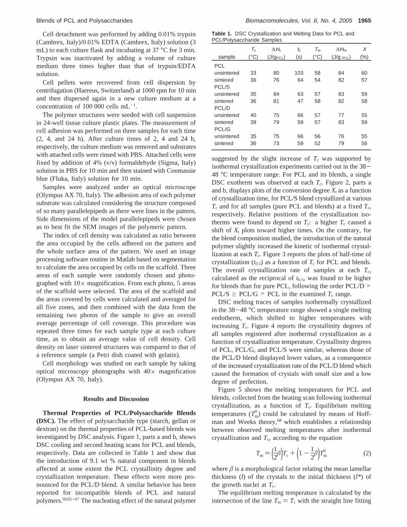
Thermal Properties of PCL/Polysaccharide Blends(DSC). The effect of polysaccharide type (starch, gellan ordextran) on the thermal properties of PCL-based blends wasinvestigated by DSC analysis. Figure 1, parts a and b, showsDSC cooling and second heating scans for PCL and blends,respectively. Data are collected in Table 1 and show thatthe introduction of 9.1 wt % natural component in blendsaffected at some extent the PCL crystallinity degree andcrystallization temperature. These effects were more pro-nounced for the PCL/D blend. A similar behavior has beenreported for incompatible blends of PCL and naturalpolymers.59,65-67 The nucleating effect of the natural polymer
suggested by the slight increase ofTc was supported byisothermal crystallization experiments carried out in the 38-48 °C temperature range. For PCL and its blends, a singleDSC exotherm was observed at eachTc. Figure 2, parts aand b, displays plots of the conversion degreeXt as a functionof crystallization time, for PCL/S blend crystallized at variousTc and for all samples (pure PCL and blends) at a fixedTc,respectively. Relative positions of the crystallization iso-therms were found to depend onTc: a higherTc caused ashift of Xt plots toward higher times. On the contrary, forthe blend composition studied, the introduction of the naturalpolymer slightly increased the kinetic of isothermal crystal-lization at eachTc. Figure 3 reports the plots of half-time ofcrystallization (t0.5) as a function ofTc for PCL and blends.The overall crystallization rate of samples at eachTc,calculated as the reciprocal oft0.5, was found to be higherfor blends than for pure PCL, following the order PCL/D>PCL/Sg PCL/G > PCL in the examinedTc range.
DSC melting traces of samples isothermally crystallizedin the 38-48 °C temperature range showed a single meltingendotherm, which shifted to higher temperatures withincreasingTc. Figure 4 reports the crystallinity degrees ofall samples registered after isothermal crystallization as afunction of crystallization temperature. Crystallinity degreesof PCL, PCL/G, and PCL/S were similar, whereas those ofthe PCL/D blend displayed lower values, as a consequenceof the increased crystallization rate of the PCL/D blend whichcaused the formation of crystals with small size and a lowdegree of perfection.
Figure 5 shows the melting temperatures for PCL andblends, collected from the heating scan following isothermalcrystallization, as a function ofTc. Equilibrium meltingtemperatures (Tm
0 ) could be calculated by means of Hoff-man and Weeks theory,68 which establishes a relationshipbetween observed melting temperatures after isothermalcrystallization andTc, according to the equation
whereâ is a morphological factor relating the mean lamellarthickness (l) of the crystals to the initial thickness (l* ) ofthe growth nuclei atTc.
The equilibrium melting temperature is calculated by theintersection of the lineTm ) Tc with the straight line fitting
Table 1. DSC Crystallization and Melting Data for PCL andPCL/Polysaccharide Samples
sampleTc
(°C)∆Hc
(J/gPCL)tc(s)
Tm
(°C)∆Hm
(J/g PCL)X
(%)
PCLunsintered 33 80 103 58 84 60sintered 36 76 64 54 82 57PCL/Sunsintered 35 84 63 57 83 59sintered 36 81 47 58 82 58PCL/Dunsintered 40 75 66 57 77 55sintered 39 79 59 57 83 59PCL/Gunsintered 35 75 66 56 76 55sintered 36 73 59 52 79 56
Tm ) (12â)Tc + (1 - 12
â)Tm0 (2)
Blends of PCL and Polysaccharides Biomacromolecules, Vol. 6, No. 4, 2005 1965
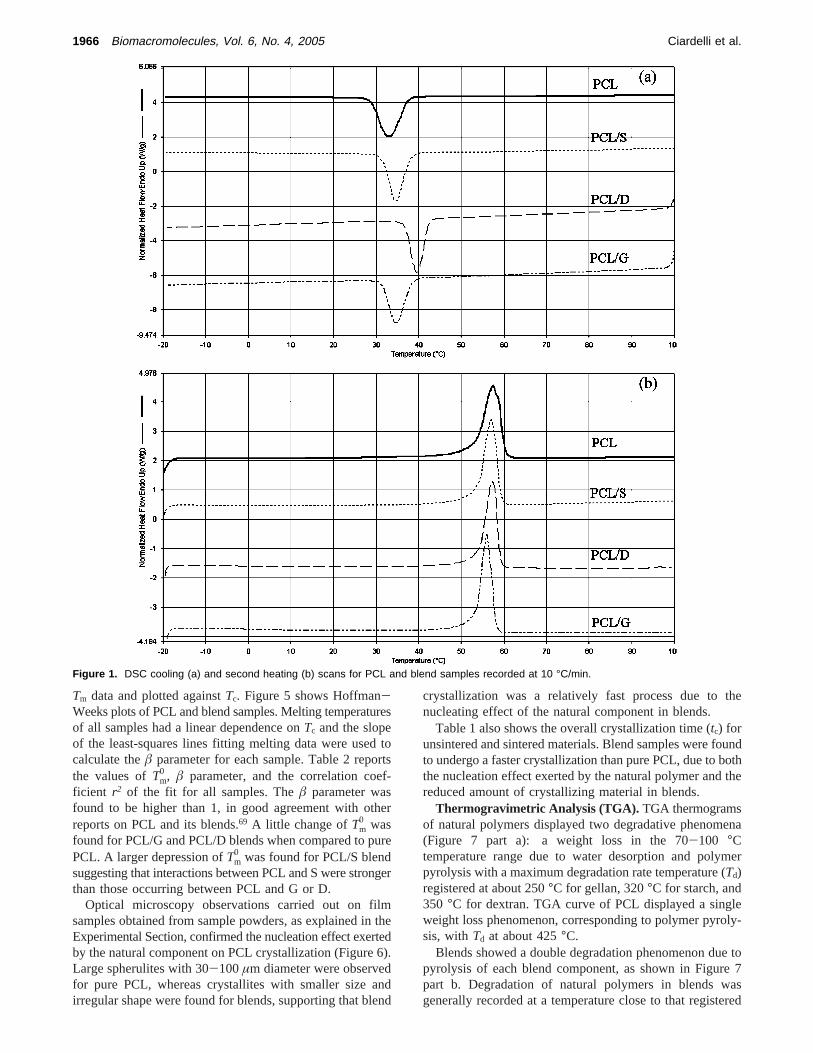
Tm data and plotted againstTc. Figure 5 shows Hoffman-Weeks plots of PCL and blend samples. Melting temperaturesof all samples had a linear dependence onTc and the slopeof the least-squares lines fitting melting data were used tocalculate theâ parameter for each sample. Table 2 reportsthe values ofTm
0 , â parameter, and the correlation coef-ficient r2 of the fit for all samples. Theâ parameter wasfound to be higher than 1, in good agreement with otherreports on PCL and its blends.69 A little change ofTm
0 wasfound for PCL/G and PCL/D blends when compared to purePCL. A larger depression ofTm
0 was found for PCL/S blendsuggesting that interactions between PCL and S were strongerthan those occurring between PCL and G or D.
Optical microscopy observations carried out on filmsamples obtained from sample powders, as explained in theExperimental Section, confirmed the nucleation effect exertedby the natural component on PCL crystallization (Figure 6).Large spherulites with 30-100µm diameter were observedfor pure PCL, whereas crystallites with smaller size andirregular shape were found for blends, supporting that blend
crystallization was a relatively fast process due to thenucleating effect of the natural component in blends.
Table 1 also shows the overall crystallization time (tc) forunsintered and sintered materials. Blend samples were foundto undergo a faster crystallization than pure PCL, due to boththe nucleation effect exerted by the natural polymer and thereduced amount of crystallizing material in blends.
Thermogravimetric Analysis (TGA). TGA thermogramsof natural polymers displayed two degradative phenomena(Figure 7 part a): a weight loss in the 70-100 °Ctemperature range due to water desorption and polymerpyrolysis with a maximum degradation rate temperature (Td)registered at about 250°C for gellan, 320°C for starch, and350 °C for dextran. TGA curve of PCL displayed a singleweight loss phenomenon, corresponding to polymer pyroly-sis, withTd at about 425°C.
Blends showed a double degradation phenomenon due topyrolysis of each blend component, as shown in Figure 7part b. Degradation of natural polymers in blends wasgenerally recorded at a temperature close to that registered
Figure 1. DSC cooling (a) and second heating (b) scans for PCL and blend samples recorded at 10 °C/min.
1966 Biomacromolecules, Vol. 6, No. 4, 2005 Ciardelli et al.
for pure polysaccharides. Pyrolysis of blended PCL occurredat a 15-25 °C lower temperature than that of pure PCL,
probably as a consequence of interactions occurring betweenPCL and the degradation products from the natural compo-nent. Moreover, for PCL/D blend, the pyrolysis peak wasbroader and degradation phenomena started at a lowertemperature than for PCL and other blends.
TGA analysis of blends did not detect evident waterdesorption phenomena, as a consequence of their low content(9.1 wt %) of the strongly hydrophilic natural polymer.
In conclusion, thermal stability of 90.9/9.1 (wt/wt) PCL/polysaccharide blends was found to be limited by the typeof blended natural polymer and it was high enough to justifyour attempts at processing PCL and PCL/S, PCL/G, andPCL/D blends through sintering.
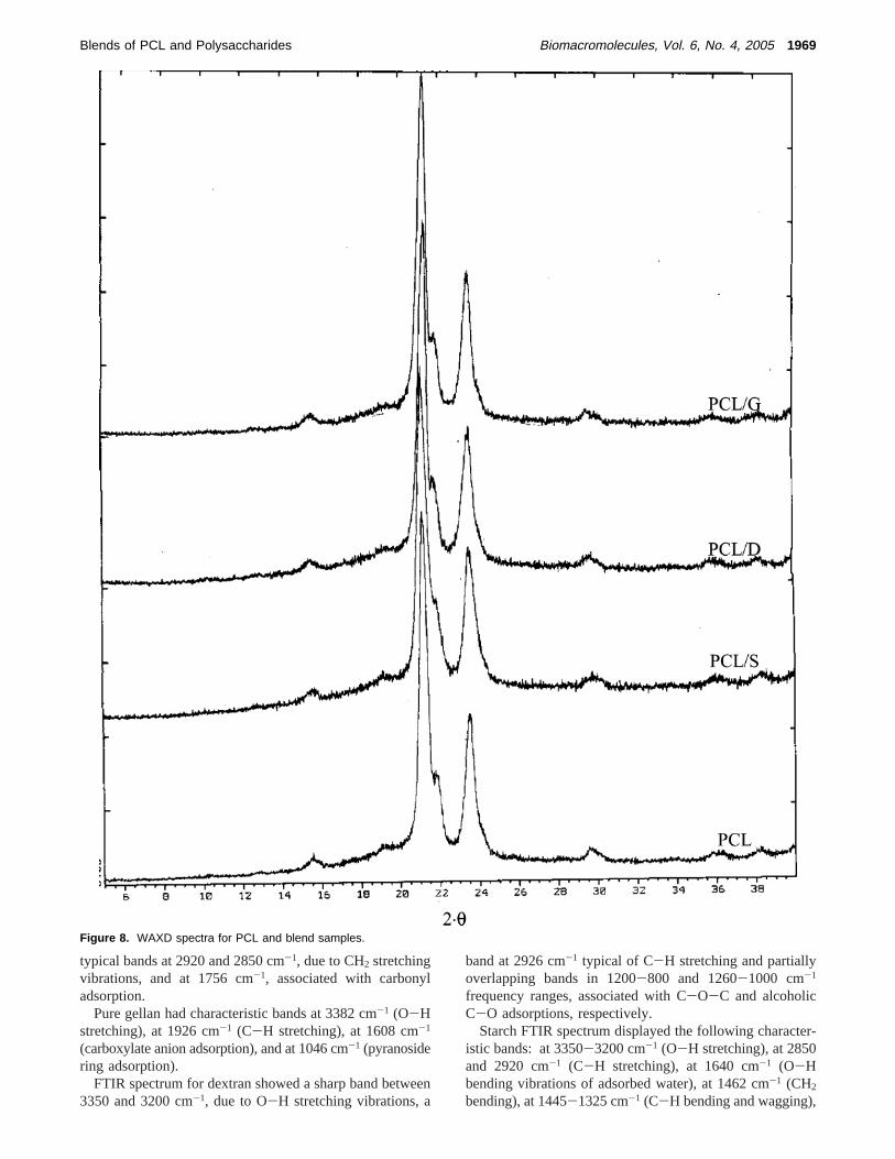
X-ray Diffraction Analysis (WAXD). X-ray diffractionpatterns of pure PCL and blends (Figure 8) displayed theirmain peaks at 2θ equal to 21.2°, 21.8°, and 23.5° which arethose typical of an orthorhombic crystalline unit cell.70 The
Figure 2. Plots of relative crystallinity against crystallization time:(a) PCL/S samples isothermally crystallized in the 40-48 °C tem-perature range; (b) PCL and blend samples isothermally crystallizedat Tc ) 44 °C. t0 is the initial time, i.e., the time at which temperaturereaches the fixed value Tc.
Figure 3. Half-time of crystallization for PCL and blends isothermallycrystallized in the 32-50 °C temperature range.
Figure 4. Crystallinity degrees calculated from the heating scanperformed after isothermal crystallization in the 38-50 °C temperaturerange, for PCL and blend samples.
Figure 5. Plots of Tm (recorded during the heating scan followingisothermal crystallization at Tc) as a function of Tc and Hoffman-Weeks plots for PCL and blends.
Table 2. Equilibrium Melting Temperatures Tm0 and â Parameters
for PCL and Its Blends
sample Tm0 (°C) â r2
PCL 67 1.7 0.991PCL/S 61 2.8 0.991PCL/D 64 2.1 0.989PCL/G 65 1.6 0.989
Blends of PCL and Polysaccharides Biomacromolecules, Vol. 6, No. 4, 2005 1967

peak relative intensity was similar for all samples, evidencinga symilar crystallinity degree for blends and pure PCL. Thisfinding is not contradictory with respect to DSC results, asX-ray analysis was performed on as produced microparticles,
whereas thermal properties were collected from the secondheating DSC scans after deleting sample thermal history.
FTIR-ATR Analysis. Figure 9 shows FTIR-ATR spectraof pure components and blends. PCL spectrum showed
Figure 6. Optical microscopy images of PCL (a), PCL/D (b), PCL/S (c), and PCL/G (d) thin films obtained by sandwiching particles among aglass slide and a cover glass at 100 °C for 5 min, followed by cooling at room temperature. Bars indicate 25 µm.
Figure 7. Derivative thermogravimetric (DTG) curves for (a) pure components (b) PCL and blends.
1968 Biomacromolecules, Vol. 6, No. 4, 2005 Ciardelli et al.
typical bands at 2920 and 2850 cm-1, due to CH2 stretchingvibrations, and at 1756 cm-1, associated with carbonyladsorption.
Pure gellan had characteristic bands at 3382 cm-1 (O-Hstretching), at 1926 cm-1 (C-H stretching), at 1608 cm-1
(carboxylate anion adsorption), and at 1046 cm-1 (pyranosidering adsorption).
FTIR spectrum for dextran showed a sharp band between3350 and 3200 cm-1, due to O-H stretching vibrations, a
band at 2926 cm-1 typical of C-H stretching and partiallyoverlapping bands in 1200-800 and 1260-1000 cm-1
frequency ranges, associated with C-O-C and alcoholicC-O adsorptions, respectively.
Starch FTIR spectrum displayed the following character-istic bands: at 3350-3200 cm-1 (O-H stretching), at 2850and 2920 cm-1 (C-H stretching), at 1640 cm-1 (O-Hbending vibrations of adsorbed water), at 1462 cm-1 (CH2
bending), at 1445-1325 cm-1 (C-H bending and wagging),
Figure 8. WAXD spectra for PCL and blend samples.
Blends of PCL and Polysaccharides Biomacromolecules, Vol. 6, No. 4, 2005 1969
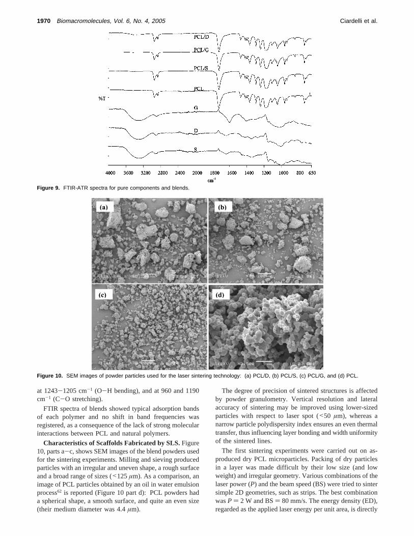
at 1243-1205 cm-1 (O-H bending), and at 960 and 1190cm-1 (C-O stretching).
FTIR spectra of blends showed typical adsorption bandsof each polymer and no shift in band frequencies wasregistered, as a consequence of the lack of strong molecularinteractions between PCL and natural polymers.
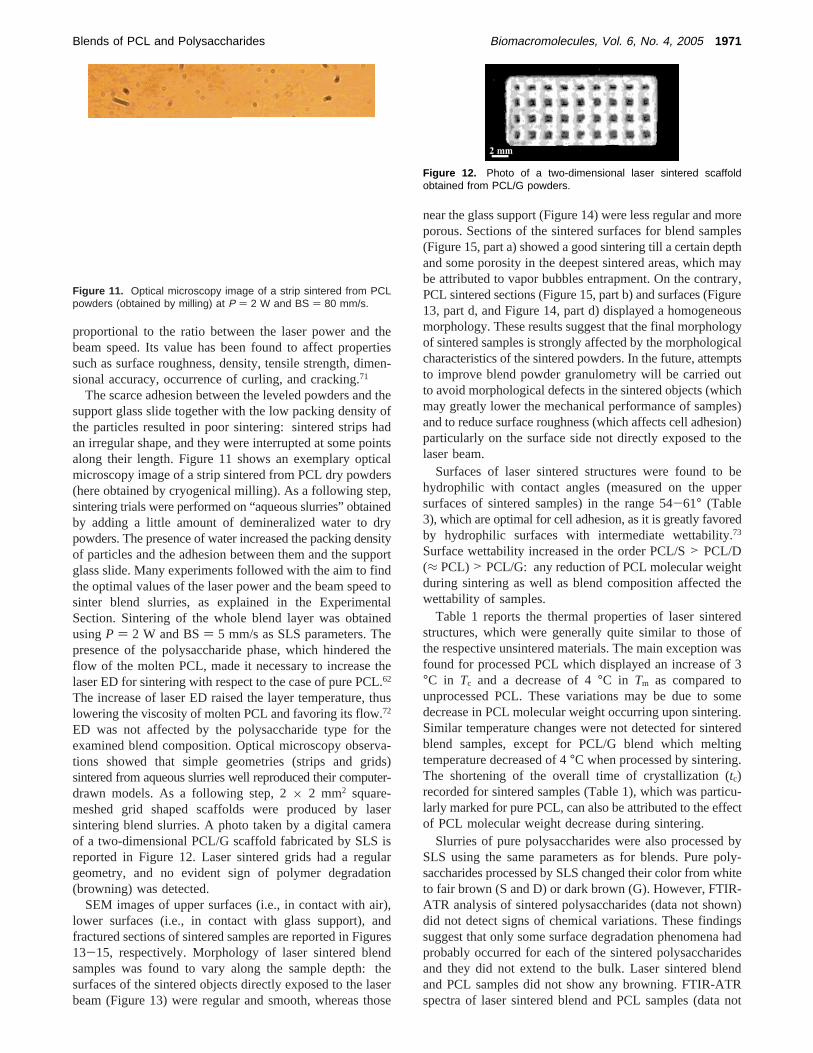
Characteristics of Scaffolds Fabricated by SLS.Figure10, parts a-c, shows SEM images of the blend powders usedfor the sintering experiments. Milling and sieving producedparticles with an irregular and uneven shape, a rough surfaceand a broad range of sizes (<125µm). As a comparison, animage of PCL particles obtained by an oil in water emulsionprocess62 is reported (Figure 10 part d): PCL powders hada spherical shape, a smooth surface, and quite an even size(their medium diameter was 4.4µm).
The degree of precision of sintered structures is affectedby powder granulometry. Vertical resolution and lateralaccuracy of sintering may be improved using lower-sizedparticles with respect to laser spot (<50 µm), whereas anarrow particle polydispersity index ensures an even thermaltransfer, thus influencing layer bonding and width uniformityof the sintered lines.
The first sintering experiments were carried out on as-produced dry PCL microparticles. Packing of dry particlesin a layer was made difficult by their low size (and lowweight) and irregular geometry. Various combinations of thelaser power (P) and the beam speed (BS) were tried to sintersimple 2D geometries, such as strips. The best combinationwasP ) 2 W and BS) 80 mm/s. The energy density (ED),regarded as the applied laser energy per unit area, is directly
Figure 9. FTIR-ATR spectra for pure components and blends.
Figure 10. SEM images of powder particles used for the laser sintering technology: (a) PCL/D, (b) PCL/S, (c) PCL/G, and (d) PCL.
1970 Biomacromolecules, Vol. 6, No. 4, 2005 Ciardelli et al.
proportional to the ratio between the laser power and thebeam speed. Its value has been found to affect propertiessuch as surface roughness, density, tensile strength, dimen-sional accuracy, occurrence of curling, and cracking.71
The scarce adhesion between the leveled powders and thesupport glass slide together with the low packing density ofthe particles resulted in poor sintering: sintered strips hadan irregular shape, and they were interrupted at some pointsalong their length. Figure 11 shows an exemplary opticalmicroscopy image of a strip sintered from PCL dry powders(here obtained by cryogenical milling). As a following step,sintering trials were performed on “aqueous slurries” obtainedby adding a little amount of demineralized water to drypowders. The presence of water increased the packing densityof particles and the adhesion between them and the supportglass slide. Many experiments followed with the aim to findthe optimal values of the laser power and the beam speed tosinter blend slurries, as explained in the ExperimentalSection. Sintering of the whole blend layer was obtainedusingP ) 2 W and BS) 5 mm/s as SLS parameters. Thepresence of the polysaccharide phase, which hindered theflow of the molten PCL, made it necessary to increase thelaser ED for sintering with respect to the case of pure PCL.62
The increase of laser ED raised the layer temperature, thuslowering the viscosity of molten PCL and favoring its flow.72
ED was not affected by the polysaccharide type for theexamined blend composition. Optical microscopy observa-tions showed that simple geometries (strips and grids)sintered from aqueous slurries well reproduced their computer-drawn models. As a following step, 2× 2 mm2 square-meshed grid shaped scaffolds were produced by lasersintering blend slurries. A photo taken by a digital cameraof a two-dimensional PCL/G scaffold fabricated by SLS isreported in Figure 12. Laser sintered grids had a regulargeometry, and no evident sign of polymer degradation(browning) was detected.
SEM images of upper surfaces (i.e., in contact with air),lower surfaces (i.e., in contact with glass support), andfractured sections of sintered samples are reported in Figures13-15, respectively. Morphology of laser sintered blendsamples was found to vary along the sample depth: thesurfaces of the sintered objects directly exposed to the laserbeam (Figure 13) were regular and smooth, whereas those
near the glass support (Figure 14) were less regular and moreporous. Sections of the sintered surfaces for blend samples(Figure 15, part a) showed a good sintering till a certain depthand some porosity in the deepest sintered areas, which maybe attributed to vapor bubbles entrapment. On the contrary,PCL sintered sections (Figure 15, part b) and surfaces (Figure13, part d, and Figure 14, part d) displayed a homogeneousmorphology. These results suggest that the final morphologyof sintered samples is strongly affected by the morphologicalcharacteristics of the sintered powders. In the future, attemptsto improve blend powder granulometry will be carried outto avoid morphological defects in the sintered objects (whichmay greatly lower the mechanical performance of samples)and to reduce surface roughness (which affects cell adhesion)particularly on the surface side not directly exposed to thelaser beam.
Surfaces of laser sintered structures were found to behydrophilic with contact angles (measured on the uppersurfaces of sintered samples) in the range 54-61° (Table3), which are optimal for cell adhesion, as it is greatly favoredby hydrophilic surfaces with intermediate wettability.73
Surface wettability increased in the order PCL/S> PCL/D(≈ PCL)> PCL/G: any reduction of PCL molecular weightduring sintering as well as blend composition affected thewettability of samples.
Table 1 reports the thermal properties of laser sinteredstructures, which were generally quite similar to those ofthe respective unsintered materials. The main exception wasfound for processed PCL which displayed an increase of 3°C in Tc and a decrease of 4°C in Tm as compared tounprocessed PCL. These variations may be due to somedecrease in PCL molecular weight occurring upon sintering.Similar temperature changes were not detected for sinteredblend samples, except for PCL/G blend which meltingtemperature decreased of 4°C when processed by sintering.The shortening of the overall time of crystallization (tc)recorded for sintered samples (Table 1), which was particu-larly marked for pure PCL, can also be attributed to the effectof PCL molecular weight decrease during sintering.
Slurries of pure polysaccharides were also processed bySLS using the same parameters as for blends. Pure poly-saccharides processed by SLS changed their color from whiteto fair brown (S and D) or dark brown (G). However, FTIR-ATR analysis of sintered polysaccharides (data not shown)did not detect signs of chemical variations. These findingssuggest that only some surface degradation phenomena hadprobably occurred for each of the sintered polysaccharidesand they did not extend to the bulk. Laser sintered blendand PCL samples did not show any browning. FTIR-ATRspectra of laser sintered blend and PCL samples (data not
Figure 11. Optical microscopy image of a strip sintered from PCLpowders (obtained by milling) at P ) 2 W and BS ) 80 mm/s.
Figure 12. Photo of a two-dimensional laser sintered scaffoldobtained from PCL/G powders.
Blends of PCL and Polysaccharides Biomacromolecules, Vol. 6, No. 4, 2005 1971

shown) were found to be analogous to those of theirunsintered counterparts, evidencing that sintering did not giverise to any detectable variation in the chemical structure ofmaterials. Blending each polysaccharide with a large amountof PCL, which is a more heat resistant polymer thanpolysaccharides, could protect the polysaccharide phase from
direct contact with the laser beam and, therefore, fromdegradation.
Laser sintered blend samples were also analyzed by anIR Imaging System. Figure 16, parts a and b, shows the false-color contour map acquired from a 1 mm× 1 mm zone ofa sintered PCL/S sample and its medium spectrum, respec-
Figure 13. Upper surface of laser sintered samples: (a) PCL/D, (b) PCL/G, (c) PCL/S, and (d) PCL.
Figure 14. Lower surface of laser sintered samples: (a) PCL/D, (b) PCL/G, (c) PCL/S, and (d) PCL.
1972 Biomacromolecules, Vol. 6, No. 4, 2005 Ciardelli et al.
tively. A correlation map was obtained from the mediumspectrum (Figure 16, part c), suggesting a good homogeneityof PCL/S blend. An analogous result was obtained forsintered PCL/G blend whereas sintered PCL/D displayedphase segregation (data not shown). These findings suggestthe lower compatibility and higher interfacial energy of thePCL/D system, when compared to PCL/G and PCL/S blends.
Cell Attachment. Figure 17 shows percentage areaoccupied by cells for blend and PCL scaffolds and for acontrol layer (gelatin) as a function of culture time. The ratiobetween cell density on structures and on the gelatin layerat any time may be taken as an index of cell adhesionefficiency.62
Cell adhesion on blend scaffolds increased with time,displaying values in the order PCL/S> PCL/G > PCL/D.The introduction of gellan and starch greatly increasedfibroblast attachment with respect to PCL. Starch was themost effective polysaccharide in enhancing PCL biocom-
patibility among the tested ones: cells adhered to PCL/Sscaffolds at a slightly lower rate than to gelatin control layerand at a similar extent after 24 h culture time. The celladhesion rate to PCL/G scaffolds was only slightly lowerthan to the control layer. On the contrary, PCL/D scaffoldsdisplayed the lowest fibroblast attachment rate among blendscaffolds: a comparison between PCL and PCL/D blendshowed that the introduction of D only slightly increasedcell attachment within the first 4 h, whereas it greatlydecreased it after 24 h culture time. Poor cell adhesion for
Figure 15. SEM images of the fractured surfaces of laser sinteredsamples: (a) PCL/D 90.9/9.1 wt/wt; (b) PCL.
Table 3. Water Contact Angle for PCL and PCL/PolysaccharideLaser Sintered Samples
sample contact angle
PCL 58° ( 4PCL/S 52° ( 4PCL/D 66° ( 1PCL/G 62° ( 3
Figure 16. (a) IR false color contour image of a 1 mm × 1 mm areafor a PCL/S laser sintered sample. (b) “Medium” spectrum of the areaanalyzed at point (a). (c) Correlation map of the area in (a) withrespect to the “medium” spectrum shown in (b).
Blends of PCL and Polysaccharides Biomacromolecules, Vol. 6, No. 4, 2005 1973
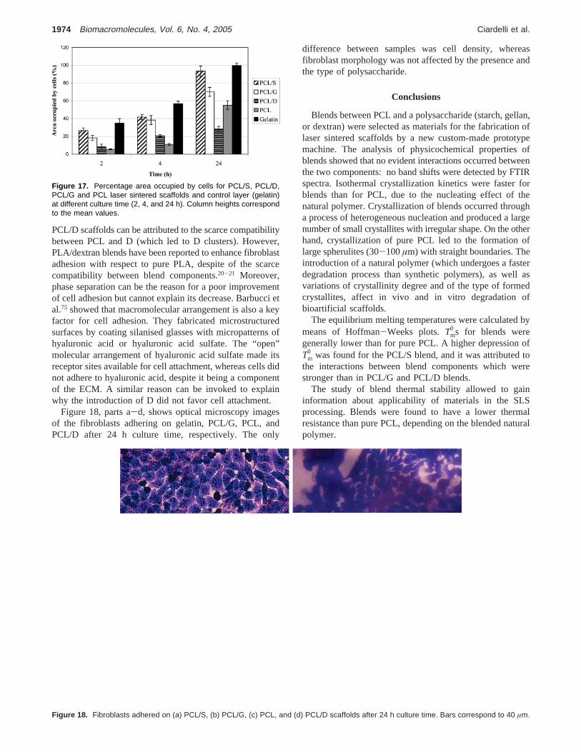
PCL/D scaffolds can be attributed to the scarce compatibilitybetween PCL and D (which led to D clusters). However,PLA/dextran blends have been reported to enhance fibroblastadhesion with respect to pure PLA, despite of the scarcecompatibility between blend components.20-21 Moreover,phase separation can be the reason for a poor improvementof cell adhesion but cannot explain its decrease. Barbucci etal.75 showed that macromolecular arrangement is also a keyfactor for cell adhesion. They fabricated microstructuredsurfaces by coating silanised glasses with micropatterns ofhyaluronic acid or hyaluronic acid sulfate. The “open”molecular arrangement of hyaluronic acid sulfate made itsreceptor sites available for cell attachment, whereas cells didnot adhere to hyaluronic acid, despite it being a componentof the ECM. A similar reason can be invoked to explainwhy the introduction of D did not favor cell attachment.
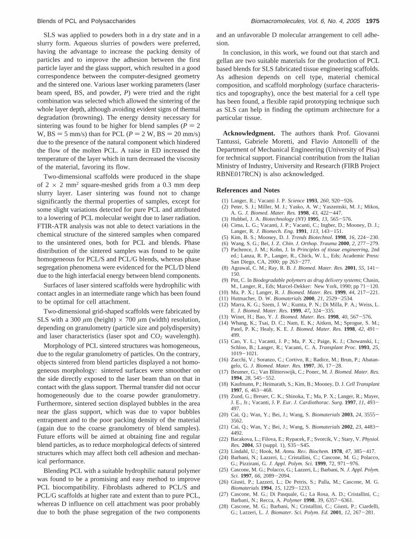
Figure 18, parts a-d, shows optical microscopy imagesof the fibroblasts adhering on gelatin, PCL/G, PCL, andPCL/D after 24 h culture time, respectively. The only
difference between samples was cell density, whereasfibroblast morphology was not affected by the presence andthe type of polysaccharide.
Conclusions
Blends between PCL and a polysaccharide (starch, gellan,or dextran) were selected as materials for the fabrication oflaser sintered scaffolds by a new custom-made prototypemachine. The analysis of physicochemical properties ofblends showed that no evident interactions occurred betweenthe two components: no band shifts were detected by FTIRspectra. Isothermal crystallization kinetics were faster forblends than for PCL, due to the nucleating effect of thenatural polymer. Crystallization of blends occurred througha process of heterogeneous nucleation and produced a largenumber of small crystallites with irregular shape. On the otherhand, crystallization of pure PCL led to the formation oflarge spherulites (30-100µm) with straight boundaries. Theintroduction of a natural polymer (which undergoes a fasterdegradation process than synthetic polymers), as well asvariations of crystallinity degree and of the type of formedcrystallites, affect in vivo and in vitro degradation ofbioartificial scaffolds.
The equilibrium melting temperatures were calculated bymeans of Hoffman-Weeks plots.Tm
0 s for blends weregenerally lower than for pure PCL. A higher depression ofTm
0 was found for the PCL/S blend, and it was attributed tothe interactions between blend components which werestronger than in PCL/G and PCL/D blends.
The study of blend thermal stability allowed to gaininformation about applicability of materials in the SLSprocessing. Blends were found to have a lower thermalresistance than pure PCL, depending on the blended naturalpolymer.
Figure 17. Percentage area occupied by cells for PCL/S, PCL/D,PCL/G and PCL laser sintered scaffolds and control layer (gelatin)at different culture time (2, 4, and 24 h). Column heights correspondto the mean values.
Figure 18. Fibroblasts adhered on (a) PCL/S, (b) PCL/G, (c) PCL, and (d) PCL/D scaffolds after 24 h culture time. Bars correspond to 40 µm.
1974 Biomacromolecules, Vol. 6, No. 4, 2005 Ciardelli et al.
SLS was applied to powders both in a dry state and in aslurry form. Aqueous slurries of powders were preferred,having the advantage to increase the packing density ofparticles and to improve the adhesion between the firstparticle layer and the glass support, which resulted in a goodcorrespondence between the computer-designed geometryand the sintered one. Various laser working parameters (laserbeam speed, BS, and powder,P) were tried and the rightcombination was selected which allowed the sintering of thewhole layer depth, although avoiding evident signs of thermaldegradation (browning). The energy density necessary forsintering was found to be higher for blend samples (P ) 2W, BS) 5 mm/s) than for PCL (P ) 2 W, BS) 20 mm/s)due to the presence of the natural component which hinderedthe flow of the molten PCL. A raise in ED increased thetemperature of the layer which in turn decreased the viscosityof the material, favoring its flow.
Two-dimensional scaffolds were produced in the shapeof 2 × 2 mm2 square-meshed grids from a 0.3 mm deepslurry layer. Laser sintering was found not to changesignificantly the thermal properties of samples, except forsome slight variations detected for pure PCL and attributedto a lowering of PCL molecular weight due to laser radiation.FTIR-ATR analysis was not able to detect variations in thechemical structure of the sintered samples when comparedto the unsintered ones, both for PCL and blends. Phasedistribution of the sintered samples was found to be quitehomogeneous for PCL/S and PCL/G blends, whereas phasesegregation phenomena were evidenced for the PCL/D blenddue to the high interfacial energy between blend components.
Surfaces of laser sintered scaffolds were hydrophilic withcontact angles in an intermediate range which has been foundto be optimal for cell attachment.
Two-dimensional grid-shaped scaffolds were fabricated bySLS with a 300µm (height)× 700 µm (width) resolution,depending on granulometry (particle size and polydispersity)and laser characteristics (laser spot and CO2 wavelength).
Morphology of PCL sintered structures was homogeneous,due to the regular granulometry of particles. On the contrary,objects sintered from blend particles displayed a not homo-geneous morphology: sintered surfaces were smoother onthe side directly exposed to the laser beam than on that incontact with the glass support. Thermal transfer did not occurhomogeneously due to the coarse powder granulometry.Furthermore, sintered section displayed bubbles in the areanear the glass support, which was due to vapor bubblesentrapment and to the poor packing density of the material(again due to the coarse granulometry of blend samples).Future efforts will be aimed at obtaining fine and regularblend particles, as to reduce morphological defects of sinteredstructures which may affect both cell adhesion and mechan-ical performance.
Blending PCL with a suitable hydrophilic natural polymerwas found to be a promising and easy method to improvePCL biocompatibility. Fibroblasts adhered to PCL/S andPCL/G scaffolds at higher rate and extent than to pure PCL,whereas D influence on cell attachment was poor probablydue to both the phase segregation of the two components
and an unfavorable D molecular arrangement to cell adhe-sion.
In conclusion, in this work, we found out that starch andgellan are two suitable materials for the production of PCLbased blends for SLS fabricated tissue engineering scaffolds.As adhesion depends on cell type, material chemicalcomposition, and scaffold morphology (surface characteris-tics and topography), once the best material for a cell typehas been found, a flexible rapid prototyping technique suchas SLS can help in finding the optimum architecture for aparticular tissue.
Acknowledgment. The authors thank Prof. GiovanniTantussi, Gabriele Moretti, and Flavio Antonelli of theDepartment of Mechanical Engineering (University of Pisa)for technical support. Financial contribution from the ItalianMinistry of Industry, University and Research (FIRB ProjectRBNE017RCN) is also acknowledged.
References and Notes
(1) Langer, R.; Vacanti J. P.Science1993, 260, 920-926.(2) Peter, S. J.; Miller, M. J.; Yasko, A. W.; Yaszemski, M. J.; Mikos,
A. G. J. Biomed. Mater. Res.1998, 43, 422-447.(3) Hubbel, J. A.Biotechnology (NY)1995, 13, 565-576.(4) Cima, L. G.; Vacanti, J. P.; Vacanti, C.; Ingber, D.; Mooney, D. J.;
Langer, R.J. Biomech. Eng.1991, 113, 143-151.(5) Kim, B. S.; Mooney, D. J.Trends Biotechnol.1998, 16, 224-230.(6) Wang, S. G.; Bei, J. Z.Chin. J. Orthop. Trauma2000, 2, 277-279.(7) Pachence, J. M.; Kohn, J. InPrinciples of tissue engineering, 2nd
ed.; Lanza, R. P., Langer, R., Chick, W. L., Eds; Academic Press:San Diego, CA, 2000; pp 263-277.
(8) Agrawal, C. M.; Ray, R. B.J. Biomed. Mater. Res.2001, 55, 141-150.
(9) Pitt, C. InBiodegradable polymers as drug deliVery systems; Chasin,M., Langer, R., Eds; Marcel-Dekker: New York, 1990; pp 71-120.
(10) Ma, P. X.; Langer, R.J. Biomed. Mater. Res.1999, 44, 217-221.(11) Hutmacher, D. W.Biomaterials2000, 21, 2529-2534.(12) Marra, K. G.; Szem, J. W.; Kumta, P. N.; Di Milla, P. A.; Weiss, L.
E. J. Biomed. Mater. Res.1999, 47, 324-335.(13) Winet, H.; Bao, Y.J. Biomed. Mater. Res.1998, 40, 567-576.(14) Whang, K.; Tsai, D. C.; Nam, E. K.; Aitken, M.; Sprogue, S. M.;
Patel, P. K.; Healy, K. E.J. Biomed. Mater. Res.1998, 42, 491-499.
(15) Cao, Y. L.; Vacanti, J. P.; Ma, P. X.; Paige, K. J.; Chowanski, I.;Schloo, B.; Langer, R.; Vacanti, C. A.Transplant Proc.1993, 25,1019-1021.
(16) Zacchi, V.; Soranzo, C.; Cortivo, R.; Radice, M.; Brun, P.; Abatan-gelo, G.J. Biomed. Mater. Res.1997, 36, 17-28.
(17) Beumer, G.; Van Blitterswijk, C.; Ponec, M.J. Biomed. Mater. Res.1994, 28, 545-552.
(18) Kaufmann, P.; Heimarath, S.; Kim, B.; Mooney, D. J.Cell Transplant1997, 6, 463-468.
(19) Zund, G.; Breuer, C. K.; Shinoka, T.; Ma, P. X.; Langer, R.; Mayer,J. E., Jr.; Vacanti, J. P.Eur. J. Cardiothorac. Surg.1997, 11, 493-497.
(20) Cai, Q.; Wan, Y.; Bei, J.; Wang, S.Biomaterials2003, 24, 3555-3562.
(21) Cai, Q.; Wan, Y.; Bei, J.; Wang, S.Biomaterials2002, 23, 4483-4492.
(22) Bacakova, L.; Filova, E.; Rypacek, F.; Svorcik, V.; Stary, V.Physiol.Res.2004, 53 (suppl. 1), S35-S45.
(23) Lindahl, U.; Hook, M.Annu. ReV. Biochem.1978, 47, 385-417.(24) Barbani, N.; Lazzeri, L.; Cristallini, C.; Cascone, M. G.; Polacco,
G.; Pizzirani, G.J. Appl. Polym. Sci.1999, 72, 971-976.(25) Cascone, M. G.; Polacco, G.; Lazzeri, L.; Barbani, N.J. Appl. Polym.
Sci.1997, 66, 2089-2094.(26) Giusti, P.; Lazzeri, L.; De Petris, S.; Palla, M.; Cascone, M. G.
Biomaterials1994, 15, 1229-1233.(27) Cascone, M. G.; Di Pasquale, G.; La Rosa, A. D.; Cristallini, C.;
Barbani, N.; Recca, A.Polymer1998, 39, 6357-6361.(28) Cascone, M. G.; Barbani, N.; Cristallini, C.; Giusti, P.; Ciardelli,
G.; Lazzeri, L.J. Biomater. Sci. Polym. Ed.2001, 12, 267-281.
Blends of PCL and Polysaccharides Biomacromolecules, Vol. 6, No. 4, 2005 1975
(29) Barbani, N.; Lazzeri, L.; Lelli, L.; Bonaretti, A.; Seggiani, M.;Narducci, P.; Pizzirani, G.; Giusti, P.J. Mater. Sci., Mater. Med.1994, 5, 882-886.
(30) Lazzeri, L.; Barbani, N.; Cascone, M. G.; Lupinacci, D.; Giusti, P.;Laus, M.J. Mater. Sci., Mater. Med.1994, 5, 862-867.
(31) Cascone, M. G.; Sim, B.; Downes, S.Biomaterials1995, 16, 569-574.
(32) Gref, R.; Rodrigues, J.; Couvreur, P.Macromolecules2002, 35,9861-9867.
(33) Rodriguez, J. S.; Santos-Magalhaes, N. S.; Coelho, L. C. B. B.,Couvreur, P.; Ponchel, G.; Gref, R.J. Controlled Release2003, 92:103-112.
(34) Lemarchand, C.; Couvreur, P.; Besnard, M.; Costantini, D.; Gref,R. Pharm. Res.2003, 20, 1284-1292.
(35) Woodward, S. C.; Brewer, P. S.; Moatamed, F.J. Biomed. Mater.Res.1985, 44, 437-444.
(36) Pitt, C. G.; Gratzei, M. M.; Kimmei, G. L.; Surles, J.; Schindler, A.Biomaterials1981, 2, 215-220.
(37) Kweon, H. Y.; Yoo, M. K.; Park, I. K.; Kim, T. H.; Lee, H. C.; Lee,H. S.; Oh, J. S.; Akaike, T.; Cho, C. S.Biomaterials2003, 24,801-808.
(38) Gan, Z.; Luang, Q.; Zhang, J.; Jing, X.Polym. Degrad. Stab.1997,56, 209-213.
(39) Labow, R. S.; Meek, E.; Matheson, L.; Santerre, J. P.Biomaterials2002, 23, 3969-3975.
(40) Wang, G. B.; Labow, R. S.; Santerre, J. P.J. Biomed. Mater. Res.1997, 36, 407-417.
(41) Woei, K.; Hutmacher, D. W.; Schantz, J. T.; Seng, C.; Too, H. P.;Chye, T.; Phan, T. T.; Teoh, S. H.Tissue Eng.2001, 7, 441-455.
(42) Benoit, M. A.; Baras, B.; Gillard, J.Int. J. Pharm.1999, 184, 73-84.
(43) Coombes, A. G. A.; Rizzi, S. C.; Williamson, M.; Barralet, J. E.;Downes, S.; Wallace, W. A.Biomaterials2004, 25, 315-325.
(44) Waddell, R. L.; Marra, K. G.; Collins, K. L.; Leung, J. T.; Doctor,J. S.Biotechnol. Prog.2003, 19, 1767-1774.
(45) Verreck, G.; Chun, I.; Li, Y.; Kataria, R.; Zhang, Q.; Rosenblatt, J.;Decorte, A.; Heymans, K.; Adriaensen, J.; Bruining, M.; VanRemoortere, M.; Borghys, H.; Meert, T.; Peeters, J.; Brewster, M.E. Biomaterials2005, 26, 1307-1315.
(46) Serrano, M. C.; Pagani, R.; Vallet-Regi, M.; Pena, J.; Ramila, A.;Izquierdo, I.; Portoles, M. T.Biomaterials2004, 25, 5603-5611.
(47) Hutmacher, D. W.; Schantz, J. T.; Zein, I.; Ng, K. W.; Tech, S. H.;Tan, K. C.J. Biomed. Mater. Res.2001, 55, 203-216.
(48) Jane, J.Mater. Sci., Pure Appl. Chem.1995, A32 (4), 751.(49) Gomes, M. E.; Malafaya, P.; Ribeiro, A.; Reis, R. L.; Cunha, A. M.
Biomaterials2000, 22, 883-889.(50) Pereira, C. S.; Cunha, A. M.; Reis, R. L.; Va`squez, B.; San Roma`n,
J. J. Mater. Sci. Mater. Med.1998, 9, 825-833.(51) Reis, R. L.; Cunha, A. M.; Bevis, M. J.J. Appl. Med. Polym.1998,
2, 49-53.
(52) Sworn, G.; InHandbook of hydrocolloids; Phillips, G. O., Williams,P. A., Eds; CRC Press LLC: Boca Raton, FL, 2000; pp 117-135.
(53) Chandrasekaran, R.; Radha, A.Trends Food Sci. Technol.1995, 6,143-147.
(54) Nunes, M. C.; Batista, P.; Raymundo, A.; Alves, M. M.; Sousa, I.Colloids Surf. B: Biointerfaces2003, 31, 21-29.
(55) Barbani, N.; Bertoni, F.; Ciardelli, G.; Cristallini, C.; Cascone, M.G.; Coluccio, M. L.; Giusti, P.JABBs2003, 1, 221.
(56) Ohkawa, K.; Kitagawa, T.; Yamamoto, H.;Macromol. Mater. Eng.2004, 289, 33-40.
(57) Berrada, S.; Amedee, J.; Avramoglou, T.; Jozefonvicz, J.; HarmandF. J. Biomater. Sci. Polym. Ed.1994, 6, 211-222.
(58) Matzinos, P.; Tserki, V.; Kontoyiannis, A.; Panayiotou, C.Polym.Degrad. Stab.2002, 77, 17-24.
(59) Rutot, D.; Duquesne, E.; Ydens, I.; Degee, P.; Dubois, P.Polym.Degrad. Stab.2001, 73, 561-566.
(60) Demirgoz, D.; Elvira, C.; Mano, J. F.; Cunha, A. M.; Piskin, E.;Reis, R. L.;Polym. Degrad. Stab.2000, 70, 161-170.
(61) Mano, J. F.; Koniarova, D.; Reis, R. L.J. Mater. Sci.: Mater. Med.2003, 14, 127-135.
(62) Ciardelli, G.; Chiono, V.; Cristallini, C.; Barbani, N.; Ahluwalia, A.;Vozzi, G.; Previti, A.; Tantussi, G.; Giusti, P.J. Mater. Sci. Mater.Med.2004, 15, 303-308.
(63) Pham, D. T.; Gault, R. S.Int. J. Mach. Tools Manuf.1998, 38, 1257-1287.
(64) Pitt, C. G.; Chasalow, F. I.; Hibionada, Y. M.; Klimas, D. M.;Schlinder, A.J. Appl. Polym. Sci.1981, 26, 3779-3787.
(65) Wu, C. S.Polymer Degrad. Stab.2003, 80, 127-134.(66) Dubois, P.; Narayan, R.Macromol. Symp.2003, 198, 233-243.(67) Duquesne, E.; Rutot, D.; Degee, P.; Dubois, P.Macromol. Symp.
2001, 175, 33-43.(68) Wunderlich, B.Macromolecular Physics. Crystal melting; Wunder-
lich, B., Ed; Academic Press: New York, 1980; Vol 3, pp 33-36.(69) Leczano, E. G.; Coll, S. C.; Prolongo, M. G.Polymer1996, 37 (16),
3603-3609.(70) Hu, H.; Dorset, D. L.Macromolecules1990, 23, 4604-4607.(71) Ho, H. C. H.; Cheung, W. L.; Gibson, I.Ind. Eng. Chem. Res. 2003,
42, 1850-1862.(72) Rosa, D. S.; Guedes, C. G. F.; Pedroso, A. G.; Calil, M. R.Mater.
Sci. Eng. C2004, 24, 659-662.(73) Saltzman, W. M. InPrinciples of tissue engineering; Lanza, R. P.,
Langer, R., Chick, W. L., Eds.; R. G. Landers Company: GeorgetownTX, 1997; pp 225-246.
(74) Skoglund, P.; Fransson, A.J. Appl. Polym. Sci.1996, 61, 2455-2465.
(75) Barbucci, R.; Pasqui, D.; Wirsen, A.; Affrossman, S.; Curtis, A.;Tetto, C.J. Mater. Sci. Mater. Med.2003, 14, 721-725.
BM0500805
1976 Biomacromolecules, Vol. 6, No. 4, 2005 Ciardelli et al.


![Copolymerization of [epsiv]-caprolactone and morpholine-2 ... · Makromol. Chem. 193, 1927-1942 (1992) 1927 Copolymerization of &-caprolactone and morpholine-2,5-dione derivatives](https://static.fdocuments.in/doc/165x107/5ad096377f8b9ae2138dec54/copolymerization-of-epsiv-caprolactone-and-morpholine-2-chem-193-1927-1942.jpg)