Bithalamic Hyperintensity on T2-Weighted MR: Vascular ... · Bithalamic Hyperintensity on...
7
Bithalamic Hyperintensity on T2-Weighted MR: Vascular Causes and Evaluation with MR Angiography D. Antonio Bell, Wayne L. Davis, Anne G. Osborn, and H. Ric Harnsberger PURPOSE: To determine whether MR angiography can be used to differentiate between the two vascular causes of bithalamic hyperintensity on T2-weighted MR images: "top of the basilar" artery occlusion and deep cerebral vein thrombosis. METHODS: A retrospective review identified six patients with bithalamic T2 hyperintensity of vascular causes. MR angiography was performed in four patients, MR angiography and conventional angiography in one patient , and conventional angiography in one patient. Data pertaining to clinical presentation and hospital course were collected. MR angiographic techniques were multislab overlapping three-dimensional time-of-flight, 2-D time-of-flight , and 2-D phase-contrast. RESULTS: Three cases of top of the basilar artery occlusion and three cases of deep cerebral vein thrombosis were recognized. In all cases, T2 hyperintensity in a vascular distribution suggested cerebral occlusive disease. Infarction involving the thalami and basal ganglia was present in two cases of deep cerebral vein thrombosis. Infarction of the thalami, mesodiencephalic region, and cerebellar hemispheres was present in two cases of basilar artery occlusion. Bithalamic infarction alone was seen in one case of deep cerebral vein thrombosis and one case of basilar artery occlusion. In the five cases in which MR angiography was used, this technique accurately distinguished the vessels involved (arterial or venous). CONCLUSION: MR angiography is a useful adjunct to MR imaging in the evaluation of bithalamic T2 hyperintensity. It does help distinguish between the two vascular causes: top of basilar artery occlusion and deep cerebral vein thrombosis. Index terms: Magnetic resonance angiography (MRA); Thalamus; Brain, magnetic resonance; Arteries, cerebral; Veins, cerebral AJNR Am J Neuroradio/15: 893 - 899, May 1994 Bithalamic hyperintensity on T2-weighted magnetic resonance (MR) images has been de- scribed in a relatively small number of pathologic states. Causes include birth asphyxia, bithalamic glioma, bilateral germ cell tumors, carbon mon- oxide poisoning, Wernicke encephalopathy , and vascular events such as "top of the basilar" syn- drome and deep cerebral vein thrombosis (1-9). Patients with basilar artery occlusion and deep cerebral vein thrombosis may have dramatic clin- ical presentations, with potentially devastating neurologic consequences (9, 10). Vascular terri- Received May 4, 1993; accepted for publication July 27. From the Department of Radiology, Neuroradiology Section, Univer- sity of Utah School of Medicine, Salt Lake City. Address reprint requests to Wayne L. Davis, MD, Department of Radiology, Neuroradiology Section, Universit y of Utah Health Sciences Center, 50 N Medical Dr , Salt Lake City, UT 84132. AJNR 15:893-899, May 1994 0195-6108/ 94/ 1505-0893 © American Society of Neuroradiology 893 tories overlap in these two diseases, so that im- aging and clinical findings may be similar. Im- mediate aggressive intervention in the form of intraarterial thrombolytic therapy is advocated for basilar artery occlusion and is potentially life- saving (11). On the other hand, therapy for deep cerebral vein thrombosis, although controversial, generally consists of anticoagulation, rectification of risk factors, and supportive care (9, 12). Any delay in diagnosis and treatment of these types of cerebrovascular disease may significantly in- crease their morbidity and mortality. Conventional angiography is an accurate but invasive method of distinguishing between the two vascular causes of paramedian thalamic in- farction. MR angiography provides noninvasive assessment of the cerebral vasculature, demon- strating blood flow in patent vessels (13-15) . We report our experience with three types of MR angiography (multislab overlapping three-dimen- sional time-of-flight, 2-D time-of-flight, and 2-D
-
Upload
doannguyet -
Category
Documents
-
view
226 -
download
0
Transcript of Bithalamic Hyperintensity on T2-Weighted MR: Vascular ... · Bithalamic Hyperintensity on...
Bithalamic Hyperintensity on T2-Weighted MR: Vascular Causes and Evaluation with MR Angiography
D. Antonio Bell, Wayne L. Davis, Anne G. Osborn , and H. Ric Harnsberger
PURPOSE: To determine whether MR angiography can be used to differentiate between the two vascular causes of bithalamic hyperintensity on T2-weighted MR images: "top of the basilar" artery
occlusion and deep cerebral vein thrombosis. METHODS: A retrospective review identified six
patients with bithalamic T2 hyperintensity of vascular causes. MR angiography was performed in four patients, MR angiography and conventional angiography in one patient, and conventional
angiography in one patient. Data pertaining to clinical presentation and hospital course were
collected. MR angiographic techniques were multislab overlapping three-dimensional time-of-flight, 2-D time-of-flight, and 2-D phase-contrast. RESULTS: Three cases of top of the basilar artery occlusion and three cases of deep cerebral vein thrombosis were recognized . In all cases, T2
hyperintensity in a vascular distribution suggested cerebral occlusive disease. Infarction involving the thalami and basal ganglia was present in two cases of deep cerebral vein thrombosis. Infarction
of the thalami , mesodiencephalic region, and cerebellar hemispheres was present in two cases of
basilar artery occlusion. Bithalamic infarction alone was seen in one case of deep cerebral vein thrombosis and one case of basilar artery occlusion. In the five cases in which MR angiography
was used, this technique accurately distinguished the vessels involved (arterial or venous). CONCLUSION: MR angiography is a useful adjunct to MR imaging in the evaluation of bithalamic T2 hyperintensity. It does help distinguish between the two vascular causes: top of basilar artery occlusion and deep cerebral vein thrombosis.
Index terms: Magnetic resonance angiography (MRA); Thalamus; Brain, magnetic resonance; Arteries, cerebral ; Veins, cerebral
AJNR Am J Neuroradio/15:893- 899, May 1994
Bithalamic hyperintensity on T2-weighted magnetic resonance (MR) images has been described in a relatively small number of pathologic states. Causes include birth asphyxia, bithalamic glioma, bilateral germ cell tumors, carbon monoxide poisoning, Wernicke encephalopathy, and vascular events such as "top of the basilar" syndrome and deep cerebral vein thrombosis (1-9).
Patients with basilar artery occlusion and deep cerebral vein thrombosis may have dramatic clinical presentations, with potentially devastating neurologic consequences (9, 10). Vascular terri-
Received May 4, 1993; accepted for publication July 27. From the Department of Radiology, Neuroradiology Section, Univer
sity of Utah School of Medicine, Salt Lake City.
Address reprint requests to Wayne L. Davis, MD, Department of
Radiology, Neuroradiology Section, University of Utah Health Sciences
Center, 50 N Medical Dr, Salt Lake City, UT 84132.
AJNR 15:893-899, May 1994 0195-6108/ 94/ 1505-0893 © American Society of Neuroradiology
893
tories overlap in these two diseases, so that imaging and clinical findings may be similar. Immediate aggressive intervention in the form of intraarterial thrombolytic therapy is advocated for basilar artery occlusion and is potentially lifesaving (11). On the other hand, therapy for deep cerebral vein thrombosis, although controversial , generally consists of anticoagulation, rectification of risk factors, and supportive care (9, 12). Any delay in diagnosis and treatment of these types of cerebrovascular disease may significantly increase their morbidity and mortality.
Conventional angiography is an accurate but invasive method of distinguishing between the two vascular causes of paramedian thalamic infarction. MR angiography provides noninvasive assessment of the cerebral vasculature, demonstrating blood flow in patent vessels (13-15) . We report our experience with three types of MR angiography (multislab overlapping three-dimensional time-of-flight, 2-D time-of-flight, and 2-D
894 BELL
phase-contrast) in the evaluation of three patients with basilar artery occlusion and two patients with deep cerebral vein thrombosis, all of whom presented with bithalamic T2 hyperintensity.
Subjects and Methods
Six patients (three men, three women; age range 25-62 years) with vascular causes of bithalamic T2 hyperintensity were identified in an 11 -month period (March 1992 to January 1993). There were three cases of top of the basilar artery occlusion and three cases of deep cerebral vein thrombosis. All of the patients had an MR exam; five patients had concurrent MR angiography. A retrospective chart review was performed to determine initial clinical presentation and subsequent hospital course. The MR and MR angiographic findings were reviewed with particular attention to the pattern of abnormal T2 hyperintensity within the brain and associated vascular abnormalities.
The MR exams were performed on a 1.5-T unit. All patients were scanned within 48 hours of acute neurologic deterioration . MR imaging in four patients consisted of a parasagittal Tl-weighted localizer (600-750/ 11-16/ 1 [repetition time/ echo time/ excitations]), whole brain axial T1-weighted (750/ 11 / 1), and whole brain axial dual-echo fast spin-echo (3200-5650/ 17, 1 02/ 1). Additionally , two patients received intravenous gadopentetate dimeglumine at a dosage of 0.1 mmol/kg, followed by whole brain axial and coronal Tl-weighted imaging (516-600/ 16/ 1).
Three MR angiographic techniques were used: 1) 2-D time-of-flight of the head for venous flow (radio frequencyspoiled gradient echo with flip angle of 50 degrees, fixed inferior saturation band, 45/ 7.7 / 1) and of the neck for arterial flow (radio frequency-spoiled gradient echo with flip angle of 50 degrees, superior concatenated saturation of venous flow, 45/ 7.7 / 1); 2) sagittal 2-D phase contrast of the head for venous flow (gradient echo with flip angle of 20 degrees, 33-39/ 8.5-10.2/ 4-8, velocity encoding 10-30 em/ sec); and 3) multislab overlapping 3-D time-of-flight of the head for arterial flow (25-31 / 6.7-7.4/ 1.0). The least available technique, multislab overlapping 3-D time-offlight, has been described in detail elsewhere (13-15). Two patients underwent conventional angiography, and in one of these two patients MR angiography was not done.
Results
Clinical and MR and MR angiographic findings are presented in the Table. The clinical presentations for top of the basilar artery occlusion tended to be more severe (coma, n = 2; nausea , vomiting, vertigo, n = 1) than for deep cerebral vein thrombosis (confusion, n = 2; coma, n = 1). Risk factors for top of the basilar artery occlusion included oral contraceptives, vertebral artery dissection, cocaine use, and uncontrolled hypertension. Two patients with deep cerebral vein thrombosis were noted to be dehydrated (patients 1
AJNR: 15, May 1994
and 6). In patient 6 , dehydration was secondary to Crohn disease. Patient 2 had a vertex calvarial metastasis with known superior sagittal sinus thrombosis.
All six patients showed bithalamic hyperintensity on T2-weighted MR. Patient 4 had T2 hyperintensity in the thalami , occipital lobes, mesencephalon, pons, and left superior cerebellar hemisphere. Patient 5 had T2 hyperintensity in the thalami, mesencephalon, and superior cerebellar hemispheres (Figs 1 A-1 D). Both of these patients showed absence of flow in the top of the basilar artery on MR angiography, suggesting occlusion.
Patients 2 and 6 with deep cerebral vein thrombosis were found to have T2 hyperintensity in the thalami and basal ganglia. In patient 2 (Figs 2A-2E), sequential MR angiography revealed persistent thrombosis in the superior sagittal sinus, with new lack of flow in the straight sinus and deep cerebral veins, suggesting extension of thrombosis. Two-dimensional phase-contrast MR angiography more clearly defined the venous abnormality in this patient. The 2-D time-of-flight technique revealed high signal in the superior sagittal sinus, which proved to be a flow mimic as the corresponding MR revealed signal characteristics typical of extracellular methemoglobin in the superior sagittal sinus. Conventional angiography revealed deep cerebral vein thrombosis in patient 6.
Patients 1 and 3 had bithalamic T2 hyperintensity only. MR showed that patient 3 had basilar artery occlusion. This finding was confirmed by conventional angiography (Figs 3A-3F). Patient 1 had dural sinus and deep cerebral vein thrombosis, shown equally well by time-of-flight and phase-contrast techniques.
The clinical course tended to be more severe in basilar artery occlusion: patients 4 and 5 died , and patient 3 was left with akinetic mutism, a severe neurologic syndrome characterized by gross psychomotor retardation and mood disturbance. As a result of deep cerebral vein thrombosis, one patient died (patient 6), whereas the remainder recovered fully or with only mild deficits.
Discussion
Bithalamic hyperintensity on T2-weighted MR images is by itself a relatively nonspecific finding (1-9, 16). When a vascular cause for bithalamic lesions is suggested by a sudden, dramatic clinical presentation, MR angiography can help sort these patients into arterial and venous groups. Because

AJNR: 15, May 1994
Clinical characteristics and imaging findings in six patients
Patient Age/Sex
44/ F
2 55/ M
3 30/F
4 25/M
Clinical Presentation
Confusion
Confusion
Obtundation
Risk Factors
Dehydration
Adenocarcinoma of
the lung, vertex
calvarial metastasis
Oral contraceptives
Nausea, vomiting, Cocaine use
vertigo
BITHALAMIC HYPERINTENSITY 895
MR MR Angiography
Bithalamic T2 hyper- Dural sinus and deep
intensity
T2 hyperintensity in
the thalami and
basal ganglia
Bithalamic T2 hyper-
intensity
cerebral vein
thrombosis
Dural sinus and deep
cerebra l vein
thrombosis
Occlusion of distal
basi lar artery and
PI segments of the
posterior cerebral
arteries, right ver
tebral artery dis
section (confirmed
by conventional
angiography)
T2 hyperintensity in Occlusion of distal
the thalami, mid- basilar artery, PI
brain, pons, occipi
tal lobes, and left
superior cerebellar
hemisphere
segments of the
posterior cerebral
arteries, and the
superior cerebellar
arteries
Outcome
Recovered, residual
cognitive deficits
and broad-based
gait
Recovered, neuro
logic function to
baseline
Recovered , residual
akinetic mutism
Died in hospital
5 62/M Coma Uncontrolled hyper- T2 hyperintensity in Occlusion of basilar Died in hospital
tension the thalami, mid- artery, PI seg-
6 32/F Coma, seizures Crohn disease
treatment options vary considerably depending on an arterial or venous cause, MR with MR angiography may have a profound impact on the type of therapy chosen.
Bithalamic infarction has been reported in association with basilar artery occlusion or deep cerebral vein thrombosis (7, 9). We found that these vascular diseases can present with similar abnormalities on MR, because the arterial and venous vascular distributions overlap in the thalami. Though arteriography may accurately characterize the vascular abnormality, MR with MR angiography shows the underlying vascular disease and noninvasively provides information regarding the severity of tissue infarction. In reviewing the literature, we found four reports of the use of MR angiography specifically to detect cerebrovascular occlusive disease (17-20).
T2 hyperintensity in the mesodiencephalic region, occipital lobes, and posterior fossa is well
brain, and superior
cerebellar hemi-
spheres
ments of the pos
terior cerebral ar
teries, and the su
perior cerebellar ar-
teries, right
vertebral artery dis
section
T2 hyperintensity in Deep cerebral vein
the thalami and thrombosis (by
basal ganglia conventional an
giography)
Died in hospital
described in cases of basilar artery occlusion and strongly suggests this diagnosis (21). However, changes characteristic of infarction by MR are not present for approximately 12 hours, potentially delaying diagnosis and intervention (21 ). Other signs, such as high signal within the affected vessel on T1-weighted images or lack-offlow void phenomenon on T2-weighted images, have been proposed as indicators that would allow earlier diagnosis (21 ). MR angiography provides assessment of vascular flow and thereby may provide a basis for immediate diagnosis and earlier intervention and the potential of decreasing infarct size in this frequently catastrophic illness (11).
T2 hyperintensity develops in the thalami and basal ganglia in patients with deep cerebral vein thrombosis, corresponding to the venous vascular territories, and infarctions that are usually hemorrhagic develop (9). MR alone is not suffi-
896 BELL AJNR: 15, May 1994
A 8
D E F
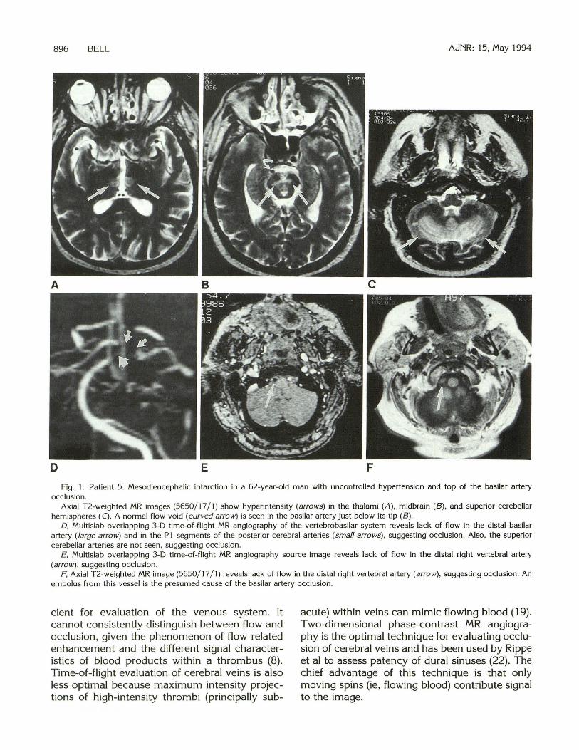
Fig. 1. Patient 5 . Mesodiencephalic infarction in a 62-year-old man with uncontrolled hypertension and top of the basilar artery occlusion.
Axial T2-weighted MR images (5650/ 17 / 1) show hyperintensity (arrows) in the thalami (A), midbrain (B), and superior cerebellar hemispheres (C). A normal flow void (curved arrow) is seen in the basilar artery just below its tip (B).
D, Multislab overlapping 3-D time-of-flight MR angiography of the vertebrobasilar system reveals lack of flow in the distal basilar artery (large arrow) and in the P1 segments of the posterior cerebral arteries (small arrows), suggesting occlusion. Also, the superior cerebellar arteries are not seen, suggesting occlusion.
£ , Multislab overlapping 3-D time-of-flight MR angiography source image reveals lack of flow in the distal right vertebral artery (arrow), suggesting occlusion.
F, Axial T2-weighted MR image (5650/ 17 / 1) reveals lack of flow in the distal right vertebral artery (arrow) , suggesting occlusion. An embolus from this vessel is the presumed cause of the basilar artery occlusion .
cient for evaluation of the venous system. It cannot consistently distinguish between flow and occlusion, given the phenomenon of flow-related enhancement and the different signal characteristics of blood products within a thrombus (8). Time-of-flight evaluation of cerebral veins is also less optimal because maximum intensity projections of high-intensity thrombi (principally sub-
acute) within veins can mimic flowing blood (19). Two-dimensional phase-contrast MR angiography is the optimal technique for evaluating occlusion of cerebral veins and has been used by Rippe et al to assess patency of dural sinuses (22). The chief advantage of this technique is that only moving spins (ie, flowing blood) contribute signal to the image.
AJNR: 15, May 1994
A B
BITHALAMIC HYPERINTENSITY 897
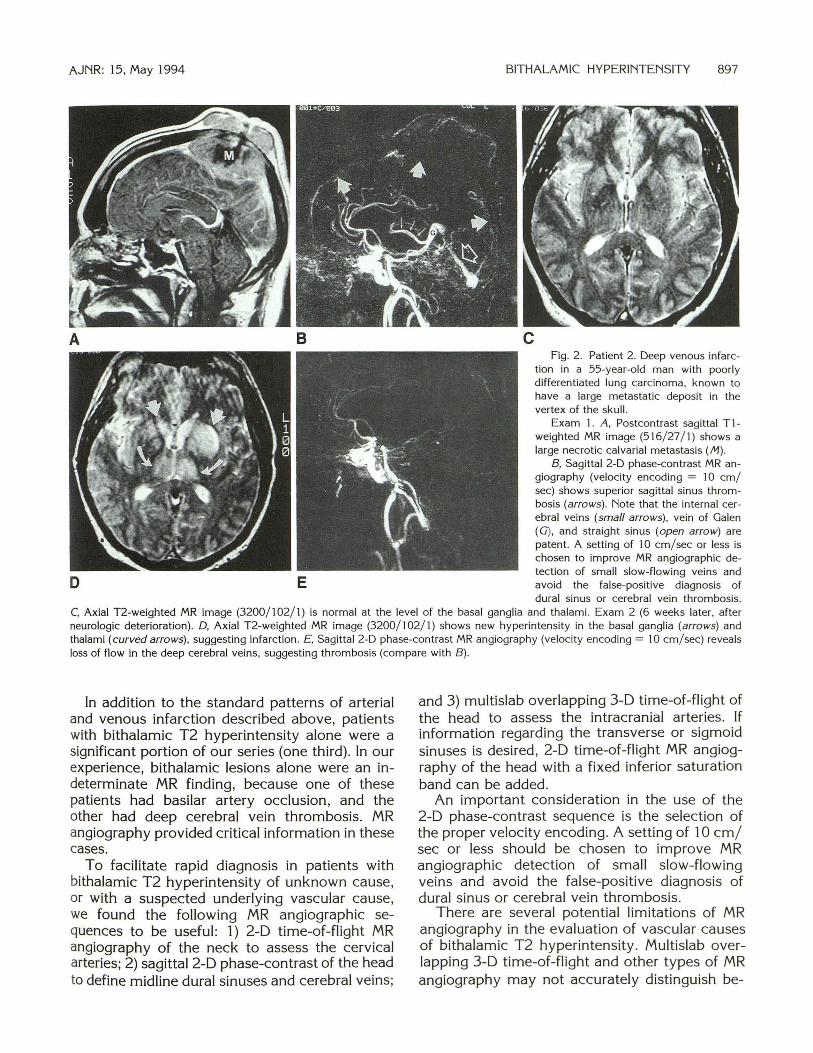
c Fig. 2. Patient 2. Deep venous infarc
tion in a 55-year-old man with poorly differentiated lung carcinoma, known to have a large metastatic deposit in the vertex of the skull.
Exam 1 . A, Postcontrast sagittal T ]weighted MR image (516/27/1) shows a large necrotic calvarial metastasis (;\1).
B, Sagittal 2-D phase-contrast MR angiography (velocity encoding = 10 em/ sec) shows superior sagittal sinus thrombosis (arrows). Note that the internal cerebral veins (small arrows), vein of Galen (G), and straight sinus (open arrow) are patent. A setting of 10 em/ sec or less is chosen to improve MR angiographic detection of small slow-flowing veins and
D E avoid the false-positive diagnosis of dural sinus or cerebral vein thrombosis.
C, Axial T2-weighted MR image (3200/102/1) is normal at the level of the basal ganglia and thalami. Exam 2 (6 weeks later, after neurologic deterioration). D, Axial T2-weighted MR image (3200/102/1) shows new hyperintensity in the basal ganglia (arrows) and thalami (curved arrows), suggesting infarction. £, Sagittal 2-D phase-contrast MR angiography (velocity encoding = 10 em/sec) reveals loss of flow in the deep cerebral veins, suggesting thrombosis (compare with B) .
In addition to the standard patterns of arterial and venous infarction described above, patients with bithalamic T2 hyperintensity alone were a significant portion of our series (one third). In our experience, bithalamic lesions alone were an indeterminate MR finding, because one of these patients had basilar artery occlusion, and the other had deep cerebral vein thrombosis. MR angiography provided critical information in these cases.
To facilitate rapid diagnosis in patients with bithalamic T2 hyperintensity of unknown cause, or with a suspected underlying vascular cause, we found the following MR angiographic sequences to be useful: 1) 2-D time-of-flight MR angiography of the neck to assess the cervical arteries; 2) sagittal 2-D phase-contrast of the head to define midline dural sinuses and cerebral veins;
and 3) multislab overlapping 3-D time-of-flight of the head to assess the intracranial arteries. If information regarding the transverse or sigmoid sinuses is desired , 2-D time-of-flight MR angiography of the head with a fixed inferior saturation band can be added .
An important consideration in the use of the 2-D phase-contrast sequence is the selection of the proper velocity encoding. A setting of 10 em/ sec or less should be chosen to improve MR angiographic detection of small slow-flowing veins and avoid the false-positive diagnosis of dural sinus or cerebral vein thrombosis.
There are several potential limitations of MR angiography in the evaluation of vascular causes of bithalamic T2 hyperintensity. Multislab overlapping 3-D time-of-flight and other types of MR angiography may not accurately distinguish be-

898 BELL AJNR: 15, May 1994
Fig. 3. Patient 3. Bithalamic infarction in a 30-year-old woman with basilar artery occlusion. A , AxiaLT2-weighted MR image (3200/ 102/1) shows bithalamic hyperintensity (arrows) , indicating infarction. B, Multislab overlapping 3-D time-of-flight MR angiography of the vertebrobasilar system reveals lack of flow in the distal basilar
artery and in the P1 segments of the posterior cerebral arteries (open arrow), indicating occlusion . Flow in the P2 segments and more distal branches of the posterior cerebral arteries (arrows) is maintained by contributions from the posterior communicating arteries.
C, Sagittal 2-D phase-contrast MR angiography shows normal venous structures, with flow in the superior sagittal sinus thrombosis (S), straight sinus (open arrow), vein of Galen (G), and internal cerebral veins (small arrows).
D, Conventional left vertebral arteriogram with subtraction shows occlusion of the distal basilar artery (arrow). The superior cerebellar arteries are patent, with duplication on the right (curved arrows). This duplication is only vaguely seen in B, possibly because of the effects of in-plane spin saturation .
E, 2-D time-of-flight MR angiography of the cervical arteries shows abnormal flow in the proximal right vertebral artery (arrowheads) , suggesting dissection.
F, This was confirmed by conventional angiography (curved arrow). The patient was also noted to have a right aortic arch (A , ascending portion) with non-mirror-image branching (origin of aberrant left subclavian artery depicted by arrows).
G, Note the stasis of contrast (arrowheads) in the early venous phase of a right brachiocephalic trunk injection. An embolus from the site of dissection is the presumed cause of the basilar artery occlusion.
tween total vessel occlusion and extremely slow flow (15). In cases of severe underlying atherosclerosis of the vertebrobasilar system, turbulent flow may sufficiently dephase spins so that no
flow is detected, simulating occlusion (15). Occlusion may also be simulated in these patients by slow-flowing spins that become saturated within a given imaging volume, resulting in loss of signal.
AJNR: 15, May 1994
MR angiography may not detect thrombosis of smaller cortical veins, a serious condition commonly associated with hemorrhagic infarction (19). Absence of a vein or sinus by MR angiography may represent occlusion . However, because of variants in venous anatomy, confirmation of the diagnosis is possible only when clot or evidence of infarction is noted on the corresponding MR image (19). Motion artifact is another significant challenge in arterial or venous MR angiography. Image quality can be significantly affected in these patients, who are often critically ill and unable to cooperate fully.
In summary, MR angiography allows for accurate differentiation between the two vascular causes of bithalamic T2 hyperintensity by MR: basilar artery occlusion and deep cerebral vein thrombosis. Multislab overlapping 3-D time-offlight and 2-D phase-contrast MR angiography are the most useful for the evaluation of major intracranial arteries and midline dural sinuses and deep cerebral veins, respectively, and should be considered when bithalamic lesions are noted or when mesodiencephalic occlusive disease is suspected.
Acknowledgments
We thank Dr Steven Crawford, who provided a case for this series.
References
1. Voit T , Lemburg P, Neuen E, Lumenta C, Stork W. Damage of
thalamus and basal ganglia in asphyxiated full - term neonates. Neu
ropediatrics 1987;18:176-181.
2. Partlow GD, del Carpio-O'Donovan R, Melanson D, Peters TM. Bilat
eral thalamic glioma: review of eight cases with personality change
and mental deterioration. AJNR Am J Neuroradiol 1992; 13:1225-
1230 3. Kobayashi T , Yoshida J , Kida Y. Bilateral germ cell tumors involving
the basal ganglia and thalamus. Neurosurgery 1989;24:579-583
4. Chang KH, Han MH, Kim HS, Wie BA, Han MC. Delayed encephalop-
BITHALAMIC HYPERINTENSITY 899
athy after acute carbon monoxide intoxication: MR imaging features
and distribution of cerebral white matter lesions. Radiology
1992;1 84: 117-1 22
5. Tuchman RF, Moser FG, Moshe SL. Carbon monoxide poisoning:
bilateral lesions in the thalamus on MR imaging of the brain . Pediatr
Radio/1990;20:478-479
6. Gallucci M, Bozzao A, Splendiani A, Masciocchi C, Passariello R.
Wernicke encephalopathy: MR findings in five patients. AJNR Am J
Neuroradio/1990; 11:887-892
7. Gomez CR, Saul RF, Selhorst JB, et al. Meso-diencephalic infarction:
a not so rare form of stroke. Ita! J Neurol Sci 1990; 11:551-557
8. Ashforth RA, Melanson D. Ethier R. MR of deep cerebra l venous thrombosis. Can J Neurol Sci 1989; 16:4 17-421
9. Erbguth F, Brenner P, Schuierer G, Druschky K-F, Neundorfer B.
Diagnosis and treatment of deep cerebral vein thrombosis. Neurosurg Rev 1991;14:145-148
10. Caplan LR. "Top of the basilar" syndrome. Neurology 1980;30:72-79
11. Hacke W, Zeumer H, Ferbert A , Bruckmann H, del Zoppo GJ . Intra
arterial thrombolytic therapy improves outcome in patients with acute vertebrobasilar occlusive disease. Stroke 1988; 19:1216-1222
12. Granato DB, Archer CR , Awwad EE. Magnetic resonance imaging of
cerebral venous thrombosis secondary to "low-dose" birth control
pills. C/in Imaging 1989; 13:220-224
13. Parker DL, Yuan C, Blatter DD. MR angiography by multiple thin slab
3D acquisition. Magn Reson Med 1991 ;17:434-451
14. Blatter DD, Parker DL, Robison RO. Cerebral MR angiography by
multiple overlapping thin slab acquisition . Part I. Quantitative analysis
of vessel visibility. Radiology 1991 ; 179:805-811
15. Blatter DD, Parker DL, Ahn SS, et al. Cerebral MR angiography with
multiple overlapping thin slab acquisition. Part II. Early clinical ex
perience. Radiology 1992; 183:379-389
16. Marcu H, Hacker H, Vonofakos D. Bilateral reversible thalamic lesions
on computed tomography. Neuroradiology 1979;18:201-204
17. Yogi T J , Balzer JO, Stemmler J , Bergman C, Egger E, Lissner J. MR
angiography in children with cerebral neurovascular diseases: findings
in 31 cases. AJR Am J Roentgenol 1992;4:817 -823
18. Rippe DJ, Boyko OB, Spritzer CE, et al. Demonstration of dural sinus
occlusion by the use of MR angiography. AJNR Am J Neuroradio/
1990;11:199-201
19. Mattie HP, Wentz KU , Edelman RR , et al. Cerebral venography with
MR. Radiology 1991;178:453-458
20. Heiserman JE, Drayer BP , Keller PJ, Fram EK. Intracrania l vascular
stenosis and occlusion: evaluation with three-dimensional time-of
flight MR angiography. Radiology 1992; 185:667-673
21. Biller J , Yuh WTC, Mitchell GW, Bruno A , Adams HP. Early diagnosis
of basilar artery occlusion using MRI. Stroke 1988; 19:297-306
22. Rippe DJ, Boyko OB, Spri tzer CE, et al. Demonstration of dural sinus
occlusion by the use of MR angiography. AJNR Am J Neuroradiol
1990;11 :199-20 1