
Bisdionin C—A Rationally Designed, Submicromolar …davapc1.bioch.dundee.ac.uk/pdf/bisc.pdf ·...
5
Published: March 23, 2011 r2011 American Chemical Society 428 dx.doi.org/10.1021/ml200008b | ACS Med. Chem. Lett. 2011, 2, 428–432 LETTER pubs.acs.org/acsmedchemlett Bisdionin C—A Rationally Designed, Submicromolar Inhibitor of Family 18 Chitinases Alexander W. Sch€ uttelkopf, † Ole A. Andersen, † Francesco V. Rao, † Matthew Allwood, ‡ Christina L. Rush, † Ian M. Eggleston, ‡,|| and Daan M. F. van Aalten* ,† † Division of Molecular Microbiology and ‡ Division of Biological Chemistry and Molecular Microbiology, College of Life Sciences, University of Dundee, Dow Street, Dundee, DD1 5EH, Scotland C hitin, a polymer of β-(1,4)-linked N-acetylglucosamine, is an important structural component of the fungal cell wall as well as the exoskeletons of arthropods. Chitinases, the enzymes that hydrolyze chitin, are required for proper morphogenesis of these organisms through their function in cell separation in fungi 1,2 and in molting of arthropods, 3 respectively. Conse- quently, chitinase inhibitors are thought to have antifungal and insecticidal potential. 3,4 Additionally, humans possess two active chitinases despite our inability to metabolize chitin. 5,6 Human macrophage chitotriosidase (HCHT) has been found to be overexpressed in fungal/bacterial infections and in the lysosomal storage disorder Gaucher's disease, 7,8 while the upregulation of acidic mammalian chitinase (AMCase) in lung tissue is related to asthma progression. Indeed, inhibition of this enzyme has been shown to result in reduced recruitment of inflammatory cells in a mouse model of asthma. 9 These enzymes, which belong to CAZy family GH18, 10 share an unusual catalytic mechanism involving a nucleophilic attack by the N-acetyl group of the 1 sugar itself rather than a protein carboxylate as observed in other retaining glycosidases 11,12 (sugar binding sites are numbered relative to the scissile glyco- sidic bond between the 1 and þ1 sugars 13 ). GH18 chitinases can be subdivided at a sequence/structural level into “bacterial- type” and “plant-type” family members, which differ in substrate specificity and active site architecture, with the former possessing a relatively deep active site groove, whereas the latter have a more shallow and accessible open active site. 1416 Known inhibitors of these chitinases include cyclic dipep- tides, 17,18 cyclic pentapeptides, 1921 thiazolines, 22 the pseudo- trisaccharide allosamidin, 23 and piperazine derivatives. 24 Most of these compounds are natural products or their derivatives, and their use for either in vivo studies or as ligand design leads is significantly impeded by their limited availability and their chemical complexity and subsequent poor synthetic accessibility. We recently identified xanthine derivatives as promising leads for GH18 inhibitors 25 and subsequently developed a micromolar chitinase inhibitor comprised of two linked caffeine molecules with desirable druglike properties, a crystallographically defined binding mode, and excellent synthetic accessibility. 26 In this work, we present the further development of this class of compounds through in silico and in vitro screens, focusing on modifying the caffeine-caffeine linker and resulting in bisdionin C, the first rationally designed submicromolar inhibitor of GH18 chitinases. The present structural and docking data focus on ChiB1 from the opportunistic fungal pathogen Aspergillus fumi- gatus (AfChiB1) as an experimentally accessible, representative bacterial-type GH18 chitinase, while inhibition data have been determined for a number of chitinases. We recently described a micromolar inhibitor of family 18 chitinases (Figure 1, n = 2), which was identified in a virtual screen for xanthine derivatives 26 after previous work had shown a Received: January 11, 2011 Accepted: March 11, 2011 ABSTRACT: Chitinases of the GH18 family play important roles in a variety of pathogenic organisms and have also been shown to be involved in human asthma progression, making these enzymes potential drug targets. While a number of potent GH18 chitinase inhibitors have been described, in general, these compounds suffer from limited synthetic accessibility or unfavorable medicinal- chemical properties, making them poor starting points for the development of chitinase-targeted drugs. Exploiting available structural data, we have rationally designed bisdionin C, a sub- micromolar inhibitor of GH18 enzymes, that possesses desirable druglike properties and tractable chemical synthesis. A crystallographic structure of a chitinase-bisdionin C complex shows the two aromatic systems of the ligand interacting with two conserved tryptophan residues exposed in the active site cleft of the enzyme, while at the same time forming extensive hydrogen- bonding interactions with the catalytic machinery. The observed mode of binding, together with inhibition data, suggests that bisdionin C presents an attractive starting point for the development of specific inhibitors of bacterial-type, but not plant-type, GH 18 chitinases. KEYWORDS: GH18 Chitinase, xanthine, ligand design
Transcript of Bisdionin C—A Rationally Designed, Submicromolar …davapc1.bioch.dundee.ac.uk/pdf/bisc.pdf ·...
Published: March 23, 2011
r 2011 American Chemical Society 428 dx.doi.org/10.1021/ml200008b |ACS Med. Chem. Lett. 2011, 2, 428–432
LETTER
pubs.acs.org/acsmedchemlett
Bisdionin C—A Rationally Designed, Submicromolar Inhibitor ofFamily 18 ChitinasesAlexander W. Sch€uttelkopf,† Ole A. Andersen,† Francesco V. Rao,† Matthew Allwood,‡ Christina L. Rush,†
Ian M. Eggleston,‡,|| and Daan M. F. van Aalten*,†
†Division of Molecular Microbiology and ‡Division of Biological Chemistry and Molecular Microbiology, College of Life Sciences,University of Dundee, Dow Street, Dundee, DD1 5EH, Scotland
Chitin, a polymer of β-(1,4)-linkedN-acetylglucosamine, is animportant structural component of the fungal cell wall as
well as the exoskeletons of arthropods. Chitinases, the enzymesthat hydrolyze chitin, are required for proper morphogenesis ofthese organisms through their function in cell separation infungi1,2 and in molting of arthropods,3 respectively. Conse-quently, chitinase inhibitors are thought to have antifungal andinsecticidal potential.3,4 Additionally, humans possess two activechitinases despite our inability to metabolize chitin.5,6 Humanmacrophage chitotriosidase (HCHT) has been found to beoverexpressed in fungal/bacterial infections and in the lysosomalstorage disorder Gaucher's disease,7,8 while the upregulation ofacidic mammalian chitinase (AMCase) in lung tissue is related toasthma progression. Indeed, inhibition of this enzyme has beenshown to result in reduced recruitment of inflammatory cells in amouse model of asthma.9
These enzymes, which belong to CAZy family GH18,10 sharean unusual catalytic mechanism involving a nucleophilic attack bythe N-acetyl group of the �1 sugar itself rather than a proteincarboxylate as observed in other retaining glycosidases11,12
(sugar binding sites are numbered relative to the scissile glyco-sidic bond between the �1 and þ1 sugars13). GH18 chitinasescan be subdivided at a sequence/structural level into “bacterial-type” and “plant-type” family members, which differ in substratespecificity and active site architecture, with the former possessinga relatively deep active site groove, whereas the latter have a moreshallow and accessible open active site.14�16
Known inhibitors of these chitinases include cyclic dipep-tides,17,18 cyclic pentapeptides,19�21 thiazolines,22 the pseudo-trisaccharide allosamidin,23 and piperazine derivatives.24 Most ofthese compounds are natural products or their derivatives, andtheir use for either in vivo studies or as ligand design leads issignificantly impeded by their limited availability and theirchemical complexity and subsequent poor synthetic accessibility.
We recently identified xanthine derivatives as promising leadsfor GH18 inhibitors25 and subsequently developed a micromolarchitinase inhibitor comprised of two linked caffeine moleculeswith desirable druglike properties, a crystallographically definedbinding mode, and excellent synthetic accessibility.26 In thiswork, we present the further development of this class ofcompounds through in silico and in vitro screens, focusing onmodifying the caffeine-caffeine linker and resulting in bisdioninC, the first rationally designed submicromolar inhibitor of GH18chitinases. The present structural and docking data focus onChiB1 from the opportunistic fungal pathogen Aspergillus fumi-gatus (AfChiB1) as an experimentally accessible, representativebacterial-type GH18 chitinase, while inhibition data have beendetermined for a number of chitinases.
We recently described a micromolar inhibitor of family 18chitinases (Figure 1, n = 2), which was identified in a virtualscreen for xanthine derivatives26 after previous work had shown a
Received: January 11, 2011Accepted: March 11, 2011
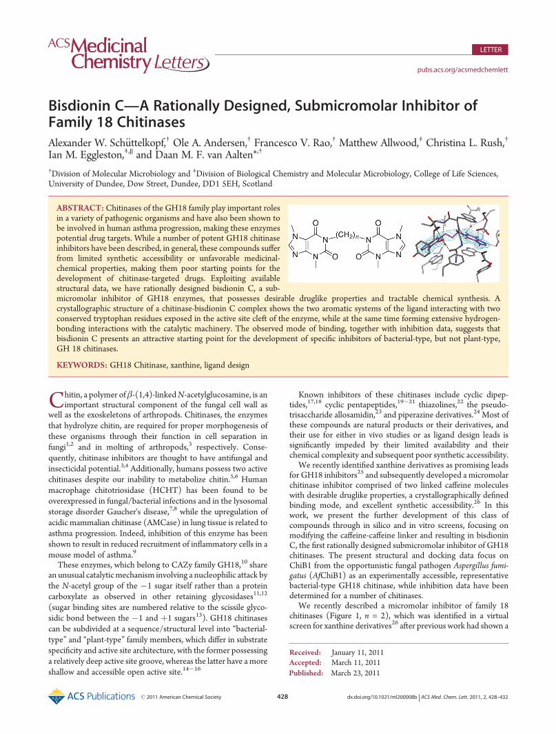
ABSTRACT: Chitinases of the GH18 family play important rolesin a variety of pathogenic organisms and have also been shown tobe involved in human asthma progression, making these enzymespotential drug targets. While a number of potent GH18 chitinaseinhibitors have been described, in general, these compounds sufferfrom limited synthetic accessibility or unfavorable medicinal-chemical properties, making them poor starting points for thedevelopment of chitinase-targeted drugs. Exploiting availablestructural data, we have rationally designed bisdionin C, a sub-micromolar inhibitor of GH18 enzymes, that possesses desirable druglike properties and tractable chemical synthesis. Acrystallographic structure of a chitinase-bisdionin C complex shows the two aromatic systems of the ligand interacting with twoconserved tryptophan residues exposed in the active site cleft of the enzyme, while at the same time forming extensive hydrogen-bonding interactions with the catalytic machinery. The observed mode of binding, together with inhibition data, suggests thatbisdionin C presents an attractive starting point for the development of specific inhibitors of bacterial-type, but not plant-type,GH 18 chitinases.
KEYWORDS: GH18 Chitinase, xanthine, ligand design

429 dx.doi.org/10.1021/ml200008b |ACS Med. Chem. Lett. 2011, 2, 428–432
ACS Medicinal Chemistry Letters LETTER
number of these molecules, including caffeine, binding efficientlyto the active site of AfChiB1.25 Upon determining the crystalstructure of a complex ofAfChiB1 with this compound, it becameapparent that the ligand adopts an unexpected strained confor-mation in the active site. In particular, the “primary” caffeine(which occupies the �1 subsite) is in an energetically less favo-rable flipped conformation as compared to the previouslydescribed crystal structures of complexes with free xanthinederivatives.25 It was hypothesized that modifying the xanthine-xanthine linker, in particular its length, could alleviate the strainand thus yield a more potent chitinase inhibitor, giving rise to aseries of potential chitinase inhibitors, termed the bisdioninshere, which are based on the dicaffeine scaffold with variablelinker lengths (Figure 1).
To test the effects of linker length on inhibition in vitro,bisdionins C�E were synthesized using established methodol-ogy starting from appropriate xanthine and R,ω-dibromoalkanebuilding blocks.27,28 The inhibitory potency of the bisdioninseries was tested against AfChiB1 as well as HCHT and AMCaseactivity (from mouse lung homogenate) (Table 2). At theexpense of introducing a single additional methylene group, inall cases, bisdionin C shows an improvement in inhibition of atleast one order of magnitude as compared to bisdionin B. Thelarger bisdionin variants offer no improvement over bisdionin B.
Like bisdionin B, bisdionin C is a druglike molecule as assessedby Lipinski's rule of five:29 It has six hydrogen bond acceptors andno hydrogen bond donors, a molecular mass of 400.4 Da, anda predicted logP of approximately zero (calculated using theMolinspiration property calculation service, http://www.molinspiration.com/). The drastic increase in potency from a rela-tively small increase in compound size also makes it a more efficientchitinase inhibitor (ligand efficiency for AfChiB1 is approximately�0.41 kcal mol�1 atom�1). This compares favorably with allosami-din, a well-known potent natural product GH18 inhibitor, whichviolates several of the rule of five criteria by having 10 hydrogen bonddonors, 13 acceptors, and a molecular mass of 622.6 Da, as well ashaving anundesirably low logPof�4.7 and a poorer ligandefficiencyfor AfChiB1 of approximately �0.25 kcal mol�1 atom�1.30
To investigate the binding mode of bisdionin C, the structureof AfChiB1 in complex with the inhibitor was refined against2.0 Å synchrotron diffraction data to an Rwork/Rfree of 18.3% and21.6%, respectively (Table 1). The active site reveals clearelectron density for the bisdionin C ligand (Figure 2). Whilethe overall binding mode is similar to that of bisdionin B, withone of the caffeines occupying the �1 subsite sandwichedbetween Trp384 and a flipped-down Trp137 and the secondcaffeine stacking with Trp52, occupying approximately the �3subsite, there are a number of striking differences between thetwo ligands. These differences together explain the significantlyimproved potency of bisdionin C over bisdionin B.
First, a comparison of intercaffeine distances of bisdionin B(≈8.2 Å) and bisdionin C (≈9.6 Å) suggests that the latter
molecule should be a better mimic of a chitooligosaccharidesubstrate, which, bound to a GH18 active site, has an n...nþ2sugar ring distance of≈10 Å (measured in a complex structure ofHCgp-39, PDB id 1HJW; Houston et al.31). The bisdionin Ccomplex reveals that the longer linker indeed allows the ligand,while maintaining the position of the secondary caffeine, to insertits primary caffeine more deeply into the AfChiB1 active sitepocket (Figure 2). In doing so, it displaces the buried watermolecule that, in the bisdionin B complex, mediates the hydro-gen-bonding interaction between N9 of the primary caffeine andthe hydroxyl of Tyr245. In general terms, the tighter fit of thecaffeine into the�1 pocket of the protein explains the improvedbinding of bisdionin C.
Strikingly, the primary caffeine of bisdionin C is flipped by180� around the N1-linker bond as compared to bisdionin B,reverting it to the binding mode previously observed for smallerxanthine derivatives like caffeine or pentoxifylline.25 In additionto the deeper insertion into the binding pocket, this orientationalso allows the caffeine moiety to make an additional hydrogenbond (a water-mediated H-bond accepted by O2), while thewater-mediated hydrogen bond to Tyr245 of the bisdionin Bcomplex is replaced with a direct interaction. These features,together with the fact that smaller xanthine derivatives unrest-rained by a second ring system unambiguously adopt this bindingmode, suggest that it is energetically more favorable as comparedto that observed for bisdionin B. At the same time, it is note-worthy that the active site orientation of the ostensibly less tightlybound secondary caffeine is essentially identical between bisdio-nin B and C, whereas it is the more buried primary caffeine thatundergoes a drastic change in orientation. As has been suggestedbefore,26 this observationmay be explained in part by the fact that
Figure 1. Chemical structure of bisdionin; n denotes the number ofmethylene groups in the linker connecting the two xanthine ring systems,compoundswith n=1�5 have beennamedbisdioninA�E.The ring atomnumbering scheme is given for one of the two xanthine rings.
Table 1. Data Collection and Refinement Statistics for theAfChiB1-Bisdionin C Complexa
resolution range (Å) 30.0�2.0
total measurements 265212 (24551)
unique reflections 91066 (9004)
completeness 0.998 (0.992)
Rsym 0.089 (0.570)
redundancy 2.9 (2.7)
I/σ(I) 12.3 (2.4)
no. of protein residues 790
no. of ligand residues 2
no. of solvent molecules 691
Rwork, Rfree 0.183, 0.216
average B factor (Å2)
overall 28.8
protein 27.8
ligand 28.4
solvent 36.8
rmsd bonds (Å) 0.010
rmsd angles (�) 1.1
Ramachandran plot statistics (%)
most favored region 91.6
additional allowed region 8.0
generously allowed region 0.4
disallowed region 0.0aValues in parentheses refer to the highest resolution shell of 0.07 Åwidth. Ramachandran statistics were determined by PROCHECK.39

430 dx.doi.org/10.1021/ml200008b |ACS Med. Chem. Lett. 2011, 2, 428–432
ACS Medicinal Chemistry Letters LETTER
the bisdionin B-like binding mode likely allows for improvedπ-π stacking between the primary xanthine ring and its sand-wiching tryptophan residues, which could alleviate the energeticpenalty imposed by the flipped xanthine.
The two xanthine rings of bisdionin C are almost perfectlycoplanar with their tryptophan stacking partners. This is inmarked contrast to bisdionin B, where an unfavorable xanthine-xanthine angle precludes coplanarity with the tryptophans andinstead adopts two conformations in the active site, each of whichapproximates coplanarity on one side of the molecule, resultingin nonideal π-π stacking interactions and in fact slight clasheswith the protein. The longer linker of bisdionin C increasesflexibility, and its odd number of methylene groups is morecompatible with the two xanthines being at an angle suitable foroptimal stacking in the chitinase active site. This is furthersupported by the fact that the aromatic rings of bisdionin C areessentially coplanar with the “unrestrained” ring systems found inthe AfChiB1-caffeine complex structure,25 which shows two caf-feine molecules bound in the �1 and the �3 subsites.
The three tryptophan residues (52, 137, and 384 in AfChiB1)that are the hallmark features of the AfChiB1-bisdionin Cinteraction are conserved in the majority of bacterial-typeGH18 chitinases, suggesting that bisdionin C might be a potentinhibitor of other members of this subfamily, including the twohuman chitinases. Indeed, these two enzymes are inhibited bybisdionin C, albeit with an approximately 1.5 orders of magni-tude higher IC50 (Table 2). Considering the expected degree ofsimilarity between the active sites of these three enzymes,26 thesource of this significant reduction in affinity is unclear. At thesame time, two of these tryptophan residues are missing fromplant type GH18 enzymes, which would suggest that bisdionin Cshould be a poor inhibitor of this GH18 subfamily. To test this,bisdionin IC50 values for AfChiA1, a representative plant typechitinase, were determined (Table 2). As expected, bisdioninsB�D show no significant inhibition of AfChiA1. Surprisingly,bisdionin E was found to inhibit AfChiA1 weakly; it is possiblethat due to the longer tether its secondary caffeine is able tointeract with an unknown new AfChiA1-specific binding site.Being specific for the bacterial-type chitinases, bisdionin C couldthus provide a chemical tool to dissect the different functions ofplant type and bacterial type enzymes in biological systems.
Through in vitro screening of bisdionin compounds withvarying linker lengths, we have identified bisdionin C as a potent
family 18 chitinase inhibitor. The crystal structure of a AfChiB1-bisdionin C complex elucidates how the addition of a single CH2
group can improve ligand affinity by at least 10-fold for all testedbacterial-type chitinases: The longer linker not only allows theprimary caffeine to penetrate deeper into the active site butfortuitously also enables the ligand to engage in concurrent andclose-to-ideal π�π stacking interactions with both of its aro-matic systems. At the same time, bisdionin C retains its desirabledruglike properties and good synthetic accessibility, making it anexcellent lead for the development of drugs targeted at thebacterial-type chitinases involved in a variety of disease processes.In particular, the inhibition of AMCase, while decreased ascompared to that of AfChiB1, suggests that bisdionin C willprovide a good starting point for the development of AMCaseinhibitors, which could be useful in the treatment of allergicasthma.9 In addition, the specificity of bisdionin C for bacterial-type over plant-type GH18 chitinases will enable its use as achemical tool to dissect the different roles these subclasses ofenzymes play in, for example, fungal organisms.
’EXPERIMENTAL PROCEDURES
A. fumigatus ChiB1 (AfChiB1) and human chitotriosidase wereexpressed and purified as described previously.25,26 AMCase activitywas determined frommouse lung homogenate. Lung homogenates wereprepared from female BALB/C mice. Tissue (100 mg) was taken fromisolated lung lobes, homogenized in 2 mL of HBSS (Invitrogen), and
Figure 2. Stereoview of bisdionin C bound to AfChiB1. The ligand is shown in purple, protein residues are colored gray, and selected water moleculesare shown as red spheres. Potential hydrogen bonds are indicated by black dotted lines. Residues interacting with the ligands are labeled. The unbiased|Fo| � |Fc|, jcalc electron density map covering the ligand is shown contoured at 2.75σ.
Table 2. Experimental IC50 Values for Inhibition of Family 18Chitinases by the Bisdionins
IC50 (μM)
bisdionin (n) AfChiB1 HCHT AMCase AfChiA1
B (2) 4.8( 1.4 110( 10 90( 4 >1000
C (3) 0.20( 0.01 8.3( 0.7 3.4( 0.2 >1000
D (4) 9.0( 2.0 91( 17 ND >1000
E (5) 5.7( 0.8 260( 70 ND 820( 50
431 dx.doi.org/10.1021/ml200008b |ACS Med. Chem. Lett. 2011, 2, 428–432
ACS Medicinal Chemistry Letters LETTER
centrifuged (800g, 10 min), and the resulting supernatant was collected.AfChiA1 was expressed and purified as described by Rush et al.32.The bisdionins B�E (Itaharo and Imamura;27 Figure 1) with alkyl
linkers ranging from two (n= 2, bisdionin B) to fivemethylene units (n= 5,bisdioninE) were synthesized according to themethod ofCavallaro et al.,28
whereby a suspension of theobromine (2 equiv) and potassium carbonate(2 equiv) in dry DMF was heated to 120 �C under argon for1 h, followed by treatment with 1 equiv of the appropriate R,ω-dibro-moalkane. The reaction mixture was poured into water and neutralizedwith 0.1MHCl. The precipitate formedwas then collectedbyfiltration andpurified by precipitation from a chloroform solution with ether. Thecompoundswere characterized by 1H and 13CNMRand electrospraymassspectrometry, and their purity was confirmed by analytical RP-HPLC.The enzyme activity was determined as described before,26,32 using
4-methylumbelliferyl-β-D-N,N0-diacetylchitobiose (4MU-GlcNAc2; forAfChiB1 and lung homogenate) and 4-methylumbelliferyl-β-D-N,N0,N00-triacetylchitotriose (4MU-GlcNAc3; for HCHT andAfChiA1), bothpurchased from Sigma, as substrates. All IC50 determinations werecarried out with triplicate measurements for each inhibitor concentra-tion. Data analysis was performed in GRAFIT 5.0.33
Crystals of AfChiB1 were grown as described before.25 After theywere washed in 0.1 M sodium citrate, pH 5.5, and 1.4 M Li2SO4 toremove bound Tris, they were soaked in mother liquor containing≈125-fold molar excess of bisdionin C for up to 30 min. Aftercryoprotection in 3 M Li2SO4, data were collected at BM14, EuropeanSynchrotron Radiation Facility (ESRF) (Grenoble, France). Data wereprocessed using the HKL suite34 and truncated using CCP4 software.35
After phasing by manual molecular placement of the apo-AfChiB1structure (PDB id 1W9P), refinement was performed using REFMAC36
and model building in O.37 Starting coordinates and topologies forbisdionin C were generated by PRODRG.38 Data and model statisticsare given in Table 1. Figures were generated using PyMOL (www.pymol.org). Atomic coordinates and structure factors have been depos-ited with the Protein Data Bank (www.rcsb.org).
’AUTHOR INFORMATION
Corresponding Author*Tel: þ44 1382 384979. E-mail: [email protected].
Present Addresses
)Department of Pharmacy and Pharmacology, University ofBath, Bath, BA2 7AY, England.
Funding SourcesThis work was supported by a Wellcome Trust Senior ResearchFellowship and an MRC Programme Grant (D.vA.), a WellcomeTrust Project Grant (I.M.E. and D.vA.), a Wellcome Trust Ph.D.Studentship (M.A.), and a BBSRC CASE Studentship (F.V.R.).We thank the staff at ESRF BM14 for their support.
’REFERENCES
(1) Kuranda, M. J.; Robbins, P. W. Chitinase is required for cell-separation during growth of Saccharomyces cerevisiae. J. Biol. Chem. 1991,266, 19758–19767.(2) Takaya, N.; Yamazaki, D.; Horiuchi, H.; Ohta, A.; Takagi, M.
Cloning and characterisation of a chitinase-encoding gene chiA fromAspergillus nidulans, disruption of which decreases germination fre-quency and hyphal growth. Biosci., Biotechnol., Biochem. 1998, 62, 60–65.(3) Merzendorfer, H.; Zimoch, L. Chitin metabolism in insects:
Structure, function and regulation of chitin synthases and chitinases.J. Exp. Biol. 2003, 206, 4393–4412.(4) Cohen, E. Chitin synthesis and degradation as targets for
pesticide action. Arch. Insect Biochem. Physiol. 1993, 22, 245–261.
(5) Hollak, C. E. M.; vanWeely, S.; van Oers, M. H. J.; Aerts, J. M. F.G. Marked elevation of plasma chitotriosidase activity—A novel hall-mark of Gaucher disease. J. Clin. Invest. 1994, 93, 1288–1292.
(6) Boot, R. G.; Renkema, G. H.; Verhoek, M.; Strijland, A.; Bliek, J.;de Meulemeester, T. M. A. M. O.; Mannens, M. M. A. M.; Aerts, J. M. F.G. The human chitotriosidase gene—Nature of inherited enzymedeficiency. J. Biol. Chem. 1998, 273, 25680–25685.
(7) Labadaridis, I.; Dimitriou, E.; Theodorakis, M.; Kafalidis, G.;Velegraki, A.; Michelakakis, H. Chitotriosidase in neonates with fungaland bacterial infections. Arch. Dis. Child. 2005, 90, F531–F532.
(8) Guo, Y.; He, W.; Boer, A. M.; Wevers, R. A.; de Bruijn, A. M.;Groener, J. E.; Hollak, C. E.; Aerts, J. M.; Galjaard, H.; van Diggelen,O. P. Elevated plasma chitotriosidase activity in various lysosomalstorage disorders. J. Inherited Metab. Dis. 1995, 18, 717–722.
(9) Yang, C. J.; Liu, Y. K.; Liu, C. L.; Shen, C. N.; Kuo, M. L.; Su,C. C.; Tseng, C. P.; Yen, T. C.; Shen, C. R. Inhibition of acidicmammalian chitinase by RNA interference suppresses ovalbumin-sensi-tized allergic asthma. Hum. Gene Ther. 2009, 20, 1597–1606.
(10) Cantarel, B. L.; Coutinho, P. M.; Rancurel, C.; Bernard, T.;Lombard, V.; Henrissat, B. The Carbohydrate-Active EnZymes database(CAZy): An expert resource for Glycogenomics.Nucleic Acids Res. 2009,37, D233–D238.
(11) Terwisscha van Scheltinga, A. C.; Armand, S.; Kalk, K. H.;Isogai, A.; Henrissat, B.; Dijkstra, B. W. Stereochemistry of chitinhydrolysis by a plant chitinase/lysozyme and X-ray structure of acomplex with allosamidin. Biochemistry 1995, 34, 15619–15623.
(12) van Aalten, D. M. F.; Komander, D.; Synstad, B.; Gåseidnes, S.;Peter, M. G.; Eijsink, V. G. H. Structural insights into the catalyticmechanism of a family 18 exo-chitinase. Proc. Natl. Acad. Sci. U.S.A.2001, 98, 8979–8984.
(13) Davies, G. J.; Wilson, K. S.; Henrissat, B. Nomenclature forsugar-binding subsites in glycosyl hydrolases. Biochem. J. 1997,321, 557–559.
(14) Perrakis, A.; Tews, I.; Dauter, Z.; Oppenheim, A. B.; Chet, I.;Wilson, K. S.; Vorgias, C. E. Crystal structure of a bacterial chitinase at2.3 Å resolution. Structure 1994, 2, 1169–1180.
(15) Terwisscha van Scheltinga, A. C.; Hennig, M.; Dijkstra, B. W.The 1.8 Å resolution structure of hevamine, a plant chitinase/lysozyme, and analysis of the conserved sequence and structuremotifs of glycosyl hydrolase family 18. J. Mol. Biol. 1996, 262,243–257.
(16) van Aalten, D. M. F.; Synstad, B.; Brurberg, M. B.; Hough, E.;Riise, B. W.; Eijsink, V. G. H.;Wierenga, R. K. Structure of a two-domainchitotriosidase from Serratia marcescens at 1.9 Å resolution. Proc. Natl.Acad. Sci. U.S.A. 2000, 97, 5842–5847.
(17) Izumida, H.; Imamura, N.; Sano, H. A novel chitinaseinhibitor from a marine bacterium Pseudomonas sp. J. Antibiot. 1996,49, 76–80.
(18) Houston, D. R.; Eggleston, I.; Synstad, B.; Eijsink, V. G. H.; vanAalten, D. M. F. The cyclic dipeptide CI-4 inhibits family 18 chitinasesby structural mimicry of a reaction intermediate. Biochem. J. 2002,368, 23–27.
(19) Arai, N.; Shiomi, K.; Yamaguchi, Y.; Masuma, R.; Iwai, Y.;Turberg, A.; Koelbl, H.; Omura, S. Argadin, a new chitinase inhibitor,produced by Clonostachys sp. FO-7314. Chem. Pharm. Bull. 2000,48, 1442–1446.
(20) Shiomi, K.; Arai, N.; Iwai, Y.; Turberg, A.; Koelbl, H.; Omura, S.Structure of argifin, a new chitinase inhibitor produced by Gliocladiumsp. Tetrahedron Lett. 2000, 41, 2141–2143.
(21) Houston, D. R.; Shiomi, K.; Arai, N.; Omura, S.; Peter, M. G.;Turberg, A.; Synstad, B.; Eijsink, V. G. H.; van Aalten, D. M. F. High-resolution structures of a chitinase complexed with natural productcyclopentapeptide inhibitors: Mimicry of carbohydrate substrate. Proc.Natl. Acad. Sci. U.S.A. 2002, 99, 9127–9132.
(22) Macdonald, J. M.; Tarling, C. A.; Taylor, E. J.; Dennis, R. J.;Myers, D. S.; Knapp, S.; Davies, G. J.; Withers, S. G. Chitinase Inhibitionby Chitobiose and Chitotriose Thiazolines. Angew. Chem., Int. Ed. 2010,49, 2599–2602.

432 dx.doi.org/10.1021/ml200008b |ACS Med. Chem. Lett. 2011, 2, 428–432
ACS Medicinal Chemistry Letters LETTER
(23) Sakuda, S.; Isogai, A.;Matsumoto, S.; Suzuki, A.; Koseki, K. Thestructure of allosamidin, a novel insect chitinase inhibitor produced byStreptomyces sp. Tetrahedron Lett. 1986, 27, 2475–2478.(24) Cole, D. C.; Olland, A. M.; Jacob, J.; Brooks, J.; Bursavich, M.;
Czerwinski, R.; DeClercq, C.; Johnson, M.; Joseph-McCarthy, D.;Ellingboe, J. W.; Lin, L.; Nowak, P.; Presman, E.; Strand, J.; Tam, A.;Williams, C.; Yao, S.; Tsao, D.; Fitz, L. J. Identification and characteriza-tion of acidic mammalian chitinase inhibitors. J. Med. Chem. 2010,26, 6122–6128.(25) Rao, F. V.; Andersen, O. A.; Vora, K. A.; DeMartino, J. A.; van
Aalten, D. M. F. Methylxanthine drugs are chitinase inhibitors: Inves-tigation of inhibition and binding modes.Chem. Biol. 2005, 12, 973–980.(26) Schuettelkopf, A. W.; Andersen, O. A.; Rao, F. V.; Allwood, M.;
Lloyd, C.; Eggleston, I. M.; van Aalten, D. M. F. Screening-basedDiscovery and Structural Dissection of a Novel Family 18 ChitinaseInhibitor. J. Biol. Chem. 2006, 281, 27278–27285.(27) Itaharo, T.; Imamura, K. Preparation and NMR study of 7,70-
(R,ω-alkanediyl) bis(theophylline), 1,10-(R,ω-alkanediyl)bis(theobromine)and 1,10-(R,ω-alkanediyl)bis(3-methyluracil). Bull. Chem. Soc. Jpn. 1994,67, 203–209.(28) Cavallaro, R. A.; Filocamo, L.; Galuppi, A.; Galioni, A.; Brufani,
M.; Genazzani, A. A. Potentiation of cADPR-induced Ca2þ-release bymethylxanthine analogues. J. Med. Chem. 1999, 42, 2527–2534.(29) Lipinski, C. A.; Lombardo, F.; Dominy, B. W.; Feeney, P. J.
Experimental and computational approaches to estimate solubility andpermeability in drug discovery and development settings. Adv. DrugDelivery Rev. 1997, 23, 3–25.(30) Vaaje-Kolstad, G.; Houston, D. R.; Rao, F. V.; Peter, M. G.;
Synstad, B.; van Aalten, D. M. F.; Eijsink, V. G. H. Structure of theD142N mutant of the family 18 chitinase ChiB from Serratia marcescensand its complex with allosamidin. Biochim. Biophys. Acta 2004,1696, 103–111.(31) Houston, D. R.; Recklies, A. D.; Krupa, J. C.; van Aalten,
D. M. F. Structure and ligandinduced conformational change of the 39kD glycoprotein from human articular chondrocytes. J. Biol. Chem. 2003,278, 30206–30212.(32) Rush, C. L.; Sch€uttelkopf, A. W.; Hurtado-Guerrero, R.; Blair,
D. E.; Ibrahim, A. F. M.; Desvergnes, S.; Eggleston, I. M.; van Aalten,D.M. F. Natural product-guided discovery of a fungal chitinase inhibitor.Chem. Biol. 2010, in press.(33) Leatherbarrow, R. J.GraFit, Version 5; Erithacus Software Ltd.:
Horley, United Kingdom, 2001.(34) Otwinowski, Z.; Minor, W. Processing of X-ray diffraction data
collected in oscillation mode. Methods Enzymol. 1997, 276, 307–326.(35) Collaborative Computational Project Number 4. The CCP4
suite: Programs for protein crystallography. Acta Crystallogr. 1994, D50,760�763.(36) Murshudov, G. N.; Vagin, A. A.; Dodson, E. J. Refinement of
macromolecular structures by the maximum-likelihood method. ActaCrystallogr. 1997, D53, 240–255.(37) Jones, T. A.; Zou, J. Y.; Cowan, S. W.; Kjelgaard, M. Improved
methods for building models in electron density maps and the locationof errors in these models. Acta Crystallogr. 1991, D47, 110–119.(38) Schuettelkopf, A.W.; van Aalten, D.M. F. PRODRG: A tool for
high-throughput crystallography of protein-ligand complexes. ActaCrystallogr. 2004, D60, 1355–1363.(39) Laskowski, R. A.; MacArthur, M. W.; Moss, D. S.; Thornton,
J. M. PROCHECK: A program to check the stereochemical quality ofprotein structures. J. Appl. Crystallogr. 1993, 26, 283–291.


![Lag,lock,sync,slip:themany“phases” ofcoupledflagellaIn particular, differential cis-trans sensitivity to submicromolar Ca2+ in cell models and in isolated axonemes [9, 36] is](https://static.fdocuments.in/doc/165x107/611b41a975553172b357e365/laglocksyncslipthemanyaoephasesa-ofcoupledi-in-particular-diierential.jpg)


![Inhibitors against Fungal Cell Wall Remodeling Enzymesdavapc1.bioch.dundee.ac.uk/pdf/scgas2i2.pdf · Inhibitors against Fungal Cell Wall Remodeling Enzymes Ignacio Delso,*[a] Jessika](https://static.fdocuments.in/doc/165x107/5f87ac06eb97293053446e00/inhibitors-against-fungal-cell-wall-remodeling-inhibitors-against-fungal-cell-wall.jpg)


