BIOTASCOPE - El Probiótico Biotascope, we aim to cover some of the recent main articles in each...
32
Translational Science in Microbiota April 2015 • ISSUE BIOTASCOPE 1 RECONSTITUTION WITH MICROBIOTA GERM-FREE MICE REDUCED: • ORGAN WEIGHT (E.G. HEART, LIVER, LUNGS) • CARDIAC OUTPUT, OXYGEN CONSUMPTION • EXPRESSION OF GROWTH FACTORS IN CNS • INTESTINAL MOTILITY • MESENTERIC AND SYSTEMIC LYMPH NODES • SERUM IMMUNOGLOBULIN LEVELS INCREASED: • FOOD INTAKE TO SUSTAIN BODY WEIGHT • SUSCEPTIBILITY TO INFECTION • ATTENTION DEFICIT HYPERACTIVITY DISORDER STRUCTURAL AND FUNCTIONAL RECOVERY: • PROTECTIVE EFFECTS • METABOLIC EFFECTS • TROPHIC EFFECTS
Transcript of BIOTASCOPE - El Probiótico Biotascope, we aim to cover some of the recent main articles in each...

Translational Science in Microbiota
April 2015 • ISSUE
BIOTASCOPE1
RECONSTITUTION WITH MICROBIOTA
GERM-FREE MICEREDUCED:• ORGAN WEIGHT
(E.G. HEART, LIVER, LUNGS)
• CARDIAC OUTPUT, OXYGEN CONSUMPTION
• EXPRESSION OF GROWTH FACTORS IN CNS• INTESTINAL MOTILITY• MESENTERIC AND SYSTEMIC LYMPH NODES• SERUM IMMUNOGLOBULIN LEVELS
INCREASED:• FOOD INTAKE TO SUSTAIN BODY WEIGHT • SUSCEPTIBILITY TO INFECTION• ATTENTION DEFICIT HYPERACTIVITY DISORDER
STRUCTURAL AND FUNCTIONAL RECOVERY:• PROTECTIVE EFFECTS• METABOLIC EFFECTS• TROPHIC EFFECTS

> Contents
EditorialSerhat Bor 1
Very ClinicalINTESTINAL GAS • Fernando Azpiroz 2
PEDIATRIC ANTIBIOTIC-ASSOCIATED DIARRHEA • Ener Cagri Dinleyici 7
Very BasicDECIPHERING THE HUMAN GUT MICROBIOME • Francisco Guarner 11
TREATMENT OF COW’S MILK PROTEIN ALLERGY • Yvan Vandenplas, Johan Marchand, Lien Meyns 18
Essence from the literature• Tarkan Karakan 23
Whispers from Congresses• Christian G. Boggio Marzet
UNITED EUROPEAN GASTROENTEROLOGY WEEK 2014 26
NASPGHAN ANNUAL MEETING 2014 27
BIOTASCOPE
Editorial BoardEditor in Chief: Serhat Bor (Turkey)Email: [email protected]
Members of ISGoP group: Andras Arato (Hungary); Christian Boggio-Marzet (Argentina); Serhat Bor (Turkey); Ener Cagri Dinleyici (Turkey); Said Ettair (Morocco); Francisco Guarner (Spain); Aldo Maruy (Peru); Annalisa Passariello (Italy); Sohail Thobani (Pakistan); Miguel Valdovinos (Mexico); Lin Zhang (China).

>Contents
BIOTASCOPE1
Editorial
Dear Colleagues,
An international group concerning microbiota called the International Study Group of Probiotics (ISGoP), composed of different specialties from around the world, discussed the need for a new publication and realized that a widely distributed journal publishing review articles about microbiota both in adults and children was required. In view of this need, this new journal – Biotascope – has been launched for different specialties – from GPs to internists and from gastroenterologists to pediatricians – in the field of microbiota. This is a rapidly expanding and very interesting area covering health promotion and the prevention and management of disease. The number of publications on microbiota has increased dramatically in recent years, so much so, that health care professionals, including clinicians and basic scientists, are having difficulties in following new data and developments in this field.
To optimize the quality of Biotascope, internationally renowned scientists were asked to contribute under three major headings: clinical, translational and basic science. Intestinal gas is an underestimated topic and one of the most complicated, with minimal basic science research and limited success with existing therapies. Fernando Azpiroz, who is one of the leaders of the field with many published research articles, contributed to this first issue with a very interesting paper on this topic. As the main area of interest of the journal is microbiota, sine qua non, a prominent topic should be that of antibiotic-associated diarrhea (AAD). Since this problem is more common in children, one of the experts in this area, Ener Cagri Dinleyici, has summarized the latest pediatric AAD data. Francisco Guarner has covered all the main issues in his comprehensive article entitled “Deciphering the Human Gut Microbiome” including notes from the NIH Human Microbiome Project, the European MetaHIT project and a useful glossary. Last but not least, Yvan Vandenplas and his group have contributed with a paper on a common pediatric problem, namely “Treatment of Cow’s Milk Protein Allergy”.
In addition to the review articles, the latest research from relevant meetings has also been included under the title of “Whispers from Congresses”. The first two conference summaries were from the United European Gastroenterology Week (UEGW) and the North American Society for Pediatric Gastroenterology, Hepatology and Nutrition (NASPGHAN), thanks to Christian Boggio from Argentina.
In total, 2832 articles on microbiota were published in 2014 according to PubMed and it is impossible to read all of them. With Biotascope, we aim to cover some of the recent main articles in each issue – a regular feature we are calling “Essence from the Literature”. For this issue, Tarkan Karakan has taken responsibility for this section and summarized four important articles.
We hope you enjoy this novel journal with its thought-provoking editorials and insightful, focused reviews of diverse topics in microbiota as well as news from congresses and the recently published literature. Our aim is to publish review articles relevant to a wide audience, from practicing clinicians to basic scientists, from some of the best scientists in the field, in a format that is readily accessible. You will see more interesting articles such as those on fecal microbiota transplantation and post-infectious irritable bowel syndrome in future issues.
We look forward to receiving your feedback and suggestions.
Sincerely
Serhat Bor MDSection of Gastroenterology Ege University School of Medicine Izmir, Turkey
Email: [email protected]
750
1995 1996 1997 1998 1999 2000 2001 2002 2003 2004 2005 2006 2007 2008 2009 2010 2011 2012 2013 2014
1500
Num
ber
of p
ublic
atio
ns
Years
2250
3000
BIOTASCOPE
INTESTINAL GASFernando Azpiroz, M.D.Digestive System Research Unit, University Hospital Vall d’Hebron;Centro de Investigación Biomédica en Red de Enfermedades Hepáticas y Digestivas (Ciberehd);Departament de Medicina, Universitat Autònoma de Barcelona, Bellaterra (Cerdanyola del Vallès), Spain
Address for correspondence:
Fernando Azpiroz, M.D.Digestive System Research Unit - Hospital General Vall d’Hebron - 08035-Barcelona, Spaine-mail: [email protected]
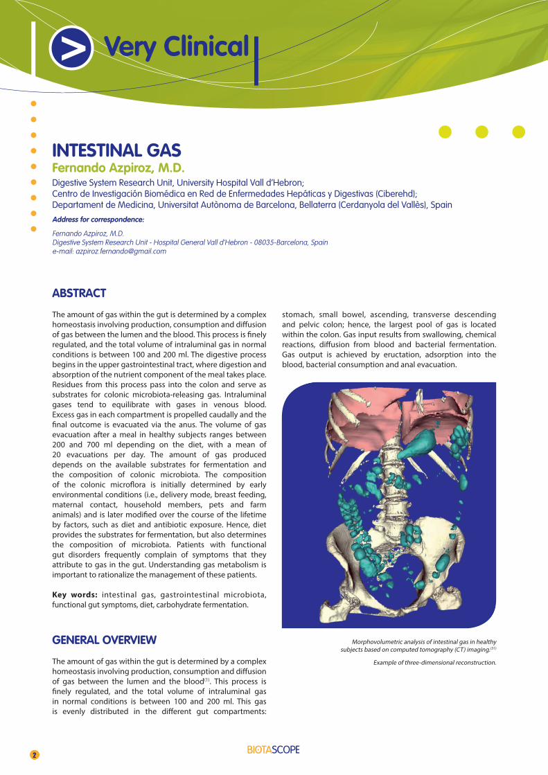
ABSTRACT The amount of gas within the gut is determined by a complex homeostasis involving production, consumption and diffusion of gas between the lumen and the blood. This process is finely regulated, and the total volume of intraluminal gas in normal conditions is between 100 and 200 ml. The digestive process begins in the upper gastrointestinal tract, where digestion and absorption of the nutrient component of the meal takes place. Residues from this process pass into the colon and serve as substrates for colonic microbiota-releasing gas. Intraluminal gases tend to equilibrate with gases in venous blood. Excess gas in each compartment is propelled caudally and the final outcome is evacuated via the anus. The volume of gas evacuation after a meal in healthy subjects ranges between 200 and 700 ml depending on the diet, with a mean of 20 evacuations per day. The amount of gas produced depends on the available substrates for fermentation and the composition of colonic microbiota. The composition of the colonic microflora is initially determined by early environmental conditions (i.e., delivery mode, breast feeding, maternal contact, household members, pets and farm animals) and is later modified over the course of the lifetime by factors, such as diet and antibiotic exposure. Hence, diet provides the substrates for fermentation, but also determines the composition of microbiota. Patients with functional gut disorders frequently complain of symptoms that they attribute to gas in the gut. Understanding gas metabolism is important to rationalize the management of these patients.
Key words: intestinal gas, gastrointestinal microbiota, functional gut symptoms, diet, carbohydrate fermentation.
GENERAL OVERVIEW
The amount of gas within the gut is determined by a complex homeostasis involving production, consumption and diffusion of gas between the lumen and the blood(1). This process is finely regulated, and the total volume of intraluminal gas in normal conditions is between 100 and 200 ml. This gas is evenly distributed in the different gut compartments:
stomach, small bowel, ascending, transverse descending and pelvic colon; hence, the largest pool of gas is located within the colon. Gas input results from swallowing, chemical reactions, diffusion from blood and bacterial fermentation. Gas output is achieved by eructation, adsorption into the blood, bacterial consumption and anal evacuation.
2
> Very Clinical
Morphovolumetric analysis of intestinal gas in healthy subjects based on computed tomography (CT) imaging.(31)
Example of three-dimensional reconstruction.

BIOTASCOPE
In humans, the stomach contains a small chamber of gas. Gas is introduced into the stomach during swallowing, and the excess gas is eliminated by belching or passage into the intestine. Chemical reactions in the duodenum and proximal jejunum, particularly neutralization of alkalis and acids, produce CO2. Gas within the colon is by-and-large determined by the metabolism of colonic bacteria, where residues from the diet not absorbed in the small bowel are fermented releasing hydrogen, CO2, methane, H2S and other gases in very low proportions.
In each compartment of the gut, intraluminal gases tend to equilibrate with gases in venous blood. CO2, hydrogen and methane produced within the gut diffuse rapidly into the blood. Oxygen and nitrogen enter into the gut within swallowed air, but whereas most oxygen is removed from the gut by absorption, nitrogen has a low diffusibility and is poorly absorbed. Excess gas in each compartment is propelled caudally into the next and the final outcome is evacuated via the anus. Due to local homeostasis, the composition of gas varies tremendously in the different compartments of the gut, and the volume and composition of gas evacuated via the anus depends largely on colonic metabolism.
COLONIC GAS METABOLISM
The digestive process begins in the upper gastrointestinal tract, where digestion and absorption of the nutrient component of the meal takes place. Residues from this process pass into the colon and serve as substrates for colonic microbiota favouring its proliferation. While the number of microorganisms in the upper gastrointestinal tract is relatively small, the colon provides an adequate niche and harbours a large population of human microbiota. Furthermore, transit in the colon is slow, allowing for a prolonged interaction between meal-derived substrates and microbiota. In this process, gas is produced depending on the amount of available substrates for fermentation, the composition of microbiota and the time for interaction (i.e., transit time). In contrast to the colon, gas released by fermentation in the upper tract is minimal under normal conditions, due to the low number of microorganisms and the relatively short residency time of substrates.
Passage of fermentable products into the colon
Physiologically, various carbohydrates are incompletely absorbed in the small bowel, pass into the colon and are fermented by microbiota-releasing gas. White rice flour is the only complex carbohydrate that is almost totally absorbed. In contrast, a fraction of the complex carbohydrate in wheat (e.g., pasta, white bread), oats, potatoes and corn resist amylase digestion and are not absorbed in the small bowel(2). Humans are genetically programmed to suppress lactase synthesis after weaning, and hence, become lactose malabsorbers; only a fraction of the worldwide population has changed their genetic program and retains the ability to digest lactose during adulthood(3). Fruits and vegetables (particularly legumes) contain indigestible oligosaccharides, such as stachyose and raffinose, that are not digested in the small bowel, enter the colon and are fermented by microbiota. Fructose, present in soft drinks in large quantities, and also used as a low-calorie sugar substitute may escape small bowel absorption and reach the colon. Fermentable fiber, including hemi glucose, pectin, gums and mucilage, passes into the colon and is a particularly good substrate for microbiota. Mannitol, sorbitol and xylitol, present in soft drinks and dietetic candies, are also incompletely absorbed in the small bowel and reach the colon. Interactions between different food-stuffs may reduce small bowel digestion/absorption: for instance, fiber increases starch malabsorption(4), and a pancreatic amylase inhibitor present in beans reduces intestinal digestion of starch(5). Endogenous mucus may also be fermented by intestinal bacteria(6). To what extent the fermentation of all these various unabsorbed substrates contributes to gas production depends on the composition of colonic microbiota, which exhibits large inter-individual variations.
3
Very Clinical
Unabsorbed residues entering the colon are fermented by gas producing bacteria. Methanogens and sulfate-reducing bacteria consume large amounts of hydrogen. Part of the colonic gases diffuse into the blood and are eliminated by breath; the remainder is evacuated via the anus.
Mean values (+SE) gas distribution along different gut compartments: stomach (ST), small bowel (SB), right (RC), transverse (TC), left (LC) and pelvic colon (PC).(31)
ST SB RC TC LC PC
50 ml
0

BIOTASCOPE
Metabolic activity of colonic microflora
The composition of the colonic microflora is initially determined by early environmental conditions (i.e., delivery mode, breast feeding, maternal contact, household members, pets and farm animals) and is later modified over the course of the lifetime by factors, such as diet and antibiotic exposure(7). Fermentation of undigested substrates involves different metabolic pathways with variable amounts of gas production, primarily carbon dioxide and hydrogen(8); of note, intestinal hydrogen is only derived from bacterial fermentation. Other colonic microorganisms consume large amounts of the hydrogen, and CO2, produced by fermentation(8-10). Intraluminal gases may be consumed following three predominant metabolic routes:a) synthesis of short chain fatty acids by acetogens, b) reduction of CO2 to methane by methanogens orc) reduction of sulfate to sulfide by sulfate-reducing microor-
ganisms(11-13).
Sulfate-reducing bacteria and methanogens compete for hydrogen and do not coexist in the same region; the feces usually contains one or other type of organism. Methanogens tend to locate in the distal colon and sulfate-reducing bacteria colonize the rest of the colon. The distribution depends on the abundance of methanogens. In subjects with a low concentration of methanogenic flora, sulfate-reducing bacteria are present in the whole colon; however, if methanogens are abundant, sulfate-reducing bacteria are confined to the proximal colon(8;11).
The composition of colonic microbiota is influenced by diet(7). For example, it has been shown that anal gas evacuation increases following the initial intake of a prebiotic, but then decreases with regular consumption(14). This adaptation may be related to reduced gas production; i.e. proliferation of organisms that use non-hydrogen releasing fermentative pathways(15) or by up-regulation of gas-consuming activity. A recent study showed that a high-flatulogenic diet increased the relative abundance of methanogens in healthy subjects(16).
Elimination of intestinal gases
Diffusion of gas between the lumen of the gut and the blood depends on the partial pressure of each gas at both sides of the barrier, its diffusibility and the time of exposure to the mucosa. Gases absorbed from each gut compartment into the blood are excreted by breath, where they can be detected by gas chromatography (breath tests). Breath hydrogen excretion after ingestion of non-absorbable carbohydrates, for instance lactulose, varies considerably among individuals, and these inter-individual differences seem related to differences in bacterial hydrogen consumption rather than
differences in production. The fraction of hydrogen that escapes consumption is determined by the type of hydrogen -consuming bacteria. Consumption is markedly enhanced by the presence of methanogens, which consume hydrogen more rapidly than other hydrogen-consuming bacteria. Some data suggest that, if methanogens were present throughout the colon, virtually no hydrogen would escape consumption. Indeed, the inability of some subjects to increase breath hydrogen excretion after intestinal carbohydrate malabsorption(17) probably reflects extremely efficient consumption of hydrogen by methanogens rather than a failure to produce hydrogen. Methanogens are present in most individuals, but in a considerable proportion (about 60%) the concentration is so low that methane is not detectable by breath tests after ingestion of non-digestible, fermentable substrates.
Sulfur-containing gases with characteristic odour(18), have high toxicity, but are efficiently metabolized by the colonic mucosa, preventing absorption into the blood. Allyl methyl sulfide, an odoriferous sulfur-containing gas derived from garlic, does not undergo mucosal metabolism, and is absorbed into the blood and excreted by expired air. Pulmonary elimination of this gas produces the characteristic breath smell related to garlic consumption(19).
Anal gas evacuation
The volume of gas evacuation in healthy subjects after a meal ranges between 200 and 700 ml depending on the diet, with a mean of 20 evacuations per day(16). Anal gas evacuation is determined by the amount of gas production and the amount of intraluminal disposal via mucosal absorption and microbiota consumption. As discussed above, the volume of gas production depends on the amount of fermentable substrates in the colon (i.e., meals and diet), and the composition and metabolic activity of the microbiota. Intracolonic gas disposal by absorption/consumption accounts for about 3/4 of the gas produced, but it is not clear to what extent this is related to absorption or consumption(20). Of note, gas-producing and gas-consuming activities of microbiota are independent, indicating that they are produced by sub-pools that may function independently from each other (20). The different volumes of anal gas evacuation by individuals consuming a standard meal may be related to differences in gas consumption rather than differences in production. Gas absorption, particularly hydrogen, depends on the rate of gas production: at low production a large proportion is absorbed and eliminated by breath, but at higher production the fraction eliminated via the anus increases and this is probably related to a faster gas transit because hydrogen absorption is not saturable(21).
4

BIOTASCOPE
INTESTINAL GAS AND SYMPTOMS
Patients with functional gut disorders, particularly irritable bowel syndrome (IBS) and functional dyspepsia, frequently complain of symptoms that they attribute to gas in the gut. In these patients it is important to identify what exactly they are complaining of, because their management should be adapted to the specific symptom(22).
Repetitive eructation
Belching is due to aerophagia: the patients inadvertently swallow air that accumulates in the hypopharinx or in the stomach, and is then released by belching with a sense of relief(23). These patients frequently have epigastric fullness and dyspeptic-type symptoms that they misinterpret as excessive gas in the stomach. In some cases the process is triggered by emotional distress. Aerophagia usually resolves, or at least improves, with a clear pathophysiological explanation of the symptoms. Underlying dyspeptic symptoms may also require treatment. Specific therapy is advised in case of psychological disorders(23).
Excessive anal gas evacuation
Some patients complain of voluminous gas evacuation or odoriferous flatus, which may become socially disabling. Odour depends on trace elements, such as sulfur-containing gases (e.g., H2S, methanethiol, and dimethyl sulfide) that are produced by sulfate -reducing bacteria in the colon(18). Excessive gas production on a normal diet is usually due to a highly flatulogenic colonic flora because of an increase in gas-producing bacteria, or more likely a deficit of gas-consuming microorganisms. Excessive gas production may be due to diseases that produce malabsorption of nutrients in the small bowel; however, this is relatively rare and easily recognized due to associated clinical manifestations. It has recently been shown that in most patients complaining of excess flatus, colonic gas products are within the normal range, although the number of gas evacuations per anus is increased(16). These data suggest that increased rectal perception may play a role in this condition.
Patients complaining of excessive and/or odoriferous gas evacuation may benefit from a low-flatulogenic diet even if gas production is within the normal range(24). Well tolerated foodstuffs include: meat, fowl, fish and eggs; carbohydrates, gluten-free bread, rice bread, and rice; some vegetables, such as lettuce and tomatoes, and some fruits, such as cherries and grapes. High-flatulogenic foodstuffs include: beans, Brussels sprouts, onions, celery, carrots, raisins, bananas, wheat germ, and fermentable fiber(1). After a one week gas-free diet, these patients usually experience frank symptom relief.
With an orderly reintroduction of other foodstuffs, they should learn to identify their offending meal components. If a strict diet does not reduce gas production, diseases that produce intestinal malabsorption should be investigated.
Rectal gas retention
Some patients complain of impaired anal evacuation and abdominal gas retention. Rectal evacuation is normally produced by a mild abdominal compression associated with a relaxation of the anal sphincters. Some patients are not able to relax the anal canal properly and experience a sensation of anal blockage during attempted evacuation of gas or feces(25). In these patients gas retention is frequently associated with constipation, prolonged duration of colonic fermentation and increased production of gas. In patients with gas retention due to impaired anal evacuation, anal incoordination can be resolved with biofeedback treatment(25), which also resolves fecal retention.
Sensation of abdominal bloating and distension
Abdominal bloating is one of the most frequent and bothersome symptoms in patients with IBS. These patients probably represent a heterogeneous group in which bloating is produced by different combinations of pathophysiological mechanisms, that in most cases are subtle and undetectable by conventional methods(26). It has been consistently shown that IBS patients who complain of bloating have impaired handling of intestinal gas due to abnormal gut reflexes. Intestinal gas, and possibly also other types of gas intestinal contents, may accumulate resulting in segmental pooling and focal gut distension. Additional evidence indicates that these patients also have intestinal hypersensitivity with increased perception of intraluminal stimuli. Recent studies showed that abnormal distension is produced by a paradoxical diaphragmatic contraction, associated with a relaxation of the anterior abdominal wall(27). In these patients, perception of gut symptoms may trigger a conditioned somatic response with abdominophrenic incoordination and distension. Hence, abdominal distension seems to be a somatic manifestation of functional gut disorders.
Since patients with bloating and distention suffer from a variant of IBS, the basic approach to therapy should be similar to that prescribed for IBS(22). In these patients various pathophysiological mechanisms interact to produce their symptoms. A hypersensitive gut may be associated with impaired anal evacuation, particularly in constipation-predominant IBS patients, and symptoms will worsen if gas production is increased by a high-flatulogenic diet or fiber supplements.
5

BIOTASCOPE
A combined treatment strategy should be considered. Experimental studies suggest that mild exercise, a traditional recommendation, facilitates intestinal gas clearance(28). Avoiding high-flatulogenic foodstuffs and fiber overload usually helps, but strict exclusion diets cannot be recommended for the long term. Treatment of constipation improves bloating and distension, possibly by preventing fecal overload. A meta-analysis analyzed the effect of cimetropium, hyoscine, meberine, otiloninum, pinaverium and trimebutine, and concluded that smooth muscle relaxants are superior to placebo in the management of IBS symptoms, specifically in improving abdominal pain and distension(29). The effect of prokinetics, antibiotics and gas-reducing substances has not been clearly established(30). Rifaximin seems to alleviate bloating but its mechanism of action has not been identified; considering the importance of a rich microbiota on gut function this treatment option seems questionable. Recently, the effect of pre- and probiotics on abdominal bloating has been studied, and the initial results are promising. Recent data indicate that abdominal distension may be improved by behavioral techniques.
References
(1) Azpiroz F. Intestinal gas. In: Feldman M, Friedman LS, Brandt LJ, editors. Sleisenger and Fordtran’s. Gastrointestinal and Liver Disease: Pathophysiology, Diagnosis, Management. 10 ed. Canada: Saunders, Elsevier; 2013. p. in press.
(2) Levitt MD, Hirsh P, Fetzer CA, Sheahan M, Levine AS. H2 excretion after ingestion of complex carbohydrates. Gastroenterology 1987 Feb;92(2):383-9.
(3) Yang J, Deng Y, Chu H, Cong Y, Zhao J, Pohl D, et al. Prevalence and presentation of lactose intolerance and effects on dairy product intake in healthy subjects and patients with irritable bowel syndrome. Clin Gastroenterol and Hepatol 2013;11:262-8.
(4) Hamberg O, Rumessen JJ, Gudmand-Hoyer E. Inhibition of starch absorption by dietary fibre. A comparative study of wheat bran, sugar-beet fibre, and pea fibre. Scand J Gastroenterol 1989 Jan;24(1):103-9.
(5) Boibin M, Flourié B Rizza RA, Go VLW, DiMagno EP. Gastrointestinal and metabolic effects of amylase inhibition in diabetics. Gastroenterology 1998;1988:387-94.
(6) Perman JA, Modler S. Glycoproteins as substrates for production of hydrogen and methane by colonic bacterial flora. Gastroenterology 1982;82:911-6.
(7) Wu GD, Chen J, Hoffmann C, Bittinger K, Chen YY, Keilbaugh SA, et al. Linking long-term dietary patterns with gut microbial enterotypes. Science 2011 Oct 7;334(6052):105-8.
(8) Suarez F, Furne J, Springfield J, Levitt M. Insights into human colonic physiology obtained from the study of flatus composition. Am J Physiol 1997 May;272:G1028-G1033.
(9) Strocchi A, Levitt MD. Factors affecting hydrogen production and consumption by human fecal flora. The critical roles of hydrogen tension and methanogenesis. J Clin Invest 1992 Apr;89(4):1304-11.
(10) Gibson GR, Cummings JH, Macfarlane GT, Allison C, Segal I, Vorster HH, et al. Alternative pathways for hydrogen dispossal during fermentation in the human colon. Gut 1990;31:679-83.
(11) Carbonero F, Benefiel AC, Gaskins HR. Contributions of the microbial hydrogen economy to colonic homeostasis. Nat Rev Gastroenterol Hepatol 2012 Sep;9(9):504-18.
(12) Flourie B, Pellier P, Florent C, Marteau P, Pochart P, Rambaud JC. Site and substrates for methane production in human colon. Am J Physiol 1991 May;260(5 Pt 1):G752-G757.
(13) Strocchi A, Furne J, Ellis C, Levitt MD. Methanogens outcompete sulphate reducing bacteria for H2 in the human colon. Gut 1994 Aug;35(8):1098-101.
(14) Azpiroz F, Barba E, Mego M, Bendenzú A, Accarino A, Merino X, et al. Metabolic adaptation of colonic microbiota to diet. United European Gastroenterology Journal 2014;2(5S):A436.
(15) Hertzler SR, Savaiano DA, Levitt MD. Fecal hydrogen production and consumption measurements. Response to daily lactose ingestion by lactose maldigesters. Dig Dis Sci 1997 Feb;42(2):348-53.
(16) Manichanh C, Eck A, Varela E, Roca J, Clemente JC, Gonzalez A, et al. Anal gas evacuation and colonic microbiota in patients with flatulence: effect of diet. Gut 2014;63:401-8.
(17) Cloarec D, Bornet F, Gouilloud S, Barry JL, Salim B, Galmiche JP. Breath hydrogen response to lactulose in healthy subjects: relationship to methane producing status. Gut 1990 Mar;31(3):300-4.
(18) Suarez FL, Springfield J, Levitt MD. Identification of gases responsible for the odour of human flatus and evaluation of a device purported to reduce this odour. Gut 1998 Jul;43(1):100-4.
(19) Suarez F, Springfield J, Furne J, Levitt M. Differentiation of mouth versus gut as site of origin of odoriferous breath gases after garlic ingestion. Am J Physiol 1999 Feb;276(2 Pt 1):G425-G430.
(20) Mego M, Bendezu A, Accarino A, Malagelada JR, Azpiroz F. Intestinal gas homeostasis: disposal pathways. Neurogastroenterol Motil 2015 Jan 4;online.
(21) Christl SU, Murgatroyd PR, Gibson GR, Cummings JH. Production, metabolism, and excretion of hydrogen in the large intestine. Gastroenterology 1992;102:1269-77.
(22) Azpiroz F, Malagelada J-R. Abdominal bloating. Gastroenterology 2005;129:1060-78.
(23) Bredenoord AJ, Smout AJ. Physiologic and pathologic belching. Clin Gastroenterol Hepatol 2007 Jul;5(7):772-5.
(24) Azpiroz F, Hernandez C, Guyonnet D, Accarino A, Santos J, Malagelada JR, et al. Effect of a low-flatulogenic diet in patients with flatulence and functional digestive symptoms. Neurogastroenterol Motil 2014 Feb 19;26(6):779-85.
(25) Azpiroz F, Enck P, Whitehead WE. Anorectal functional testing. Review of a collective experience. Am J Gastroenterol 2002;97:232-40.
(26) Accarino A, Perez F, Azpiroz F, Quiroga S, Malagelada JR. Abdominal distension results from caudo-ventral redistribution of contents. Gastroenterology 2009;136:1544-51.
(27) Burri E, Barba E, Huaman JW, Cisternas D, Accarino A, Soldevilla A, et al. Mechanisms of postprandial abdominal bloating and distension in functional dyspepsia. Gut 2014;63:395-400.
(28) Villoria A, Serra J, Azpiroz F, Malagelada J-R. Physical activity and intestinal gas clearance in patients with bloating. Am J Gastroenterol 2006;101:2552-7.
(29) Poynard T, Regimbeau C, Benhamou Y. Meta-analysis of smooth muscle relaxants in the treatment of irritable bowel syndrome. Aliment Pharmacol Ther 2001;15:355-61.
(30) Azpiroz F, Serra J. Treatment of excessive intestinal gas. Current Treatment Options in Gastroenterology 2004;7:299-305.
(31) Accarino A, Perez F, Azpiroz F, Quiroga S, Malagelada JR. Intestinal Gas and Bloating: Effect of Prokinetic Stimulation. Am J Gastroenterol. 2008 Aug;103(8):2036-42.
6

BIOTASCOPE7
> Very Clinical
ABSTRACT Antibiotics are frequently prescribed drugs, particularly in children. Antibiotic-associated diarrhea (AAD) is a common side effect and is defined as otherwise unexplained diarrhea that occurs in association with the administration of antibiotics. AAD occurs in 11–40% of children between a day of starting antibiotics and up to two months after cessation of treatment. The severity of AAD may range from mild to severe, and serious complications may also occur, such as pseudomembranous colitis. Although no infectious agent is found in most cases, the bacterial agent commonly associated with AAD, particularly in the most severe episodes, is Clostridium difficile. Antibiotics change the balance (diversity and number of bacteria) of intestinal microbiota and can cause the overgrowth of pathogens, resulting in AAD. Treatment modalities for AAD include discontinuing antibiotic treatment and supportive care. AAD might result in an increased length of hospital stay, increased costs of care, and an increased risk of developing other nosocomial infections. There are clinical studies and meta-analyses demonstrating the efficacy of probiotics for the prevention of AAD, and these effects are strain-specific, particularly Lactobacillus GG and Saccharomyces boulardii. The addition of probiotics will prevent one in 7–10 cases of AAD; however, cost effectiveness studies are required to decide the issue of the routine supplementation of probiotics.
Key words: children, antibiotic-associated diarrhea, Clostridium difficile, probiotics.
INTRODUCTION
Antibiotics are the key treatment choice for bacterial infection in the outpatient and hospital setting and are one of the most commonly prescribed drugs in children. In spite of having a clear role for the treatment of infections, short- and long-term complications related to antibiotic use have been described(1). In addition, worldwide overuse and inappropriate use of antibiotics can create life-threatening conditions, as these could result in an increased prevalence of resistant microorganisms(2).
The intestinal microbiota contains more than a thousand different species of microorganisms, and recent research has shown that these microorganisms are integral in the stability of
the anatomical and functional integrity of the gastrointestinal tract(3). Antibiotics have been shown to affect the intestinal microbiota. The adverse effects of antibiotics include various gastrointestinal symptoms including nausea, vomiting, flatulence, abdominal bloating and diarrhea. Antibiotic-associated diarrhea (AAD) is one of the side effects that occur during the use of antibiotics; the incidence varies according to regional and antibiotic groups. AAD diarrhea is unexplained diarrhea occurring between two hours to two months after starting antibiotics, in which diarrhea is defined as more than two unformed stools for ≥2 days(4-5).
In the general population, the incidence of AAD ranges from 5–62% and 11–40% in children; AAD occurs between the initiation of therapy and up to two months after the end of treatment [6]. Turck et al.(7) conducted a clinical study over 11 consecutive months in 650 children aged between one month and 15 years who received antibiotic treatment. In this patient population, the incidence of AAD was 11%, and 68% of these cases occurred during the antibiotic treatment. Vanderhoof et al.(8) reported AAD in 26% of the patients receiving placebo in their placebo‐controlled study evaluating the effects of Lactobacillus GG (LGG) on the prevention of AAD in children. Arvola et al.(9) reported an AAD incidence of 16% in the first two weeks after initiating antibiotic treatment in children treated with oral antibiotics for an acute respiratory infection. In the study conducted by Turck and colleagues(6), the time between the start of antibiotic treatment and the onset of AAD was 5.3 ± 3.5 days, and the duration of AAD was 4.0 ± 3.0 days; none of the patients with AAD required hospitalization. The incidence of these AAD episodes was significantly greater in children younger than two years old. However, a second study conducted in Thailand could not demonstrate an association between younger ages or a high dose of antibiotics and the development of AAD(10).
All antibiotics may elicit AAD, but some antibiotics are associated with a higher risk of AAD. The risk of AAD is irrespective of the dose administrated and the route (oral or parenteral) of administration. The incidence of AAD was 23% for amoxicillin-clavulanate, 9% for cephalosporins, 8% for macrolides, 6% for trimethoprim/sulfamethoxazole and 16% for erythromycin/sulfafurazole according to Turck et al.’s study(6). In the study conducted in Thailand, amoxicillin-
PEDIATRIC ANTIBIOTIC-ASSOCIATED DIARRHEAEner Cagri DinleyiciEskisehir Osmangazi University Faculty of MedicineDepartment of PediatricsTR-26480 Eskisehir, Turkey
Email: [email protected]

BIOTASCOPE
clavulanate was the most commonly prescribed antibiotic, and the incidence of AAD was 16.7% in the amoxicillin/clavulanate group, 6.9% in the amoxicillin group and 11.1% in the erythromycin group(10). Erdeve and colleagues(11) performed a study in 466 children in Turkey, and the AAD rate was 25.6% in children receiving sulbactam-ampicillin; the highest rate of AAD occurred in children younger than six years of age.
PATHOPHYSIOLOGY
There are several potential mechanisms for the pathophysiology of AAD; however, these remain controversial. The development of AAD is related to the mucosal integrity, disrupted intestinal microbiota balance (composition including number and diversity of the microorganisms) and also changes in vitamin/mineral metabolism. In the overwhelming majority of cases, AAD is the result of dysbiosis caused by antibiotics. The most unfavorable enteral effect of antibiotics is the qualitative and quantitative change of gastrointestinal microbiota. A significant reduction in the number of commensal bacteria is the most striking consequence of antibiotics. The reduction of the intestinal strains Bacteroides and Bifidobacterium of the lumen results in increased numbers of facultative anaerobes (Fusobacterium, Clostridia, and Eubacteria). The decrease of commensal bacteria is responsible for the diminished carbohydrate metabolism. Antibiotics disrupt the integrity of the normal colonic mucosa, affect carbohydrate metabolism, and as a result of antimicrobial activity in the colon, lead to the development of osmotic diarrhea and/or pathogenic bacteria associated diarrhea. Antibiotics also reduce the short-chain fatty acids (SCFAs) and the accumulation of non-absorbed carbohydrates in the intestinal lumen. SCFAs, particularly butyrate, are important trophic factors for the colonic mucosa, and their deficiency may cause decreased absorption of electrolytes and water. Some antibiotics, mostly orally administered and poorly absorbing antibiotics, may cause direct damage to the intestinal mucosa with a shortening of villi, which leads to malabsorption. Several antibiotics, such as clindamycin, erythromycin and gentamycin, disturb the transport mechanisms of intestinal epithelial cells. Some antibiotics directly affect the motility of the gastrointestinal tract; for example, erythromycin acts as a motilin-receptor agonist, and clavulanate stimulates small bowel motility. The most important pathogen in AAD is Clostridium difficile, which is a typical nosocomial pathogen. However, data concerning the potential role of C. difficile in AAD in children are contradictory and even more difficult to interpret given that newborns and young infants are often carriers. Less frequently, Staphylococcus aureus, C. perfringens, Klebsiella oxytoca and Salmonella strains may also be detected in AAD(12-16).
CLINICAL FINDINGS
The clinical spectrum of AAD can range from 1–2 days with mild diarrhea to forms with severe or fatal complications, which are mainly related to C. difficile infection(17). Symptoms and signs can also occur later, up to six weeks after the discontinuation of the treatment. AAD does not seem to correlate with the duration of antibiotic therapy, and symptoms may start even on the first day of intake(18). In most cases, diarrhea with loose or watery stools lasts a few days without any severe general symptoms, and the patients recover without complications. In moderate cases, abdominal cramping, fever, leukocytosis, hypoalbuminemia, fecal leukocytosis and colonic thickening may be observed(19). C. difficile accounts for the majority of cases of colitis in AAD and pseudomembranous colitis in severe cases; however, the actual incidence is not known in children. Severe diarrhea, liquid stool with mucus and blood, fecal leucocytes >5/high power field, altered flora and the presence of gram-positive bacilli with oval subterminal spores were sensitive predictors for the diagnosis of C. difficile infection. Barakat et al.(20) evaluated children with antibiotic-associated bloody diarrhea and used sigmoidoscopy to reveal varying types of erythema (patchy, ring, diffuse), ulcers (aphthoid, diffuse) and only 26% of pseudomembrane formation. Acute bloody diarrhea was stopped in all patients 2 to 6 days after the discontinuation of antibiotics.
PREVENTION
The beneficial effects of probiotics for the prevention of AAD have been shown in children. The effects of probiotics which are thought to prevent AAD include the regulation/restoration of intestinal microbiota, ensuring the continuity of carbohydrate fermentation, competition with opportunistic pathogenic microorganisms, inhibiting the growth of microorganisms, stimulating the immune system, blocking pathogenic colonization and increasing the production of mucin in the intestine, thus contributing to the prevention of epithelial barrier function(16, 17, 22). The mechanism of action of probiotics is not fully elucidated, and these effects are strain specific.
Szajewska et al.(22) performed a meta-analysis for the effects of probiotics in the prevention of AAD in children; they evaluated only randomized, double-blind, placebo-controlled clinical trials. Probiotics decreased the risk of AAD from 28.5% to 11.9% (risk ratio [RR] 0.44; 95% confidence interval [CI]: 0.25 to 0.77), and a subgroup analysis showed that the reduction of the risk of AAD was associated with the use of LGG, S. boulardii, and a combination of B. lactis and S. thermophilus. For every seven children that will develop diarrhea while being treated
8

BIOTASCOPE
with antibiotics, one fewer will develop AAD if they also receive probiotics. McFarland(23) evaluated six randomized controlled trials with LGG and AAD and reported a relative risk of 0.31, whereas the probiotic mixtures containing other bacteria in the same analysis displayed a relative risk of 0.51.
S. boulardii (250 mg/day) was associated with a lower prevalence of diarrhea occurring during or up to two weeks after the antibiotic therapy in children with otitis media and/or respiratory tract infection. S. boulardii also reduced the risk of AAD, which was caused either by C. difficile or of an unknown etiology, compared with placebo(24). Another randomized controlled trial showed that S. boulardii had a reduced risk of diarrhea (6.2% vs 18% of patients; RR 0.3; 95% CI: 0.2 to 0.5) in children receiving ampicillin-sulbactam or azithromycin(11). In a meta-analysis of S. boulardii, McFarland(17) evaluated 10 randomized controlled trials and reported an AAD relative risk of S. boulardii of 0.47 in the group. Although yogurt containing probiotic bacteria and food is often consumed, Conway et al.(25) showed that the supplementation of Lactobacillus containing yogurt has no effect on the development of AAD.
In 2011, a Cochrane analysis assessed the effect of probiotics on AAD among 2941 patients (the majority of the studies were conducted in ambulatory outpatients). The administration of probiotics along with antibiotics was shown to provide a significant reduction in the incidence of AAD(26). A Cochrane review reporting the incidence of diarrhea in nine studies in children suggested that probiotics are effective for preventing AAD (RR 0.49; 95% CI: 0.32 to 0.74)(26). The effects were particularly noticeable in patients receiving beta lactam and penicillin (73% reduction), whereas there was a 51% reduction in patients receiving cephalosporins and a 50% reduction in the macrolide group, which were not significantly different. For every 10 treated patients, one case of diarrhea can be prevented (number needed to treat 10; 95% CI: 7 to 18). Regarding safety, no trials reported a serious adverse event although only half of the trials included had reported adverse events(26).
In 2014, McFarland(27) evaluated the strain specific effects of probiotics on the prevention of AAD. This meta-analysis indicated that a significant protective efficacy for AAD was found when the 16 different types of probiotics were combined (RR 0.43); there was also a significant reduction of pediatric C. difficile infection (RR 0.34). S. boulardii and LGG showed a significant efficacy for pediatric AAD (RR for S. boulardii was 0.43 and RR for LGG was 0.44).
TREATMENT
Most of the AAD cases responded only to the discontinuation or change of the antibiotic. The implicated antibiotic should be discontinued or changed if required, and supportive management with fluid and electrolytes should also be provided(4). Oral metronidazole, oral vancomycin or fecal transplantation are the treatment choices for C. difficile infection(28). Currently, Shan et al.(29) showed that 5 days of S. boulardii treatment had beneficial effects for AAD treatment (lower stool frequency and a shorter duration of diarrhea) and the prevention of AAD. This study shows promising results for the prevention of AAD in children and in the treatment of AAD.
GUIDELINES
In 2011, the Third International Panel of Yale Workshop on Probiotics identified grade A evidence for the prevention of AAD in outpatient and hospitalized patients and B/C evidence for the prevention of CDAD(30). The practice guidelines of the World Gastroenterology Organization (WGO) stated that there was strong evidence of efficacy in the prevention of AAD in children with S. boulardii or LGG (evidence level: 1A and 1B, respectively). The Bifidobacterium lactis Bb12 + S. thermophilus combination is also effective in the prevention of AAD in children (evidence level 1B)(31).
CONCLUSION
AAD, in addition to having a negative effect on the quality of life, increases the length of hospital stay and costs. Generally, antibiotic treatment is stopped or changed when it occurs in inpatient or outpatient children and this also affects the success of the treatment and might lead to a prolonged hospital stay. Long-term hospitalization, particularly in the intensive care unit, might result in an increased risk for nosocomial infection and mortality. AAD causes anxiety for parents, which is justified by the risk of dehydration associated with AAD, particularly in children younger than 2 years. In outpatient settings, antibiotic treatment was stopped by the parents when AAD was observed, and second line antibiotic treatments for these children are broader than first line antibiotic treatments. The addition of probiotics can prevent one in 7–10 cases of AAD; however, cost effectiveness studies are required to decide the issue of routine supplementation of probiotics.
9

BIOTASCOPE
References
(1) Cox LM, Blaser MJ. Antibiotics in early life and obesity. Nat Rev Endocrinol. 2015;11(3):182-190.
(2) Davies J, Davies D. Origins and evolution of antibiotic resistance. Microbiol Mol Biol Rev. 2010;74(3):417-33.
(3) Versalovic J. The human microbiome and probiotics: implications for pediatrics. Ann Nutr Metab. 2013;63(Suppl 2):42-52.
(4) Alam S, Mushtaq M. Antibiotic associated diarrhea in children. Indian Pediatr. 2009;46(6):491-6.
(5) Wiström J, Norrby SR, Myhre EB, Eriksson S, Granström G, Lagergren L, et al. Frequency of antibiotic-associated diarrhoea in 2462 antibiotic treated hospitalized patients: a prospective study. J Antimicrob Chemother. 2001;47(1):43-50.
(6) Johnston BC, Goldenberg JZ, Vandvik PO, Sun X, Guyatt GH. Probiotics for the prevention of pediatric antibiotic-associated diarrhea. Cochrane Database Syst Rev. 2011;9(11):CD004827.
(7) Turck D, Bernet JP, Marx J, Kempf H, Giard P, Walbaum O, et al. Incidence and risk factors of oral antibiotic-associated diarrhea in an outpatient pediatric population. J Pediatr Gastroenterol Nutr. 2003;37(1):22-6.
(8) Vanderhoof JA, Whitney DB, Antonson DL, Hanner TL, Lupo JV, Young RJ. Lactobacillus GG in the prevention of antibiotic-associated diarrhea in children. J Pediatr. 1999;135(5):564-8.
(9) Arvola T, Laiho K, Torkkeli S, Mykkänen H, Salminen S, Maunula L, et al. Prophylactic Lactobacillus GG reduces antibiotic-associated diarrhea in children with respiratory infections: a randomized study. Pediatrics. 1999;104(5):e64.
(10) Damrongmanee A, Ukarapol N. Incidence of antibiotic-associated diarrhea in a pediatric ambulatory care setting. J Med Assoc Thai 2007;90(3):513-7.
(11) Erdeve O, Tiras U, Dallar Y. The probiotic effect of Saccharomyces boulardii in a pediatric age group. J Trop Pediatr. 2004;50(4):234-6.
(12) Högenauer C, Hammer HF, Krejs GJ, Reisinger EC. Mechanisms and management of antibiotic-associated diarrhea. Clin Infect Dis. 1998;27(4):702-10.
(13) Hickson M. Probiotics in the prevention of antibiotic-associated diarrhoea and Clostridium difficile infection. Therap Adv Gastroenterol. 2011;4(3):185-97.
(14) Bartlett JG. Antibiotic-associated diarrhea. N Engl J Med. 2002;346(5):334-9.
(15) Britton RA, Young VB. Interaction between the intestinal microbiota and host in Clostridium difficile colonization resistance. Trends Microbiol. 2012;20(7):313-9.
(16) Doron SI, Hibberd PL, Gorbach SL. Probiotics for prevention of antibiotic-associated diarrhea. J Clin Gastroenterol. 2008;42(Suppl 2):S58-63.
(17) McFarland LV. Systematic review and meta-analysis of Saccharomyces boulardii in adult patients. World J Gastroenterol. 2010;16(18):2202-22.
(18) Mylonakis E, Ryan ET, Calderwood SB. Clostridium difficile—Associated diarrhea: A review. Arch Intern Med. 2001;161(4):525-33.
(19) Bartlett JG, Gerding DN. Clinical recognition and diagnosis of Clostridium difficile infection. Clin Infect Dis. 2008;46 (Suppl 1):S12-8.
(20) Barakat M, El-Kady Z, Mostafa M, Ibrahim N, Ghazaly H. Antibiotic-associated bloody diarrhea in infants: clinical, endoscopic, and histopathologic profiles. J Pediatr Gastroenterol Nutr. 2011;52(1):60-4.
(21) Dinleyici EC, Kara A, Ozen M, Vandenplas Y. Saccharomyces boulardii CNCM I-745 in different clinical conditions. Expert Opin Biol Ther. 2014;14(11):1593-609.
(22) Szajewska H, Ruszczyński M, Radzikowski A. Probiotics in the prevention of antibiotic-associated diarrhea in children: a meta-analysis of randomized controlled trials. J Pediatr. 2006;149(3):367-72.
(23) McFarland LV. Meta-analysis of probiotics for the prevention of antibiotic associated diarrhea and the treatment of Clostridium difficile disease. Am J Gastroenterol. 2006;101(4):812-22.
(24) Kotowska M, Albrecht P, Szajewska H. Saccharomyces boulardii in the prevention of antibiotic-associated diarrhoea in children: a randomized double-blind placebo-controlled trial. Aliment Pharmacol Ther. 2005;21(5):583-90.
(25) Conway S, Hart A, Clark A, Harvey I. Does eating yogurt prevent antibiotic-associated diarrhoea? A placebo-controlled randomised controlled trial in general practice. Br J Gen Pract. 2007;57(545):953-9.
(26) Johnston BC, Goldenberg JZ, Vandvik PO, Sun X, Guyatt GH. Probiotics for the prevention of pediatric antibiotic-associated diarrhea. Cochrane Database Syst Rev. 2011;9(11):CD004827.
(27) McFarland LV. Deciphering meta-analytic results: a mini-review of probiotics for the prevention of paediatric antibiotic-associated diarrhoea and Clostridium difficile infections. Benef Microbes. 2014 Jun 2 [Epub ahead of print].
(28) Cammarota G, Ianiro G, Gasbarrini A. Fecal microbiota transplantation for the treatment of Clostridium difficile infection: a systematic review. J Clin Gastroenterol. 2014;48(8):693-702.
(29) Shan LS, Hou P, Wang ZJ, Liu FR, Chen N, Shu LH, et al. Prevention and treatment of diarrhoea with Saccharomyces boulardii in children with acute lower respiratory tract infections. Benef Microbes. 2013;4(4):329-34.
(30) Floch MH, Walker WA, Madsen K, Sanders ME, Macfarlane GT, Flint HJ, et al. Recommendations for probiotic use-2011 update. J Clin Gastroenterol. 2011;45(Suppl):S168-71.
(31) Guarner F, Khan AG, Garisch J, Eliakim R, Gangl A, Thomson A, et al. World Gastroenterology Organisation Global Guidelines: probiotics and prebiotics October 2011. J Clin Gastroenterol. 2012;46(6):468-81.
10

BIOTASCOPE11
> Very Basic
ABSTRACT Animal studies have shown that gut microbes play key roles in nutrition, metabolism and immune function, and have widespread influence beyond the gastrointestinal tract. Understanding the impact of gut microbial communities on human health is widely perceived as one of the most exciting advancements in biomedicine in recent years. Large-scale research projects (Human Microbiome Project, MetaHIT, etc.) are providing novel insights on the structure and function of the microbial communities in the human gut. The field is progressing rapidly owing to the availability of high-throughput molecular sequencing techniques combined with powerful bioinformatics for taxonomic identification and comparative analysis of datasets. Such studies have pointed out that loss of biodiversity in the human gut microbiota is associated with far reaching consequences on host health. Perturbed gut microbial colonization might be the origin of some chronic non-communicable diseases of increasing incidence in modern society, including metabolic, inflammatory and neoplastic disorders. Further understanding of the importance of developing and maintaining gut microbiota diversity may lead to targeted interventions for health promotion, disease prevention and management. Diet, functional foods and gut microbiota transplantation are the principal tools utilized in clinical practice for improving host-microbial symbiosis, and warrant further investigation for their ability to restore microbial richness in various disease states.
Key words: microbiota, antibiotics, metagenomics, symbiosis, 16SrRNA gene.
Key questions:
1 - Are gut microbes relevant for human health?
2 - What do we know about the structure and function of the human gut microbiota?
3 - Are changes in microbiota composition associated with human disease?
4 - Can we improve host-microbe relationships for health outcomes?
GLOSSARY OF TERMS
Dysbiosis: an imbalance of the normal gut microbiota composition.
Enterotype: a classification of the human gut microbial communities into three groups or types, on the basis of the bacteriological composition of the ecosystem (diversity and abundance of the predominant genera).
Metagenome: the total genetic content of the combined genomes of the constituents of an ecological community.
Metagenomics: the study of all the genetic material recovered directly from environmental samples bypassing the need to isolate and culture individual community members.
Metatranscriptomics: the study of all actively transcribed ribosomal and messenger RNA from a community as a whole.
Microbiome: the collective genome of the microbial symbionts in a host animal.
Microbiota: the collection of microbial communities colonizing a particular ecological niche.
Operational taxonomic unit (OTU): definition of a species in bacteriology based on 16S rRNA sequence similarity (see phylotype).
Pathobionts: microbial partners that under normal circumstances live as symbionts but have the potential to cause disease in the host.
Phylotype: a microbial group defined by 16S rRNA sequence similarity rather than by phenotypic characteristics. A similarity of approximately 97% indicates a species-level.
Symbionts: the microbial partners in symbiosis.
Symbiosis: close and persistent interactions between living organisms of different species. Biological interactions may be mutualistic (both partners derive a benefit), commensalistic (one partner benefits without affecting the other), or parasitic (one benefits while the other is harmed).
DECIPHERING THE HUMAN GUT MICROBIOMEFrancisco Guarner, MD, PhDDigestive System Research Unit, University Hospital Vall d’Hebron, Passeig Vall d’Hebron, 119-129; 08035 Barcelona, Spain
Email: [email protected]
TAKE-HOME MESSAGES- Symbiosis between microbes and the animal host is critical for normal growth and development and for maintaining lifelong health.- New sequencing technologies and powerful bioinformatic tools allow the description of the full spectrum of the microbial communities that inhabit the human
intestinal tract, as well as their functional contributions to host health.- The composition of a “healthy” human gut microbiota remains to be elucidated, but some optimal characteristics of the microbial communities may be depicted: • Functional profile (metabolic and trophic provisions) • Compositional profile (diversity of species) • Ecologic stability (resistance to stress and resilience)- Loss of gut bacterial richness is associated with intestinal inflammation and may play a role in the chronic diseases observed in developed societies.

BIOTASCOPE
LIVING IN A MICROBIAL WORLD
Bacteria have been on Earth for 3.5 billion years, appearing approximately 1 billion years after the Earth’s crust was formed(1). Fossils and associated geochemical markers of biologic activity indicate that microbial organisms inhabited the oceans in Archean times, and that Cyanobacterium resembling cells were the origin of free oxygen gas in the atmosphere. Today, microbial communities are ubiquitous and truly essential for maintaining the conditions for life on Earth. Because of their enormous global size, microbial communities have a massive impact across the globe and their contributions affect many aspects of life, not only human or animal infections, but mostly the cycling of the critical elements for maintaining life. Generation of atmospheric gases, synthesis of organic materials from inorganic sources, corruption of organic to inorganic materials, corrosion, degradation, and bioremediation are vital ecological functions for global carbon, oxygen and nitrogen cycles, which are the critical cycles for life on Earth.
Animals appear in the fossil record much later than bacteria, during the Cambrian period about 600 million years ago. Not surprisingly, animals co-evolved in permanent association with microbial communities maternally inherited at birth or acquired from the environment. Permanent associations that evolve over long periods of time are usually beneficial for all partners, host and microbes, and are grouped under the term ‘symbiosis’ (the microbial partners are called ‘symbionts’). Animals provide habitat and nutrients whereas microbes contribute to their body functions. For mammals, the genes encoding enzymes for biosynthesis of essential amino acids or vitamins were lost early in evolution. Microbial symbionts have, through evolution, adapted to provide the required organic compounds (essential amino acids and vitamins) and the ability to obtain energy from different sources(2). For instance, in ruminants eating poorly digestible low protein diets the amino acid supply largely depends on the microbial activities in their fore-stomachs.
Chronic microbial colonization that inflicts no evident harm on the host has only attracted minor attention during the past century. It was recognized on the basis of observations from microscopy, but most aspects of symbiont origins and functions remained unexplored before the age of molecular techniques because of the difficulties in the cultivation and isolation of a large majority of these microbial species. Development of novel gene sequencing technologies as well as availability of powerful bioinformatic analysis tools have allowed a dramatic proliferation of research studies on symbiont communities over the past few years.
THE HUMAN GUT MICROBIOTA
The term microbiota refers to the collection of microbial communities colonizing a particular ecological niche, and the microbiome is the collective genome of the microbial symbionts in a given host. Microbial colonizers are not casual bystanders, or potential invaders when host immunity is compromised. Gut microbial communities constitute an important functional part of animals. This was clearly proven some decades ago by experiments using axenic (germ-free) rodents and birds(3).
Animals bred under germ-free conditions have extraordinary nutritional requirements and are highly susceptible to infections.
Germ-free animals have increased nutritional requirements in order to sustain body weight, are highly susceptible to infections and show structural and
functional deficiencies. Reconstitution of germ-free animals with a microbiota restores most of these deficiencies, suggesting that gut bacteria provide
important and specific tasks to the host’s homeostasis.
Organ weights (e.g., heart, lung, and liver), cardiac output, intestinal wall thickness, gastrointestinal motility, serum gamma-globulin levels, and lymph nodes, among other characteristics, are all reduced or atrophic in germ-free animals. Germ-free mice display greater locomotor activity and reduced anxiety when compared with mice with a conventional
12
REDUCED:• ORGAN WEIGHT
(E.G. HEART, LIVER, LUNGS)• CARDIAC OUTPUT,
OXYGEN CONSUMPTION• EXPRESSION OF GROWTH
FACTORS IN CNS• INTESTINAL MOTILITY• MESENTERIC AND
SYSTEMIC LYMPH NODES• SERUM IMMUNOGLOBULIN LEVELS
INCREASED:• FOOD INTAKE TO SUSTAIN
BODY WEIGHT • SUSCEPTIBILITY TO INFECTION• ATTENTION DEFICIT
HYPERACTIVITY DISORDER
STRUCTURAL AND FUNCTIONAL RECOVERY:• PROTECTIVE EFFECTS• METABOLIC EFFECTS• TROPHIC EFFECTS
RECONSTITUTION WITH MICROBIOTA
GERM-FREE MICE

BIOTASCOPE
gut microbiota. Reconstitution of germ-free animals with a microbiota restores most of these deficiencies, suggesting that gut microbes provide important and specific tasks for host homeostasis. Most interestingly, fecal transplants can transfer disease phenotypes such as obesity(4), insulin resistance(5), intestinal inflammation(6), and anxiety(7). Thus, microbial colonization of animals is critical for nutrition, body growth, induction and regulation of immunity, endocrine homeostasis, maturation of the central nervous system, and even, behavior.
Human beings are also associated with large and diverse microbial populations that live on body surfaces and in cavities connected with the external environment. In humans, microorganisms colonize all epithelial surfaces, but the gastrointestinal tract harbors the largest microbial population. The human gastrointestinal tract houses around two hundred trillion microbial cells, most of them belonging to the domain Bacteria.
The human gastrointestinal tract is the organ system responsible for digesting foodstuffs, absorbing nutrients, and expelling waste. The stomach and small bowel perform most of the digestive and absorptive tasks in around 3 to 4 hours. Then, the ‘waste’ is retained in the large bowel for an average of 2 days, under perfect conditions for feeding microbes. Thus, the human colon is by far the largest ecological niche for microbial communities in the human body, and harbors billions of microbial cells per gram of luminal contents. Several hundred grams of microbes living in the large bowel should affect host physiology and pathology in different ways; this is currently the focus of extensive research in order to fully understand their impact in medicine. Our knowledge on gut bacteria was largely limited to community members with potential pathogenicity by either translocation or production of toxins. However, we do not know which microbes provide functional contributions, and how do they work? This knowledge is essential for improving symbiosis between the host and guests.
GUT MICROBIAL COMMUNITIES
Large-scale research projects have been aimed at deciphering the structure and function of the human microbiota, namely the National Institutes of Health’s (NIH) Human Microbiome Project (HMP) and the European MetaHIT project. Thanks to the advances in sequencing technologies as well as in the bioinformatic tools needed to analyze massive amounts of data, those projects, as well as other research initiatives, are providing a deeper insight on the microbial communities
that inhabit the human body, and allow the identification of changes that are associated with disease states.
The novel approach for the analysis of microbial communities in environmental or biological samples is called “metagenomics”, and is defined as the study of all the genetic material recovered directly from the sample, by-passing the need to isolate and culture individual community members.
13
Profiling the human gut microbiota. On the left side, see the approaches used when culture of an individual microorganism or the amplification of its genome is conceivable. On the right side, when most of the bacteria in the sample are not cultivable, approaches including metagenomics and metatranscriptomics are applied to the whole microbial community in the sample in order to collect information regar-ding microbial diversity, gene content and gene expression.
(Source: from figure 2 in: Manichanh, C. et al.,The gut microbiota in IBD. Nat Rev Gastroen-terol Hepatol. 2012 Oct;9(10):599-608.(26))

BIOTASCOPE
Bacterial composition in the lumen varies from cecum to rectum, while the community of mucosa-associated bacteria is highly stable from terminal ileum to large bowel in a given individual. Factors such as diet, drug intake, travelling, and colonic transit time have an impact on microbial composition of fecal samples over time in a unique host(9, 10). While intra-individual fluctuations in the composition of the microbiota can be remarkable, the
microbial ecosystem tends to return to its typical compositional pattern. Most strains are resident for decades in a given individual. Microbial diversity changes with age, increasing from infancy to adulthood and decreasing in the elderly, particularly in centenarian individuals. There are striking differences in composition and diversity between Westernized and non-Westernized populations. The fecal microbiota of adults is less diverse in metropolitan areas
14
The metagenome is the collective genetic content of the combined genomes of the constituents of an ecological community. The standard procedure consists of the extraction of DNA from the sample, followed by amplification of the small subunit ribosomal RNA gene (16S rRNA) with universal primers, and sequencing of all copies of the gene in the sample. The 16S rRNA gene is present in all prokaryotic cells (Bacteria and Archea) and contains both conserved and variable regions. Similarities and differences in the sequence of nucleotides of the 16S rRNA gene allow taxonomic identification ranging from the domain and phylum level to the species level. Currently, around 3 million aligned and annotated 16S rRNA sequences are available in DNA databases (http://rdp.cme.msu.edu/). Taxonomic identification is based on comparison of
16S rRNA sequences in the sample with reference sequences in the database. In this way, studies on the 16S rRNA gene provide information about microbial composition in a given sample, i.e. diversity and relative abundance of community members.
Studies have highlighted that only 7 to 9 of the 55 phyla of the domain Bacteria are detected in fecal or mucosal samples from the human gut. Around 90% of all taxa belong to just two phyla: Bacteroidetes and Firmicutes. Other phyla that have been consistently found in the human distal gut are Proteobacteria, Actinobacteria, Fusobacteria, and Verrucomicrobia(8). At genus level, Bacteroides, Faecalibacterium and Bifidobacterium are the most abundant, but their relative proportion is highly variable across individuals. Only very few species of Archea (mostly Methanobrevibacter smithii) are represented.
Genus abundance variation box plot for the 30 most abundant genera of the human gut microbiota as determined by metagenomic sequencing of human fecal samples. Genera are colored by their respective phylum (see inset for color key). Inset shows phylum abundance box plot (Source: from Figure 1b in: Arumugam M et al, Enterotypes of the human gut microbiome. Nature. 2011;473(7346):174-80)(12).

BIOTASCOPE
of North America than in rural non-Westernized populations of Africa and South America(11).
At strain level, each individual harbors a distinctive pattern of microbial communities. However, network analysis of fecal communities at genus level across different individuals suggested that the microbial ecosystem conforms well-balanced microbial symbiotic states driven by groups of co-occurring genera. Multidimensional scaling and principal coordinates analysis of samples from American, European, and Japanese subjects revealed that all individual samples gathered around three robust clusters according to their similarity in composition. Clustering was not driven by age, gender, nationality, or body mass index. These clusters were designated as ‘enterotypes’ (12). Each enterotype is identifiable by variation in the levels of one of three genera: Bacteroides (enterotype 1), Prevotella (enterotype 2), and Ruminococcus (enterotype 3).
Enterotype partitioning suggests the existence of a limited number of well-balanced host-microbial symbiotic states. The discreteness of these balanced states suggests that the fundamental structure of the human gut microbiota is primarily determined by interactions within the community members. Genome size and number of coding genes are much smaller in prokaryotes than in eukaryotes. Thus, single microbial species do not have enough genetic resources on their own, and are likely to have obligate dependencies on other species. Therefore, multispecies communities with complex nutritional and social interdependencies are the natural lifestyle for most prokaryotes.
The clinical implications of enterotypes are under investigation. A study exploring the associations between diet and gut microbiota composition, based on food frequency questionnaires collected over long periods, indicated that diet affects the proportions of Prevotella versus Bacteroides in Western populations(13). The Bacteroides enterotype was associated with diets enriched in protein and fat. In contrast, the Prevotella enterotype was linked to diets with predominance of carbohydrates and sugars. Thus, the presence of stable gut microbial communities may be linked to long-term dietary patterns.
FUNCTIONAL GENOMICS
The molecular approach is not limited to 16S rRNA sequencing. The decreasing cost and increasing speed of DNA sequencing, coupled with advances in computational analyses of large datasets, have made it feasible to analyze entire genomes. The resulting information describes the collective genetic content of the community from which functional and metabolic networks can be inferred. Importantly, whole-genome sequencing provides information about nonbacterial members in the community, including viruses, yeasts, and protists. Full metagenomic
analysis of human fecal samples has identified up to 10 million nonredundant microbial genes(14). A large majority (95%) of the identifiable genes are bacterial, with a small proportion of virus-like or eukaryotic genes. Each individual carries an average of 600,000 nonredundant microbial genes in the gastrointestinal tract, and around 300,000 genes are common, in the sense that are present in about 50% of individuals(14, 15).
Functional screening relies on sequencing all genetic material in the community, including taxonomically unknown members, and matching the sequences to known functional genes. Such studies have generated fascinating information about functions within the microbial communities of the human gut. The extensive nonredundant catalogue of microbial genes encodes groups of proteins engaged in up to 20,000 biological functions related to life in the intestinal habitat(15). Some functions are common to free-living bacteria, like the main metabolic pathways (e.g. central carbon metabolism, amino-acid synthesis), and some important protein complexes (i.e. RNA and DNA polymerases, ATP synthase, general secretory apparatus). Some other gene clusters encode functions that may be especially important for microbial life within the gut, such as those involved in adhesion to the host proteins (i.e. collagen, fibrinogen, fibronectin) or in harvesting sugars from the glycolipids secreted by epithelial cells.
Interestingly, despite the highly divergent compositions of gut microbiota across individuals in terms of taxonomy, functional gene profiles are rather similar in healthy subjects. Most functional pathways are common and expressed in similar abundance among fecal microbiotas from different human individuals(16). Such data imply that there is functional redundancy across taxonomic diversity, i.e. same or similar functional pathways are present in different microbial species. This concept is likely to be very relevant for a definition of a ‘normal’ or ‘healthy’ gut microbial ecosystem in humans: functional profiling may eventually become the optimal approach rather than listing of species or strains.
ANTIBIOTICS, DYSBIOSIS, AND RISK OF DISEASE
Every day, 10 to 30 out of 1,000 inhabitants of developed countries consume a defined daily dose of antibiotics as ambulatory patients(17). Although most courses of antibiotics result in no immediate signs or symptoms, there is a concern that altering the composition of the microbiota will interfere with some of its functions. Antibiotic-associated diarrhea is the most commonly recognized complication of antibiotics, and develops in 15% to 25% of patients receiving antibiotics. Most episodes of diarrhea induced by antibiotics are mild and self-limiting, resolving within a few days. However, an increasing number of cases develop more severe forms, including Clostridium difficile-associated diarrhea. Antibiotics directed against the opportunistic pathogen can decrease the load of the pathogen and toxin production, inducing clinical remission.
15

BIOTASCOPE
However, if the microbiota is unable to restore microbial homeostasis preventing the overgrowth of C. difficile, the patient will develop recurrent episodes of infection(18). In such cases, fecal microbiota transplantation is an acceptable treatment method with high cure rates(19). This disorder is the perfect paradigm for how antibiotics can disturb the protective function of the gut microbiota in humans, and underscores the need for restoring the microbial ecosystem for a definitive cure.
Use of antibiotics induces a decrease in microbial diversity (i.e. loss of richness in the ecosystem) and overgrowth of resistant species, which may even result in an overall increase of microbial load(20). Perturbations of the gut microbial ecosystem during infancy, combined with genetic susceptibility, may have a long-lasting impact on the immune system, leading to disease or predisposition to disease later in life. Indeed, it has been shown that repeated use of antibiotics during infancy may increase the risk of inflammatory bowel diseases(21), metabolic disorders(22), and atopic diseases(23).
Pathologies such as C. difficile-associated diarrhea, inflammatory bowel diseases, some functional bowel disorders, obesity, type 2 diabetes, nonalcoholic steatohepatitis, advanced chronic liver disease, and others have been linked to changes in the composition of the gut microbiota(23, 24). Consistency among studies is still poor for some of these examples, possibly because of lack of fully standardized methodology. In addition, such associations do not necessarily indicate a causative role for the microbiota in the pathogenesis of a disease, as they could instead be a consequence of the disease. Follow-up studies and, particularly, intervention studies aimed at restoring the normal composition of the gut microbiota are needed.
Richness of the gut microbial ecosystem appears to be a critical characteristic for a healthy gut microbiota. Low diversity is associated with an imbalance between pro- and anti-inflammatory species, and may trigger host inflammation. Microbial gene counts can be used as an accurate biomarker of microbial diversity or richness, as this strategy can assess the presence and abundance of genes from known as well as unknown taxa, including not only bacteria but also viruses and eukaryotes. Interestingly, individuals with low microbial gene counts (below 480,000) are characterized by more marked overall adiposity, insulin resistance, leptin resistance, dyslipidemia and a more pronounced inflammatory phenotype when compared with high gene count individuals(25). Moreover, several metabolic parameters were slightly altered in otherwise healthy individuals with low microbial gene counts. Obese individuals with low gene counts gained more weight over time and had a propensity towards metabolic comorbidities. Low diversity appears to be a risk factor for the development of metabolic syndrome (e.g. type 2 diabetes, dyslipidemia, steatohepatitis).
From a functional point of view, low diversity is associated with a reduction in butyrate-producing bacteria, increased mucus degradation potential, reduced hydrogen and methane production combined with increased hydrogen sulfide formation. The gene-poor microbiota thus appears to be less healthy(25).
In conclusion, molecular studies provide an in depth insight into the microbial communities that inhabit the human gut, and allow the identification of changes that are associated with disease. A better knowledge of the contributions of microbial symbionts to host health will certainly help in the design of novel interventions to improve symbiosis and combat disease.
16
Reduced diversity of species in the fecal microbiota from patients with Crohn’s disease. The vertical axis shows numbers of dominant species (OTUs) identified in the fecal microbiota of healthy subjects and patients with Crohn’s disease. A substantial reduction of species belonging to Firmicutes class has been consistently found in patients with Crohn’s disease.
(Source: from figure 4 in: Manichanh, C. et al., The gut microbiota in IBD. Nat Rev Gastroenterol Hepatol. 2012 Oct;9(10):599-608.(26))
BIOTASCOPE
References
(1) Schopf JW. Microfossils of the Early Archean Apex chert: new evidence of the antiquity of life. Science. 1993;260:640-6.
(2) Moran NA. Symbiosis. Curr Biol. 2006;16(20):R866-71.
(3) Wostmann BS. The germfree animal in nutritional studies. Ann Rev Nutr. 1981;1:257-79.
(4) Ridaura VK, Faith JJ, Rey FE, Cheng J, Duncan AE, Kau AL, et al. Gut microbiota from twins discordant for obesity modulate metabolism in mice. Science. 2013;341(6150):1241214.
(5) Suez J, Korem T, Zeevi D, Zilberman-Schapira G, Thaiss CA, Maza O, et al. Artificial sweeteners induce glucose intolerance by altering the gut microbiota. Nature. 2014;514(7521):181-6.
(6) Devkota S, Wang Y, Musch MW, Leone V, Fehlner-Peach H, Nadimpalli A, et al. Dietary-fat-induced taurocholic acid promotes pathobiont expansion and colitis in Il10-/- mice. Nature. 2012;487(7405):104-8.
(7) Bercik P, Denou E, Collins J, Jackson W, Lu J, Jury J, et al. The intestinal microbiota affect central levels of brain-derived neurotropic factor and behavior in mice. Gastroenterology. 2011;141(2):599-609.
(8) Eckburg PB, Bik EM, Bernstein CN, Purdom E, Dethlefsen L, Sargent M, et al. Diversity of the human intestinal microbial flora. Science. 2005;308(5728):1635-8.
(9) David LA, Maurice CF, Carmody RN, Gootenberg DB, Button JE, Wolfe BE, et al. Diet rapidly and reproducibly alters the human gut microbiome. Nature. 2014;505(7484):559-63.
(10) Lozupone CA, Stombaugh JI, Gordon JI, Jansson JK, Knight R. Diversity, stability and resilience of the human gut microbiota. Nature. 2012;489(7415):220-30.
(11) Yatsunenko T, Rey FE, Manary MJ, Trehan I, Dominguez-Bello MG, Contreras M, et al. Human gut microbiome viewed across age and geography. Nature. 2012;486(7402):222-7.
(12) Arumugam M, Raes J, Pelletier E, Le Paslier D, Yamada T, Mende DR, et al. Enterotypes of the human gut microbiome. Nature. 2011;473(7346):174-80.
(13) Wu GD, Chen J, Hoffmann C, Bittinger K, Chen YY, Keilbaugh SA, et al. Linking long-term dietary patterns with gut microbial enterotypes. Science. 2011;334(6052):105-8.
(14) Li J, Jia H, Cai X, Li J, Jia H, Cai X, et al. An integrated catalog of reference genes in the human gut microbiome. Nat Biotechnol. 2014;32:834-41.
(15) Qin J, Li R, Raes J, Arumugam M, Burgdorf KS, Manichanh C, et al. A human gut microbial gene catalogue established by metagenomic sequencing. Nature. 2010;464:59-65.
(16) Human Microbiome Project Consortium. A framework for human microbiome research. Nature. 2012;486(7402):215-21.
(17) Goossens H, Ferech M, Coenen S, Stephens P; European Surveillance of Antimicrobial Consumption Project Group. Comparison of outpatient systemic antibacterial use in 2004 in the United States and 27 European countries. Clin Infect Dis. 2007;44(8):1091-5.
(18) Britton RA, Young VB. Role of the intestinal microbiota in resistance to colonization by Clostridium difficile. Gastroenterology. 2014;146(6):1547-53.
(19) van Nood E, Vrieze A, Nieuwdorp M, Fuentes S, Zoetendal EG, de Vos WM, et al. Duodenal infusion of donor feces for recurrent Clostridium difficile. N Engl J Med. 2013;368(5):407-15.
(20) Panda S, El khader I, Casellas F, López Vivancos J, García Cors M, Santiago A, et al. Short-term effect of antibiotics on human gut microbiota. PLoS One. 2014;9(4):e95476.
(21) Hviid A, Svanström H, Frisch M. Antibiotic use and inflammatory bowel diseases in childhood. Gut. 2011;60(1):49-54.
(22) Trasande L, Blustein J, Liu M, Corwin E, Cox LM, Blaser MJ. Infant antibiotic exposures and early-life body mass. Int J Obes (Lond). 2013;37(1):16-23.
(23) Cho I, Blaser MJ. The human microbiome: at the interface of health and disease. Nat Rev Genet. 2012;13(4):260-70.
(24) Mayer EA, Savidge T, Shulman RJ. Brain-gut microbiome interactions and functional bowel disorders. Gastroenterology. 2014;146(6):1500-12.
(25) Le Chatelier E, Nielsen T, Qin J, Prifti E, Hildebrand F, Falony G, et al. Richness of human gut microbiome correlates with metabolic markers. Nature. 2013;500(7464):541-6.
(26) Manichanh C, Borruel N, Casellas F, Guarner F. The gut microbiota in IBD. Nat Rev Gastroen terol Hepatol. 2012 Oct;9(10):599-608.
17

BIOTASCOPE18
TREATMENT OF COW’S MILK PROTEIN ALLERGYYvan Vandenplas1, Johan Marchand1,2, Lien Meyns2
1 UZ Brussel, Vrije Universiteit Brussel, Brussels, Belgium2 AZ Jan Portaels, Vilvoorde, BelgiumAddress for correspondence:Yvan Vandenplas, Department of Pediatrics, UZ Brussel, Laarbeeklaan 101, 1090 Brussels, Belgium.E-mail: [email protected]
ABSTRACT Introduction: The diagnosis and treatment of cow’s milk protein allergy (CMPA) is still a challenge.
Methods: A systematic literature search was performed using Embase, Medline, The Cochrane Database of Systematic Reviews and Cochrane Central Register of Controlled Clinical Trials for the diagnosis and treatment of CMPA.
Results: Since none of the symptoms of CMPA are specific and since there is no sensitive diagnostic test (except a challenge test), the diagnosis of CMPA remains difficult. A “symptom-based score” has been developed to raise awareness, but this still needs validation. An elimination diet and a challenge test is the recommended diagnostic approach. The recommended dietary treatment is an extensive cow’s milk based hydrolysate. Amino acid based formula is recommended in the most severe cases. However, soy infant formula and hydrolysates from other protein sources (rice) are gaining popularity, as they taste better and are cheaper than the extensive cow’s milk based hydrolysates. Data suggest that an extensive rice hydrolysate offers a valid alternative. A recent meta-analysis confirmed the safety of soy and estimates that about 10–15% of CMPA infants become allergic to soy.
Conclusions: An accurate diagnosis of CMA is still a challenge. The revival of soy and the development of rice hydrolysates challenge the broad use of cow’s milk based extensive hydrolysates and amino acid formula as a first management approach.
Key words: infants, cow’s milk protein allergy.
INTRODUCTION The prevalence of allergic diseases worldwide is rising dramatically in both developed and developing countries. These allergic diseases include: asthma, rhinitis, anaphylaxis, drug, food and insect allergy, atopic dermatitis or eczema, urticaria and angioedema. This increase is especially problematic in children, who are bearing the greatest burden of the rising trend which has occurred over the last two decades.
A food allergy is: “an adverse health effect arising from a specific immune response that occurs reproducibly on exposure to a
given food”. Cow’s milk protein allergy (CMPA), which is also commonly referred to as cow’s milk allergy, is the leading cause of food allergy in infants and children younger than three years(1). While there is indirect data favoring an increase in CMPA prevalence, knowledge of the time trend of CMPA prevalence is very limited and there are no unequivocal data to suggest an increase2).
An important differentiation in the management of milk hypersensitivities is that of allergy or intolerance(3). Allergy is the adverse immune response to constituents within the milk, whereas intolerance is a non-allergic food sensitivity as the result of lactase deficiency, the dietary enzyme required to digest lactose, the predominant sugar in milk.
Hypo-allergenic formula does have a different meaning in different parts of the world. In Europe, a hypo-allergenic formula is a formula with reduced allergenicity, thus with hydrolyzed protein. In the USA, a hypo-allergenic formula is a formula that is effective in at least 90% (with a 95% confidence interval) of children with CMPA.
ETIOLOGY
Food allergens are defined as the specific components of food or ingredients within food recognized by allergen-specific immune cells which then elicit specific immunologic reactions, resulting in characteristic symptoms. Food allergens are typically proteins, but sometimes may also be chemical haptens.
Food allergy symptoms commonly associated with immunoglobulin E (IgE)-mediated reactions include: urticaria, angioedema, vomiting, diarrhea, eczema or atopic dermatitis, rhinitis and anaphylaxis. Symptoms associated with non-IgE mediated reactions include: vomiting, constipation, hemosiderosis, malabsorption, villous atrophy, eosinophilic proctocolitis, enterocoloitis and eosinophilic esophagitis. However, in some infants, irritability and colic may be the only symptoms of food allergy(4, 5).
CMPA is an immunologic response to milk protein, with a Danish cohort study suggesting that 54% of milk allergies are IgE-mediated, and the remaining 46% are classified as non-IgE
> Very Basic

BIOTASCOPE
mediated(6). This however, depends on the definition of non-IgE-mediated allergy. While the incidence of CMPA is classically reported to be approximately 3–5%, a larger percentage of infants (10–15%) manifest symptoms relate to the ingestion of cow’s milk.
The risk of developing allergic sensitization, atopic dermatitis and asthma is increased in children with a positive family history for atopy in first-degree relatives. However, it has not been demonstrated that there is an increased risk for CMPA if there is a positive family history.
Allergic symptoms often develop in a common sequence and pattern in what is termed the “allergic march” with progression of atopic disease from eczema to asthma, and then to allergic rhinoconjunctivitis(7). It is thought to be the result of a regional allergic response which then leads to systemic allergic inflamma-tion. While genetic and environmental factors predispose indivi-duals to developing the allergic march, data support four possible interventions to prevent progression of the allergic march:
• exclusive breast feeding during the first few months of life, or, alternatively
• use of hydrolyzed infant formula
• supplements of dietary prebiotics or probiotics
• treatment with inhalant allergen immunotherapy (subcutaneous or sublingual).
EPIDEMIOLOGY
Comparable international epidemiological evidence on CMPA prevalence is lacking, predominantly due to methodological and geographical differences in clinical evaluation(5). European prospective cohort studies from the last 15 years suggest that the prevalence of CMPA is between 1.9 and 4.9%; this is consistent with a 2002 meta-analysis of 229 articles on CMPA which found that CMPA is the most common food allergy in early childhood, with an incidence of 2 to 3% in the first year of life(8).
The perception of milk allergy is much higher than the prevalence of confirmed CMPA, with patient reports suggesting hypersensitive reactions to cow milk in preschoolers ranging between 1–17.5% [3]. As a result, it is desirable to undertake controlled elimination or milk challenge procedures before switching to more expensive formula.
Most infants with CMPA develop symptoms within the first month after the introduction of CMP-based formula. The majority of infants have two or more symptoms from two or more organ systems. CMPA in infancy has a good prognosis with a remission rate of approximately 85 to 90% at 3 years. In particular, gastrointestinal symptoms have a good prognosis(8).
While the majority of infants present with two or more symptoms, this may be an artefact of practitioners not identifying allergy in the presence of only a single symptom.
It is interesting to note however, that the vast majority of data come from specialized centers, and the epidemiology of CMPA in primary care is unclear.
SYMPTOMS AND DIAGNOSIS
The most frequent symptoms of CMPA are listed in Table 1.
Main symptoms
• Dysphagia, dyspepsia• Colic, abdominal pain• Vomiting, regurgitation, nausea• Anorexia, refusal to feed, early satiety• Diarrhea ± intestinal protein or blood loss• Constipation ± perianal rash• Failure to thrive• Occult blood loss; Iron-deficiency anemia• Food impaction
Respiratory symptoms
• Runny nose• Chronic coughing• Wheezing/stridor• Breathing difficulties
Skin symptoms
• Urticaria• Atopic eczema• Angioedema
General symptoms
• Anaphylaxis• Shock like symptoms with severe metabolic acidosis,
vomiting and diarrhea (FPIES)
Table 1: Symptoms and signs related to CMPA [adapted from 5].
Except for anaphylaxis, there is not one symptom that is specific for CMPA. CMPA rarely develops after the age of 12 months, and most often develops within two months after the introduction of cow’s milk to the diet. CMPA is also more likely if more than one organ system is involved. Recently, a symptom-based score was developed to raise awareness for symptoms related to the ingestion of cow’s milk(9). An initial score of ≥12 decreasing to <6 under an elimination diet was related to a positive predictive value of 80%, equating to a positive challenge test(10). A challenge test is considered as the golden standard diagnostic test, but does not in fact prove that the immune system is involved. Although a double-blind challenge test is more accurate, most guidelines accept an open challenge to confirm the diagnosis of CMPA.
19
Very Basic

BIOTASCOPE20
TREATMENT
Where infants are formula fed, either exclusively or as a supplement to breastfeeding, it is common for pediatricians to change the formula when symptoms of intolerance occur(4). A number of alternatives to cow’s milk-based formulae exist and include(3):
• Amino acid formula (AAF)
• Partially hydrolyzed formula (pHF)
• Extensively hydrolyzed formula (eHF), casein or whey
• Rice partial and extensively hydrolyzed formula
• Soy formula (SF)
• Soy hydrolyzed formula (SHF)
• Other mammalian milks (e.g., sheep’s milk, goat’s milk, camel’s milk); some of these are adapted to the nutritional needs of infants, while others are not.
Milk formulas can be hydrolyzed in order to remove allergenic epitopes(11). pHF have been developed with the aim of minimizing the number of sensitizing epitopes within milk proteins, while at the same time retaining peptides with
sufficient size and immunogenicity to stimulate the induction of oral tolerance (and thus, they are not suitable in treatment). eHF have been extensively hydrolyzed in order to destroy allergenic epitopes; in which most of the nitrogen is in the form of free amino acids and peptides <1500 kDa(7). AAF formula have been developed to overcome the hypersensitivity that can arise from the residual proteins in eHF.
A practical algorithm for the treatment of CMPA is proposed in Figure 1(12). The suspected diagnosis is based on clinical symptoms. The Cow Milk Symptom Score (CoMiSS) may increase awareness to better detect these infants(9). The suspicion of the diagnosis is based on the presence of symptoms involving the gastrointestinal and respiratory tracts, the skin, and general manifestations such as crying and irritability. These symptoms are grouped together in CoMiSS, which still needs validation before it can be considered as a tool to contribute to the diagnosis of CMPA. Whether diagnostic investigations (e.g., IgE, specific RAST and the skin prick test) should be performed depends on local possibilities and routine, but these investigations are not recommended as they do not contribute to the diagnosis - although they do contribute to the prognosis.
Cow’s milk protein allergy CMPA? CMPA based on symptoms ?
+/- specific IgE / SPT (CoMiSS > 12°)
Formula fed babyNo anaphylaxisBreastfed baby
Formula fed babyAnaphylaxis
(often spec IgE positive or pos SPT)
eHF 2- 4 weeksContinue BF, mother on CM-free dietand calcium for 2-4 weeks
2-4 weeks AAF
Symptoms improve or disappear
Reintroduction /Challenge cow milk
Long-term management• Elimination of all cow milk sources• Consider: Breast milk as the first option eHF (/ /AAF for at least 6 months or until 9 to 12 months of age• Monitor for tolerance development
Reconsidercompliance
Consult dieticianNot CMPA?
Not CMPA Not CMPA
YesYes NoNo
YesNo
YesNo?
Symptoms improve or disappear Symptoms improve or disappear
Not CMPA° CoMiSS: awareness tool for cow’s milk related symptoms (Acta Paed 2015) needs still validation SPT: skin prick test ; BF: breastfeeding ; AAF: amino acid based formula ; E(R)HF: extensive (rice) hydrolysate formula
Figure 1(12)

BIOTASCOPE
In exclusively breastfed infants, breastfeeding should be continued and the mother should be put on a strict cow’s milk-free diet. If this does not result in an improvement, the diagnosis of CMPA cannot be confirmed, providing that the mother is compliant with the elimination diet. In cases where symptoms improve, cow’s milk should be reintroduced to the diet of the mother to confirm the diagnosis.
In formula fed infants presenting with anaphylaxis (which is a rare condition), amino acid based formula is recommended for a period of 2–4 weeks. If there is no improvement, the infant does not have CMPA. If there is an improvement, a challenge test is not recommended in this situation. Long term management should plan, at the right time, a challenge with extensive hydrolysates before cow’s milk is reintroduced (all in a hospital environment).
The vast majority of infants will be formula fed and present with a combination of the symptoms listed in the CoMiSS. In these infants, an extensive hydrolysate is recommended for 2–4 weeks. If the symptoms do not improve, a diagnosis of CMPA is unlikely. However, since a small percentage of CMPA infants will still react to the peptides present in an extensive hydrolysate, a trial with an amino acid based formula can be considered in this subgroup. In cases where symptoms improve, a challenge with cow’s milk under medical supervision is recommended. If an eHF is not available, if the infant refused to drink it, or if it is too expensive, a rice hydrolysate or soy infant formula are second choice options. Goat’s milk infant formula is not an option in CMPA.
While eHF and AAF remove allergenicity, in CMPA prevention of the loss of immunogenicity also prevents the immune system from developing tolerance to milk proteins(11). As a result, pHF is commonly used for prevention of allergy. In CMPA treatment, as pHFs contain larger peptides than eHF, they trigger activation of symptoms in a relatively large percentage of already sensitized infants and are therefore not recommended where there is a risk of severe CMPA symptoms(11). AAF is tolerated by >95% of those allergic to cow’s milk, while pHF is tolerated by approximately 50-66% of milk-allergic individuals(13). However, while pHF is not considered “hypoallergenic” by these criteria, it is acknowledged that these formula have a reduced allergenicity and therefore have a place, and are frequently used by practitioners, in the prevention of infant allergy. Decisions about when and how formula should be changed can vary between practitioners, and as such a number of guidelines aimed at harmonizing diagnosis and treatment strategy exist.
There are qualitative and quantitative differences in the composition of gut microbiota between patients affected by CMPA and healthy infants(14). These findings lead to the concept that specific beneficial bacteria from the human intestinal microflora, and designated probiotics, could restore intestinal
homeostasis and prevent or alleviate allergy, at least in part by interacting with the intestinal immune cells. Food antigens and intestinal microflora constitute the majority of the antigen load in the intestine, and confronted with these antigens the default reaction of the immune system is systemic unresponsiveness. This phenomenon is known as oral tolerance and is a key feature of intestinal immunity(15). Local influences of probiotics include: hydrolysis of antigenic peptides in the gut lumen, modulation of intestinal permeability and reduction of systemic penetration of antigens, increased local IgA production and modulation of local inflammation, and stimulation of epithelial cell growth and differentiation(14). Administration of Lactobacillus GG (LGG) to food-allergic children (aged <2 years, challenge-proven and mild-to-moderate eczema) significantly improved the eczema score(16). Studies in infants with eczema who received formula supplemented with LGG showed a beneficial effect in decreasing gastrointestinal symptoms(17). For instance, after a challenge study in infants allergic to cow’s milk proteins, fecal IgA levels were detected to be higher, and tumor necrosis factor (TNF)-α levels were lower in the LGG group compared with placebo(18). The significant increase in memory B cells in LGG-treated infants could be of particular importance(19). Moreover, LGG is able to induce interferon (IFN)-γ secretion in infants with CMPA and in infants with IgE-associated dermatitis, but not in infants without CMPA. This supports the view that the pattern of intestinal microflora may be aberrant in infants with an atopic predisposition, and the beneficial effects of probiotics are evident only in this group(20). The addition of LGG to an eHF significantly improved recovery of the inflamed colonic mucosa compared with eHF alone in infants with blood in the stool and CMPA-induced colitis, as indicated indirectly by greater decreases in fecal calprotectin and in the number of infants with persistence of occult blood in stools after 1 month(21).
The second objective in the treatment of CMPA is tolerance acquisition. Hol et al.,(22) showed that supplementation of a combination of Lactobacillus casei CRL431 and Bifidobacterium lactis Bb-12 to an eHF failed to induce additional or accelerated cow’s milk tolerance during 12 months of treatment in infants with CMPA. In contrast, we recently demonstrated that an eHF containing LGG accelerated the development of tolerance acquisition in infants affected by CMPA. Infants (aged 1–12 months) consecutively referred for strongly suspected CMPA, but still receiving cow’s milk proteins, were invited to participate in the study. Subjects were randomly allocated to one of the two groups of dietary interventions: group 1 received an eHF and group 2 received an eHF containing LGG (at least 1.4 × 10⁷ CFU/100 mL). After 12 months, the double-blind, placebo-controlled food challenge (DBPCFC) was negative in 15 of 28 infants in the control group (53.6%) and in 22 of 27 infants receiving the eHF containing LGG (81.5%, p = 0.027). These findings suggest an innovative approach for infants affected by CMPA is able to reduce the time of tolerance acquisition(23).
21

BIOTASCOPE22
References
(1) Sicherer SH. Epidemiology of food allergy. J All Clin Immunol. 2011;127(3):594-602.
(2) Sackesen C. Epidemiology of cow’s milk allergy: has it changed? Clin Transl Allergy. 2011;1(Suppl 1):S50.
(3) Fiocchi A, Brozek J, Schünemann H, Bahna SL, von Berg A, Beyer K, et al. World Allergy Organization (WAO) Diagnosis and Rationale for Action against Cow’s Milk Allergy (DRACMA) Guidelines. World Allergy Organ J. 2010;3(4):157-61.
(4) American Academy of Pediatrics. Committee on Nutrition. Hypoallergenic infant formulas. Pediatrics. 2000;106(2 Pt 1):346-9.
(5) Koletzko S, Niggemann B, Arato A, Dias JA, Heuschkel R, Husby S, et al. Diagnostic approach and management of cow’s-milk protein allergy in infants and children: ESPGHAN GI Committee practical guidelines. J Pediatr Gastroenterol Nutr. 2012;55(2):221-9.
(6) Høst A, Halken S. A prospective study of cow milk allergy in Danish infants during the first 3 years of life. Clinical course in relation to clinical and immunological type of hypersensitivity reaction. Allergy. 1990;45(8):587-96.
(7) Gordon BR. The allergic march: can we prevent allergies and asthma? Otolaryngol Clin North Am. 2011;44(3):765-77.
(8) Høst A. Frequency of cow’s milk allergy in childhood. Ann Allergy Asthma Immunol. 2002;89(6 Suppl 1):33-7.
(9) Vandenplas Y, Dupont C, Eigenmann P, Host A, Kuitunen M, Ribes-Koninck C, et al. A workshop report on the development of the Cow’s Milk-related Symptom Score awareness tool for young children. Acta Paediatr. 2015;104(4):334-9.
(10) Vandenplas Y, Steenhout P, Planoudis Y, Grathwohl D; Althera Study Group. Treating cow’s milk protein allergy: a double-blind randomized trial comparing two extensively hydrolysed formulas with probiotics. Acta Paediatr. 2013;102(10):990-8.
(11) Crittenden RG, Bennett LE. Cow’s milk allergy: a complex disorder. J Am Coll Nutr. 2005;24(6 Suppl);582S-91S.
(12) Vandenplas Y, Alarcon P, Alliet P, De Greef E, De Ronne N, Hoffman I, et al. Algorithms for managing infant constipation, colic, regurgitation and cow’s milk allergy in formula-fed infants. Acta Paediatr. 2015 Feb 3 [Epub ahead of print].
(13) Bahna SL. Hypoallergenic formulas: optimal choices for treatment versus prevention. Ann Allergy Asthma Immunol. 2008;101(5):453-9.
(14) Canani RB, Di Costanzo M. Gut microbiota as potential therapeutic target for the treatment of cow’s milk allergy. Nutrients. 2013;5(3):651-62.
(15) Mowat AM. Anatomical basis of tolerance and immunity to intestinal antigens. Nat Rev Immunol. 2003;3(4):331-41.
(16) Majamaa H, Isolauri E. Probiotics: A novel approach in the management of food allergy. J Allergy Clin Immun. 1997;99(2):179-85.
(17) Isolauri E, Arvola T, Sütas Y, Moilanen E, Salminen S. Probiotics in the management of atopic eczema. Clin. Exp Allergy 2000;30(11):1604-10.
(18) Isolauri E. Studies on Lactobacillus GG in food hypersensitivity disorders. Nutr Today Suppl. 1996:31:285-315.
(19) Nermes M, Kantele JM, Atosuo TJ, Salminen S, Isolauri E. Interaction of orally administered Lactobacillus rhamnosus GG with skin and gut microbiota and humoral immunity in infants with atopic dermatitis. Clin Exp Allergy. 2010;41(3):370-7.
(20) Pohjavuori E, Viljanen M, Korpela R, Kuitunen M, Tiittanen M, Vaarala O, et al. Lactobacillus GG effect in increasing IFN-γ production in infants with cow’s milk allergy. J Allergy Clin Immun. 2004;114(1):131-6.
(21) Baldassarre ME, Laforgia N, Fanelli M, Laneve A, Grosso R, Lifschitz C. Lactobacillus GG improves recovery in infants with blood in the stools and presumptive allergic colitis compared with extensively hydrolyzed formula alone. J Pediatr. 2010;156(3):397-401.
(22) Hol J, van Leer EH, Elink Schuurman BE, de Ruiter LF, Samsom JN, Hop W, et al. The acquisition of tolerance toward cow’s milk through probiotic supplementation: a randomized, controlled trial. J Allergy Clin Immunol. 2008;121(6):1448-54.
(23) Berni Canani R, Nocerino R, Terrin G, Coruzzo A, Cosenza L, Leone L, et al. Effect of extensively hydrolyzed casein formula supplemented with Lactobacillus GG on tolerance acquisition in infants with cow’s milk allergy: a randomized trial. J Allergy Clin Immun. 2012;129(2):580-2.

BIOTASCOPE23
> ESSENCE FROM THE LITERATURE
Gut microbiota in newborns is characterized by facultative anaerobes and later anaerobic genera, which are selectively controlled by feeding patterns, gestational age, and mode of delivery. The microbiota in the first days of life is associated with the gut microbiota profile in later life and many disease states in adults. Arboleya S et al. from Spain investigated the effect of antibiotic exposure and mode of delivery on the gut microbiota of preterm infants. Investigators enrolled 13 Caucasian full-term vaginally delivered breast-fed (FTVDBF) infants (7 males/6 females) and 27 very low birth weight (VLBW) preterm infants (12 males/15 females). Preterm infants (7 delivered vaginally and 20 by cesarean section) had no necrotizing enterocolitis (NEC) or systemic infection.
None of the FTVDBF infants received antibiotics, but three mothers had intrapartum antibiotic prophylaxis (IAP) with a single dose of ampicillin. Among the preterm infants, 14 mothers received IAP and 12 infants received antibiotics. Only 5 of the 27 preterm infants and mother pairs were free of antibiotic exposure. Fecal samples were collected between 24–48 hours of life, and at 10, 30 and 90 days of age (a total of four samples). There were notable differences between the composition of intestinal microbiota of FTVDBF and VLBW infants. Preterm infants had lower levels of Bacteroidaceae, Clostridiaceae, Micrococcaceae, Pasteurellaceae, and Porphyromonadaceae and higher levels of Bifidobacteriaceae, Comamonadaceae, Propionibacteriaceae, Streptococcaceae, unclassified Actinobacteria, unclassified Bacilli or unclassified Lactobacillales, and, especially, Lactobacillaceae, than FTVDBF infants. At 10 days of age, preterm infants had a significant
reduction in the percentage of these bacteria than did FTVDBF babies. This pattern was relatively stable until 90 days of life.
The impact of delivery mode (cesarean versus vaginal) on gut microbiota was surprisingly negligible in preterm infants (although previous reports indicated a negative impact of cesarean delivery on the microbiota of full-term infants). However, antibiotic exposure had a strong impact on gut microbiota in both preterm and term infants. IAP in the mothers had an equal or even higher effect than direct administration of antibiotics to the infant during the first days of life. But the effect on microbiota was apparent only after 30 days. The infants not exposed to antibiotics (either directly or via their mothers) had higher relative amounts of Comamonadaceae, Staphylococcaceae, and unclassified Bacilli than the other three groups. However, the non-antibiotic-exposed infants did not differ from infants that received antibiotics but whose mothers did not receive them. After 90 days of age, most of these differences had disappeared, with Ruminococcaceae being the only family with statistically significant differences among groups.
In conclusion, Arboleya S et al. showed that antibiotic exposure, even to mothers only, affected the microbiota of preterm infants in a deleterious way. Given the importance of healthy gut colonization in the early days of life and subsequent probable effects on adulthood, antibiotic exposure should be restricted in these vulnerable preterm infant populations.
Tarkan KarakanGazi University, Section of Gastroenterology, Ankara, TurkeyE-mail: [email protected]
Intestinal Microbiota Development in Preterm Neonates and Effect of Perinatal Antibiotics
Arboleya S, Sánchez B, Milani C, Duranti S, Solís G, Fernández N, de Los Reyes-Gavilán CG, Ventura M, Margolles A, Gueimonde M. J Pediatr. 2015 Mar;166(3):538-44. doi: 10.1016/j.jpeds.2014.09.041.

BIOTASCOPE
H. pylori is one of the most prevalent infections in humans, infecting approximately 50% of World population. The eradication regimens with multiple antibiotics possess side-effects including diarrhea, dyspepsia, taste disorders. Probiotics are recently advocated for their prophylactic role in antibiotic-associated diarrhea and related adverse events. Limited data also indicate a possible suppression of H. pylori in vitro and vivo conditions.
Based on these observations, researchers from China performed a meta-analysis of clinical human trials of probiotics in H. pylori eradication therapies. They have searched 711 studies and after detailed assessment they analyzed 14 clinical trials published between 2000 and 2014.
The pooled odds ratio (ORs) for the eradication rates in the intention-to-treat (ITT) and per-protocol (PP) analysis of the probiotic group versus the control group were 1.67 (95% confidence interval [CI]: 1.38–2.02) and 1.68 (95% CI: 1.35–2.08), respectively, using the fixed-effects model. Low heterogeneity was demonstrated between studies in both the ITT (I2 = 0.00%) and PP analyses (I2 = 0.00%). There was no difference between 7 and >7 days of eradication therapy, using single or multiple probiotic preparations. Five studies included Asian and nine studies included Caucasian patients. These studies also had overlapping CIs. However, the sensitivity of the Asian studies was greater than that of the Caucasian studies (Asian: OR = 1.78, 95% CI: 1.40–2.26; Caucasian: OR = 1.48, 95% CI: 1.06–2.05).
The pooled OR for the incidence of total adverse effects was significantly lower in the probiotic group (OR = 0.49, 95% CI: 0.26–0.94) using the random effects model due to significant heterogeneity (I2 = 85.7%). The studies were than divided into two categories according to the probiotic strains used. Significant heterogeneity was observed in the four studies that included Lactobacillus and in another six studies without Lactobacillus. Individual adverse effects, such as taste disorders, metallic taste, diarrhea, vomiting, nausea, and epigastric pain, were also analyzed. Probiotic supplementation significantly reduced the incidence of diarrhea (OR = 0.21, 95% CI: 0.06–0.74), whereas the incidence of taste disorders, metallic taste, vomiting, nausea, and epigastric pain did not differ significantly between the probiotic group and the control group.
In conclusion, this meta-analysis suggests that probiotic supplementation during H. pylori eradication therapy in adults may have beneficial effects on the eradication rate, particularly in Asian patients, and the incidence of total adverse effects, particularly diarrhea.
META-ANALYSIS OF THE EFFICACY OF PROBIOTICS IN HELICOBACTER PYLORI ERADICATION THERAPY
Zhu R, Chen K, Zheng YY, Zhang HW, Wang JS, Xia YJ, Dai WQ, Wang F, Shen M, Cheng P, Zhang Y, Wang CF, Yang J, Li JJ, Lu J, Zhou YQ, Guo CY.
World J Gastroenterol. 2014 Dec 21;20(47):18013-21. doi: 10.3748/wjg.v20.i47.18013.
24

BIOTASCOPE
Recent studies have suggested that the gut microbiome may be an important factor in the development of colorectal cancer. Abnormalities in the gut microbiome have been reported in patients with colorectal cancer; however, this microbial community has not been explored as a potential screen for early-stage disease. Zackular JP et al. from Ann Arbor, Michigan characterized the gut microbiome in patients from three clinical groups representing the stages of colorectal cancer development: healthy, adenoma, and carcinoma. Analysis of the gut microbiome from stool samples revealed both an enrichment and depletion of several bacterial populations associated with adenomas and carcinomas. Most importantly, there was a significant difference in the gut microbiome of people with colonic adenomas compared with those with healthy colons. This has considerable importance in secondary prevention, because screening for early-stage colorectal cancer hinges on the ability to detect early disease. They found that failure to detect at least 1 of the 5 operational taxonomic units (OTUs) served as a signal of the presence of adenoma. The probability of having an adenoma rose more than 50-fold with this added information about the microbiome. The likelihood ratio of a positive guaiac fecal occult blood test (gFOBT) was
41 (95% CI, 34.75–47.25), which was lower than the likelihood ratio of a positive microbiome test. For a person who is 65 years of age with a positive gFOBT, the post-test probability of adenoma was 6.46%, indicating a 1 in 15 chance of having an adenoma. This contrasts with the 10.67% probability of adenoma (1 in 9 chance) using a positive microbiome test in the same 65-year-old. This shows that microbiome test is more sensitive than gFOBT. Fusobacterium is one of the most abundant bacteria found in colon cancer patients in previous studies. However, in this study, researchers found that Fusobacterium is present in 19 out of 30 colon cancers. Using the relative abundance data for the remaining panel of microbial biomarkers, such as Porphyromonas, Bacteroides, and Enterobacteriaceae, they were able to accurately classify cancer patients. This strongly suggests that there may be multiple underlying mechanisms by which the microbiome is involved in colorectal cancer and that colorectal cancer is likely a polymicrobial disease. This study opens new horizons for colon cancer prevention in addition to the traditional gFOBT, which is a relatively late disease marker. Further studies of validation are urgently needed.
THE HUMAN GUT MICROBIOME AS A SCREENING TOOL FOR COLORECTAL CANCER
Zackular JP, Rogers MA, Ruffin MT 4th, Schloss PD. Cancer Prev Res (Phila). 2014 Nov;7(11):1112-21. doi: 10.1158/1940-6207.CAPR-14-0129.
25
Investigators from China searched for differences in gut microbiota between 79 acute-on-chronic liver failure (ACLF) and 50 healthy controls. Previous data have shown that bacterial translocation plays an important role in the pathophysiology of ACLF. They found a marked difference between the ACLF group and controls. The overall microbial diversity and richness were decreased in ACLF. ACLF patients had lower abundance of Bacteroidaceae, Ruminococcaceae, and Lanchnospiraceae, but higher abundance of Pasteurellaceae, Streptococcaceae, and Enterecoccaceae. In addition, the relative abundance of Lachnospiraceae was obviously decreased in ACLF patients with hepatic encephalopathy. Antibiotics had no major impact on gut microbiota in ACLF patients. The relative abundance of Pasteurellaceae and MELD score were independent factors
predicting mortality rate. There were strong correlations between Ruminococcaceae and Lachnospiraceae and levels of inflammatory cytokines. These data indicate that the gut microbiome components are significantly different between healthy controls and patients with ACLF, and are associated with ACLF-related mortality. The gut microbiota remains relatively stable in the short term after onset of ACLF, which has important implications for use of the microbiota as a diagnostic tool. Additionally, inflammatory cytokines (e.g. interleukin [IL]-6, tumor necrosis factor alphas [TNF-α], and IL-2) were associated with specific microbial families. These findings are encouraging for the design of diagnostic biomarkers and targeted probiotics for decreasing mortality in ACLF.
GUT DYSBIOSIS IN ACUTE-ON-CHRONIC LIVER FAILURE AND ITS PREDICTIVE VALUE FOR MORTALITY
Chen Y, Guo J, Qian G, Fang D, Shi D, Guo L, Li L. J Gastroenterol Hepatol. 2015 Feb 25. doi: 10.1111/jgh.12932. [Epub ahead of print]

BIOTASCOPE26
Oral presentations: Clostridium difficile, targeted therapy for inflammatory bowel disease, and gut function in health and disease
Clostridium difficile (C. difficile) infection (CDI) may cost the EU up to €3 billion per year, and this is expected to double in the next 40 years, as CDIs increase. CDI is misdiagnosed in ~25% of cases, and more than 50% of European hospitals do not utilize the most accurate testing procedure. Optimized diagnostics and testing for all hospitalized patients is crucial for optimal treatment; even with improved diagnosis, CDI is difficult to treat.
Alterations in gut microbe levels caused by deficiencies in some signaling proteins can exacerbate inflammatory bowel disease (IBD). Research into novel therapies to treat IBD is ongoing. Investigations include the role of antibiotic peptides and cytokines such as interleukin (IL)-6; the role of mucosal lipocalin-2 in antimicrobial immunity and its potential use as a biomarker in IBD; and the role of IL-23 in protective mucosal immune responses and host-microbe interactions in microbiota-related inflammation.
A healthy gastrointestinal (GI) tract requires a mucosal border that functions effectively. A variety of factors affect mucosal border functioning, including gut microflora composition, the integrity of the intestinal mucosal barrier, and nervous and immune system interactions. A study from Harvard Medical School has demonstrated that antibiotics have a detrimental effect on the gut microbiota of healthy volunteers, and how administration of Saccharomyces boulardii (S. boulardii) can restore gut microbiota.
Patients with irritable bowel syndrome (IBS) typically exhibit disturbed gut microbiota composition, but the exact etiology and pathophysiology is unknown. Proposed mechanisms include brain-gut-axis and autonomic nervous system dysregulation, altered levels of gastrointestinal hormones
and neuropeptides, abnormal gastrointestinal motility, low-grade intestinal inflammation possibly caused by alterations in gut microbiota, and environmental and psychological factors.
Symposium: Saccharomyces boulardii CNCM I-745 in antibiotic associated diarrhea
A symposium entitled “New insights into the clinical and economic impact of Saccharomyces boulardii CNCM I-745” held at UEGW explored the global implications of S. boulardii research with respect to the way antibiotics and probiotics are prescribed, the clinical impact of antibiotics and probiotics on the gut microbiome, and the economic impact of probiotic use on the healthcare system.
In healthy individuals, antibiotic treatment causes significant changes to the microbiome, including alterations in the ratio of good to bad gut microbes. These changes can lead to disorders such as diarrhea and infection. Use of S. boulardii in combination with antibiotic therapy reduces these alterations, and could be key to restoring and maintaining healthy gut function.
A questionnaire investigating the knowledge, attitudes, and current practices of >1670 physicians with respect to probiotics showed that most physicians felt they were informed on probiotics, and knowledge of the efficacy and clinical benefits associated with S. boulardii and L. rhamnosus GG in antibiotic associated and infectious diarrhea was demonstrated in 46% and 30% of physicians surveyed.
Economic evaluation of S. boulardii use for prophylaxis of AAD results in savings from both the payer, societal and hospital perspectives, ranging from €14.5–94.3 per patient. In hospitalized patients, use of S. boulardii for AAD prophylaxis leads to substantial savings, enough to offset acquisition costs, regardless of the perspective of the analysis.
Christian G. Boggio Marzet, MDPediatric Gastroenterology & Nutrition Section. Hospital “Dr. I.Pirovano”. Buenos Aires. Argentina
Address for correspondence:Prof. Christian Boggio MarzetGastroenterología & Nutrición Pediátrica. Hospital Gral. de Agudos “Dr. I.Pirovano”. Av.Monroe 3555. Buenos Aires (1430) Argentina
E-mail: [email protected]
UNITED EUROPEAN GASTROENTEROLOGY WEEK 2014The United Gastroenterology Week (UEGW) took place October 18–22, 2014, in Vienna, Austria.The congress hosted 12,868 attendees, 2,374 attendees for postgraduate course, and presented 3551 abstracts on a wide range of topics. Top three participant countries were Italy, UK and Russian Federation and according to top three countries for abstract submissions, Japan took the first place followed by UK and Italy.
> Whispers from Congresses

BIOTASCOPE27
Posters: pre-clinical and clinical use of probiotic supplementation
Several posters dealt with probiotic supplementation in a variety of scenarios such as colitis, gastrointestinal dysfunction, metabolic syndrome, antibiotic-associated infections, and long-term proton pump inhibitor therapy. In an obese mouse model of type 2 diabetes mellitus, S. boulardii had beneficial effects, while use of S. boulardii in an experimental model of stress and non-steroidal anti-inflammatory-induced colitis significantly reduced Escherichia coli counts in feces and spleen, and decreased colonic lesions, the colonic expression of pro-inflammatory markers, and plasma levels of IL-1 and tumor necrosis factor. GI neuromuscular dysfunction and
enteric nervous system anomalies secondary to chronic herpes simplex virus-1 are improved by dietary S. boulardii, leading to beneficial effects in patients with enteric mobility disorders associated with mild inflammation. In patients with metabolic syndrome, Lactobacillus casei (L. casei) supplementation had no effect on parameters of glucose metabolism, inflammation, and innate immune response. In a group of hospitalized children in Bulgaria, the use of L. reuteri DSM 17938 did not alter the rate of C. difficile colonization, and GI side effects were not different versus placebo. In this population, the incidence of antibiotic-associated diarrhea (AAD) was too low to draw any conclusions as to the efficacy of L. reuteri in preventing AAD. In contrast, the use of L. paracasei F19 in patients receiving long-term proton pump inhibitor therapy prevented the onset of bowel symptoms.
Whispers from Congresses
In the land of Coca-Cola and CNN Broadcasting News, the North America Society for Pediatric Gastroenterology, Hepatology and Nutrition (NASPGHAN) celebrated the Annual Meeting during October 23–26 2014 in Atlanta, Georgia, USA. The Annual Meeting was the best attended meeting in the history of NASPGHAN, with more than 1400 attendees, almost twice that of 2012. A total of 508 abstracts were selected for this meeting and published as a Supplement of the Journal of Pediatric Gastroenterology, Hepatology and Nutrition(1).
The meeting began with a Postgraduate Course, organized by the Professional Education Committee. This course provided a comprehensive overview of the main topics: liver, endoscopy, nutrition, intestinal inflammation and a potpourri of common gastrointestinal problems. Small group learning luncheons provided for in depth discussion of clinical cases with Professors.
The Annual Meeting began with the Meet the Professor Breakfast, a special session for doctors to keep in closer contact with experts in a range of aspects of pediatric gastroenterology, hepatology and nutrition. The first plenary session was dedicated to basic and clinical science chaired by Dr. Carlo Di Lorenzo (NASPGHAN President) and Dr. Athos Bousvaros (Past-President). Dr. Barnard and Dr. Balistreri give a comprehensive review of the topics. There was an interesting clinical session about how to manage the diet in different gastrointestinal (GI) conditions: inflammatory bowel
disease, irritable bowel syndrome, allergy, and short bowel syndrome. The session entitled “The role of the microbiome in NASH” by Dr. Mouzaki addressed the effects of intestinal microbiota on nutrient metabolism and evaluated the inflammatory impact of microbiota on the liver of patients with nonalcoholic steatohepatitis (NASH), demonstrating the therapeutic potential of microbiota in this situation.
Another clinical session dedicated to gastroenterology in the Neonatal Intensive Care Unit, was highlighted in the conference “Probiotics in Necrotizing enterocolitis” by Dr. Philip Sherman. He provided an update on the composition and function of the gut microbiota during the first two years of life. He considered the potential impacts of an altered gut microbiome, highlighting the evidence for altering the gut microbiota to prevent necrotizing enterocolitis and to treat allergic colitis.
There was a spectacular conference called “Virtual Celiac Disease: The Spectrum of Gluten-related Disorders” presented by Dr. Hill with an expert panel composed of Dr. Fasano, Dr. Guandalini, Dr. Hoffenberg and others. The panel took part in a comprehensive discussion about the challenge in differentiating celiac disease from wheat allergy and non-celiac gluten sensitivity, and choosing appropriate tests to correctly diagnose gluten-related disorders, focusing on developing strategies for dealing with diagnostic dilemmas in gluten related disorders.
NASPGHAN ANNUAL MEETING 2014

BIOTASCOPE28
The Research Session presented by Richard Grand and Peter Durie talked about “Clinical and Translational Research in IBD” and “Pediatric Pancreatic Research”.
The Latin America Society for Pediatric Gastroenterology, Hepatology and Nutrition (LASPGHAN) meeting was organized by Dr. Miguel Saps, the Chair of the NASPGHAN International Committee, Dr. Larrosa Haro, the Mexican Councilor of NASPGHAN, and Dr. Orsi, a LASPGHAN Past President. There were two up-to-date topics related to Autoimmune Hepatitis and Diagnosis and Treatment of Clostridium difficile given by Dr. Feldstein and Sylvester. There was an interesting debate about gastroesophageal reflux with “for” and “against” positions about treating, studying or ignoring it, issued by Dr. Rodriguez and Dr. Garza. Dr. Orsi gave a Latin-American view of this topic. Finally there were four presentations of the best Latin-American abstracts. The first, with the highest score, was for the International Study Group on Probiotics presented by Dr. Boggio Marzet who analyzed the results of an international survey about probiotics from 10 countries. He concluded that physicians’ knowledge in the field of probiotics is crucial for a full understanding of human microbiota in health and disease. The study highlighted the importance of creating awareness of scientific information available on probiotics. The other abstracts were about non-acid reflux as a cause of esophagitis in children with esophageal atresia, and the role of esophageal intraluminal impedance, serum levels of vitamin D in children with normal nutritional status and obesity, and correlation between liver damage classification and body composition in children with chronic hepatic disease.
On Saturday morning there was an interesting Clinical Session called “The role of the microbiome in IBD” by Dr. Kugathasan. He reviewed the literature about the gut microbiome in IBD over the last decade evaluating whether diet and host genetics determine the gut microbiome and critically reviewed whether the microbiome influences the diagnosis and treatment of IBD. The question arising was whether emerging discoveries of the microbiome can help clinicians manage IBD in day-to-day practice. Other topics in IBD were the role of drugs and the initiation of maintenance treatment in moderate-to-severe Crohn´s disease in reference to immunomodulators versus biologic agents.
On Saturday afternoon there were two clinical sessions, the first about research into pediatric gut motility with Dr. Heuckeroth, who discussed some of the current clinical dilemmas in the care of children with intestinal motility disorders and reviewed the type of information we need to improve our ability to care for children with serious intestinal motility disorders, focusing on options for diagnosing, treating and working in a collaborative manner to advance pediatric intestinal motility disorder research. The second was a session on Hot Topics in Pediatric
Motility. Dr. Gisela Chelimsky presented on the autonomic nervous system and understanding the relationship of postural orthostatic tachycardia syndrome (POTS) to functional GI disorders (FGIDs), focusing on the central mechanisms of pain modulation, and its relationship with autonomic function.
The new NASPGHAN-ESPGHAN guidelines for the treatment of functional constipation and changes from previous guidelines were explained by Dr. Nurko, and Dr. Miranda highlighted current pharmacological treatment options for chronic abdominal pain, problems with clinical trials, and potential novel targets for pharmacological therapy.
The 7th Annual Pediatric “Hands on” Endoscopy Session took place during the NASPGHAN Annual Meeting. The sessions were free for attendees and 12 NASPGHAN faculty members instructed more than 250 participants on the techniques of hemostatic clipping, polypectomy, electrocoagulation and single-balloon enteroscopy at 6 stations. At the “Hands-on” Motility Workshop held during the Annual Meeting, participants learned about the equipment, software and the technique used to perform anorectal manometry, the indications for anorectal manometry in children, and pitfalls to watch out for while performing the test. Participants also had the opportunity to discuss several representative normal and abnormal anorectal manometry tracings in small groups, led by an expert in the field.The 2nd NASPGHAN symposium for registered dietitians was also held as part of the annual NASPGHAN meeting. The all-day session was split into didactic sessions in the morning and break-out sessions in the afternoon.
To sum up, the NASPGHAN Annual Meeting was, just like every year, the perfect place to update, discuss and meet with colleagues around the world in the field of pediatric gastroenterology, hepatology and nutrition. The next NASPGHAN meeting will be held in Washington from October 6–11, 2015. See you soon!
References
(1) North American Society for Pediatric Gastroenterology, Hepatology and Nutrition Annual Meeting, October 23–26, 2014 Atlanta, GA, Scientific Abstracts, J Pediatr Gastroenterol Nutr 2014:59(Suppl 2).

BIOTASCOPE29
© 2015 Springer Healthcare
Although every effort has been made to ensure that drug doses and other information are presented accurately in this publication, the ultimate responsibility rests with the prescribing physician. Neither the publisher nor the editors and authors can be held responsible for errors or for any consequences arising from the use of the information contained herein. Any product mentioned in this publication should be used in accordance with the prescribing information prepared by the manufacturers.
All rights reserved. No part of this publication may be reproduced, stored in a retrieval system or transmitted in any form or by any means electronic, mechanical, photocopying, recording or otherwise without the prior written permission of the copyright holder.
The content of this publication has been developed independently by the editors and authors, and has not been subject to peer review. The editors and authors have received fees for their participation.
Editorial and production services have been provided by Springer Healthcare.
The development and distribution of this publication is financed by Biocodex.

Floratil®
Enterol®
Ultra-Levure®
Perenterol®
Codex®
Ultra-Levura®
Econorm®
Precosa®Florastor®
UL 250®
Perenteryl®
Florestor®
Bioflora®
Enflor®
Inteflora®
Reflor®
Bioflor®