
Biosensors for pandemics - Confstreaming...Biosensors for pandemics February 02-03, 2021 Gold...
50
Transcript of Biosensors for pandemics - Confstreaming...Biosensors for pandemics February 02-03, 2021 Gold...
B io s e ns o r s f o r p a n d e m ic s F e b r u a r y 0 2 - 0 3 , 2 0 2 1
On behalf of the Organising and the International Scientific Committees we take great
pleasure in welcoming you for the 2nd edition of the Biosensors for Pandemics Online
International Conference.
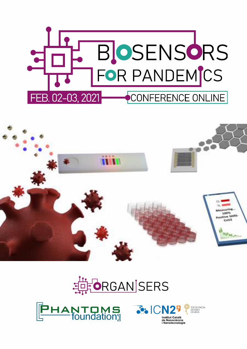
“Biosensors for Pandemics: Reliable and efficient nanotech-based diagnostics in emergency
situations” is an Online conference that will address main problems scientific community is
facing nowadays for COVID-19 pandemics.
The idea is to get together worldwide well-known experts in biosensing technologies who are
working currently in COVID-19 diagnostics or have a great potential for application of their
technologies in the near future. This conference is also addressed to specialists in virology,
epidemiology and other health areas and communication technologies that are crucial for
guiding biosensing community and their diagnostics ideas using biosensors in the right way.
A virtual round table providing answers to participants and sharing ideas between speakers
is also previewed.
We are indebted to the following Scientific Institutions and Companies for their help and/or
financial support: Institut Catala de Nanociencia I Nanotecnologia (ICN2) and Graphene
Flagship.
We also would like to thank all the speakers and participants that join us this year.
We truly hope that Biosensors for Pandemics serves as an international platform for
communication between science and business.
Hope to see you again online in the next edition of Biosensors for Pandemics.
Biosensors for Pandemics Organising Committee
2

B io s e ns o r s f o r p a n d e m ic s F e b r u a r y 0 2 - 0 3 , 2 0 2 1
PAGE
4 COMMITTEES
5 SPONSORS
5 PARTNER & MEDIA PARTNER
6 ABSTRACTS - INDEX
8 ABSTRACTS - SPEAKERS
9 PLENARY
11 KEYNOTES
28 INVITED
33 ORALS
41 ABSTRACTS - FLASH POSTERS
45 ABSTRACTS - ePOSTERS

B io s e ns o r s f o r p a n d e m ic s F e b r u a r y 0 2 - 0 3 , 2 0 2 1
Organising Committee
Phantoms Foundation (Spain) Antonio Correia
ICREA/ICN2 (Spain) Arben Merkoçi
CSIC/ICN2 (Spain) Laura Lechuga
International Scientific Committee
ICREA/ICN2 (Spain) Stephan Roche
Technical Committee
Phantoms Foundation (Spain) Concepción Narros Hernandez Phantoms Foundation (Spain) Joaquín Ramón Laca Maderal Phantoms Foundation (Spain) José Luis Roldán
4
B io s e ns o r s f o r p a n d e m ic s F e b r u a r y 0 2 - 0 3 , 2 0 2 1
Gold Sponsor
https://icn2.cat/en/
The Institut Catala de Nanociencia I Nanotecnologia (ICN2) is a flagship research institute within
the CERCA network of centres created and supported by the Autonomous Government of
Catalonia to raise Catalan science to the premier international level. The patrons of ICN2 are
the Government of Catalonia (Generalitat), the Consejo Superior de Investigaciones Científicas
(CSIC), and the Autonomous University of Barcelona (UAB). Its core activities are Frontier Basic
and Applied Research in Nanoscience and Nanotechnology, Technology Transfer, and Public
Outreach. Currently, ICN2 numbers some 200 staff from over 30 countries, of which about 170
are researchers
Silver Sponsor
https://graphene-flagship.eu/
Funded by the European Commission, the Graphene Flagship aims to secure a major role for
Europe in the ongoing technological revolution, helping to bring graphene innovation out of the
lab and into commercial applications. The Graphene Flagship gathers nearly 170 academic
and industrial partners from 22 countries, all exploring different aspects of graphene and related
materials. Bringing diverse competencies together, the Graphene Flagship facilitates
cooperation between its partners, accelerating the timeline for industry acceptance of
graphene technologies. The European Commission’s FET Flagships enable research projects on
an unprecedented scale. With €1 billion budgets, the Graphene Flagship, Human Brain Project
and Quantum Flagship serve as technology accelerators, helping Europe to compete with other
global markets in research and innovation. With an additional €20 million investment, the
European Commission has now funded the creation of an experimental pilot line for graphene-
based electronics, optoelectronics and sensors
5
B io s e ns o r s f o r p a n d e m ic s F e b r u a r y 0 2 - 0 3 , 2 0 2 1
Speakers/FlashPosters/ePosters list Alphabetical order
Authors Session Page Débora Albuquerque (INESC-MN, Portugal)
Quantitative point-of-care device for serological testing of anti-SARS-CoV-2 IgM Oral 34 Antonio Alcamí (CBMSO,CSIC-UAM, Spain)
Monitoring SARS-CoV-2 aerosol transmission in hospitals Keynote 12 Kiana Aran (Cardea Bio / Keck Graduate Institute, USA)
Next Generation Graphene Transistors for Biological Threat Invited 29 Fabiana Arduini (Università degli Studi di Roma “Tor Vergata”, Italy)
Electrochemical miniaturized (bio)sensors to support the sustainable management of
COVID-19 outbreak Invited 30 Vincent Bouchiat (Grapheal, France)
Graphene-on-polymer biosensors for rapid screening and field diagnostics Invited - Jorge Camacho (ITEFI-CSIC, Spain)
ULTRACOV: An intelligent ultrasound scanner for lung condition evaluation in COVID-19 Keynote 13 Enric Cuevas-Ferrando (Institute of Agrochemistry and Food Technology (IATA-CSIC),
Spain)
Novel SARS-CoV-2 viability RT-qPCR assessment on complex matrices ePoster 46 Cecília de Carvalho Castro e Silva (MackGraphe, Brazil)
Integration of Aptamers on Graphene Field Effect Transistors for the Ultra-Sensitive
Detection of SARS-CoV-2 Spike Protein Invited 31 Maria Dimaki (Technical University of Denmark, Denmark)
Optimising Lateral Flow Assays for diagnostic applications ePoster 47 Pilar Domingo-Calap (Universitat de Valencia - CSIC, Spain)
Unraveling the possible fecal-oral transmission route of SARS-CoV-2 Keynote 14 M.Carmen Estevez (ICN2 / CSIC, Spain)
Nanophotonic biosensors: superior diagnostic tools for COVID-19 management Invited 32 César Fernández (IQAC / IMB-CNM-CSIC, Spain)
Analytical tools for the multiplex rapid detection of SARS-CoV-2 Keynote 15 Carlos Fernández-Lodeiro (University of Vigo, Spain)
Green synthesis of plasmonic metal nanoparticles for future biosensor applications FlashPoster 42 Manuel Fuentes (Centro de Investigación del Cancer. IBSAL.IBMCC:CSIC-USAL, Spain)
Deciphering Biomarkers for acute respiratory distress syndrome by novel nanoproteomics
approaches Oral 35 Wei Gao (California Institute of Technology, USA)
SARS-CoV-2 RapidPlex: A Graphene-Based Multiplexed Telemedicine Platform for COVID-19
Diagnosis Keynote 16 Elisa González-Romero (University of Vigo, Spain)
How chemistry talks with electricity: from proof-of-concept to portable devices fabrication
for environmental pandemic problem solving FlashPoster 43 Hossam Haick (Technion, Israel)
Non-Invasive Intelligent Nanosensors for Monitoring Pandemics Keynote 17 Chuck Henry (Colorado State University, USA)
Disposable Capillary-Driven Immunoassays for COVID-19 Keynote 18
6
B io s e ns o r s f o r p a n d e m ic s F e b r u a r y 0 2 - 0 3 , 2 0 2 1
Authors Session Page Dean Ho (NUS, Singapore)
Harnessing Digital Medicine for Pandemic Preparedness and Next Generation Healthcare Keynote 19 Liming Hu (ICN2, UAB, Spain)
AuNP-based lateral flow assay for detection of SARS-CoV-2 nucleoprotein: implicit design
challenges in a new pandemic ePoster 48 Bergoi Ibarlucea (TU Dresden, Germany)
Rapid antigen detection with antibody-doped hydrogels on impedimetric biosensors Oral 36 Maria-Cristina Navas (Universidad de Antioquia, Colombia)
Production and characterization of pseudotyped particles to develop an immunosensor for
SARS CoV 2 ePoster 49 Melanie Ott (University of California San Francisco, USA)
Amplification-free detection of SARS-CoV-2 with CRISPR-Cas13a and mobile phone
microscopy Keynote 20 Aydogan Ozcan (UCLA, USA)
Deep Learning-enabled Computational Imaging and Sensing Plenary 10 Dipanjan Pan (University of Maryland, USA)
Emerging Biosensing Approaches to SARS-CoV-2 from Molecular Targeting to Digital Detection Keynote 21 Isabel Pastoriza-Santos (University of Vigo, Spain)
Surface-enhanced Raman scattering based biosensors Keynote 22 Guillermo Rodrigo (Instituto de Biología Integrativa de Sistemas (I2SysBio)- CSIC, Spain)
CRISPR-based detection of SARS-CoV-2 Oral 37 Giulio Rosati (ICN2, Spain)
A plug & print platform for SARS-CoV-2 rapid detection on inkjet-printed nanobiosensors
with smartphone readout Oral 38 Antonio Ruiz (University College London, Institute for Materials Discovery, UK)
Towards the development of low-cost smart textiles for the real-time monitoring of
bacterial and fungi colonies FlashPoster 44 Gloria Sanchez (IATA-CSIC, Spain)
Tracking SARS-CoV-2 in Wastewater as a Real Time Viral Hotspot Indicator Keynote 23 María Sanromán (DIPC, Spain)
Colorimetric Detection of SARS-CoV-2 based on Multiple AuNPs Aggregation Oral 39 Hadi Shafiee (Harvard Medical School, USA)
Virus detection using nanoparticles, deep learning, and smartphones Keynote - Amy Shen (Okinawa Institute of Technology Graduate University, Japan)
Antibody detection of SARS-CoV-2 spike protein by an integrated plasmonic microfluidic chip Keynote 24 Javier Tamayo (IMN-CSIC, Spain)
Towards ultrasensitive detection of SARS-Cov-2 within 48 after infection by means of a high
throughput optoplasmonic technology Keynote 25 Juan Ramón Tejedor (CINN-CSIC, Spain)
Enhanced detection of SARS-CoV2 RNA species using signal amplification technologies in
the context of DNA duplexes and DNA-RNA hybrids. Keynote 26 Jing Wang (ETH Zürich, Switzerland)
Improving the sensor performance for SARS-CoV-2 Keynote 27 Zhugen Yang Yang (Cranfield University, UK)
Paper-origami device for rapid diagnosis and testing sewage for early warning of pandemic Oral 40
7

B io s e ns o r s f o r p a n d e m ic s F e b r u a r y 0 2 - 0 3 , 2 0 2 1

BIOSENSORS FOR PANDEMICS 2021
Deep Learning-enabled Computational Imaging and Sensing
Aydogan Ozcan UCLA, Los Angeles, CA, USA
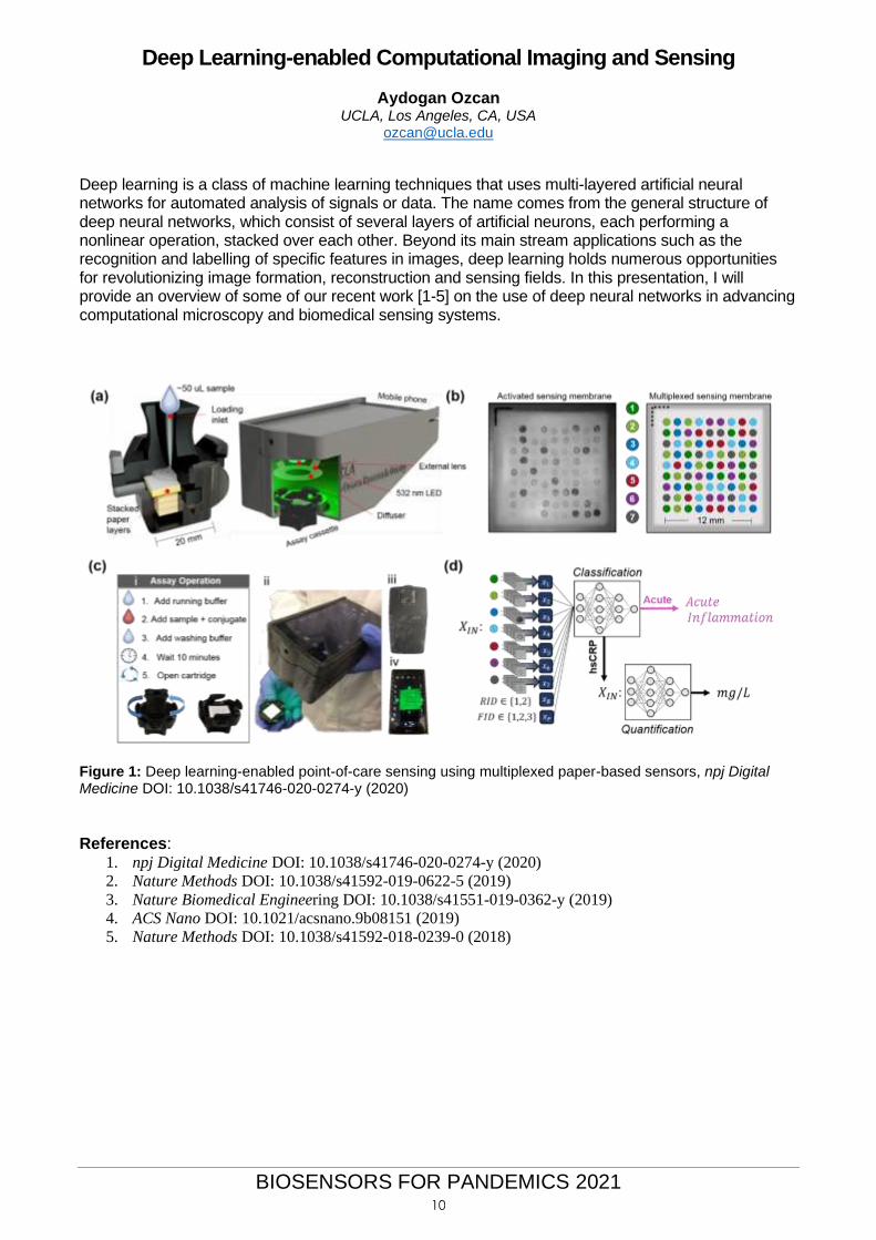
Deep learning is a class of machine learning techniques that uses multi-layered artificial neural networks for automated analysis of signals or data. The name comes from the general structure of deep neural networks, which consist of several layers of artificial neurons, each performing a nonlinear operation, stacked over each other. Beyond its main stream applications such as the recognition and labelling of specific features in images, deep learning holds numerous opportunities for revolutionizing image formation, reconstruction and sensing fields. In this presentation, I will provide an overview of some of our recent work [1-5] on the use of deep neural networks in advancing computational microscopy and biomedical sensing systems.
Figure 1: Deep learning-enabled point-of-care sensing using multiplexed paper-based sensors, npj Digital Medicine DOI: 10.1038/s41746-020-0274-y (2020)
References:
1. npj Digital Medicine DOI: 10.1038/s41746-020-0274-y (2020)
2. Nature Methods DOI: 10.1038/s41592-019-0622-5 (2019)
3. Nature Biomedical Engineering DOI: 10.1038/s41551-019-0362-y (2019)
4. ACS Nano DOI: 10.1021/acsnano.9b08151 (2019)
5. Nature Methods DOI: 10.1038/s41592-018-0239-0 (2018)
10

BIOSENSORS FOR PANDEMICS 2021
Monitoring SARS-CoV-2 aerosol transmission in hospitals Antonio Alcamí1 Alberto Rastrojo1, Bruno Hernáez1, Rocío Martín1, Laura Tabera1, Fernando Carrasco1, Begoña Aguado1, Javier Queiruga2, Vega Mauleon2, Cristina Calvo2, Sonia Alcolea3, María Luz García-García3 1Centro de Biología Molecular Severo Ochoa (CSIC-UAM), Nicolás Cabrera 1, Madrid, Spain; 2Hospital Universitario La Paz, Paseo de la Castellana 261, Madrid, Spain; 3Hospital Universitario Severo Ochoa, Avenida Orellana s/n, Leganés, Spain [email protected]
Abstract Severe acute respiratory syndrome coronavirus 2 (SARS-CoV-2) aerosols consist of small droplets exhaled from infected individuals and can remain in the air for long periods. It has been accepted that the spread of coronavirus disease 2019 (COVID-19) appears to occur through airborne transmission. Experimental production of aerosols has demonstrated that SARS-CoV-2 remains infectious in aerosols for hours [1,2]. Viral genetic material has been demonstrated in aerosols collected from hospitals [3,4,5] and recently it was demonstrated viral infectivity in aerosols collected from a hospital [6]. We optimized the methodology to capture by filtration a high diversity of airborne viruses and adapted this technology to the identification of SARS-CoV-2-containing aerosols by RT-qPCR and droplet digital PCR. We have monitored SARS-CoV-2 aerosols in different areas of two major hospitals in Madrid, Spain. La Paz University Hospital was monitored during the first COVID-19 pandemic wave, and Severo Ochoa University Hospital was monitored after the first pandemic wave and during the second wave. Air samples were taken from different locations of the hospitals: hot areas (Emergencies, Triage, Intensive Care Units), areas where transmission may occur (entrance, waiting rooms, health personnel rooms) and outside the hospital building. This technology allowed us to detect SARS-CoV-2 genomes in air samples from the hospitals during the COVID-19 pandemic. The presence of SARS-CoV-2 aerosols in different areas of the hospitals over several months will be presented. REFERENCES [1] Fears A.C. et al. Emerg Infect Dis. 26 (2020) 2168-2171. [2] van Doremalen, N. et al. N. Engl. J. Med. 382 (2020) 1564-1567. [3] Liu Y. et al. Nature 582 (2020) 557-560. [4] Zhou J. et al. Clin. Infect. Dis. 8 (2020) ciaa905. [5] Chia, P.Y. et al. Nat Commun. 11 (2020) 2800. [6] Lednicky J.A. et al. Int. J. Infect. Dis. 100 (2020) 476-482.
12
BIOSENSORS FOR PANDEMICS 2021
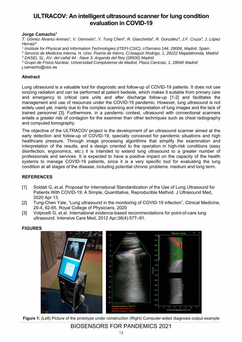
ULTRACOV: An intelligent ultrasound scanner for lung condition evaluation in COVID-19
Jorge Camacho1 T. Gómez Álvarez-Arenas1, V. Genovés1, Y. Tung Chen2, R. Giacchetta3, R. González3, J.F. Cruza3, J. López Herraiz4 1 Institute for Physical and Information Technologies (ITEFI-CSIC), c/Serrano 144, 28006, Madrid, Spain. 2 Servicio de Medicina Interna, H. Univ. Puerta de Hierro, C/Joaquín Rodrigo, 1, 28222 Majadahonda, Madrid 3 DASEL SL, AV. del cañal 44 - Nave 3. Arganda del Rey (28500) Madrid 4 Grupo de Física Nuclear, Universidad Complutense de Madrid, Plaza Ciencias, 1, 28040 Madrid [email protected] Abstract Lung ultrasound is a valuable tool for diagnostic and follow-up of COVID-19 patients. It does not use ionizing radiation and can be performed at patient bedside, which makes it suitable from primary care and emergency to critical care units and after discharge follow-up [1-2] and facilitates the management and use of resources under the COVID-19 pandemic. However, lung ultrasound is not widely used yet, mainly due to the complex scanning and interpretation of lung images and the lack of trained personnel [3]. Furthermore, in a pandemic context, ultrasound with conventional scanners entails a greater risk of contagion for the examiner than other techniques such as chest radiography and computed tomography.
The objective of the ULTRACOV project is the development of an ultrasound scanner aimed at the early detection and follow-up of COVID-19, specially conceived for pandemic situations and high healthcare pressure. Through image processing algorithms that simplify the examination and interpretation of the results, and a design oriented to the operation in high-risk conditions (easy disinfection, ergonomics, etc.) it is intended to extend lung ultrasound to a greater number of professionals and services. It is expected to have a positive impact on the capacity of the health systems to manage COVID-19 patients, since it is a very specific tool for evaluating the lung condition at all stages of the disease, including potential chronic problems. medium and long term. REFERENCES [1] Soldati G, et.al. Proposal for International Standardization of the Use of Lung Ultrasound for
Patients With COVID-19: A Simple, Quantitative, Reproducible Method. J Ultrasound Med, 2020 Apr 13.
[2] Tung-Chen Yale, “Lung ultrasound in the monitoring of COVID-19 infection”, Clinical Medicine, 20-4, 62-65, Royal College of Physicians, 2020
[3] Volpicelli G, et.al. International evidence-based recommendations for point-of-care lung ultrasound. Intensive Care Med, 2012 Apr;38(4):577–91.
FIGURES
Figure 1: (Left) Picture of the prototype under construction (Right) Computer-aided diagnosis output example
13
BIOSENSORS FOR PANDEMICS 2021
Unraveling the possible fecal-oral transmission route of SARS-CoV-2 Pilar Domingo-Calap Instituto de Biología Integrativa de Sistemas, Universitat de València-CSIC, Catedrático Agustín Escardino 9, 46980 Paterna, Spain pilar.domingo@uv,es
SARS-CoV-2 is primarily a respiratory virus, although it has been shown that it can replicate in the intestinal mucosa and be excreted via the stool. Molecular data have demonstrated the presence of the coronavirus RNA in feces [1]. However, detection of infectious viral particles is still incipient and based on a very small number of patients [2]. Here, I am investigating the presence of SARS-CoV-2 infectious particles in feces, as a potential route of viral transmission. Fecal-oral transmission would be of special interest in underdeveloped areas without adequate sanitation and sewage conditions. Interestingly, this could imply that wastewater may contain infectious viral particles, as recently suggested [3], but still requires a detailed study to validate this potential transmission route. REFERENCES [1] Sravanthi Parasa, Madhav Desai, et al. JAMA Netw Open, 3 (2020) e2011335. [2] Fei Xiao, Jing Sun, et al. Emerg Infect Dis, 26 (2020) 1920-1922. [3] Min Kan, Jianjian Wei, et al. Ann Intern Med, 1 (2020) M20-0928.
14
BIOSENSORS FOR PANDEMICS 2021
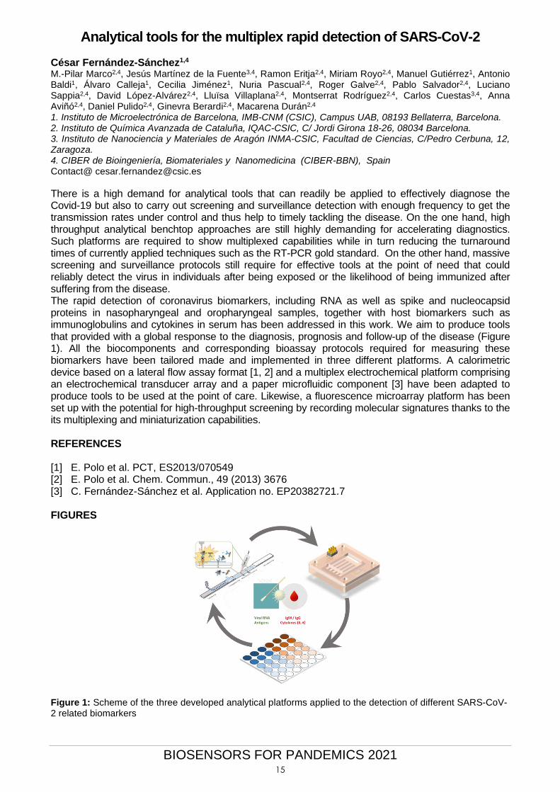
Analytical tools for the multiplex rapid detection of SARS-CoV-2 César Fernández-Sánchez1,4
M.-Pilar Marco2,4, Jesús Martínez de la Fuente3,4, Ramon Eritja2,4, Miriam Royo2,4, Manuel Gutiérrez1, Antonio Baldi1, Álvaro Calleja1, Cecilia Jiménez1, Nuria Pascual2,4, Roger Galve2,4, Pablo Salvador2,4, Luciano Sappia2,4, David López-Alvárez2,4, Lluïsa Villaplana2,4, Montserrat Rodríguez2,4, Carlos Cuestas3,4, Anna Aviñó2,4, Daniel Pulido2,4, Ginevra Berardi2,4, Macarena Durán2,4 1. Instituto de Microelectrónica de Barcelona, IMB-CNM (CSIC), Campus UAB, 08193 Bellaterra, Barcelona. 2. Instituto de Química Avanzada de Cataluña, IQAC-CSIC, C/ Jordi Girona 18-26, 08034 Barcelona. 3. Instituto de Nanociencia y Materiales de Aragón INMA-CSIC, Facultad de Ciencias, C/Pedro Cerbuna, 12, Zaragoza. 4. CIBER de Bioingeniería, Biomateriales y Nanomedicina (CIBER-BBN), Spain Contact@ [email protected]
There is a high demand for analytical tools that can readily be applied to effectively diagnose the Covid-19 but also to carry out screening and surveillance detection with enough frequency to get the transmission rates under control and thus help to timely tackling the disease. On the one hand, high throughput analytical benchtop approaches are still highly demanding for accelerating diagnostics. Such platforms are required to show multiplexed capabilities while in turn reducing the turnaround times of currently applied techniques such as the RT-PCR gold standard. On the other hand, massive screening and surveillance protocols still require for effective tools at the point of need that could reliably detect the virus in individuals after being exposed or the likelihood of being immunized after suffering from the disease. The rapid detection of coronavirus biomarkers, including RNA as well as spike and nucleocapsid proteins in nasopharyngeal and oropharyngeal samples, together with host biomarkers such as immunoglobulins and cytokines in serum has been addressed in this work. We aim to produce tools that provided with a global response to the diagnosis, prognosis and follow-up of the disease (Figure 1). All the biocomponents and corresponding bioassay protocols required for measuring these biomarkers have been tailored made and implemented in three different platforms. A calorimetric device based on a lateral flow assay format [1, 2] and a multiplex electrochemical platform comprising an electrochemical transducer array and a paper microfluidic component [3] have been adapted to produce tools to be used at the point of care. Likewise, a fluorescence microarray platform has been set up with the potential for high-throughput screening by recording molecular signatures thanks to the its multiplexing and miniaturization capabilities. REFERENCES [1] E. Polo et al. PCT, ES2013/070549 [2] E. Polo et al. Chem. Commun., 49 (2013) 3676 [3] C. Fernández-Sánchez et al. Application no. EP20382721.7 FIGURES
Figure 1: Scheme of the three developed analytical platforms applied to the detection of different SARS-CoV-2 related biomarkers
15
BIOSENSORS FOR PANDEMICS 2021
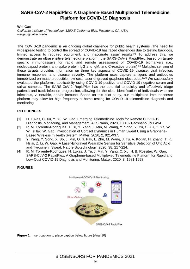
SARS-CoV-2 RapidPlex: A Graphene-Based Multiplexed Telemedicine Platform for COVID-19 Diagnosis
Wei Gao California Institute of Technology, 1200 E California Blvd, Pasadena, CA, USA [email protected]
The COVID-19 pandemic is an ongoing global challenge for public health systems. The need for widespread testing to control the spread of COVID-19 has faced challenges due to testing backlogs, limited access to required equipment, and inaccurate assay results.[1] To address this, we demonstrate an ultrasensitive telemedicine platform, the SARS-CoV-2 RapidPlex, based on target-specific immunoassays for rapid and remote assessment of COVID-19 biomarkers (i.e., nucleocapsid protein, anti-spike protein IgG and IgM, and C-reactive protein).[2] Multiplex sensing of these targets provides information on three key aspects of COVID-19 disease: viral infection, immune response, and disease severity. The platform uses capture antigens and antibodies immobilized on mass-producible, low-cost, laser-engraved graphene electrodes.[3,4] We successfully evaluated the platform's applicability using COVID-19-positive and COVID-19-negative serum and saliva samples. The SARS-CoV-2 RapidPlex has the potential to quickly and effectively triage patients and track infection progression, allowing for the clear identification of individuals who are infectious, vulnerable, and/or immune. Based on this pilot study, our multiplexed immunosensor platform may allow for high-frequency at-home testing for COVID-19 telemedicine diagnosis and monitoring. REFERENCES [1] H. Lukas, C. Xu, Y. Yu, W. Gao, Emerging Telemedicine Tools for Remote COVID-19
Diagnosis, Monitoring, and Management, ACS Nano, 2020, 10.1021/acsnano.0c08494. [2] R. M. Torrente-Rodríguez, J. Tu, Y. Yang, J. Min, M. Wang, Y. Song, Y. Yu, C. Xu, C. Ye, W.
W. IsHak, W. Gao, Investigation of Cortisol Dynamics in Human Sweat Using a Graphene-Based Wireless mHealth System, Matter, 2020, 2, 921-937.
[3] Y. Yang, Y. Song, X. Bo, J. Min, O. S. Pak, L. Zhu, M. Wang, J. Tu, A. Kogan, H. Zhang, T. K. Hsiai, Z. Li, W. Gao, A Laser-Engraved Wearable Sensor for Sensitive Detection of Uric Acid and Tyrosine in Sweat, Nature Biotechnology, 2020, 38, 217-224.
[4] R. M. Torrente-Rodríguez, H. Lukas, J. Tu, J. Min, Y. Yang, C. Xu, H. B. Rossiter, W. Gao, SARS-CoV-2 RapidPlex: A Graphene-based Multiplexed Telemedicine Platform for Rapid and Low-Cost COVID-19 Diagnosis and Monitoring, Matter, 2020, 3, 1981-1998.
FIGURES
Figure 1: Insert caption to place caption below figure (Arial 10)
16
BIOSENSORS FOR PANDEMICS 2021
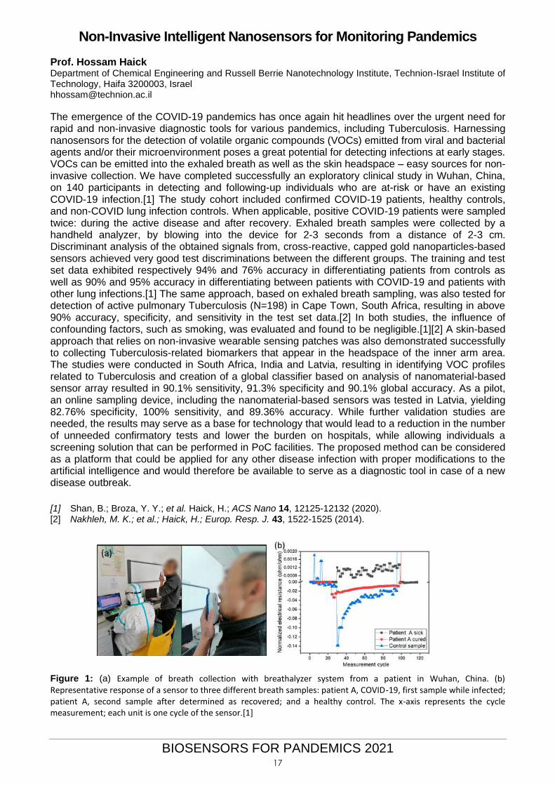
Non-Invasive Intelligent Nanosensors for Monitoring Pandemics Prof. Hossam Haick Department of Chemical Engineering and Russell Berrie Nanotechnology Institute, Technion-Israel Institute of Technology, Haifa 3200003, Israel [email protected]
The emergence of the COVID-19 pandemics has once again hit headlines over the urgent need for rapid and non-invasive diagnostic tools for various pandemics, including Tuberculosis. Harnessing nanosensors for the detection of volatile organic compounds (VOCs) emitted from viral and bacterial agents and/or their microenvironment poses a great potential for detecting infections at early stages. VOCs can be emitted into the exhaled breath as well as the skin headspace – easy sources for non-invasive collection. We have completed successfully an exploratory clinical study in Wuhan, China, on 140 participants in detecting and following-up individuals who are at-risk or have an existing COVID-19 infection.[1] The study cohort included confirmed COVID-19 patients, healthy controls, and non-COVID lung infection controls. When applicable, positive COVID-19 patients were sampled twice: during the active disease and after recovery. Exhaled breath samples were collected by a handheld analyzer, by blowing into the device for 2-3 seconds from a distance of 2-3 cm. Discriminant analysis of the obtained signals from, cross-reactive, capped gold nanoparticles-based sensors achieved very good test discriminations between the different groups. The training and test set data exhibited respectively 94% and 76% accuracy in differentiating patients from controls as well as 90% and 95% accuracy in differentiating between patients with COVID-19 and patients with other lung infections.[1] The same approach, based on exhaled breath sampling, was also tested for detection of active pulmonary Tuberculosis (N=198) in Cape Town, South Africa, resulting in above 90% accuracy, specificity, and sensitivity in the test set data.[2] In both studies, the influence of confounding factors, such as smoking, was evaluated and found to be negligible.[1][2] A skin-based approach that relies on non-invasive wearable sensing patches was also demonstrated successfully to collecting Tuberculosis-related biomarkers that appear in the headspace of the inner arm area. The studies were conducted in South Africa, India and Latvia, resulting in identifying VOC profiles related to Tuberculosis and creation of a global classifier based on analysis of nanomaterial-based sensor array resulted in 90.1% sensitivity, 91.3% specificity and 90.1% global accuracy. As a pilot, an online sampling device, including the nanomaterial-based sensors was tested in Latvia, yielding 82.76% specificity, 100% sensitivity, and 89.36% accuracy. While further validation studies are needed, the results may serve as a base for technology that would lead to a reduction in the number of unneeded confirmatory tests and lower the burden on hospitals, while allowing individuals a screening solution that can be performed in PoC facilities. The proposed method can be considered as a platform that could be applied for any other disease infection with proper modifications to the artificial intelligence and would therefore be available to serve as a diagnostic tool in case of a new disease outbreak.
[1] Shan, B.; Broza, Y. Y.; et al. Haick, H.; ACS Nano 14, 12125-12132 (2020). [2] Nakhleh, M. K.; et al.; Haick, H.; Europ. Resp. J. 43, 1522-1525 (2014).
Figure 1: (a) Example of breath collection with breathalyzer system from a patient in Wuhan, China. (b) Representative response of a sensor to three different breath samples: patient A, COVID-19, first sample while infected; patient A, second sample after determined as recovered; and a healthy control. The x-axis represents the cycle measurement; each unit is one cycle of the sensor.[1]
17
BIOSENSORS FOR PANDEMICS 2021
Disposable Capillary-Driven Immunoassays for COVID-19
Prof. Charles Henry Prof. David Dandy, Prof. Brian Geiss, Dr. Cody Carrell, Dr. Ilhoon Jang, Dr. Isabelle Samper, Zachary Call, Jeremy Link, Catherine McMahon Colorado State University, Fort Collins, CO, USA, 80526 [email protected]
Abstract
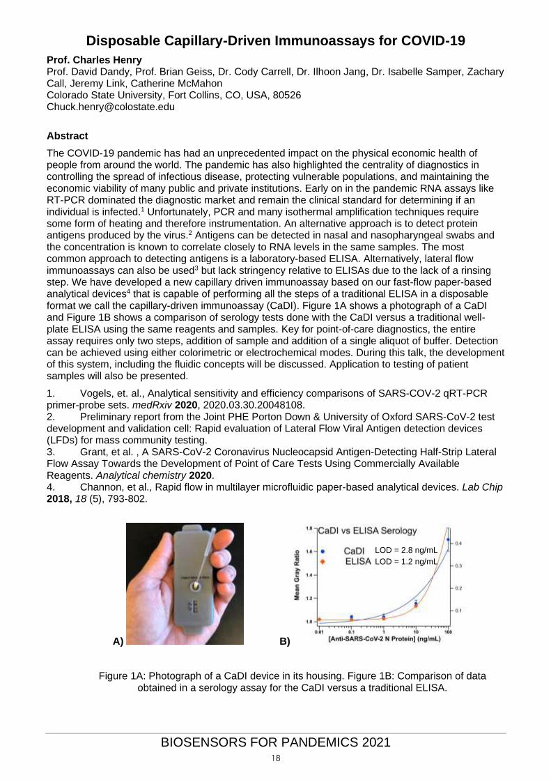
The COVID-19 pandemic has had an unprecedented impact on the physical economic health of people from around the world. The pandemic has also highlighted the centrality of diagnostics in controlling the spread of infectious disease, protecting vulnerable populations, and maintaining the economic viability of many public and private institutions. Early on in the pandemic RNA assays like RT-PCR dominated the diagnostic market and remain the clinical standard for determining if an individual is infected.1 Unfortunately, PCR and many isothermal amplification techniques require some form of heating and therefore instrumentation. An alternative approach is to detect protein antigens produced by the virus.2 Antigens can be detected in nasal and nasopharyngeal swabs and the concentration is known to correlate closely to RNA levels in the same samples. The most common approach to detecting antigens is a laboratory-based ELISA. Alternatively, lateral flow immunoassays can also be used3 but lack stringency relative to ELISAs due to the lack of a rinsing step. We have developed a new capillary driven immunoassay based on our fast-flow paper-based analytical devices4 that is capable of performing all the steps of a traditional ELISA in a disposable format we call the capillary-driven immunoassay (CaDI). Figure 1A shows a photograph of a CaDI and Figure 1B shows a comparison of serology tests done with the CaDI versus a traditional well-plate ELISA using the same reagents and samples. Key for point-of-care diagnostics, the entire assay requires only two steps, addition of sample and addition of a single aliquot of buffer. Detection can be achieved using either colorimetric or electrochemical modes. During this talk, the development of this system, including the fluidic concepts will be discussed. Application to testing of patient samples will also be presented.
1. Vogels, et. al., Analytical sensitivity and efficiency comparisons of SARS-COV-2 qRT-PCR primer-probe sets. medRxiv 2020, 2020.03.30.20048108. 2. Preliminary report from the Joint PHE Porton Down & University of Oxford SARS-CoV-2 test development and validation cell: Rapid evaluation of Lateral Flow Viral Antigen detection devices (LFDs) for mass community testing. 3. Grant, et al. , A SARS-CoV-2 Coronavirus Nucleocapsid Antigen-Detecting Half-Strip Lateral Flow Assay Towards the Development of Point of Care Tests Using Commercially Available Reagents. Analytical chemistry 2020. 4. Channon, et al., Rapid flow in multilayer microfluidic paper-based analytical devices. Lab Chip 2018, 18 (5), 793-802.
A) B)
LOD = 2.8 ng/mL
LOD = 1.2 ng/mL
Figure 1A: Photograph of a CaDI device in its housing. Figure 1B: Comparison of data obtained in a serology assay for the CaDI versus a traditional ELISA.
18
BIOSENSORS FOR PANDEMICS 2021
Harnessing Digital Medicine for Pandemic Preparedness and Next Generation Healthcare
Dean Ho Provost’s Chair Professor of Biomedical Engineering and Pharmacology Director, The N.1 Institute for Health (N.1) Director, Institute for Digital Medicine (WisDM) Head, Department of Biomedical Engineering National University of Singapore [email protected]
Drug repurposing is a widely explored approach to address indications ranging from oncology to COVID-19. However, discovering promising candidates represents the first of many steps needed to optimally harness a repurposed drug’s potential. These candidates often need to be delivered in combination with other therapies. In the quest for truly optimised drug repurposing, multiple challenges need to be overcome - The right drugs and corresponding doses need to be identified, which will have a profound impact on the drugs that ultimately comprise that combination. Using traditional approaches, this can be an insurmountable barrier given the very large drug and dose parameter space that is created. In addition, for certain diseases, a one-size-fits-all approach serves as a barrier to individualising treatment and can profoundly impair patient outcomes, as even effective drugs given at incorrect dosages can result in little to no efficacy. Furthermore, these doses may need to be modulated dynamically during the course treatment, since the patient response to treatment can also be dynamic. We will discuss our recent advances in clinical trials innovation and the clearance of first-in-class patient studies, as well as results from our ongoing clinical development studies. The ultimate objectives of WisDM and N.1, which are already being observed in the clinic, are to dynamically tailor patient-specific treatment outcomes, reduce healthcare costs, and increase accessibility to practice-changing and optimised medicine
19
BIOSENSORS FOR PANDEMICS 2021
Amplification-free detection of SARS-CoV-2 with CRISPR-Cas13a and mobile phone microscopy.
Melanie Ott University of California San Francisco, USA [email protected]
Until a safe, reliable vaccine has been broadly administered around the globe, the key to safely opening up in the age of COVID-19 — and any pandemic — will be widely available, frequent, and inexpensive testing for the disease. How can we get there? More and different types of diagnostics.
20
BIOSENSORS FOR PANDEMICS 2021
Emerging Biosensing Approaches to SARS-CoV-2 from Molecular Targeting to Digital Detection
Prof. (Dr.) Dipanjan Pan Dr. Parikshit Moitra Maha Alafeef Ketan Dighe Department of Radiology, Pediatrics, Chemical, Biochemical and Environmental Engineering, University of Maryland Baltimore and University of Maryland Baltimore County, USA [email protected]
Nanoscale structures offer unprecedented potential in pre-emptive medicine covering early detection, diagnosis and therapy of diseases. COVID-19 has claimed already been ascertained to affect >91M active cases with >1.9M deaths. Currently, COVID19 is being diagnosed primarily by three techniques, i.e. reverse-transcription polymerase chain reaction (RT-PCR), gene sequencing and chest computed tomography (CT). However, limitations of sample collection and transportation, as well as kit performance with inadequate access to advanced instrumental techniques, often cannot report COVID-19 at its initial presentation leading to the spread of this infectious disease to a wider community. Moreover, researchers found at least three central variants, distinguishable by amino acid changes, among 160 different complete human SARS-CoV-2 genome sequences. This limits the universal applicability of the currently available commercial COVID-19 kits. We have been developing novel approaches for screening of active COVID-19 cases with plasmonic and other quantitative biosensing techniques. These unique approached for selective sensing of SARS-CoV-2 eliminates the possibility of misinterpretation arisen due to the genomic variants of this virus which is the most concerning limitation of the current COVID-19 sensing kits. Our sensing approaches can detect the specific target nucleic acid sequences without signal cross talk with a detection limit to a single virus level with time of response to be around 2-3 mins. REFERENCES [1] Alafeef, Dighe K, Moitra P, Pan D.* Ultrasensitive and Quantitative Rapid Detection of SARS-
CoV-2 using Antisense Oligonucleotides Directed Electrochemical Biosensor Chip. ACS Nano 2020, 14 (12), 17028–17045.
[2] Moitra P, Alafeef M, Dighe K, Pan D.* Selective Naked-Eye Detection of SARS-CoV-2 Mediated by N gene targeted Antisense Oligonucleotide Capped Plasmonic Nanoparticles. ACS Nano. 2020, 14(6):7617-7627.
21
BIOSENSORS FOR PANDEMICS 2021
Surface-enhanced Raman scattering based biosensors
Isabel Pastoriza-Santos CINBIO, Universidade de Vigo, Lagoas-Marcosende, 36310 Vigo, Spain [email protected]
Abstract Over the years, colloidal plasmonic nanoparticles have emerged as important building blocks of modern nanoscience and nanotechnology to deal with a wide range of applications including electronics, energy, medicine, catalysis, biosensing, imaging and therapy. The unique optical properties of plasmonic nanostructures have led to the development of routes to synthesize metal nanoparticles with tailored size and morphology as well as to assemble them in a control maner. Assemblies of metal nanoparticles often exhibit collective properties, which are highly enhanced over those of the individual particles. Surface-enhanced Raman scattering (SERS) spectroscopy is an ultrasensitive analytical technique that can be applied non-invasively for the detection and imaging of a wide range of biomolecules. SERS allows identification of the specific spectral fingerprint of a probe analyte in contact with a plasmonic nanostructure and its sensitivity can go as far as the single-molecule level. Importantly, SERS offers multiplexing capability, requires no sample preparation and provides high spatial resolution. In this communication, we report the fabrication of different SERS-active platforms based on plasmonic nanoparticles/assemblies for biosensing (either label-free or indirect).1-3 Among others, we present the fabrication and performance of plasmonic microfluidic chips for label-free and ultrasensitive detection and a lateral flow immunoassay for the ultrasensitive detection of penumolysin employing plasmonic SERRS tag as labelled probe. REFERENCES [1] D. García-Lojo, S. Gómez-Graña, V. F. Martin, D. Solís, J. Taboada, J. Pérez-Juste and
I. Pastoriza-Santos. ACS Applied Materials & Interfaces, 2020, 12, 41, 46557-46564. [2] S. De Marchi, L. Vázquez-Iglesias, G. Bodelón, I. Pérez-Juste, L. A. Fernández, J. Pérez-
Juste and I. Pastoriza-Santos. Chem. Mater., 2020, 32, 13, 5739–5749. [3] L. Blanco-Covián, V. Montes-García, A. Girard, M. T. Fernández-Abedul, J. Pérez-Juste,
I. Pastoriza-Santos, K. Faulds, D. Graham and M. C. Blanco-López. Nanoscale, 2017, 9, 2051–2058.
[4] G. Bodelón, V. Montes-García, C. Fernández-López, I. Pastoriza-Santos, J. Pérez-Juste and L.M. Liz-Marzán. Small, 2015, 11, 4149–4157.
22
BIOSENSORS FOR PANDEMICS 2021
Tracking SARS-CoV-2 in Wastewater as a Real Time Viral Hotspot Indicator
Gloria Sánchez1 Walter Randazzo1, Alba Pérez-Cataluña1, Enric Cuevas-Ferrando1, Azahara Díaz-Reolid1, Irene Falcó1, Inés
Girón-Guzmán1, Ana Allende2 1Department of Preservation and Food Safety Technologies, Institute of Agrochemistry and Food Technology, IATA-CSIC, Av. Agustín Escardino 7, Paterna, 46980, Valencia, Spain. 2Research Group on Quality and Safety of Fruits and Vegetables, Department of Food Science and Technology, CEBAS-CSIC, Campus Universitario de Espinardo, 25, 30100, Murcia, Spain; [email protected]
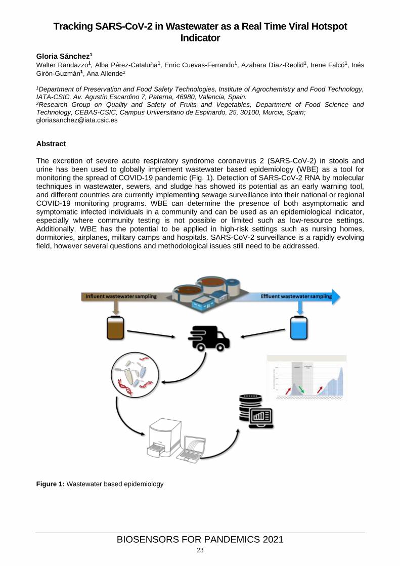
Abstract The excretion of severe acute respiratory syndrome coronavirus 2 (SARS-CoV-2) in stools and urine has been used to globally implement wastewater based epidemiology (WBE) as a tool for monitoring the spread of COVID-19 pandemic (Fig. 1). Detection of SARS-CoV-2 RNA by molecular techniques in wastewater, sewers, and sludge has showed its potential as an early warning tool, and different countries are currently implementing sewage surveillance into their national or regional COVID-19 monitoring programs. WBE can determine the presence of both asymptomatic and symptomatic infected individuals in a community and can be used as an epidemiological indicator, especially where community testing is not possible or limited such as low-resource settings. Additionally, WBE has the potential to be applied in high-risk settings such as nursing homes, dormitories, airplanes, military camps and hospitals. SARS-CoV-2 surveillance is a rapidly evolving field, however several questions and methodological issues still need to be addressed.
Figure 1: Wastewater based epidemiology
23
BIOSENSORS FOR PANDEMICS 2021
Antibody detection of SARS-CoV-2 spike protein by an integrated plasmonic microfluidic chip
Amy Q. Shen Riccardo Funari and Kang-Yu Chu Okinawa Institute of Science and Technology, Address, 1919-1 Tancha, Onna-son, Okinawa, [email protected]
The worldwide outbreak of severe acute respiratory syndrome related coronavirus 2 (SARS-CoV-2)
has led to active research in related diagnostics and medical treatments. While quantitative reverse
transcription polymerase chain reaction (qRT--PCR) is currently the best methodology to detect the
virus at the early stage of the infection, serological tests for specific antiviral antibodies are
complementary as they identify false negative qRT--PCR responses, track how effectively the
patient's immune system is fighting the infection, and are potentially helpful for plasma transfusion
therapies. Motivated by finding a reliable and cost-effective alternative to existing serological
methodologies, we report the development of an opto-microfluidic platform to quantify the
concentration of anti-SARS-CoV-2 spike protein antibodies in diluted human plasma by capturing the
wavelength shift of the localized surface plasmon resonance (LSPR) peak of gold nanostructures in
the microfluidic device upon binding interactions with SARS-CoV-2 spike protein within 30 minutes,
without labelling agents. This label-free microfluidic platform achieves a limit of detection of ~0.5 pM,
falling under the clinical relevant concentration range. We demonstrate that our opto-microfluidic
platform offers a promising point-of-care testing tool to complement standard serological assays and
make SARS-CoV-2 quantitative diagnostics easier, cheaper, and faster [1].
REFERENCES [1] Riccardo Funari, Kang-Yu Chu, Amy Q. Shen, Biosensors and Bioelectronics, 169, (2020).
112578. FIGURES
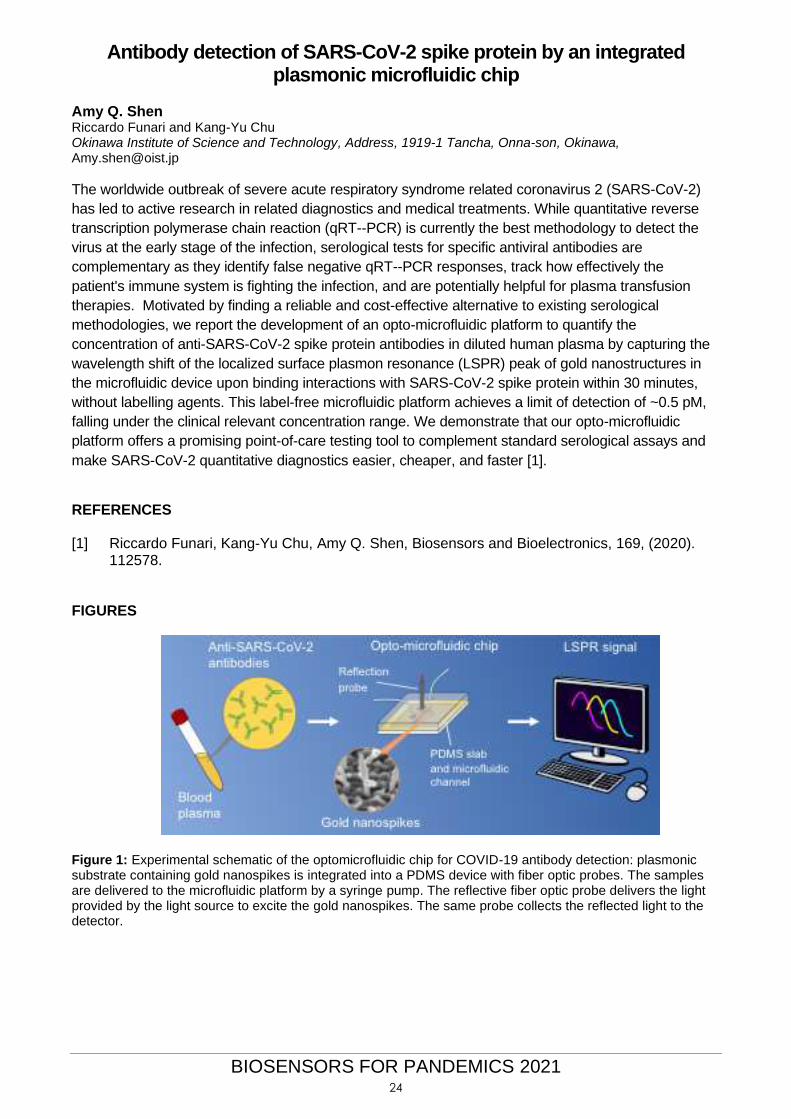
Figure 1: Experimental schematic of the optomicrofluidic chip for COVID-19 antibody detection: plasmonic substrate containing gold nanospikes is integrated into a PDMS device with fiber optic probes. The samples are delivered to the microfluidic platform by a syringe pump. The reflective fiber optic probe delivers the light provided by the light source to excite the gold nanospikes. The same probe collects the reflected light to the detector.
24
BIOSENSORS FOR PANDEMICS 2021
Towards ultrasensitive detection of SARS-Cov-2 within 48 after infection by means of a high throughput optoplasmonic technology
Javier Tamayo P.M. Kosaka, S. García-López, R. Sato, M.L. Yubero, M. Calleja Instituto de Micro y Nanotecnología(IMN-CNM), CSIC, Tres Cantos, Madrid, Spain Contact@ [email protected]
Early detection of SARS-Cov-2 infection is the best way to prevent spread of the disease. However, as we are observing, any shortage of laboratory diagnostic capacity at national or local level will hamper epidemic response. Nucleic acid amplification tests (NAAT) have become the gold-standard for detecting low-concentrations of the virus in blood. However, these methods are technically demanding and cost-prohibitive in developing countries. Immunoassays are more affordable and can be more easily adapted for point-of-care diagnosis. However, the sensitivity so far of these methods has been too low. We here report the development of a sandwich immunoassay based on novel optoplasmonic transduction[1-3] for detecting the SARS-Cov-2 nucleocapsid N-protein and the spike S-protein from a nasal swabbing material. The limit of detection of the immunoassay is below 10−15 g/mL that is equivalent to few virions in 10 mL of plasma. This is 5 orders of magnitude better than last generation of approved immunoassays and 2 orders of magnitude better than NAT methods. This LoD reduces the undetectable phase after infection to just 24-48h. The technology meets potential to be produced en masse at low cost and capability for miniaturization to be used at point-of-care. Final remark: a linear decrease in the detection time implies exponential decrease in the pandemic dissemination. REFERENCES [1] Kosaka, P.M., et al. Nature Nanotechnology 9 (2014), 1047. [2] Kosaka, P. M., Pini, V., Calleja, M., & Tamayo, J. PLoS One 12 (2017), e0171899. [3] Kosaka, P. M., Calleja, M., & Tamayo, J. Seminars in Cancer Biology 52, (2018), 26.
25
BIOSENSORS FOR PANDEMICS 2021
Enhanced detection of SARS-CoV2 RNA species using signal amplification technologies in the context of DNA duplexes and DNA-RNA
hybrids. Juan Ramón Tejedor1,2,3 Gabriel Martín4, Cristina Mangas3, Pablo Santamarina-Ojeda2, 3, Raúl F. Pérez1, 3, Marta E Álvarez-Argüelles4, Santiago Melón-García4, Agustín Fernández Fernández1, 2, 3 and Mario Fernández Fraga1, 2, 3, 5, 1.Department of Nanomedicine, Cancer Epigenetics Unit, Nanomaterials and Nanotechnology Research Center (CINN-CSIC), El Entrego, Principality of Asturias, 33940, Spain. 2.Department of Cancer, Foundation for Biomedical Research and Innovation in Asturias (FINBA), Health Research Institute of Asturias (ISPA), Oviedo, Principality of Asturias, 33011, Spain. 3.Instituto Universitario de Oncología del Principado de Asturias (IUOPA), University of Oviedo, Oviedo, Principality of de Asturias, 33006, Spain. 4.Department of Microbiology, Unit of Virology, Central University Hospital of Asturias (HUCA), Oviedo, Principality of Asturias, 33011, Spain. 5.Center for Biomedical Network Research on Rare Diseases (CIBER-ER), Madrid, Madrid, 28029, Spain. [email protected] / [email protected]
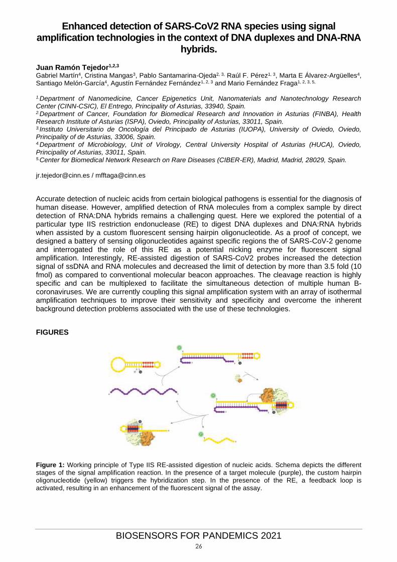
Accurate detection of nucleic acids from certain biological pathogens is essential for the diagnosis of human disease. However, amplified detection of RNA molecules from a complex sample by direct detection of RNA:DNA hybrids remains a challenging quest. Here we explored the potential of a particular type IIS restriction endonuclease (RE) to digest DNA duplexes and DNA:RNA hybrids when assisted by a custom fluorescent sensing hairpin oligonucleotide. As a proof of concept, we designed a battery of sensing oligonucleotides against specific regions the of SARS-CoV-2 genome and interrogated the role of this RE as a potential nicking enzyme for fluorescent signal amplification. Interestingly, RE-assisted digestion of SARS-CoV2 probes increased the detection signal of ssDNA and RNA molecules and decreased the limit of detection by more than 3.5 fold (10 fmol) as compared to conventional molecular beacon approaches. The cleavage reaction is highly specific and can be multiplexed to facilitate the simultaneous detection of multiple human B-coronaviruses. We are currently coupling this signal amplification system with an array of isothermal amplification techniques to improve their sensitivity and specificity and overcome the inherent background detection problems associated with the use of these technologies.
FIGURES
Figure 1: Working principle of Type IIS RE-assisted digestion of nucleic acids. Schema depicts the different stages of the signal amplification reaction. In the presence of a target molecule (purple), the custom hairpin oligonucleotide (yellow) triggers the hybridization step. In the presence of the RE, a feedback loop is activated, resulting in an enhancement of the fluorescent signal of the assay.
26
BIOSENSORS FOR PANDEMICS 2021
Improving the sensor performance for SARS-CoV-2 Jing Wang Guangyu Qiu, Zhibo Gai, Yile Tao, Lanja Saleh, Jiukai Tang, Ting Gui, Gerd A. Kullak-Ublick, Xiaole Zhang Institute of Environmental Engineering, ETH Zürich, Zürich 8093, Switzerland Laboratory for Advanced Analytical Technologies, Empa, Dübendorf 8600, Switzerland [email protected]
Abstract (Arial 11) The coronavirus disease 2019 (COVID-19) has now penetrated every populated patch of the globe and sows destruction in our daily life. Reliable and sensitive virus sensing systems are therefore of vital importance for timely infection detection and transmission prevention. We have developed a dual-functional plasmonic photothermal biosensor for detection of SARS-CoV-2 [1], which was validated by testing clinical COVID-19 patient samples, and indicated its potential applications in fast clinical infection screening and real-time environmental monitoring. We developed a simple framework to integrate the a priori dose-response relation for SARS-CoV based on mice experiments, the recent data on infection risk from a meta-analysis and the respiratory virus shedding in exhaled breath, to shed light on the dose-response relation for human [2]. The aerosol transmission infection risk was evaluated based on the dose-response model for both typical indoor environment [2] and a Chinese seafood market [3]. REFERENCES [1] Qiu, G., Gai, Z., Tao, Y., Schmitt, J., Kullak-Ublick, G.A., Wang, J. (2020) Dual-Functional
Plasmonic Photothermal Biosensors for Highly Accurate Severe Acute Respiratory Syndrome Coronavirus 2 Detection, ACS Nano, https://dx.doi.org/10.1021/acsnano.0c02439
[2] Zhang, X. and Wang, J. (2020) Dose-response Relation Deduced for Coronaviruses from COVID-19, SARS and MERS Meta-analysis Results and its Application for Infection Risk Assessment of Aerosol Transmission, Clinical Infectious Diseases, https://doi.org/10.1093/cid/ciaa1675 .
[3] Zhang, X.; Ji, Z.; Yue, Y.; Liu, H.; Wang, J. (2020) Infection risk assessment of COVID-19 through aerosol transmission: a case study of South China Seafood Market, Environmental Science and Technology, https://dx.doi.org/10.1021/acs.est.0c02895 .
FIGURES
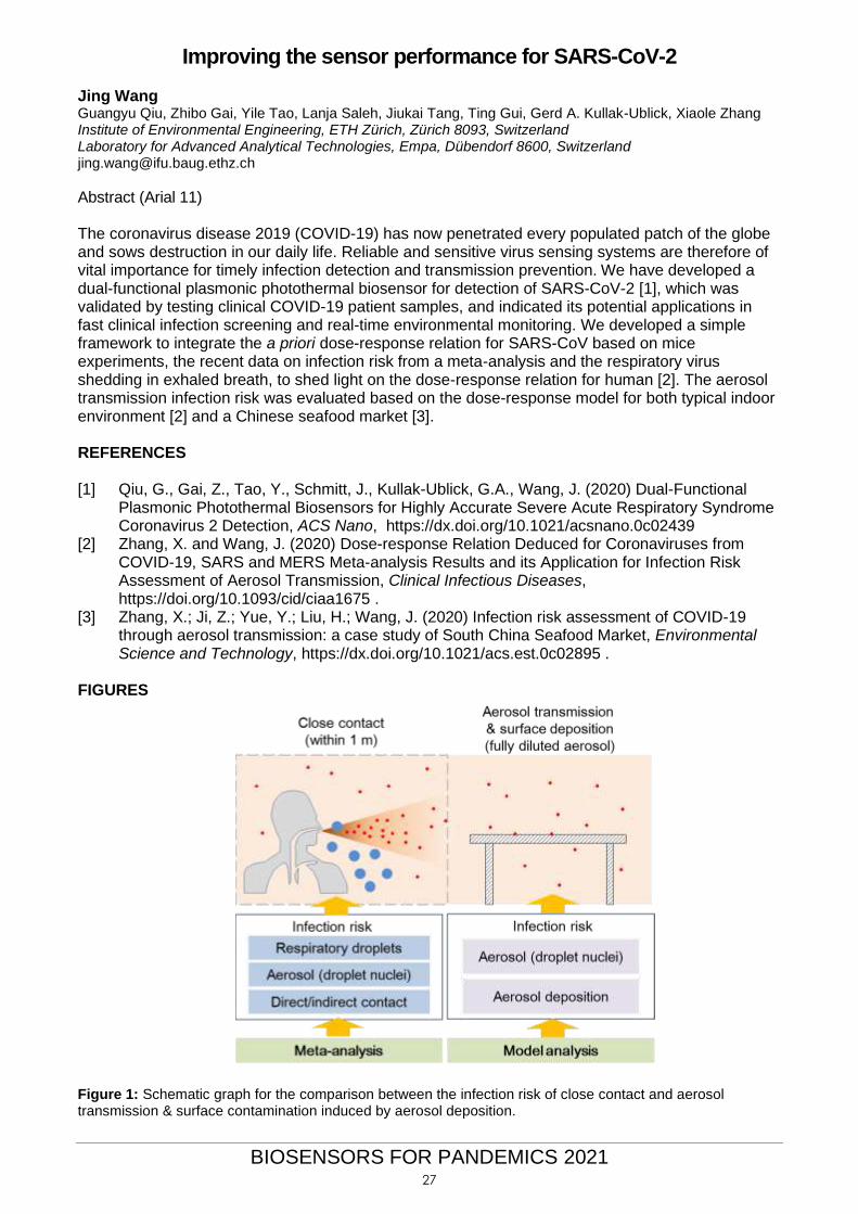
Figure 1: Schematic graph for the comparison between the infection risk of close contact and aerosol transmission & surface contamination induced by aerosol deposition.
27

BIOSENSORS FOR PANDEMICS 2021
Next Generation Graphene Transistors for Biological Threat Kiana Aran, Ph.D. Co-founder and Chief Scientific Officer Cardea Bio Inc., 8969 Kenamar Dr. Suite 104, San Diego, U.S. [email protected] Keck Graduate Institute, 535 Watson Drive, Claremont, U.S. [email protected]
The rapid transmission and severe clinical outcomes associated with SARS-CoV-2 has highlighted the importance of digital diagnostic platforms in pandemic control. Biology-gated graphene field effect transistors (gFETs) leverage the high sensitivity and biocompatibility of graphene, enabling a digital connection to biology. The multiomics and multiplexing capability of gFETs allows for the detection of multiple biomarkers and SAR-Cov-2 antibodies utilizing different epitopes of pathogens leading to higher specificity and lower limits of detection. The versatility of gFETs allows them to be incorporated into different hardware platforms from a point-of-care handheld reader to high throughput automated liquid handlers and combined with cloud-based computational power (such as ML and AI) to meet the needs of pandemics. REFERENCES [1] R. Hajian, S. Balderston, T. Tran, T. Deboer, J. Etienne, M. Sandhu, N. Wauford, J. Nokes, M.
Athaiya, J. Paredes, R. Peytavi, B. Goldsmith, N. Murthy, I.M. Conboy, K. Aran. Detection of
unamplified target genes via CRISPR/Cas9 immobilized on a graphene field-effect transistor.
2019, Nature Biomedical Engineering (Front Cover featured). DOI 10.1038/s41551-019-037
[2] C.M. Sadlowski, S. Balderston, M. Sandhu, R. Hajian, T. Tran, K. Aran. Graphene-based
biosensor for on-chip detection of bio-orthogonally labeled proteins to identify the circulating
biomarkers of aging during heterochronic parabiosis, 2018, Lab Chip (Back Cover, featured),
2018,18, 3230-3238
[3] S. Viswanathan, T.N. Narayanan, K. Aran, K. Fink, J. Paredes, P.M. Ajayan, D. Liepmann, V.
Renu-gopalakrishanan. Graphene–protein field effect biosensors: glucose sensing,
2015, Materials Today. DOI 10.1016/j. mattod.2015.04.003
[4] B. Goldsmith, L. Locascio, Y. Gao, M. Lerner, A. Walker, J. Lerner, J. Kyaw, A. Shue, S. Afsahi,
D. Pan, J. Nokes, F. Barron. Digital Biosensing by Foundry-Fabricated Graphene Sensors.
2019, Scientific Reports 9, no. 1, 434. DOI 10.1038/s41598-019-38700-w
[5] S. Afsahi, M.B. Lerner, J.M. Goldstein, J. Lee, X. Tang, D.A. Bagarozzi, D. Pan, L. Locascio, A.
Walker, F. Barron, B. Goldsmith. Novel Graphene-Based Biosensor for Early Detection of Zika
Virus Infection. 2018, Biosensors and Bioelectronics 100: 85–88. DOI
10.1016/j.bios.2017.08.051
FIGURES
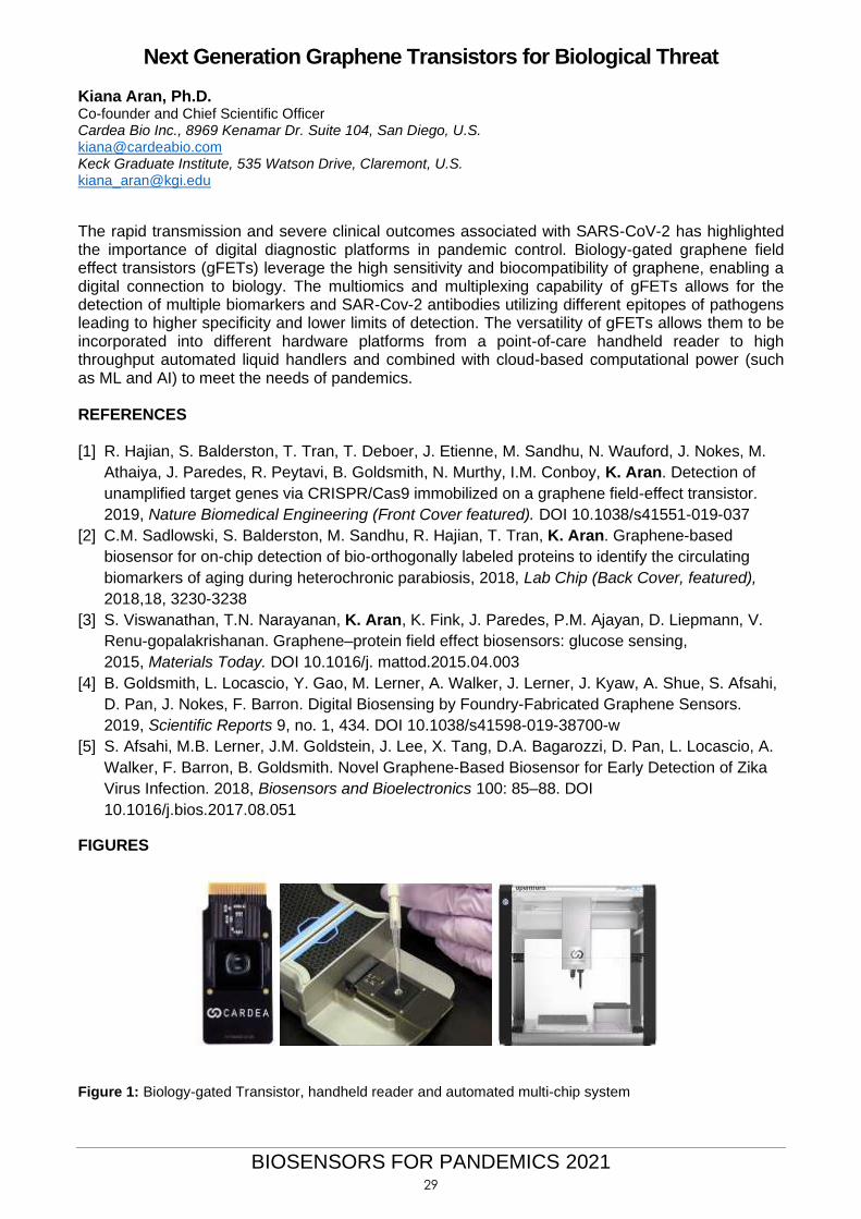
Figure 1: Biology-gated Transistor, handheld reader and automated multi-chip system
29
BIOSENSORS FOR PANDEMICS 2021
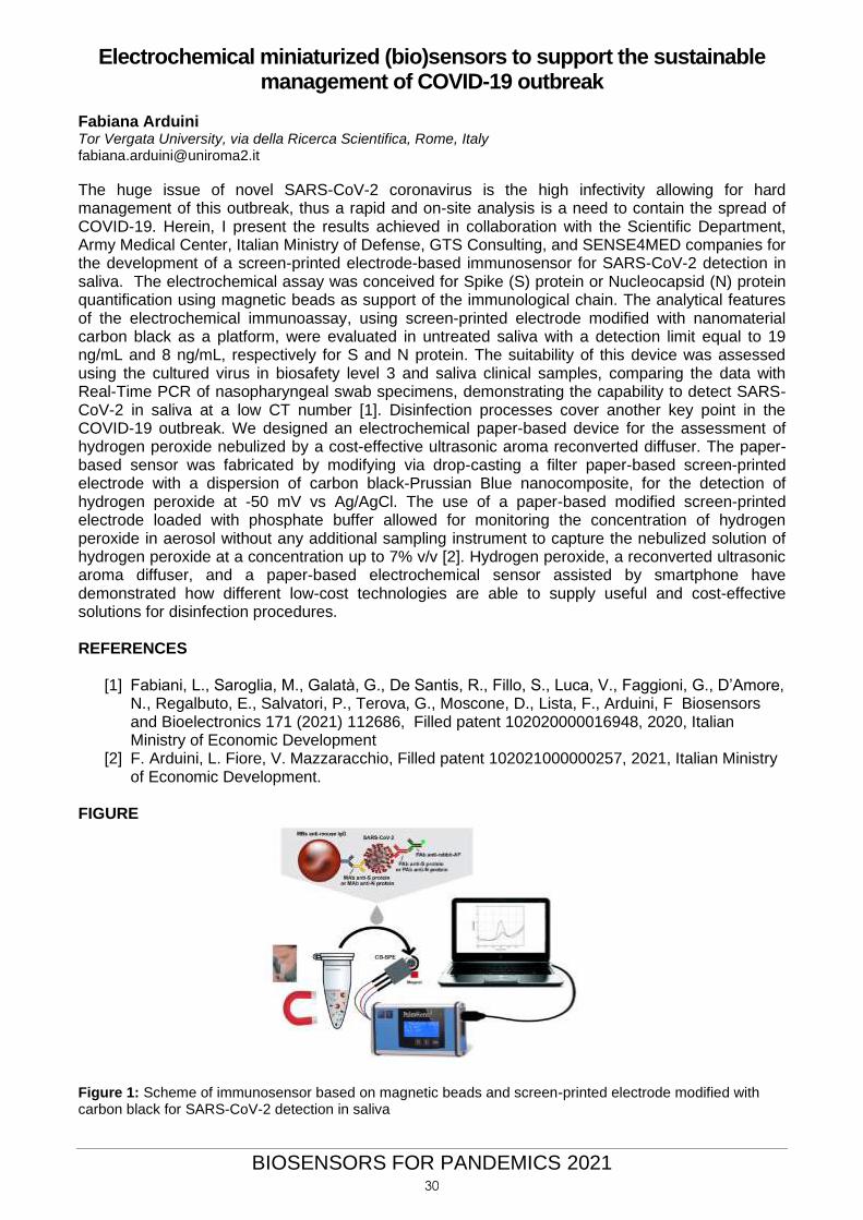
Electrochemical miniaturized (bio)sensors to support the sustainable management of COVID-19 outbreak
Fabiana Arduini Tor Vergata University, via della Ricerca Scientifica, Rome, Italy [email protected]
The huge issue of novel SARS-CoV-2 coronavirus is the high infectivity allowing for hard management of this outbreak, thus a rapid and on-site analysis is a need to contain the spread of COVID-19. Herein, I present the results achieved in collaboration with the Scientific Department, Army Medical Center, Italian Ministry of Defense, GTS Consulting, and SENSE4MED companies for the development of a screen-printed electrode-based immunosensor for SARS-CoV-2 detection in saliva. The electrochemical assay was conceived for Spike (S) protein or Nucleocapsid (N) protein quantification using magnetic beads as support of the immunological chain. The analytical features of the electrochemical immunoassay, using screen-printed electrode modified with nanomaterial carbon black as a platform, were evaluated in untreated saliva with a detection limit equal to 19 ng/mL and 8 ng/mL, respectively for S and N protein. The suitability of this device was assessed using the cultured virus in biosafety level 3 and saliva clinical samples, comparing the data with Real-Time PCR of nasopharyngeal swab specimens, demonstrating the capability to detect SARS-CoV-2 in saliva at a low CT number [1]. Disinfection processes cover another key point in the COVID-19 outbreak. We designed an electrochemical paper-based device for the assessment of hydrogen peroxide nebulized by a cost-effective ultrasonic aroma reconverted diffuser. The paper-based sensor was fabricated by modifying via drop-casting a filter paper-based screen-printed electrode with a dispersion of carbon black-Prussian Blue nanocomposite, for the detection of hydrogen peroxide at -50 mV vs Ag/AgCl. The use of a paper-based modified screen-printed electrode loaded with phosphate buffer allowed for monitoring the concentration of hydrogen peroxide in aerosol without any additional sampling instrument to capture the nebulized solution of hydrogen peroxide at a concentration up to 7% v/v [2]. Hydrogen peroxide, a reconverted ultrasonic aroma diffuser, and a paper-based electrochemical sensor assisted by smartphone have demonstrated how different low-cost technologies are able to supply useful and cost-effective solutions for disinfection procedures. REFERENCES
[1] Fabiani, L., Saroglia, M., Galatà, G., De Santis, R., Fillo, S., Luca, V., Faggioni, G., D’Amore, N., Regalbuto, E., Salvatori, P., Terova, G., Moscone, D., Lista, F., Arduini, F Biosensors and Bioelectronics 171 (2021) 112686, Filled patent 102020000016948, 2020, Italian Ministry of Economic Development
[2] F. Arduini, L. Fiore, V. Mazzaracchio, Filled patent 102021000000257, 2021, Italian Ministry of Economic Development.
FIGURE
Figure 1: Scheme of immunosensor based on magnetic beads and screen-printed electrode modified with carbon black for SARS-CoV-2 detection in saliva
30
BIOSENSORS FOR PANDEMICS 2021
Integration of Aptamers on Graphene Field Effect Transistors for the
Ultra-Sensitive Detection of SARS-CoV-2 Spike Protein
Cecilia de Carvalho Castro e Silva1,2 Carme Martinez3, Giulio Rosati2, Elías Torres Alonso4, Amaia Zurutuza4 and Arben Merkoçi2,5 1 Graphene and Nanomaterials Research Center – MackGraphe, Campus UPM, 01302-907 São Paulo, SP, Brazil. 2 Nanobioelectronics & Biosensors Group, Catalan Institute of Nanoscience and Nanotechnology (ICN2), CSIC and BIST, (ICN2), Campus UAB, 08193 Bellaterra, Barcelona, Spain. 3 Integrated Systems and Circuits Group, Centre Nacional de Microelectrònica (IMB-CNM, CSIC) Campus de la UAB · 08193 Bellaterra, Barcelona, Spain. 4 Graphenea Semiconductor S.L.U. Paseo Mikeletegi 83 20009 Donostia-San Sebastián, Spain. 5ICREA, Institució Catalana de Recerca i Estudis Avançats, Passeig Lluís Companys 23, Barcelona, Spain
Abstract Due to COVID-19 pandemic, novel analytical tools to perform SARS-CoV2 detection in a reliable and fast way are a priority. These devices should work with low sample volume and a non-invasive approach. Biosensors based on graphene field-effect transistors (GFETs) are thus a highly attractive technology. In fact, they allow real-time label-free electrical detection, scalability, relatively inexpensive mass production, miniaturization, and the possibility of on-chip integration of both sensor and measurement systems [1]. Moreover, graphene has unique properties such as high carrier mobilities and electrical conductivity, flexibility, biocompatibility, facile chemical functionalization, and large specific surface area, allowing the immobilization of high density of bioreceptors, leading to increased sensitivity [1]. Therefore, we are addressing the development of GFETs biosensors based on aptamers as bioreceptors for the label-free detection of SARS-CoV-2 Spike protein (Sp). With this aim, we employed GFET devices produced by Graphenea (GFET-S20) [2], a Spanish company. Each microchip contains two arrays of 6 GFETs, connected by a common source and a gold gate electrode integrated in the centre of the microchip. The graphene surface of the GFETs was non-covalently functionalized, allowing the immobilization of the specific aptamers for the SARS-CoV-2, followed by a blocking step. The GFETs were then electrically characterized at each surface modification step. The measurements in solution were performed with an ionic liquid gate configuration. The GFETs aptasensors allowed the detection of down to 1 femtomolar SP in PBS. The GFET aptasensor exhibits a high electrical sensitivity for the SARS-CoV2 spike protein detection due the combined effect of the graphene properties and their functionalization with aptamers. This allows us to explore this technology for the detection of the SARS-CoV-2 whole virus in real samples, such as saliva, potentially without any preliminary treatment. REFERENCES [1] Nguyen, E. P.; Silva, C. C. C. and Merkoçi, A. Nanoscale, 12 (2020) 19043. [2] https://www.graphenea.com/
31
BIOSENSORS FOR PANDEMICS 2021
Nanophotonic biosensors: superior diagnostic tools for COVID-19 management
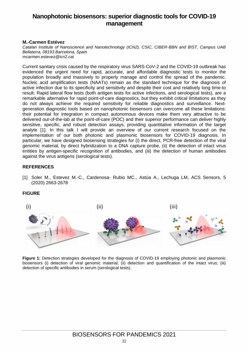
M.-Carmen Estévez Catalan Institute of Nanoscience and Nanotechnology (ICN2), CSIC, CIBER-BBN and BIST, Campus UAB Bellaterra, 08193 Barcelona, Spain [email protected]
Current sanitary crisis caused by the respiratory virus SARS-CoV-2 and the COVID-19 outbreak has evidenced the urgent need for rapid, accurate, and affordable diagnostic tests to monitor the population broadly and massively to properly manage and control the spread of the pandemic. Nucleic acid amplification tests (NAATs) remain as the standard technique for the diagnosis of active infection due to its specificity and sensitivity and despite their cost and relatively long time-to result. Rapid lateral flow tests (both antigen tests for active infections, and serological tests), are a remarkable alternative for rapid point-of-care diagnostics, but they exhibit critical limitations as they do not always achieve the required sensitivity for reliable diagnostics and surveillance. Next-generation diagnostic tools based on nanophotonic biosensors can overcome all these limitations: their potential for integration in compact autonomous devices make them very attractive to be delivered out-of-the-lab at the point-of-care (POC) and their superior performance can deliver highly sensitive, specific, and robust detection assays, providing quantitative information of the target analyte [1]. In this talk I will provide an overview of our current research focused on the implementation of our both photonic and plasmonic biosensors for COVID-19 diagnosis. In particular, we have designed biosensing strategies for (i) the direct, PCR-free detection of the viral genomic material, by direct hybridization to a DNA capture probe, (ii) the detection of intact virus entities by antigen-specific recognition of antibodies, and (iii) the detection of human antibodies against the virus antigens (serological tests). REFERENCES [1] Soler M., Estevez M.-C., Cardenosa- Rubio MC., Astúa A., Lechuga LM, ACS Sensors, 5
(2020) 2663-2678 FIGURE
Figure 1: Detection strategies developed for the diagnosis of COVID-19 employing photonic and plasmonic biosensors (i) detection of viral genomic material; (ii) detection and quantification of the intact virus; (iii) detection of specific antibodies in serum (serological tests).
32

BIOSENSORS FOR PANDEMICS 2021
Quantitative point-of-care device for serological testing of anti-SARS-
CoV-2 IgM
Débora C Albuquerque1,2
Elisabete Fernandes3; Sara Viveiros1; Sofia A.M. Martins1, Susana Cardoso1,2; Paulo P. Freitas3; Verónica C. Martins1
1 INESC-MN, Rua Alves Redol 9, Lisboa, Portugal; 2 IST ULisboa, Lisboa, Portugal; 3 INL, Braga, Portugal [email protected]
The pandemic caused by SARS-CoV-2 has originated a boom in the research and development of
in-vitro diagnostic tests, of both virological and serological nature. Among the different solutions in
the market, point-of-care (PoC) tests are becoming essential tools in expedite patient management.
Current PoC technologies for antibody testing such as lateral flow immunoassays (LFIA) are mostly
qualitative or semi-quantitative and suffer from lower sensitivities (66.0%) in comparison with central
laboratory methods, such as enzyme-linked immunosorbent assays (ELISA) and
chemiluminescence immunoassays (CLIA) (84.3% and 97.8%, respectively) [1]. We propose a
portable device with disposable chips for diagnosis and monitorization of the serological response to
Covid-19. The device is composed of a portable electronic reader and disposable biochips with an
array of magnetic sensors. Magnetic markers coupled to on-chip magnetic attraction were used to
allow an increased sensitivity (below µg/mL) of assays in short times [2]. The proposed technology
has been validated for various clinical applications [2,3]. Major assets include, low cost (< 10€/test),
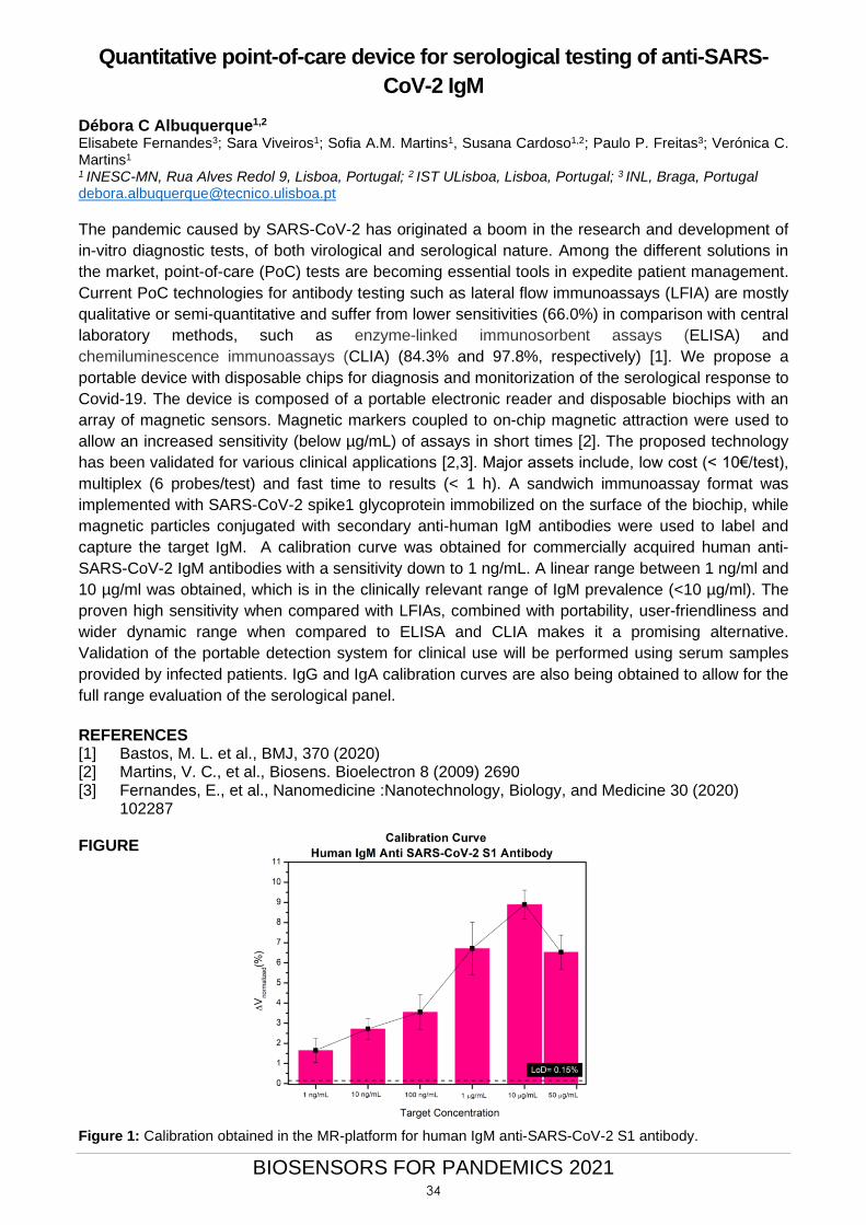
multiplex (6 probes/test) and fast time to results (< 1 h). A sandwich immunoassay format was
implemented with SARS-CoV-2 spike1 glycoprotein immobilized on the surface of the biochip, while
magnetic particles conjugated with secondary anti-human IgM antibodies were used to label and
capture the target IgM. A calibration curve was obtained for commercially acquired human anti-
SARS-CoV-2 IgM antibodies with a sensitivity down to 1 ng/mL. A linear range between 1 ng/ml and
10 µg/ml was obtained, which is in the clinically relevant range of IgM prevalence (<10 µg/ml). The
proven high sensitivity when compared with LFIAs, combined with portability, user-friendliness and
wider dynamic range when compared to ELISA and CLIA makes it a promising alternative.
Validation of the portable detection system for clinical use will be performed using serum samples
provided by infected patients. IgG and IgA calibration curves are also being obtained to allow for the
full range evaluation of the serological panel.
REFERENCES [1] Bastos, M. L. et al., BMJ, 370 (2020) [2] Martins, V. C., et al., Biosens. Bioelectron 8 (2009) 2690 [3] Fernandes, E., et al., Nanomedicine :Nanotechnology, Biology, and Medicine 30 (2020)
102287
FIGURE
Figure 1: Calibration obtained in the MR-platform for human IgM anti-SARS-CoV-2 S1 antibody.
34
BIOSENSORS FOR PANDEMICS 2021
Deciphering Biomarkers for acute respiratory distress syndrome by novel nanoproteomics approaches
Presenting Author: Manuel Fuentes García1,2 Pabo Juanes-Velasco1, Alicia Landeira-Viñuela1, Angela Patricia Hernandez1, Marina L. Garcia-Vaquero1,3, Quentin Lecrevisse1, Hector Lorenzo-Gil1, Jose Manuel Sanchez-Santos3, Javier de Las Rivas3 & Manuel Fuentes3. 1.-Department of Medicine and Cytometry General Service-Nucleus. CIBERONC, Cancer Research Centre (IBMCC/CSIC/USAL/IBSAL), Salamanca, Spain. 2.-Proteomics Unit. Cancer Research Centre (IBMCC/CSIC/USAL/IBSAL), Salamanca, Spain. 3.-Functional Genomics and BioInformatics. Cancer Research Centre (IBMCC/CSIC/USAL/IBSAL), Salamanca, Spain.
Abstract Comprehensive profiling of humoral antibody response to SARS-CoV-2 proteins and monitoring immune response is essential in understanding host immunity and developing diagnostic tests and vaccines. To address this concern, we have developed a protein microarray, based on nucleic acids programmable array (NAPPA), to determine antibody responses in COVID19 patients. All our results demonstrate the utility of this array as a platform to determine the changes of antibody response in COVID-19 patients and animal models as well as to identify potential targets for diagnosis and treatment.
35
BIOSENSORS FOR PANDEMICS 2021
Rapid antigen detection with antibody-doped hydrogels on impedimetric biosensors
Bergoi Ibarlucea Tim Thiele, Gianaurelio Cuniberti Institute for Materials Science and Max Bergmann Center of Biomaterials, Center for advancing electronics Dresden, Technical University of Dresden, Dresden, Germany [email protected]
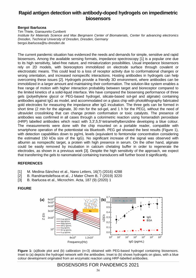
The current pandemic situation has evidenced the needs and demands for simple, sensitive and rapid biosensors. Among the available sensing formats, impedance spectroscopy [1] is a popular one due to its high sensitivity, label-free nature, and miniaturization possibilities. Usual impedance biosensors rely on 2D models, with bioreceptors immobilized on electrode surface through covalent or electrostatic means. This could lead to a loss of receptor activity due to conformational changes or wrong orientation, and increased nonspecific interactions. Hosting antibodies in hydrogels can help overcoming these issues [2]. Hydrogels provide a friendly 3D environment, where antibodies can be immobilized in a larger amount and maintaining their conformation. The solution-like system enables a free range of motion with higher interaction probability between target and bioreceptor compared to the limited kinetics of a solid-liquid interface. We have compared the biosensing performance of three gels (polyethylene glycol or PEG-based hydrogel, silicate-based sol-gel and alginate) containing antibodies against IgG as model, and accommodated on a glass chip with photolithography fabricated gold electrodes for measuring the impedance after IgG incubation. The three gels can be formed in short time (2 min for the alginate, 30 min for the sol-gel, and 1 h for the PEG), without the need of ultraviolet crosslinking that can change protein conformation or toxic catalysts. The presence of antibodies was confirmed in all cases through a colorimetric reaction using horseradish peroxidase (HRP) labelled antibodies which react with 3,3′,5,5′-tetramethylbenzidine developing a blue colour. The measurements were done with the chip mounted on a portable reader, compatible with smartphone operation of the potentiostat via Bluetooth. PEG gel showed the best results (Figure 1), with detection capabilities down to pg/mL levels (equivalent to femtomolar concentration considering the estimated 150 kDa size of the IgG). No significant increase of the signal was observed with albumin as nonspecific target, a protein with high presence in serum. On the other hand, alginate could be easily removed by incubation in calcium chelating buffer in order to regenerate the electrodes, as shown in a previous work [3]. Despite the high sensitivity of the approach, we expect that transferring the gels to nanomaterial containing transducers will further boost it significantly. REFERENCES [1] M. Medina-Sánchez et al., Nano Letters, 16(7) (2016) 4288 [2] B. Randriantsilefisoa et al., J Mater Chem B, 7 (2019) 3220 [3] B. Ibarlucea et al., Microchim. Acta, 187 (9) (2020) 1 FIGURE
8x100
1.2x101
105
2x10⁵
3x10⁵
4x10⁵
5x10⁵
6x10⁵
Z ()
Frequency(Hz)
0 pg IgG/mL
5pg IgG/mL
50pg IgG/mL
500 p IgG/mL
5ng IgG/mL
50 ng IgG/mL
5 g IgG/mL
100
101
102
103
104
105
106
0.0
0.1
0.2
0.3
0.4
0.5
0.6
0.7
IgG
BSAZ
()
IgG (pg/mL)
Basic hardware unit: SiNW-
FET
Hardware modification
Receptor PEG
200 µm 1µm
Au
(a) (b)
Figure 1: (a)Bode plot and (b) calibration (n=3) obtained with PEG-based hydrogel containing biosensors. Inset to (a) depicts the hydrogel network with the antibodies. Inset to (b) shows hydrogels on glass, with a blue colour development originated from an enzymatic reaction using HRP-labelled antibodies.
36
BIOSENSORS FOR PANDEMICS 2021
CRISPR-based detection of SARS-CoV-2 Guillermo Rodrigo M. Carmen Marqués, Raúl Ruiz, Rosa Márquez-Costa, Roser Montagud-Martínez, María Heras-Hernández I2SysBio, CSIC – Universitat de València, 46980 Paterna, Spain [email protected]
The gold-standard diagnostic technique for SARS-CoV-2 infection is based on polymerase chain reaction (PCR). However, this pandemic has highlighted the need for alternative methods that do not require precise equipment and can be closer to the patient. In this regard, CRISPR-Cas systems have emerged as suitable tools for the detection of SARS-CoV-2 genomic RNA [1,2]. In this flash presentation, we will provide an overview of our current research on this topic and what we propose to go beyond the state-of-the-art. In particular, we are exploiting the CRISPR-Cas13 system to detect virus-derived RNAs, coupling the CRISPR-Cas12 system with nanostructured materials, and developing a novel system based on CRISPR-Cas9 for multiplexed virus detection. REFERENCES [1] Broughton, J.P., et al. (2020) CRISPR-Cas12-based detection of SARS-CoV-2. Nat.
Biotechnol. 38, 870-874. [2] Joung, J., et al. (2020) Detection of SARS-CoV-2 with SHERLOCK one-pot testing. N. Engl. J.
Med. 383, 1492-1494.
37
BIOSENSORS FOR PANDEMICS 2021
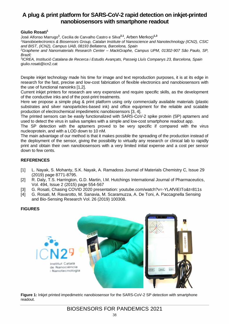
A plug & print platform for SARS-CoV-2 rapid detection on inkjet-printed nanobiosensors with smartphone readout
Giulio Rosati1 José Alfonso Marrugo1, Cecilia de Carvalho Castro e Silva2,1, Arben Merkoçi1,3 1Nanobioelectronics & Biosensors Group, Catalan Institute of Nanoscience and Nanotechnology (ICN2), CSIC and BIST, (ICN2), Campus UAB, 08193 Bellaterra, Barcelona, Spain 2Graphene and Nanomaterials Research Center – MackGraphe, Campus UPM, 01302-907 São Paulo, SP, Brazil; 3ICREA, Institució Catalana de Recerca i Estudis Avançats, Passeig Lluís Companys 23, Barcelona, Spain
Despite inkjet technology made his time for image and text reproduction purposes, it is at its edge in research for the fast, precise and low-cost fabrication of flexible electronics and nanobiosensors with the use of functional nanoinks [1,2]. Current inkjet printers for research are very expensive and require specific skills, as the development of the conductive inks and of the post-print treatments. Here we propose a simple plug & print platform using only commercially available materials (plastic substrates and silver nanoparticles-based ink) and office equipment for the reliable and scalable production of electrochemical impedimetric nanobiosensors [3, 4]. The printed sensors can be easily functionalized with SARS-CoV-2 spike protein (SP) aptamers and used to detect the virus in saliva samples with a simple and low-cost smartphone readout app. The SP detection with the aptamers proved to be very specific if compared with the virus nucleoprotein, and with a LOD down to 10 nM. The main advantage of our method is that it makes possible the spreading of the production instead of the deployment of the sensor, giving the possibility to virtually any research or clinical lab to rapidly print and obtain their own nanobiosensors with a very limited initial expense and a cost per sensor down to few cents. REFERENCES
[1] L. Nayak, S. Mohanty, S.K. Nayak, A. Ramadoss Journal of Materials Chemistry C, Issue 29
(2019) page 8771-8795. [2] R. Daly, T.S. Harrington, G.D. Martin, I.M. Hutchings International Journal of Pharmaceutics,
Vol. 494, Issue 2 (2015) page 554-567 [3] G. Rosati, Chasing COVID 2020 presentation: youtube.com/watch?v=-YLAfViEITo&t=811s [4] G. Rosati, M. Ravarotto, M. Sanavia, M. Scaramuzza, A. De Toni, A. Paccagnella Sensing
and Bio-Sensing Research Vol. 26 (2019) 100308. FIGURES
Figure 1: Inkjet printed impedimetric nanobiosensor for the SARS-CoV-2 SP detection with smartphone readout.
38
BIOSENSORS FOR PANDEMICS 2021
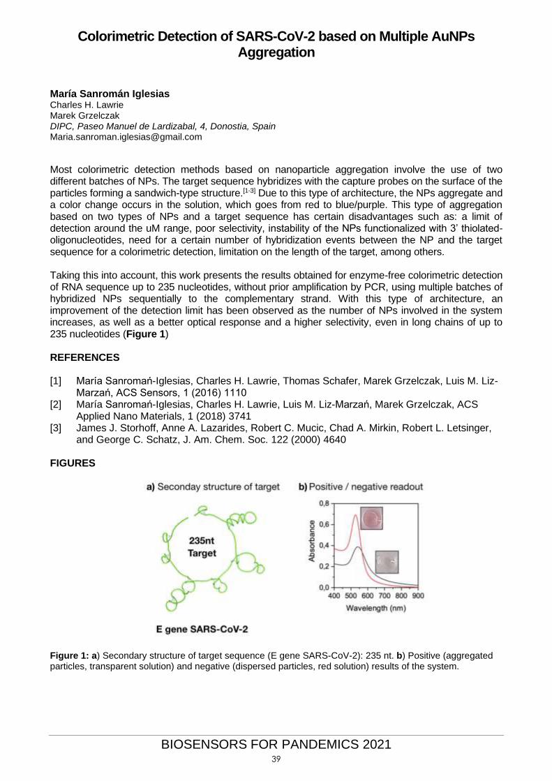
Colorimetric Detection of SARS-CoV-2 based on Multiple AuNPs Aggregation
María Sanromán Iglesias Charles H. Lawrie Marek Grzelczak DIPC, Paseo Manuel de Lardizabal, 4, Donostia, Spain [email protected]
Most colorimetric detection methods based on nanoparticle aggregation involve the use of two different batches of NPs. The target sequence hybridizes with the capture probes on the surface of the particles forming a sandwich-type structure.[1-3] Due to this type of architecture, the NPs aggregate and a color change occurs in the solution, which goes from red to blue/purple. This type of aggregation based on two types of NPs and a target sequence has certain disadvantages such as: a limit of detection around the uM range, poor selectivity, instability of the NPs functionalized with 3’ thiolated-oligonucleotides, need for a certain number of hybridization events between the NP and the target sequence for a colorimetric detection, limitation on the length of the target, among others. Taking this into account, this work presents the results obtained for enzyme-free colorimetric detection of RNA sequence up to 235 nucleotides, without prior amplification by PCR, using multiple batches of hybridized NPs sequentially to the complementary strand. With this type of architecture, an improvement of the detection limit has been observed as the number of NPs involved in the system increases, as well as a better optical response and a higher selectivity, even in long chains of up to 235 nucleotides (Figure 1) REFERENCES [1] María Sanromań-Iglesias, Charles H. Lawrie, Thomas Schafer, Marek Grzelczak, Luis M. Liz-
Marzań, ACS Sensors, 1 (2016) 1110 [2] María Sanromań-Iglesias, Charles H. Lawrie, Luis M. Liz-Marzań, Marek Grzelczak, ACS
Applied Nano Materials, 1 (2018) 3741 [3] James J. Storhoff, Anne A. Lazarides, Robert C. Mucic, Chad A. Mirkin, Robert L. Letsinger,
and George C. Schatz, J. Am. Chem. Soc. 122 (2000) 4640 FIGURES
Figure 1: a) Secondary structure of target sequence (E gene SARS-CoV-2): 235 nt. b) Positive (aggregated particles, transparent solution) and negative (dispersed particles, red solution) results of the system.
39
BIOSENSORS FOR PANDEMICS 2021
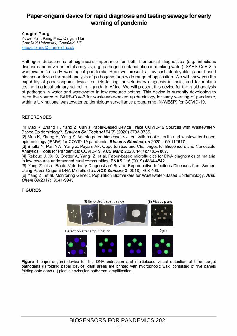
Paper-origami device for rapid diagnosis and testing sewage for early warning of pandemic
Zhugen Yang Yuwei Pan, Kang Mao, Qingxin Hui Cranfield University, Cranfield, UK [email protected] Pathogen detection is of significant importance for both biomedical diagnostics (e.g. infectious disease) and environmental analysis, e.g. pathogen contamination in drinking water), SARS-CoV-2 in wastewater for early warning of pandemic. Here we present a low-cost, deployable paper-based biosensor device for rapid analysis of pathogens for a wide range of application. We will show you the capability of paper-origami device for field-testing for veterinary diagnosis in India, and for malaria testing in a local primary school in Uganda in Africa. We will present this device for the rapid analysis of pathogen in water and wastewater in low resource setting. This device is currently developing to trace the source of SARS-CoV-2 for wastewater-based epidemiology for early warning of pandemic, within a UK national wastewater epidemiology surveillance programme (N-WESP) for COVID-19. REFERENCES [1] Mao K, Zhang H, Yang Z, Can a Paper-Based Device Trace COVID-19 Sources with Wastewater-Based Epidemiology?, Environ Sci Technol 54(7) (2020) 3733-3735. [2] Mao K, Zhang H, Yang Z. An integrated biosensor system with mobile health and wastewater-based epidemiology (iBMW) for COVID-19 pandemic. Biosens Bioelectron 2020, 169:112617. [3] Bhalla N, Pan YW, Yang Z, Payam AF: Opportunities and Challenges for Biosensors and Nanoscale Analytical Tools for Pandemics: COVID-19. ACS Nano 2020, 14(7):7783-7807. [4] Reboud J, Xu G, Gretter A, Yang Z. et al. Paper-based microfluidics for DNA diagnostics of malaria in low resource underserved rural communities. PNAS 116 (2019) 4834-4842. [5] Yang Z. et al. Rapid Veterinary Diagnosis of Bovine Reproductive Infectious Diseases from Semen Using Paper-Origami DNA Microfluidics. ACS Sensors 3 (2018): 403-409. [6] Yang Z., et al. Monitoring Genetic Population Biomarkers for Wastewater-Based Epidemiology. Anal Chem 89(2017): 9941-9945. FIGURES
Figure 1 paper-origami device for the DNA extraction and multiplexed visual detection of three target pathogens (I) folding paper device: dark areas are printed with hydrophobic wax, consisted of five panels folding onto each (II) plastic device for isothermal amplification.
P
N
1
2
3
(II) Plastic plate(I) Unfolded paper device
Detection after amplification
P
N
1
2
3
3mm
40

BIOSENSORS FOR PANDEMICS 2021
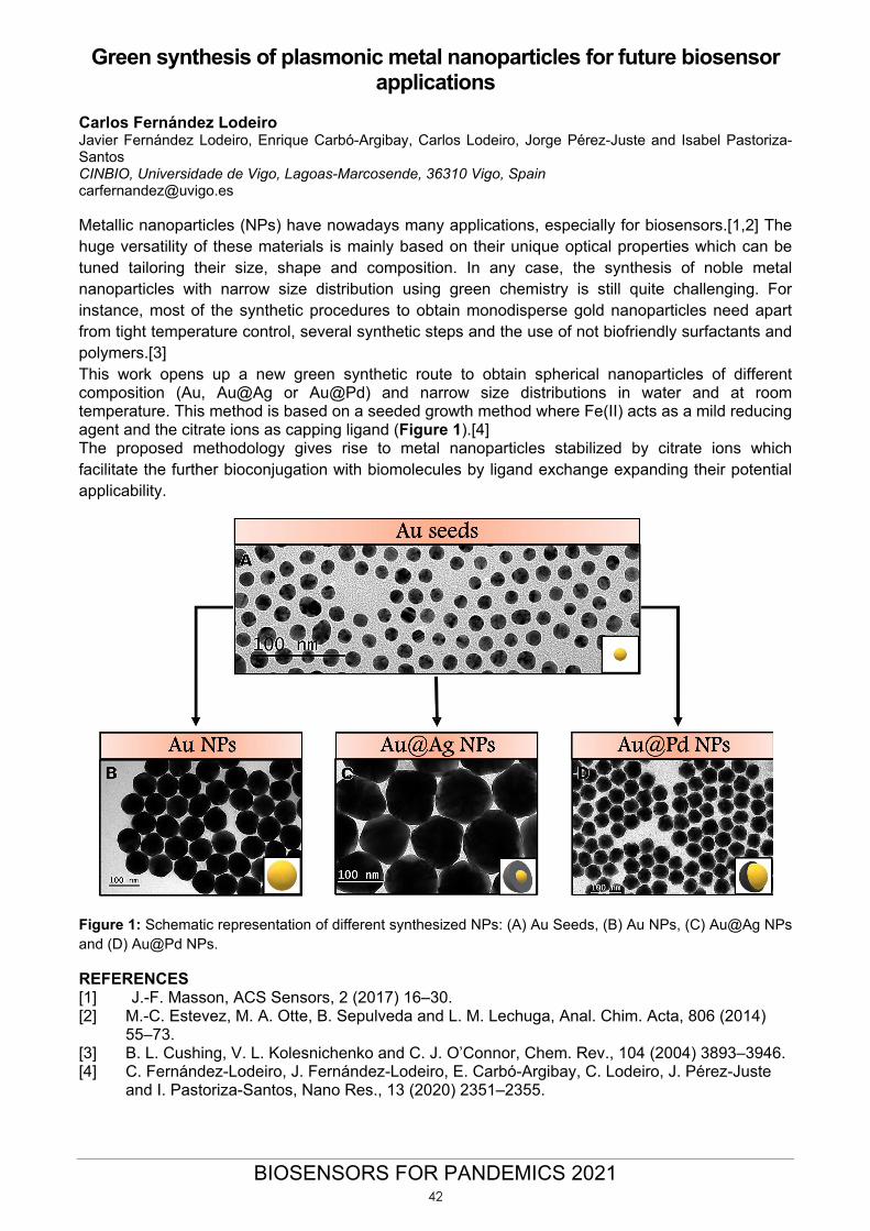
Green synthesis of plasmonic metal nanoparticles for future biosensor applications
Carlos Fernández Lodeiro Javier Fernández Lodeiro, Enrique Carbó-Argibay, Carlos Lodeiro, Jorge Pérez-Juste and Isabel Pastoriza-Santos CINBIO, Universidade de Vigo, Lagoas-Marcosende, 36310 Vigo, Spain [email protected] Metallic nanoparticles (NPs) have nowadays many applications, especially for biosensors.[1,2] The huge versatility of these materials is mainly based on their unique optical properties which can be tuned tailoring their size, shape and composition. In any case, the synthesis of noble metal nanoparticles with narrow size distribution using green chemistry is still quite challenging. For instance, most of the synthetic procedures to obtain monodisperse gold nanoparticles need apart from tight temperature control, several synthetic steps and the use of not biofriendly surfactants and polymers.[3] This work opens up a new green synthetic route to obtain spherical nanoparticles of different composition (Au, Au@Ag or Au@Pd) and narrow size distributions in water and at room temperature. This method is based on a seeded growth method where Fe(II) acts as a mild reducing agent and the citrate ions as capping ligand (Figure 1).[4] The proposed methodology gives rise to metal nanoparticles stabilized by citrate ions which facilitate the further bioconjugation with biomolecules by ligand exchange expanding their potential applicability.
Figure 1: Schematic representation of different synthesized NPs: (A) Au Seeds, (B) Au NPs, (C) Au@Ag NPs and (D) Au@Pd NPs.
REFERENCES [1] J.-F. Masson, ACS Sensors, 2 (2017) 16–30. [2] M.-C. Estevez, M. A. Otte, B. Sepulveda and L. M. Lechuga, Anal. Chim. Acta, 806 (2014)
55–73. [3] B. L. Cushing, V. L. Kolesnichenko and C. J. O’Connor, Chem. Rev., 104 (2004) 3893–3946. [4] C. Fernández-Lodeiro, J. Fernández-Lodeiro, E. Carbó-Argibay, C. Lodeiro, J. Pérez-Juste
and I. Pastoriza-Santos, Nano Res., 13 (2020) 2351–2355.
42
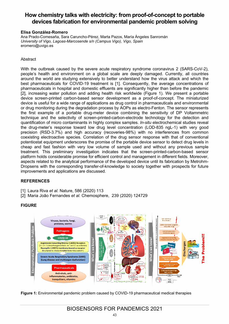
BIOSENSORS FOR PANDEMICS 2021
How chemistry talks with electricity: from proof-of-concept to portable devices fabrication for environmental pandemic problem solving
Elisa González-Romero Ana Prado-Comesaña, Sara Caruncho-Pérez, Marta Pazos, María Ángeles Sanromán University of Vigo, Lagoas-Marcosende s/n (Campus Vigo), Vigo, Spain [email protected] Abstract With the outbreak caused by the severe acute respiratory syndrome coronavirus 2 (SARS-CoV-2), people’s health and environment on a global scale are deeply damaged. Currently, all countries around the world are studying extensively to better understand how the virus attack and which the best pharmaceuticals for COVID-19 treatment is [1]. Consequently, the average concentrations of pharmaceuticals in hospital and domestic effluents are significantly higher than before the pandemic [2], increasing water pollution and adding health risk worldwide (Figure 1). We present a portable device screen-printed carbon-based sensor development as a proof-of-concept. The miniaturized device is useful for a wide range of applications as drug control in pharmaceuticals and environmental or drug monitoring during the degradation process by AOPs as electro-Fenton. The sensor represents the first example of a portable drug-meter device combining the sensitivity of DP Voltammetric technique and the selectivity of screen-printed-carbon-electrode technology for the detection and quantification of micro contaminants in highly complex samples. In-situ electrochemical studies reveal the drug-meter’s response toward low drug level concentration (LOD-835 ngL-1) with very good precision (RSD-3.7%) and high accuracy (recoveries-98%) with no interferences from common coexisting electroactive species. Correlation of the drug sensor response with that of conventional potentiostat equipment underscores the promise of the portable device sensor to detect drug levels in cheap and fast fashion with very low volume of sample used and without any previous sample treatment. This preliminary investigation indicates that the screen-printed-carbon-based sensor platform holds considerable promise for efficient control and management in different fields. Moreover, aspects related to the analytical performance of the developed device until its fabrication by Metrohm-Dropsens with the corresponding transfer-of-knowledge to society together with prospects for future improvements and applications are discussed. REFERENCES [1] Laura Riva et al. Nature, 586 (2020) 113 [2] Maria João Fernandes et al. Chemosphere, 239 (2020) 124729 FIGURE
Figure 1: Environmental pandemic problem caused by COVID-19 pharmaceutical medical therapies
43
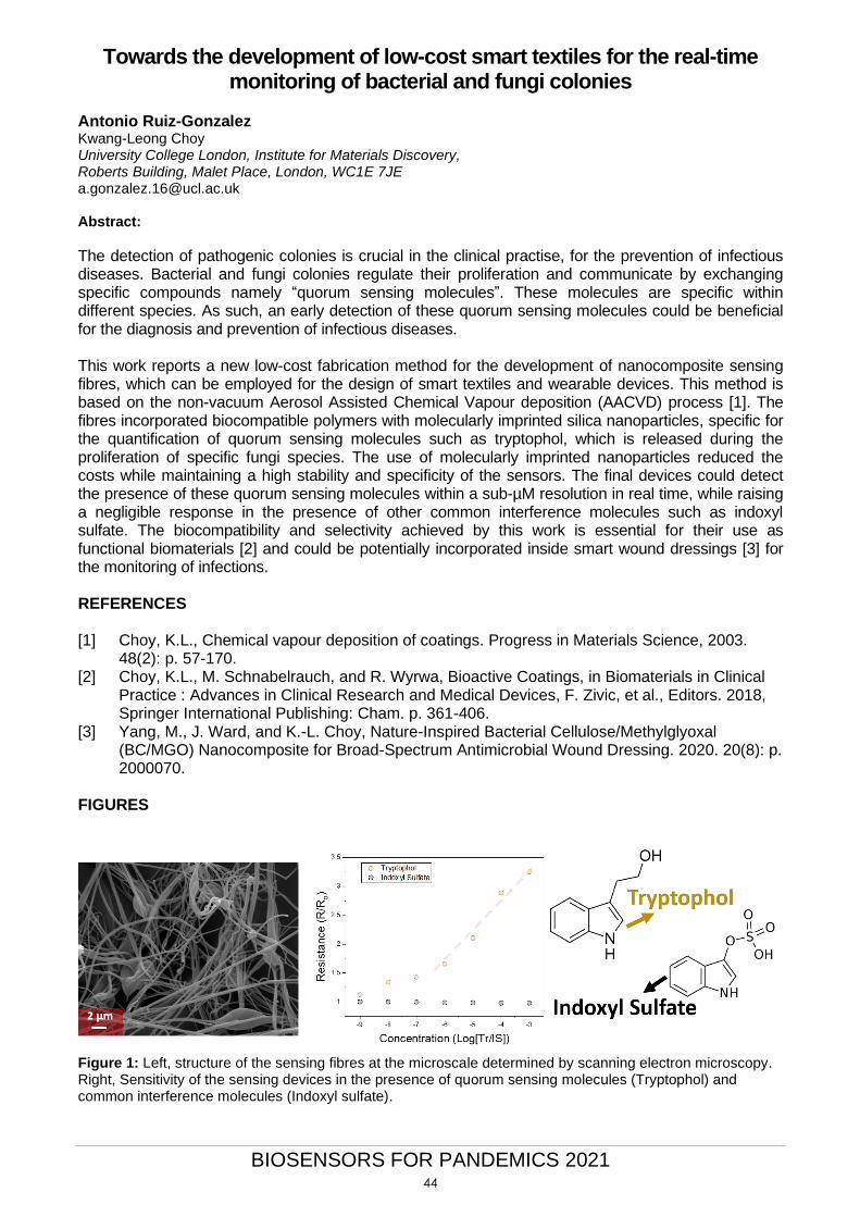
BIOSENSORS FOR PANDEMICS 2021
Towards the development of low-cost smart textiles for the real-time monitoring of bacterial and fungi colonies
Antonio Ruiz-Gonzalez Kwang-Leong Choy University College London, Institute for Materials Discovery, Roberts Building, Malet Place, London, WC1E 7JE [email protected] Abstract:
The detection of pathogenic colonies is crucial in the clinical practise, for the prevention of infectious diseases. Bacterial and fungi colonies regulate their proliferation and communicate by exchanging specific compounds namely “quorum sensing molecules”. These molecules are specific within different species. As such, an early detection of these quorum sensing molecules could be beneficial for the diagnosis and prevention of infectious diseases. This work reports a new low-cost fabrication method for the development of nanocomposite sensing fibres, which can be employed for the design of smart textiles and wearable devices. This method is based on the non-vacuum Aerosol Assisted Chemical Vapour deposition (AACVD) process [1]. The fibres incorporated biocompatible polymers with molecularly imprinted silica nanoparticles, specific for the quantification of quorum sensing molecules such as tryptophol, which is released during the proliferation of specific fungi species. The use of molecularly imprinted nanoparticles reduced the costs while maintaining a high stability and specificity of the sensors. The final devices could detect the presence of these quorum sensing molecules within a sub-µM resolution in real time, while raising a negligible response in the presence of other common interference molecules such as indoxyl sulfate. The biocompatibility and selectivity achieved by this work is essential for their use as functional biomaterials [2] and could be potentially incorporated inside smart wound dressings [3] for the monitoring of infections. REFERENCES [1] Choy, K.L., Chemical vapour deposition of coatings. Progress in Materials Science, 2003.
48(2): p. 57-170. [2] Choy, K.L., M. Schnabelrauch, and R. Wyrwa, Bioactive Coatings, in Biomaterials in Clinical
Practice : Advances in Clinical Research and Medical Devices, F. Zivic, et al., Editors. 2018, Springer International Publishing: Cham. p. 361-406.
[3] Yang, M., J. Ward, and K.-L. Choy, Nature-Inspired Bacterial Cellulose/Methylglyoxal (BC/MGO) Nanocomposite for Broad-Spectrum Antimicrobial Wound Dressing. 2020. 20(8): p. 2000070.
FIGURES
Figure 1: Left, structure of the sensing fibres at the microscale determined by scanning electron microscopy. Right, Sensitivity of the sensing devices in the presence of quorum sensing molecules (Tryptophol) and common interference molecules (Indoxyl sulfate).
44

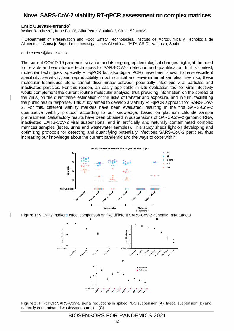
BIOSENSORS FOR PANDEMICS 2021
Novel SARS-CoV-2 viability RT-qPCR assessment on complex matrices Enric Cuevas-Ferrando1 Walter Randazzo1, Irene Falcó1, Alba Pérez-Cataluña1, Gloria Sánchez1
1 Department of Preservation and Food Safety Technologies, Instituto de Agroquímica y Tecnología de Alimentos – Consejo Superior de Investigaciones Científicas (IATA-CSIC), Valencia, Spain
The current COVID-19 pandemic situation and its ongoing epidemiological changes highlight the need for reliable and easy-to-use techniques for SARS-CoV-2 detection and quantification. In this context, molecular techniques (specially RT-qPCR but also digital PCR) have been shown to have excellent specificity, sensitivity, and reproducibility in both clinical and environmental samples. Even so, these molecular techniques alone cannot discriminate between potentially infectious viral particles and inactivated particles. For this reason, an easily applicable in situ evaluation tool for viral infectivity would complement the current routine molecular analysis, thus providing information on the spread of the virus, on the quantitative estimation of the risks of transfer and exposure, and in turn, facilitating the public health response. This study aimed to develop a viability RT-qPCR approach for SARS-CoV-2. For this, different viability markers have been evaluated, resulting in the first SARS-CoV-2 quantitative viability protocol according to our knowledge, based on platinum chloride sample pretreatment. Satisfactory results have been obtained in suspensions of SARS-CoV-2 genomic RNA, inactivated SARS-CoV-2 viral suspensions, and in artificially and naturally contaminated complex matrices samples (feces, urine and wastewater samples). This study sheds light on developing and optimizing protocols for detecting and quantifying potentially infectious SARS-CoV-2 particles, thus increasing our knowledge about the current pandemic and the ways to cope with it.
Figure 1: Viability markers effect comparison on five different SARS-CoV-2 genomic RNA targets.
Figure 2: RT-qPCR SARS-CoV-2 signal reductions in spiked PBS suspension (A), faecal suspension (B) and naturally contaminated wastewater samples (C).
46
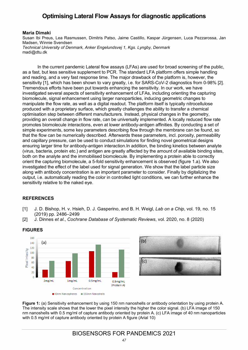
BIOSENSORS FOR PANDEMICS 2021
Optimising Lateral Flow Assays for diagnostic applications Maria Dimaki Susan Ibi Preus, Lea Rasmussen, Dimitris Patso, Jaime Castillo, Kaspar Jürgensen, Luca Pezzarossa, Jan Madsen, Winnie Svendsen Technical University of Denmark, Anker Engelundsvej 1, Kgs. Lyngby, Denmark [email protected]
In the current pandemic Lateral flow assays (LFAs) are used for broad screening of the public, as a fast, but less sensitive supplement to PCR. The standard LFA platform offers simple handling and reading, and a very fast response time. The major drawback of the platform is, however, the sensitivity [1], which has been shown to vary greatly, i.e. for SARS-CoV-2 diagnostics from 0-98% [2]. Tremendous efforts have been put towards enhancing the sensitivity. In our work, we have investigated several aspects of sensitivity enhancement of LFAs, including orienting the capturing biomolecule, signal enhancement using larger nanoparticles, inducing geometric changes to manipulate the flow rate, as well as a digital readout. The platform itself is typically nitrocellulose produced with a proprietary surface, which greatly challenges the ability to transfer a chemical optimisation step between different manufacturers. Instead, physical changes in the geometry, providing an overall change in flow rate, can be universally implemented. A locally reduced flow rate promotes biomolecule interactions, even at lower antibody-antigen affinities. By conducting a set of simple experiments, some key parameters describing flow through the membrane can be found, so that the flow can be numerically described. Afterwards these parameters, incl. porosity, permeability and capillary pressure, can be used to conduct simulations for finding novel geometrical designs ensuring larger time for antibody-antigen interaction.In addition, the binding kinetics between analyte (virus, bacteria, protein etc.) and antigen are greatly affected by the amount of available binding sites, both on the analyte and the immobilised biomolecule. By implementing a protein able to correctly orient the capturing biomolecule, a 5-fold sensitivity enhancement is observed (figure 1.a). We also investigated the effect of the label used for signal generation. We show that the label particle size along with antibody concentration is an important parameter to consider. Finally by digitalizing the output, i.e. automatically reading the color in controlled light conditions, we can further enhance the sensitivity relative to the naked eye.
REFERENCES [1] J. D. Bishop, H. v. Hsieh, D. J. Gasperino, and B. H. Weigl, Lab on a Chip, vol. 19, no. 15
(2019) pp. 2486–2499 [2] J. Dinnes et al., Cochrane Database of Systematic Reviews, vol. 2020, no. 8 (2020) FIGURES
Figure 1: (a) Sensitivity enhancement by using 150 nm nanoshells or antibody orientation by using protein A. The intensity scale shows that the lower the pixel intensity the higher the color signal. (b) LFA image of 150 nm nanoshells with 0.5 mg/ml of capture antibody oriented by protein A. (c) LFA image of 40 nm nanoparticles with 0.5 mg/ml of capture antibody oriented by protein A figure (Arial 10)
(a) (b)
(c)
47

BIOSENSORS FOR PANDEMICS 2021
AuNP-based lateral flow assay for detection of SARS-CoV-2 nucleoprotein: implicit design challenges in a new pandemic
Liming Hu a, Enric Calucho a, Celia Fuentes-Chust a, Ruslan Álvarez-Diduk a, Andrea Idili a, Claudio
Parolo a, Arben Merkoçi a,b a Nanobioelectronics & Biosensors Group, Institut Català de Nanociència i Nanotecnologia (ICN2), Campus UAB, 08193 Bellaterra, Barcelona, Spain b Institució Catalana de Recerca i Estudis Avançats (ICREA), 08010 Barcelona, Spain [email protected]
ABSTRACT 2020 was marked by a SARS-CoV-2 outbreak that has claimed more than 2 million lives around the world [1], pushed healthcare systems to their limits, shaken the economy at all levels and greatly affected people’s daily life. Roughly one year after the appearance of the first case of COVID-19, although there are vaccines available, the eradication of the pandemic is an enduring challenge due to the extremely rapid infectivity and the emergence of new strains of the virus. Therefore, there is the need of diagnostic devices dedicated to each situation: general screening, herd immunity, strain identification, etc. Lateral flow assays make ideal point-of-care testing platforms due to their simplicity, low cost and user friendliness [2]. Moreover, paper can be easily functionalized with different kind of bioreceptors (e.g., antibodies, DNA, etc.). Herein, we present a AuNP-based lateral flow device for the detection of SARS-CoV-2 nucleoprotein, from the screening of antibodies to the preliminary testing of the device [3]. In the scenario of a new pandemic, the initial lack of bioreceptors for the development of diagnostic platforms creates the conditions for a race towards the launch of the first commercially available products. Therefore, the performance of these new bioreceptors is unbeknownst to researchers during the first stages of the pandemic. Here we will show and discuss the standardization procedures and previous validation of receptor as guidelines to be used for urgent design and application of LFA. ELISA tests were carried out to determine the most efficient set of antibodies for its later transfer to a paper-based format. However, changing the environment on which antibodies work doesn’t always translate well, thus we take the opportunity to raise this issue in the context of a congress devoted to biosensors for pandemics. REFERENCES [1] WHO Coronavirus Disease (COVID-19) Dashboard; URL: https://covid19.who.int/ [2] K. J. Land, D. I. Boeras, X. S. Chen, A. R. Ramsay, R. W. Peeling, Nat. Microbiol. (2019), 4,
46. [3] Parolo, C., Sena-Torralba, A., Bergua, J.F., Calucho, E., Fuentes-Chust, C., Hu, L., Rivas, L.,
Álvarez-Diduk, R., Nguyen, E.P., Cinti, S., Quesada-González, D., and Merkoçi, A. Nature Protocols, 15 (2020) 3788-3816.
48
BIOSENSORS FOR PANDEMICS 2021
Production and characterization of pseudotyped particles to develop an immunosensor for SARS CoV 2
Maria-Camila Lopez1 Viviana Vasquez2
Javier A Jaimes3
Mauricio Rojas4
Jahir Orozco2
Maria-Cristina Navas1, *
1Grupo de Gastrohepatologia, Facultad de Medicina, Universidad de Antioquia, Lab 434 SIU, UdeA, Medellin, Colombia. 2Max Planck Tandem Group in Nanobioengineering, University of Antioquia, Complejo Ruta N, Torre A, Laboratorio 4-166, Calle 67, Nº 52-20, Medellin, 050010, Colombia. 3Department of Microbiology & immunology, College of Veterinary Medicine, Cornell University, 930 Campus Rd., VMC Box 5, Ithaca, NY 14853-640, United States of America 4Grupo de Inmunología Celular e Inmunogenetica, Facultad de Medicina, Unidad de Citometria de Flujo,
Universidad de Antioquia, Medellin, Colombia. *[email protected]
Studying SARS-CoV-2 requires biosafety level 3 (BSL-3) facilities due to its virulence and transmission risk by aerosols. We have produced viral-like particles that express the spike glycoprotein of this emergent coronavirus by taking advantage of the pseudotyped particles tools to circumvent this issue. The resultant pseudovirions display the spike protein of SARS-CoV-2 expressed on the surface of the particle and therefore could mimic the interaction between the virus and the angiotensin-converting enzyme 2 (ACE2), which has been described as the primary host cell target. This simple, but highly beneficial tool, significantly reduces biological risk in the first steps of the immunosensor development requiring only a BSL-2 facility. The pseudovirions were obtained by co-transfection of three plasmids (gag-pol of Murine Leukemia Virus (MLV), firefly luciferase gene/MLV RNA packaging signal/ 5'/3' flanking MLV long terminal repeat and SARS-CoV2 S) in HEK-293 cells and harvested 72 hours post-transfection [1]. The product was characterized by flow cytometry and indirect immunofluorescence. The spike protein was detected on the surface of the pseudovirions using an anti-SARS-CoV-2 Spike polyclonal antibody and an anti-Rabbit IgG secondary antibody cross-linked allophycocyanin, with pseudotyped particles in the presence of the secondary antibody as fluorescence control. The infectivity quantification of the pseudovirions was assessed by transduction of VERO-E6 cell using 10-fold serial dilutions; the Luciferase activity was detected 72 horas post transduction and the title was calculated based on the endpoint dilution and relative luciferase expression. Current experiments are directed to detect the pseudovirions by the interaction with a thiolated ACE2-reactive peptide linked to maleimide-coated magnetic particles, which electrochemical reaction will be followed by chronoamperometry by a biotinylated anti-ACE2 antibody conjugated with Streptavidin-Horseradish peroxidase (HRP). REFERENCES [1] Millet JK, Tang T, Nathan L, Jaimes JA, Hsu H-L., Daniel S, Whittaker GR. Journal of
Visualized Experiments, 145 (2019) 3-9
49


![WHOLE OF - SOCIETY PANDEMIC READINESS€¦ · • Pandemics ARE worldwide epidemics • Pandemics ARE unpredictable [Origin - Timing - Severity - Duration] • Pandemics HAVE happened](https://static.fdocuments.in/doc/165x107/60219e6a89cf726c976d409c/whole-of-society-pandemic-readiness-a-pandemics-are-worldwide-epidemics-a.jpg)