Bioresearch Communications
10
904 Volume 6, Issue 2, July 2020 Original Article Seasonal Variation and Molecular Characterization of Vibrio parahaemolyticus Isolated from Karnaphuni River and Estuary of Chittagong, Bangladesh Khorshed Alam 1* , Fazle Rabbi 2 , Dr. C.R Ahsan 2 , Dr. Sharmin Sultana 1 1 Department of Microbiology, University of Chittagong 2 Department of Microbiology, University of Dhaka ABSTRACT: To determine whether the environmental variables governing the dynamics of Vibrio parahaemolyticus populations, sediment samples were collected with or without 1% NaCl from the Karnaphuli River and Karnaphuli Estuary of Chittagong, Bangladesh. The MPN method was used for quantitative analysis of the organisms. The isolates were subjected to biochemical tests for confirmation and also for the molecular characterization including assessment of virulence properties. Higher occurrence of V. parahaemolyticus was observed during periods of lower salinity. Thermostable direct hemolysin (TDH) and TDH-related hemolysin (TRH) are considered as major virulence factors for the organism and are coded by the tdh and trh genes, respectively. This study was, therefore, carried out to observe the seasonal variation and to investigate the occurrence of tdh and trh containing V. parahaemolyticus, besides the brackish environments. Twelve isolates out of 15 showed identical patterns to biochemical test profiles of the reference strain. This was further confirmed by PCR using primers specific for toxR gene. Antibiogram using nine commercial antibiotics showed that these isolates were sensitive to all representative antibiotics except ampicillin and streptomycin. All V. parahaemolyticus isolates were found negative for tdh gene which was confirmed by both reverse passive latex agglutination test and PCR. The most important finding of the study was the regular occurrence of V. parahaemolyticus in river of 0% salinity, though most of them are nonpathogenic strains. However, they could not grow in nutrient agar media having no NaCl. Keywords: Vibrio parahaemolyticus, Seasonal Variation, Thermostable direct hemolysin-TDH, TDH related hemolysin-TRH, Antibiogram. Article History Received: 13 January 2020 Accepted: 09 May 2020 www.bioresearchcommunications.com Corresponding author Khorshed Alam Department of Microbiology University of Chittagong Chittagong, Chittagong-4331 Bangladesh. Email: [email protected] INTRODUCTION Vibrio parahaemolyticus, a well-known human pathogen, is the leading cause of gastroenteritis due to the consumption of seafood, primarily raw or improperly cooked shellfish worldwide (Tison and Kelly, 1984). This organism is considered to be restricted to a saline environment, and it requires Na + for survival and growth, but several studies suggest it also occurs in brackish water (Bockemühl et al., 1986), plankton, and fish (Sarkar et al., 1985) of freshwater environments. This bacterium causes approximately half of the all food poisoning cases in Taiwan, Japan and several Southeast Asian countries Bioresearch Communications Volume 06, Issue 02, July 2020 Journal Homepage: www.bioresearchcommunications.com
Transcript of Bioresearch Communications
904 Volume 6, Issue 2, July 2020
Original Article
Seasonal Variation and Molecular Characterization of Vibrio parahaemolyticus
Isolated from Karnaphuni River and Estuary of Chittagong, Bangladesh
Khorshed Alam1*
, Fazle Rabbi2, Dr. C.R Ahsan
2, Dr. Sharmin Sultana
1
1Department of Microbiology, University of Chittagong
2Department of Microbiology, University of Dhaka
ABSTRACT: To determine whether the environmental variables
governing the dynamics of Vibrio parahaemolyticus populations,
sediment samples were collected with or without 1% NaCl from the
Karnaphuli River and Karnaphuli Estuary of Chittagong, Bangladesh.
The MPN method was used for quantitative analysis of the organisms.
The isolates were subjected to biochemical tests for confirmation and
also for the molecular characterization including assessment of
virulence properties. Higher occurrence of V. parahaemolyticus was
observed during periods of lower salinity. Thermostable direct
hemolysin (TDH) and TDH-related hemolysin (TRH) are considered as
major virulence factors for the organism and are coded by the tdh and
trh genes, respectively. This study was, therefore, carried out to observe
the seasonal variation and to investigate the occurrence of tdh and trh
containing V. parahaemolyticus, besides the brackish environments.
Twelve isolates out of 15 showed identical patterns to biochemical test
profiles of the reference strain. This was further confirmed by PCR
using primers specific for toxR gene. Antibiogram using nine
commercial antibiotics showed that these isolates were sensitive to all
representative antibiotics except ampicillin and streptomycin. All V.
parahaemolyticus isolates were found negative for tdh gene which was
confirmed by both reverse passive latex agglutination test and PCR.
The most important finding of the study was the regular occurrence of
V. parahaemolyticus in river of 0% salinity, though most of them are
nonpathogenic strains. However, they could not grow in nutrient agar
media having no NaCl.
Keywords: Vibrio parahaemolyticus, Seasonal Variation,
Thermostable direct hemolysin-TDH, TDH related hemolysin-TRH,
Antibiogram.
Article History Received: 13 January 2020
Accepted: 09 May 2020
www.bioresearchcommunications.com
Corresponding author Khorshed Alam
Department of Microbiology
University of Chittagong
Chittagong, Chittagong-4331
Bangladesh.
Email: [email protected]
INTRODUCTION Vibrio parahaemolyticus, a well-known human
pathogen, is the leading cause of gastroenteritis due to
the consumption of seafood, primarily raw or
improperly cooked shellfish worldwide (Tison and
Kelly, 1984). This organism is considered to be
restricted to a saline environment, and it requires Na+
for survival and growth, but several studies suggest it
also occurs in brackish water (Bockemühl et al.,
1986), plankton, and fish (Sarkar et al., 1985) of
freshwater environments. This bacterium causes
approximately half of the all food poisoning cases in
Taiwan, Japan and several Southeast Asian countries
Bioresearch Communications Volume 06, Issue 02, July 2020
Journal Homepage: www.bioresearchcommunications.com
Khorshed Alam. et. al. Seasonal Variation and Molecular Characterization
905 Volume 6, Issue 2, July 2020
(Joseph et al., 1982, Chiou et al., 1991). It is also
reported to be an important agent of travelers'
diarrhoea and has also occasionally been associated
with extra intestinal infections, including wounds
(Blake et al., 1980). Serotyping can differentiate
isolates of V. parahaemolyticus and 13 O types and 71
K types have been identified (Iguchi et al., 1995).
Since 1996, an increased incidence of gastroenteritis in
many parts of the world has been associated with V.
parahaemolyticus serotype O3:K6 (Chiou et al., 1991,
Okuda et al., 1997). The association of O3:K6
serotype with large-scale food-borne disease outbreaks
in Taiwan, Laos, Japan, Thailand, Korea and the
United States between 1997 and 1998 suggest this
organism may have an unusual capacity to be
transmitted by foods and/or to cause human infection
and has pandemic potential (Matsumoto et al.,2000).
Since 1998, V. parahaemolyticus strains belonging to
other two serotypes, O4:K68 and O1:KUT (UT
indicates untypable), have also been isolated with
increasing frequency from diarrheal patients
(Chowdhury et al., 2000). Pathogenic strains of V.
parahaemolyticus generally produce a thermostable
direct haemolysin (TDH) that is associated with the
Kanagawa phenomenon (KP) and/or thermostable
direct haemolysin-related haemolysin (TRH). Both
TDH and TRH encoded by tdh and trh genes
respectively are now recognized as major virulence
factors in the pathogenesis of V. parahaemolyticus
(Honda and Iida, 1993, Nishibuchi and Kaper, 1995).
A separate gene, Thermolabile haemolysin (tlh) has
also been characterized (Taniguchi et al., 1986). This
gene was shown to be present in all of the V.
parahaemolyticus strains tested previously (Taniguchi
et al., 1986). The pathogenicity of V.
parahaemolyticus isolates has traditionally been
correlated with the production of the thermostable
direct haemolysin (Vp-TDH) which is responsible for
the beta-haemolysis observed when the organisms are
plated on a modified blood agar known as Wagatsuma
agar (Chun et al., 1975). More than 90% of clinical V.
parahaemolyticus isolates but less than 1% of food or
environmental strains produce TDH or possess tdh
(DePaola et al., 2000). Although V. parahaemolyticus
has been thought to be a marine bacterium because of
its indispensable Na+ requirement for survival and
growth, the occurrence of this moderately halophilic
organism in fresh water has also been observed
(Venkateswaran et al., 1989). These evidence may
suggest that the habitats of V. parahaemolyticus is
somewhere out of marine environment which inspired
us to turn our attention towards the rivers of
Bangladesh besides the estuarine environments. The
aim of this study was therefore to investigate the
occurrence of V. parahaemolyticus in the estuaries as
well as in the rivers of Bangladesh and to determine
seasonal variation, the phenotypic and genotypic traits
of these V. parahaemolyticus strains isolated from the
sediment samples of Karnaphuli river and also from an
estuary in the Chittagong near the Bay of Bengal by
biochemical characterization, tolerance of NaCl
concentration, serotyping, antibiogram, followed by
PCR for toxR, tdh, and trh and Reverse Passive Latex
Agglutination for toxin detection. Thus this study
becomes significant as it presents the molecular
characterization of the environmental V.
parahaemolyticus isolates of these regions that may be
responsible for diarrheal cases occurring sporadically.
MATERIALS AND METHODS Sampling sites
Sampling was done from two sampling sites of
different aquatic bodies in Karnafuli River and Estuary
located in Chittagong district from January 2008 to
August 2008.
Collection of environmental samples
Sediment samples from two different water bodies in
Karnaphuli River and Karnaphuli Estuary were
collected at 15-day intervals from January 2008 to
August 2008. Therefore, a total of 96 samples were
collected during the time period. Sediment samples
were collected from 10 cm depth of the shore. Several
grams of samples were aseptically collected by
wearing sterile gloves to sterile plastic bags having 1%
salt and another set was without salt. Samples were
stored at room temperature until processing in the
laboratory.
Cultural properties on X-VP plate and counting
Morphological characteristics of colonies developed
after incubation on X-VP agar plate were carefully
studied and recorded. Following overnight incubation
at 37°C, blue-green colonies on X-VP agar plates were
selected as suspected V. parahaemolyticus. They were
counted as drop plate method. Five to ten suspected V.
parahaemolyticus colonies were picked up from the X-
VP agar plate and sub cultured onto fresh X-VP plate
for pure culture.
Screening of suspected isolates on TCBS plate
Suspected V. parahaemolyticus isolates from the fresh
X-VP plate were streaked onto thiosulphate citrate bile
salt sucrose (TCBS) agar (TCBS agar; Nissui Co.,
Japan) plate for presumptive confirmation as V.
parahaemolyticus. Following overnight incubation at
37°C, isolates that produced mucoid, raised, green
colonies on TCBS agar plates were selected as V.
parahaemolyticus. The selected isolates were
preserved in preservation medium then subjected to
biochemical identification and other molecular based
assays.
Reference strains
V. parahaemolyticus strains of VP11 (new clone of
serotype O3:K6, harboring tdh derived from a patient
in Aomori Prefecture in Japan in 2000) and AQ3857
(serotype O1:K1, harboring both tdh and trh isolated
from international traveler in Hong Kong in 1983)
were used as reference strains to compare the
Khorshed Alam. et. al. Seasonal Variation and Molecular Characterization
906 Volume 6, Issue 2, July 2020
biochemical characteristics and gene analysis.
Biochemical, serological and molecular tests
according to the standard procedure reconfirmed the
strains.
Biochemical identification
After incubation, characteristic colonies on TCBS agar
plates selected as V. parahaemolyticus were subjected
to the following standard biochemical tests in order to
confirm the isolates as V. parahaemolyticus.
Biochemical tests were performed according to the
methods described in Microbiology Laboratory
Manual (Cappuccino and Sherman, 1992). The
biochemical tests were Kligler’s iron agar test, lysine
decarboxylase test, oxidase test, citrate utilization test,
motility test, ornithine decarboxylase test, indole
production test, methyl-red test, Voges-Proskauer test
and salt tolerance test.
Pure culture preparation
Pure cultures of test organisms were obtained by sub
culturing on LB agar with 3% NaCl plates after
incubation at 37°C for 18-24 h.
Salt tolerance test
All isolates were tested for their salt tolerance in
alkaline peptone agar (APA) plate containing 0%, 3%,
7% and 10% (w/v) sodium chloride. Fresh culture was
streaked onto an APA plate and growth was observed
visually after 24 h. of incubation at 37°C.
Detection of toxR gene by PCR
Isolates confirmed by the biochemical tests were
further analyzed for the presence of toxR gene of V.
parahaemolyticus by PCR using primers specific for
toxR.
Preparation of template DNA
Each isolate of V. parahaemolyticus was cultured in
LB broth containing 3% NaCl and was incubated at
37°C for overnight. The cell pellet was harvested by
centrifugation of 1 ml of culture at 10,000 rpm for 5
minutes. The supernatant was discarded and the cell
pellet was resuspended in phosphate buffered saline
(PBS), and then washed by centrifugation at 10,000
rpm for 3 minutes. This procedure was repeated for
two times. The pellet was re-suspended in TE buffer
solution and boiled for 10 minutes. Then it was put
immediately into ice for minimum 30 minutes. After
that centrifuged at 13,000 rpm for 10 minutes on a
tabletop centrifuge (Eppendorf centrifuge, 5804,
Germany) and the supernatant was collected. The
supernatant was used as the template solution for PCR.
The template DNA was then mixed with reaction
mixture.
Post PCR detection of amplified DNA by
electrophoretic analysis
The successful amplification of the toxR gene was
examined by resolving the PCR products in 1.5%
agarose gel. 0.75g agarose (Sigma) was dissolved in
50 ml of 1X Tris-borate EDTA (TBE) buffer to give a
final concentration of 1.5% agarose and was heated to
dissolve in a microwave oven (Model: D90N30 ATP,
Butterfly, China) for about 2.5 - 3 min. 1.5 μl of 10
mg/ml ethidium bromide was mixed with the molten
agarose. When the temperature came down to 50°C,
the gel was poured onto the gel tray already fixed with
appropriate combs. Following solidification of the gel,
it was submerged in 1 X TBE buffer in a gel running
tank. 8 μl PCR product was mixed with 2 μl of 10X
gel loading dye and loaded into the slots of the gel
with the aid of a micropipette. Electrophoresis was
continued with 60 volts until DNA fragments were
separated. The EtBr stained DNA bands were
observed on a UV transilluminator (Model TS-40,
USA). Photographs were taken using Gel
Documentation machine attached to a computer and
bands were analyzed with “Quality one” software
(Bio-Rad, USA). The PCR products sizes were
estimated using the 100 bp marker (Invitrogen, USA)
tdh Toxin Assay by Reverse Passive Latex
Agglutination Test (RPLA)
tdh toxin assay was done using a commercially
available Reverse Passive Latex Agglutination
(RPLA) kit (Deneke and Colwell, 1973)(Denka
Seiken, Tokyo, Japan)
Detection of tdh and trh virulence gene by PCR
Isolates showing positive results for toxR gene by PCR
were further analyzed for the presence of virulence
associated gene tdh and trh of V. parahaemolyticus by
PCR using primers specific for tdh.
Electrophoretic analysis
The PCR amplified mixture was resolved by
electrophoresis in a 1.5% agarose gel to detect the
amplicons following the method already described in
above section.
Determination of Antibiotic Susceptibility of the
test isolates
Susceptibility of each test isolate to different
antibiotics was determined in vitro by employing the
modified Kirby-Bauer (Bauer et al., 1966) method.
The zone diameters for individual antimicrobial agent
were then translated into susceptible, intermediate,
moderately susceptible, or resistant categories
according to the interpretation table (Standards and
Watts, 1999).
RESULTS Primary isolation of V. parahaemolyticus from
environment
Alkaline peptone water (APW) enriched
environmental samples were drop plate on X-VP agar
medium were incubated for 24 hours at 37°C. Then
observed for the presence of V. parahaemolyticus
colonies. On the X-VP agar medium, the suspected V.
parahaemolyticus were found to appear as
characteristic blue green colonies and count them by
MPN method. Count expressed in log cfu/gm shown in
figure 3.3 in different period of different water bodies.
Of them only four colonies were transferred to TCBS
agar plate of each sampling. On the TCBS agar plates,
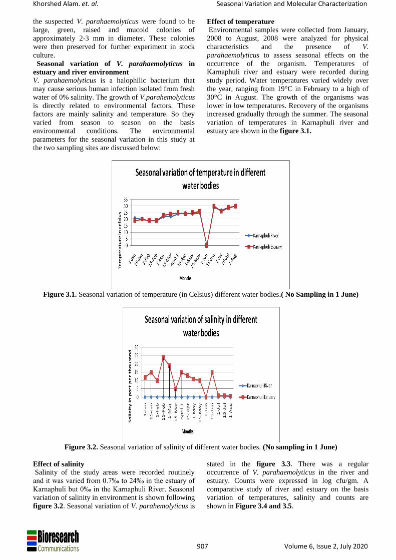
Khorshed Alam. et. al. Seasonal Variation and Molecular Characterization
907 Volume 6, Issue 2, July 2020
the suspected V. parahaemolyticus were found to be
large, green, raised and mucoid colonies of
approximately 2-3 mm in diameter. These colonies
were then preserved for further experiment in stock
culture.
Seasonal variation of V. parahaemolyticus in
estuary and river environment
V. parahaemolyticus is a halophilic bacterium that
may cause serious human infection isolated from fresh
water of 0% salinity. The growth of V.parahemolyticus
is directly related to environmental factors. These
factors are mainly salinity and temperature. So they
varied from season to season on the basis
environmental conditions. The environmental
parameters for the seasonal variation in this study at
the two sampling sites are discussed below:
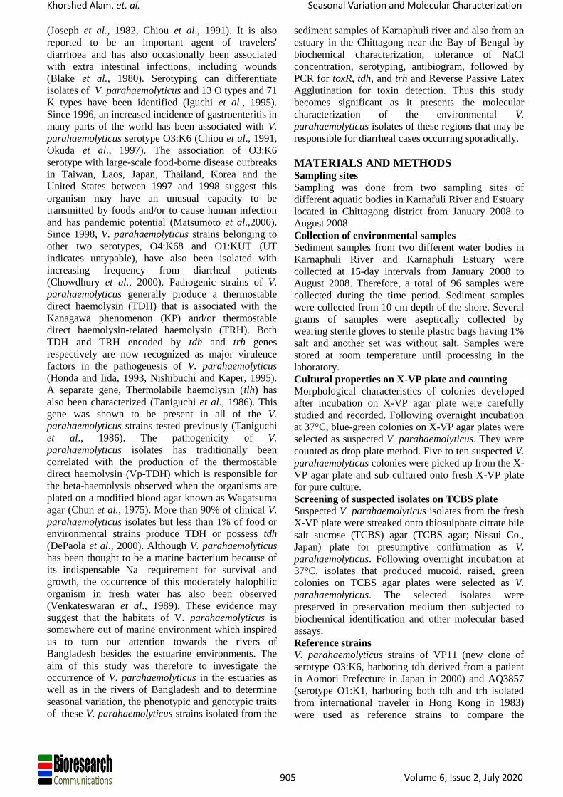
Effect of temperature
Environmental samples were collected from January,
2008 to August, 2008 were analyzed for physical
characteristics and the presence of V.
parahaemolyticus to assess seasonal effects on the
occurrence of the organism. Temperatures of
Karnaphuli river and estuary were recorded during
study period. Water temperatures varied widely over
the year, ranging from 19°C in February to a high of
30°C in August. The growth of the organisms was
lower in low temperatures. Recovery of the organisms
increased gradually through the summer. The seasonal
variation of temperatures in Karnaphuli river and
estuary are shown in the figure 3.1.
Figure 3.1. Seasonal variation of temperature (in Celsius) different water bodies.( No Sampling in 1 June)
Figure 3.2. Seasonal variation of salinity of different water bodies. (No sampling in 1 June)
Effect of salinity
Salinity of the study areas were recorded routinely
and it was varied from 0.7‰ to 24‰ in the estuary of
Karnaphuli but 0‰ in the Karnaphuli River. Seasonal
variation of salinity in environment is shown following
figure 3.2. Seasonal variation of V. parahemolyticus is
stated in the figure 3.3. There was a regular
occurrence of V. parahaemolyticus in the river and
estuary. Counts were expressed in log cfu/gm. A
comparative study of river and estuary on the basis
variation of temperatures, salinity and counts are
shown in Figure 3.4 and 3.5.
Khorshed Alam. et. al. Seasonal Variation and Molecular Characterization
908 Volume 6, Issue 2, July 2020
Figure 3.3. Seasonal variation of Vp in different water bodies. Count in Log of cfu/gm
Figure 3.4. Comparative study on Karnaphuli Estuary
Figure 3.5. Comparative study on Karnaphuli River
Biochemical identification
Stock cultures of V. parahaemolyticus were subjected
to extensive biochemical tests. Isolates that showed
positive reactions to oxidase test, citrate utilization
test, methyl red test, lysine and ornithine
decarboxylases, that demonstrated alkaline red slant
and acidic yellow butt in KIA test, indole production,
motility, that could tolerate 3%, and 7% NaCl at 37°C
that gave negative reaction to Voges-proskauer, and
couldn’t grow in 0% and 10% NaCl were identified as
V. parahaemolyticus. Results of biochemical reactions
of the 15 isolates from rivers and estuary shown in
table 3.1
0
5
10
15
Seasonal variation of Vp in different water bodies
Karnaphuli River Karnaphuli Estuary
-505
101520253035
valu
es
Comparative study on Karnaphuli Estuary

Khorshed Alam. et. al. Seasonal Variation and Molecular Characterization
909 Volume 6, Issue 2, July 2020
Table 3.1. Results showing biochemical reactions of the 15 isolates from rivers and estuary
[Keys: K= Alkaline; A= Acidic; + = Positive; - = Negative]
Among 15 isolates, 12 isolates (7 River and 5 estuary isolates) (shown in bold) gave all characteristic
biochemical reactions and hence were identified as V. parahaemolyticus.
Reference strain VP11 (new clone of serotype O3:K6) used as positive control in these biochemical tests.
Detection of toxR gene of V. parahaemolyticus by
PCR
All isolates showing positive biochemical results were
also observed for the presence or absence of toxR
gene, using toxR specific primers; since toxR gene
appears to be well conserved among vibrio species and
its regulatory function in V. parahaemolyticus has
already been described in many reports. when template
DNA from each isolate was subjected to amplification
through PCR, all isolates yielded DNA band (399bp)
specific for toxR region after 35 cycles of
amplification followed by agarose gel electrophoresis
(Figure 3.6)
Figure 3.6, 3.7. Agarose gel electrophoresis pattern of PCR amplicons of isolates from estuary and river obtained
with primers specific for toxR and tdh gene.
Area of
isolation
Strain
ID
KIA
Ox
ida
s
e
Ly
sine
Orn
ithi
ne
Mo
tilit
y
Ind
ole
Citra
te
MR
VP
NaCl %
Sla
n
t
Bu
tt
H2
S
0 3 7 10
Karnaphuli
(River)
K1 K A - + + + + + + + - - + + -
K2 K A - + + + + + + + - - + + -
K3 K A - + + + + + + + - - + + -
K4 K A - + + + + + + + - - + + -
K5 K A - + + + + - + - - - + + -
K6 K A - + + + + + + + - - + + -
K7 K A - + + + + + + + - - + + -
K8 K A - + + + + + + + - - + + -
Karnaphuli
(Estuary)
E1 K A - + + + + + + + - - + + -
E2 K A - + + + + + + + - - + + -
E3 K A - + + + + - + - - - + + -
E4 K A - + + + + + + + - - + + -
E5 K A - + + + + + + - - - + + -
E6 K A - + + + + + + + - - + + -
E7 K A - + + + + + + + - - + + -
Positive control VP 11 K A - + + + + + + + - - + + -
Test isolate
399 bp
251 bp→

Khorshed Alam. et. al. Seasonal Variation and Molecular Characterization
910 Volume 6, Issue 2, July 2020
Key: Lane A-100bp molecular weight marker.
Lane B- Positive Control strain VP11 (New clone of serotype O3:K6)(399 & 251 bp)
Lane C to J- Test isolates from estuarine and river sediment sample
Lane K-Negative control
tdh Toxin assay by Reverse Passive Latex Agglutination Test (RPLA)
The agglutination pattern judged by comparison with the following illustration:
(-) (±) (+) (++) (+++) Results classified as (+), (++) and (+++) are considered to be positive. Results classified as (± ) and (-) are
considered to be negative. Results for RPLA toxin assay of V. parahaemolyticus are presented in Table 3.2
Table 3.2: RPLA toxin assay of 12 isolated strains
Figure 3.8. Agarose gel electrophoresis pattern of PCR amplicons obtained with primers specific for trh
Key: Lane A-100 bp molecular weight marker.
Lane B-Positive control strain VP11 (New clone of serotype O3:K6)
Lane C to J- Test isolates from river and estuarine sediment
Lane K- Negative control.
Sample Strain ID Result (Toxin
production)
Karnaphuli
(River)
K1 -
K2 -
K3 -
K4 -
K6 -
K7 -
K8 -
Karnaphuli
(Estuary)
E1 -
E2 -
E6 -
E7 -
E8 -
Positive Control VP11 +
Khorshed Alam. et. al. Seasonal Variation and Molecular Characterization
911 Volume 6, Issue 2, July 2020
Detection of tdh virulence gene V. parahaemolyticus by PCR
V. parahaemolyticus isolates which yielded DNA bands (399bp) specific for toxR were further subjected to PCR
using tdh- specific primers as a reconfirmatory test for the presence of virulence gene tdh. All the isolates showed
negative results for the presence of tdh gene after 35 cycles of amplification.
An amplified fragment of 251 bp was detected after agarose gel electrophoresis for positive control (Figure 3.7)
Detection of trh virulence gene of V. parahaemolyticus by PCR
All V. parahaemolyticus isolates which yielded DNA bands (399 bp) specific for toxR were also subjected to PCR
using trh- specific (251bp) primers as a reconfirmatory test for the presence of virulence gene trh. All the isolates
showed negative results for the presence of tdh gene after 35 cycles of amplification. (Figure 3.8)
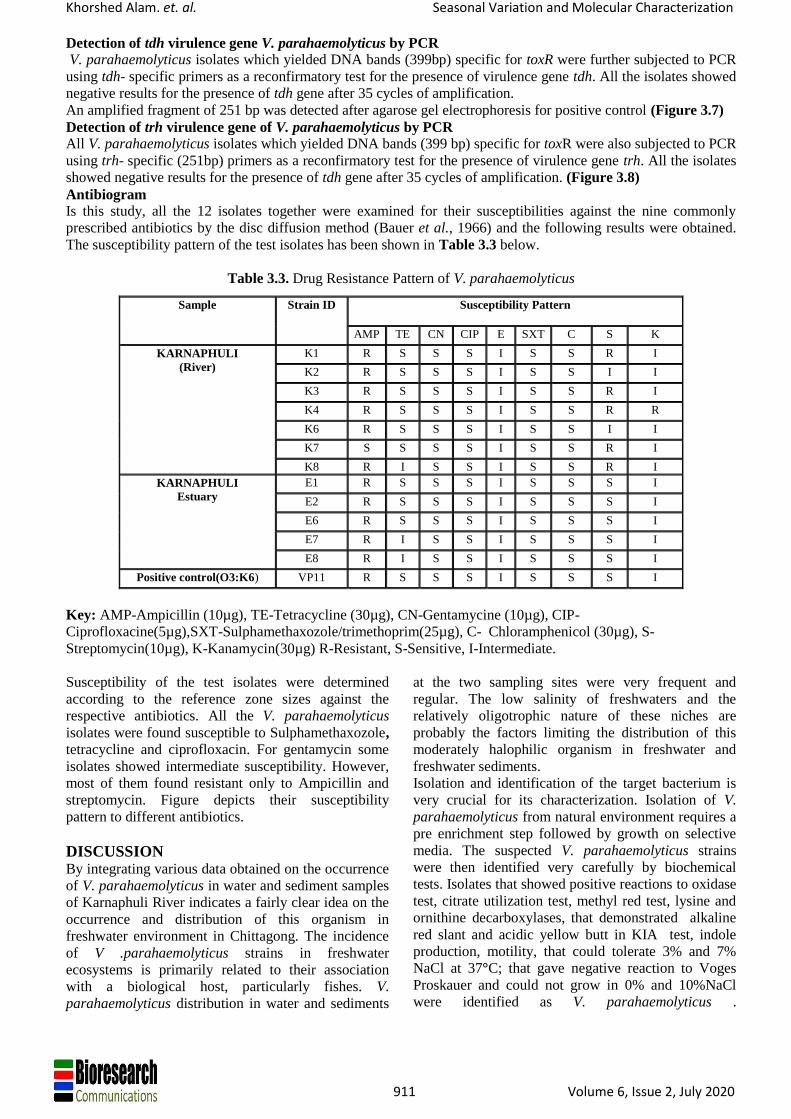
Antibiogram
Is this study, all the 12 isolates together were examined for their susceptibilities against the nine commonly
prescribed antibiotics by the disc diffusion method (Bauer et al., 1966) and the following results were obtained.
The susceptibility pattern of the test isolates has been shown in Table 3.3 below.
Table 3.3. Drug Resistance Pattern of V. parahaemolyticus
Key: AMP-Ampicillin (10µg), TE-Tetracycline (30µg), CN-Gentamycine (10µg), CIP-
Ciprofloxacine(5µg),SXT-Sulphamethaxozole/trimethoprim(25µg), C- Chloramphenicol (30µg), S-
Streptomycin(10µg), K-Kanamycin(30µg) R-Resistant, S-Sensitive, I-Intermediate.
Susceptibility of the test isolates were determined
according to the reference zone sizes against the
respective antibiotics. All the V. parahaemolyticus
isolates were found susceptible to Sulphamethaxozole,
tetracycline and ciprofloxacin. For gentamycin some
isolates showed intermediate susceptibility. However,
most of them found resistant only to Ampicillin and
streptomycin. Figure depicts their susceptibility
pattern to different antibiotics.
DISCUSSION By integrating various data obtained on the occurrence
of V. parahaemolyticus in water and sediment samples
of Karnaphuli River indicates a fairly clear idea on the
occurrence and distribution of this organism in
freshwater environment in Chittagong. The incidence
of V .parahaemolyticus strains in freshwater
ecosystems is primarily related to their association
with a biological host, particularly fishes. V.
parahaemolyticus distribution in water and sediments
at the two sampling sites were very frequent and
regular. The low salinity of freshwaters and the
relatively oligotrophic nature of these niches are
probably the factors limiting the distribution of this
moderately halophilic organism in freshwater and
freshwater sediments.
Isolation and identification of the target bacterium is
very crucial for its characterization. Isolation of V.
parahaemolyticus from natural environment requires a
pre enrichment step followed by growth on selective
media. The suspected V. parahaemolyticus strains
were then identified very carefully by biochemical
tests. Isolates that showed positive reactions to oxidase
test, citrate utilization test, methyl red test, lysine and
ornithine decarboxylases, that demonstrated alkaline
red slant and acidic yellow butt in KIA test, indole
production, motility, that could tolerate 3% and 7%
NaCl at 37°C; that gave negative reaction to Voges
Proskauer and could not grow in 0% and 10%NaCl
were identified as V. parahaemolyticus .
Sample Strain ID Susceptibility Pattern
AMP TE CN CIP E SXT C S K
KARNAPHULI
(River)
K1 R S S S I S S R I
K2 R S S S I S S I I
K3 R S S S I S S R I
K4 R S S S I S S R R
K6 R S S S I S S I I
K7 S S S S I S S R I
K8 R I S S I S S R I
KARNAPHULI
Estuary
E1 R S S S I S S S I
E2 R S S S I S S S I
E6 R S S S I S S S I
E7 R I S S I S S S I
E8 R I S S I S S S I
Positive control(O3:K6) VP11 R S S S I S S S I
C

Khorshed Alam. et. al. Seasonal Variation and Molecular Characterization
912 Volume 6, Issue 2, July 2020
Biochemically positive V. parahaemolyticus strains
were then subjected to the molecular based assay
Polymerase chain reaction (PCR). The presence of
toxR gene was used for identification of
V.parahaemolyticus. In this study, toxR gene sequence
was used in a PCR method for the specific
identification of V.parahaemolyticus. All the test
isolates showed positive results in PCR using primers
specific for toxR gene.
Thereafter molecular characterization of these V.
parahaemolyticus isolates were carried out by
detection of toxin production and presence of
virulence genes-tdh and trh. To detect tdh toxin
production by V. parahaemolyticus isolated from
environmental samples, reverse passive latex
agglutination test was carried out. In this technique,
the sensitized latex particles agglutinate in the
presence of tdh toxin produced by V.
parahaemolyticus and results in the formation of tdh
toxin. This finding deserves more characterization like
detection of relevant gene producing this enzyme by
PCR. From this point of view, PCR for tdh gene was
carried out.
In this study, PCR had been done for identifying the
distribution of tdh gene. Of all isolates, none gave
positive results in PCR using primers specific to tdh
gene. The findings correlates with the results obtained
by reverse passive latex agglutination test. Further
PCR analysis was done for the detection of another
virulence factor trh gene. Among the test strains, no
one was positive for trh gene and thus considered as
avirulent.
As antibiotic susceptibility is an important parameter
for the treatment measures and current study
antibiogram profile of the test isolates were
investigated. No significant difference was observed
between test isolates and positive control O3:K6 with
respect to antibiotic susceptibility. The isolates were
susceptible to almost all the representative antibiotics
tested including ciprofloxacine,tetracycline,
kanamycine, erythromycin
sulphamethaxazole/trimethoprim,gentamycine which
is a good indicator in the treatment of V.
parahaemolyticus associated with diarrhea and
gastroenteritis. They were resistant to only ampicillin.
Few strains showed intermediate resistance to only
gentamycine and some strains were resistant to
streptomycin. None of the isolates showed multi- drug
resistance markers like the plasmid, the class 1
integron and the SXT element. Therefore, antibiotic
resistance-associated spread of the isolates seems
unlikely and treating these strains with those
antibiotics would be possible.
Sulphamethoxazole/trimethoprim and tetracycline
seemed to be most effective with highest zone
diameter of growth inhibition when tested by disc
diffusion assay.
The study was designed to observe the seasonal
variation and characterize the isolated V.
parahaemolyticus strains from river and estuary in
Chittagong. The most important finding of the study
was the regular occurrence of V. parahaemolyticus in
river of 0% salinity though most of them are
nonpathogenic strains. The findings of this study could
be useful to compare the clonal variation between
clinical and environmental isolates belonging to the
same serogroup of V. parahaemolyticus.
CONCLUDING REMARKS Vibrio. parahaemolyticus has been thought to be a
marine bacterium because of its indispensable Na+
requirement for survival and growth. But the
occurrence of this moderately halophilic organism in
fresh water has also been observed (Bockemühl et al.,
1986) which is quite alarming due to the undeniable
dependence of people on fresh water. During
collection, a set of 1% NaCl containing samples and
another set without 1% NaCl used. Salt containing
samples showed a regular count but the count of
V.parahaemolyticus was random and infrequent in the
samples without salt. The study was thus designed to
observe the seasonal occurrence of V.
parahaemolyticusin in the rivers of Bangladesh
besides the estuarine environments and also
characterize them at the molecular level. Therefore,
Using 1% NaCl during sample collection for the
isolation of Vibrio parahaemolyticus from fresh water
without any salinity is an innovative procedure and the
occurrence of V. parahaemolyticus in the river was
found frequent and regular.
REFERENCES 1. BAUER, A., KIRBY, W., SHERRIS, J. C.,
TURCK & TURCK, M. 1966. Antibiotic
susceptibility testing by a standardized single disk
method. American journal of clinical pathology,
45, 493.
2. CAPPUCCINO, J. & SHERMAN, N. 1992.
Biochemical activities of microorganisms.
Microbiology, A Laboratory Manual. The
Benjamin/Cummings Publishing Co. California,
USA.
3. CHIOU, A., CHEN, L. & CHEN, S. 1991.
Foodborne illness in Taiwan, 1981-1989. 藥物食品檢驗局調查研究年報, 452-453.
4. CHOWDHURY, N. R., CHAKRABORTY, S.,
RAMAMURTHY, T., NISHIBUCHI, M.,
YAMASAKI, S., TAKEDA, Y. & NAIR, G. B.
2000. Molecular evidence of clonal Vibrio
parahaemolyticus pandemic strains. Emerging
infectious diseases, 6, 631.
5. CHUN, D., CHUNG, J., TAK, R. & SEOL, S. Y.
1975. Nature of the Kanagawa phenomenon of
Vibrio parahaemolyticus. Infection and immunity,
12, 81-87.

Khorshed Alam. et. al. Seasonal Variation and Molecular Characterization
913 Volume 6, Issue 2, July 2020
6. COOK, D. W., BOWERS, J. C. & DEPAOLA, A.
2002. Density of total and pathogenic (tdh+) Vibrio
parahaemolyticus in Atlantic and Gulf coast
molluscan shellfish at harvest. Journal of Food
Protection®, 65, 1873-1880.
7. DENEKE, C. F. & COLWELL, R. 1973. Studies of
the cell envelope of Vibrio parahaemolyticus.
Canadian journal of microbiology, 19, 241-245.
8. DEPAOLA, A., KAYSNER, C. A., BOWERS, J.
& COOK, D. W. 2000. Environmental
investigations of Vibrio parahaemolyticus in
oysters after outbreaks in Washington, Texas, and
New York (1997 and 1998). Applied and
environmental microbiology, 66, 4649-4654.
9. DIRITA, V. J. 1992. Co‐ordinate expression of
virulence genes by ToxR in Vibrio cholerae.
Molecular microbiology, 6, 451-458.
10. HLADY, W. G. & KLONTZ, K. C. 1996. The
epidemiology of Vibrio infections in Florida,
1981–1993. Journal of Infectious Diseases, 173,
1176-1183.
11. HONDA, T. & IIDA, T. 1993. The pathogenicity
of Vibrio parahaemolyticus and the role of the
thermostable direct haemolysin and related
haemolysins. Reviews in Medical Microbiology, 4,
106-113.
12. HONDA, T., NI, Y. & MIWATANI, T. 1989.
Purification of a TDH-related hemolysin produced
by a Kanagawa phenomenon-negative clinical
isolate of< i> Vibrio parahaemolyticus</i> 06:
K46. FEMS microbiology letters, 57, 241-245.
13. HONDA, T., TAGA, S., TAKEDA, T.,
HASIBUAN, M., TAKEDA, Y. & MIWATANI,
T. 1976. Identification of lethal toxin with the
thermostable direct hemolysin produced by Vibrio
parahaemolyticus, and some physicochemical
properties of the purified toxin. Infection and
immunity, 13, 133-139.
14. IGUCHI, T., KONDO, S. & HISATSUNE, K.
1995. Vibrio parahaemolyticus O serotypes from
O1 to O13 all produce R‐type lipopolysaccharide:
SDS‐PAGE and compositional sugar analysis.
FEMS microbiology letters, 130, 287-292.
15. JOSEPH, S. W., COLWELL, R. R. & KAPER, J.
B. 1982. Vibrio parahaemolyticus and related
halophilic vibrios. Critical reviews in
microbiology, 10, 77-124.
16. KITA-TSUKAMOTO, K., OYAIZU, H., NANBA,
K. & SIMIDU, U. 1993. Phylogenetic relationships
of marine bacteria, mainly members of the family
Vibrionaceae, determined on the basis of 16S
rRNA sequences. International journal of
systematic bacteriology, 43, 8-19.
17. NISHIBUCHI, M. & KAPER, J. B. 1995.
Thermostable direct hemolysin gene of Vibrio
parahaemolyticus: a virulence gene acquired by a
marine bacterium. Infection and Immunity, 63,
2093.
18. OGAWA, H., TOKUNOU, H., KISHIMOTO, T.,
FUKUDA, S., UMEMURA, K. & TAKATA, M.
1989. Ecology of Vibrio parahaemolyticus in
Hiroshima Bay. Hiroshima J. Vet. Med, 4, 47-57.
19. OKUDA, J., ISHIBASHI, M., HAYAKAWA, E.,
NISHINO, T., TAKEDA, Y.,
MUKHOPADHYAY, A. K., GARG, S.,
BHATTACHARYA, S., NAIR, G. B. &
NISHIBUCHI, M. 1997. Emergence of a unique
O3: K6 clone of Vibrio parahaemolyticus in
Calcutta, India, and isolation of strains from the
same clonal group from Southeast Asian travelers
arriving in Japan. Journal of Clinical Microbiology,
35, 3150-3155.
20. SARKAR, B., NAIR, G. B., SIRCAR, B. & PAL,
S. 1983. Incidence and level of Vibrio
parahaemolyticus associated with freshwater
plankton. Applied and environmental microbiology,
46, 288-290.
21. STANDARDS, N. C. F. C. L. & WATTS, J. L.
1999. Performance standards for antimicrobial
disk and dilution susceptibility tests for bacteria
isolated from animals: approved standard: NCCLS
documents M31-A, NCCLS.
22. TADA, J., OHASHI, T., NISHIMURA, N.,
SHIRASAKI, Y., OZAKI, H., FUKUSHIMA, S.,
TAKANO, J., NISHIBUCHI, M. & TAKEDA, Y.
1992. Detection of the thermostable direct
hemolysin gene (< i> tdh</i>) and the thermostable
direct hemolysin-related hemolysin gene (< i>
trh</i>) of< i> Vibrio parahaemolyticus</i> by
polymerase chain reaction. Molecular and cellular
probes, 6, 477-487.
23. TANIGUCHI, H., HIRANO, H., KUBOMURA, S.,
HIGASHI, K. & MIZUGUCHI, Y. 1986.
Comparison of the nucleotide sequences of the
genes for the thermostable direct hemolysin and the
thermolabile hemolysin from< i> Vibrio
parahaemolyticus</i>. Microbial pathogenesis, 1,
425-432.
24. TISON, D. L. & KELLY, M. T. 1984. < i>
Vibrio</i> species of medical importance.
Diagnostic microbiology and infectious disease, 2,
263-276.
25. VENKATESWARAN, K., KIIYUKIA, C.,
TAKAKI, M., NAKANO, H., MATSUDA, H.,
KAWAKAMI, H. & HASHIMOTO, H. 1989.
Characterization of toxigenic vibrios isolated from
the freshwater environment of Hiroshima, Japan.
Applied and environmental microbiology, 55,
2613-2618.


![Advances in Bioresearch Adv. Biores., Vol 5 (2) June …ABR Vol 5 [2] June 2014 172 | P a g e ©2014 Society of Education, India Advances in Bioresearch Adv. Biores., Vol 5 (2) June](https://static.fdocuments.in/doc/165x107/5ea3f181ead1bf7e282936f0/advances-in-bioresearch-adv-biores-vol-5-2-june-abr-vol-5-2-june-2014-172.jpg)