Biophosphonate related osteonecrosis of the jaws
18
Biophosphonate-Related Osteonecrosis of the Jaws Salvatore L. Ruggiero, DMD, MD a,b, * , Sook-Bin Woo, DMD, MMSc c,d a Department of Oral and Maxillofacial Surgery, Stony Brook School of Dental Medicine, Stony Brook, NY, USA b New York Center of Orthognathic and Maxillofacial Surgery, 2001 Marcus Avenue, Lake Success, NY 11042, USA c Division of Oral Medicine and Dentistry, Brigham and Women’s Hospital, 75 Francis Street, Boston, MA 02115, USA d Harvard School of Dental Medicine, 188 Longwood Avenue, Boston, MA 02115, USA In 2003 and 2004, the first reports of patients who developed necrosis of the jawbones while taking bisphosphonates appeared in the literature; most patients were on this drug for treatment of cancer and some for osteoporosis [1–3]. Since then, more than 500 cases have been identified and the number of these cases continues to grow. This article reviews the action of bi- sphosphonates, the condition called bisphosphonate-associated osteonecrosis of the jaws, strategies to minimize occurrence, and treatment of this condition. Nomenclature A universally accepted term for this new condition has not been estab- lished, which has caused some degree of confusion. This complication has been referred to in the literature as BRONJ (bisphosphonate-related osteo- necrosis of the jaw), BRON (bisphosphonate-related osteonecrosis), BON (bisphosphonate osteonecrosis), BAONJ (bisphosphonate-associated osteo- necrosis of the jaw), and simply ONJ (osteonecrosis of the jaw). Based on the clear association between bisphosphonate therapy and jaw necrosis that has been established in numerous retrospective studies, the American Association of Oral and Maxillofacial Surgeons (AAOMS) has decided to * Corresponding author. New York Center of Orthognathic and Maxillofacial Surgery, 2001 Marcus Avenue, Lake Success, NY 11042. E-mail address: [email protected] (S.L. Ruggiero). 0011-8532/08/$ - see front matter Ó 2008 Elsevier Inc. All rights reserved. doi:10.1016/j.cden.2007.09.002 dental.theclinics.com Dent Clin N Am 52 (2008) 111–128
-
Upload
andres-cardona-franco -
Category
Documents
-
view
2.283 -
download
1
description
Transcript of Biophosphonate related osteonecrosis of the jaws
Dent Clin N Am 52 (2008) 111–128
Biophosphonate-Related Osteonecrosisof the Jaws
Salvatore L. Ruggiero, DMD, MDa,b,*,Sook-Bin Woo, DMD, MMScc,d
aDepartment of Oral and Maxillofacial Surgery, Stony Brook School of Dental Medicine,
Stony Brook, NY, USAbNew York Center of Orthognathic and Maxillofacial Surgery,
2001 Marcus Avenue, Lake Success, NY 11042, USAcDivision of Oral Medicine and Dentistry, Brigham and Women’s Hospital,
75 Francis Street, Boston, MA 02115, USAdHarvard School of Dental Medicine, 188 Longwood Avenue, Boston, MA 02115, USA
In 2003 and 2004, the first reports of patients who developed necrosis ofthe jawbones while taking bisphosphonates appeared in the literature; mostpatients were on this drug for treatment of cancer and some for osteoporosis[1–3]. Since then, more than 500 cases have been identified and the numberof these cases continues to grow. This article reviews the action of bi-sphosphonates, the condition called bisphosphonate-associated osteonecrosisof the jaws, strategies to minimize occurrence, and treatment of thiscondition.
Nomenclature
A universally accepted term for this new condition has not been estab-lished, which has caused some degree of confusion. This complication hasbeen referred to in the literature as BRONJ (bisphosphonate-related osteo-necrosis of the jaw), BRON (bisphosphonate-related osteonecrosis), BON(bisphosphonate osteonecrosis), BAONJ (bisphosphonate-associated osteo-necrosis of the jaw), and simply ONJ (osteonecrosis of the jaw). Based onthe clear association between bisphosphonate therapy and jaw necrosisthat has been established in numerous retrospective studies, the AmericanAssociation of Oral and Maxillofacial Surgeons (AAOMS) has decided to
* Corresponding author. New York Center of Orthognathic and Maxillofacial Surgery,
2001 Marcus Avenue, Lake Success, NY 11042.
E-mail address: [email protected] (S.L. Ruggiero).
0011-8532/08/$ - see front matter � 2008 Elsevier Inc. All rights reserved.
doi:10.1016/j.cden.2007.09.002 dental.theclinics.com

112 RUGGIERO & WOO
adopt the term BRONJ for this entity [4], and this term will be usedthroughout this article.
Action of bisphosphonates
The actions and pharmacology of bisphosphonates have been well re-viewed and only a summary is offered here [5–7]. Bisphosphonates are ana-logs of inorganic pyrophosphates. Pyrophosphates are well-known indentistry, because they are a component of tartar-control toothpaste and in-hibit calcium precipitation [8]. Unlike pyrophosphates, bisphosphonatescontain a carbon rather than oxygen molecule. This P-C-P structure allowsthe molecule to bind to the hydroxyapatite crystals with high affinity, whilethe presence of nitrogen in one of the side chains confers markedly increasedpotency to the drug (Fig. 1). The early forms of bisphosphonate, such asetidronate and clodronate, do not contain nitrogen and therefore are lesspotent; they are available only in the oral preparation. The later forms ofbisphosphonates contain nitrogen and some are available as intravenouspreparations, making them much more bioavailable (Table 1). Pamidronateand alendronate contain nitrogen in an alkyl chain and are 10 to 100 timesmore potent than etidronate and clodronate. The most potent of the bi-sphosphonates (such as zoledronic acid and risedronate) contain nitrogenwithin a heterocyclic ring. The half-life of bisphosphonates is approximately10 years, and therefore prolonged use of this drug causes substantial drugaccumulation within the skeleton. The drug is tightly bound to the apatitecrystals until it is released during osteoclastic-mediated bone resorption.
Antiresorptive activity
One of the most important and powerful effects of bisphosphonates isinhibition of osteoclastic activity, and herein lies one of its most importantapplications in clinical practice, both for managing osteoporosis and cancersin the skeletal system. When bone resorption occurs, bisphosphonates arereleased from the hydroxyapatite crystal and are taken up by osteoclasts.Metabolites of non-nitrogen containing bisphosphonates (such as etidronateand clodronate) are cytotoxic to the osteoclasts and lead to their death. Ni-trogen-containing bisphosphonates, however, act by way of the mevalonate
Fig. 1. Structure of pyrophosphate (left) and bisphosphonate (right). For bisphosphonates, R1
acts as a ‘‘bone hook’’ for attachment of the molecule to bone, whereas R2 determines potency.
113OSTEONECROSIS OF THE JAWS
pathway (for cholesterol synthesis), inhibiting protein prenylation, a processessential for normal functioning of vital intracellular proteins, ultimatelyleading to osteoclast apoptosis (programmed cell death) [7,9]. Bisphospho-nates also inhibit differentiation of osteoclasts and stimulates osteoblaststo produce osteoclast-inhibiting factor [10]. Therefore, the net result is re-duced numbers of osteoclasts and reduced bone resorption. Because boneresorption is coupled to osteoblastic bone formation for remodeling, boneturnover (ie, resorption and deposition) becomes severely suppressed. How-ever, the bone continues to mineralize and could become brittle and lesselastic. In one case report, bisphosphonate taken at high doses led to anosteopetrotic-like state [11].
Tumoricidal activity
Many studies have shown that nitrogen-containing bisphosphonates alsoreduce the activity of cancer cells and control metastases [9,10,12]. This pro-cess may be related to inhibition of protein prenylation leading to disruptionof intracellular activity within the cancer cells [9]. However, the alteration ofthe microenvironment itself, caused by reduced bone resorption alone, couldalso account for control of metastases [13]. Bisphosphonates also reduce ad-hesion, invasion, and viability of cancer cells and may activate gamma deltaT cells, which have tumoricidal activity [14].
Antiangiogenic activity
In vitro, zoledronic acid inhibits angiogenesis mediated through basic fi-broblast growth factor and may induce apoptosis of endothelial cells [15].Antiangiogenic activity may also occur through lowering circulating levelsof vascular endothelial growth factor and platelet-derived growth factor,both of which are proangiogenic [16,17].
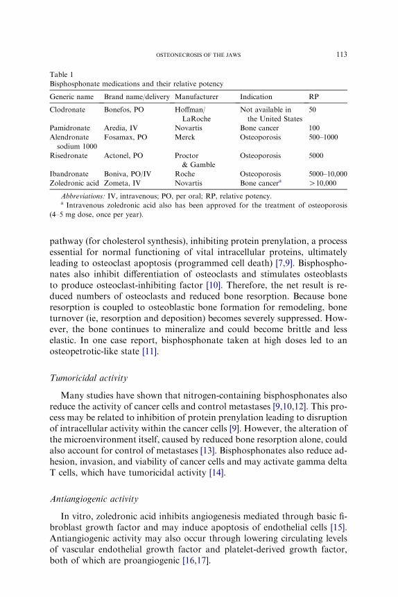
Table 1
Bisphosphonate medications and their relative potency
Generic name Brand name/delivery Manufacturer Indication RP
Clodronate Bonefos, PO Hoffman/
LaRoche
Not available in
the United States
50
Pamidronate Aredia, IV Novartis Bone cancer 100
Alendronate
sodium 1000
Fosamax, PO Merck Osteoporosis 500–1000
Risedronate Actonel, PO Proctor
& Gamble
Osteoporosis 5000
Ibandronate Boniva, PO/IV Roche Osteoporosis 5000–10,000
Zoledronic acid Zometa, IV Novartis Bone cancera O10,000
Abbreviations: IV, intravenous; PO, per oral; RP, relative potency.a Intravenous zoledronic acid also has been approved for the treatment of osteoporosis
(4–5 mg dose, once per year).

114 RUGGIERO & WOO
Clinical applications
Bisphosphonates are widely used in the management of four main condi-tions: osteoporosis, Paget’s disease of bone, multiple myeloma, and cancersthat have metastasized to the bones. This article focuses only on the onco-logic aspects of bisphosphonate use.
Multiple myeloma is amalignancy of plasma cells that primarily affects theskeletal system. Patients develop multiple lytic bone lesions containing malig-nant plasma cells, and as a result may develop skeletal-related events (SRE),such as bone pain, fractures, necessity for surgery and radiation, and hyper-calcemia. Patients also develop hypergammaglobulinemia from secretory ac-tivity of these cells, which can lead to renal failure. Clinical trials show thatbisphosphonates significantly reduce these SREs, and pamidronate and zole-dronic acid are widely used for treating multiple myeloma [18,19]. Otheragents for treating this disease include thalidomide, glucocorticoids such asdexamethasone, and proteasome inhibitors such as bortezomib, usually incombination [20,21]. These agents also have antiangiogenic properties.
Similar SREs are noted in patients who have metastatic cancers (withmetastatic breast and prostate cancers being the most common), and bi-sphosphonates also have been shown to significantly reduce SREs in thesepatients [22,23].
Early reports of bisphosphonate-associated osteonecrosis of the jaws
The earliest reports of bona fide cases of BRONJ occurred in 2003 [1–3],and have been followed by many more case reports and large case series [24–27]. All cases have occurred in the maxilla or mandible except for a singlecase that occurred after ear surgery [28].
Etiopathogenesis
The exact mechanism for the development of BRONJ is unclear. The cur-rent hypothesis focuses on severe suppression of bone turnover, coupledwith the unique conditions affecting the jaws and not other bones. Theseconditions include the following: (1) the jaw bones are separated from theoral environment from a very thin mucosa (only several mm thick for themost part) and this barrier is readily breached even with simple physiologicactivities such as mastication, (2) the oral cavity is filled with bacteria andthe jaws are often involved in infection through either the periodontal liga-ment or the pulp, (3) dentoalveolar surgery is a common procedure (eg, ex-tractions, periodontal surgeries, apicoectomies) in which bone is exposed toa bacteria-rich environment, and (4) the rate of turnover of the jawbones ishigher than that for the long bones [29].
One possible mechanism of action is that after a sufficient concentrationof bisphosphonates has accumulated, the jawbones become hypodynamicand turn over at a low rate. When infection or surgery requires increased
115OSTEONECROSIS OF THE JAWS
bone turnover for healing, this does not occur. How the bone actually be-comes necrotic and exposed is unclear at this time. However, there is a con-dition that mimics BRONJ that occurs spontaneously, involves only a fewmillimeters of necrotic bone that heals without incident, and occurs inhealthy adults. This benign sequestration of the lingual plate is a well-recog-nized phenomenon and its putative mechanism of action is trauma to thelingual periosteum, loss of vascular supply of bone supplied by that areaof periosteum, death of bone with sequestration, and healing [30]. The lin-gual plate/mylohyoid area is also a common site for spontaneously occur-ring BRONJ.
Risk factors for bisphosphonate-associated osteonecrosis of the jaws
Not all patients who use bisphosphonates develop BRONJ. The factorsthat affect its development are:
1) The use of nitrogen-containing bisphosphonates, particularly zoledronicacid [31]. Zoledronic acid used alone produced 9.5-fold and 4.5-foldgreater risk compared with pamidronate used alone or with zoledronicacid, respectively [32]. However, BRONJ has been seen in a patient whohad myeloma who took only oral clodronate, a first-generation non–nitrogen-containing bisphosphonate [33].
2) The cumulative dose of bisphosphonates. This factor also indirectly re-lates to the patient’s underlying condition, because patients who havecancer receive much higher doses of bisphosphonates than those whohave osteoporosis. For example, a patient who has osteoporosis who re-quires intravenous bisphosphonate therapy receives 4 to 5 mg of zole-dronic acid per year compared with a patient who has myeloma whoreceives 4 mg of zoledronic acid every 4 weeks. The median time of ex-posure to zoledronic acid with development of BRONJ ranged from 9 to30 months and is significantly shorter compared with other bisphosph-onate regimens [25,31,34,35]. However, the exposure can be as little as 3months. The risk for developing BRONJ increases over time, with a cu-mulative hazard of 1% within the first year of treatment with zoledronicacid, and 15% at 4 years [31].
3) Dentoalveolar surgery. Approximately 60% of all cases are associatedwith either tooth extraction or other surgery (eg, periodontal and apicalsurgery and implant placement) [24,25,36]. However, tooth extractionsare the most common inciting factor [26]. One study showed that theprevalence of BRONJ in patients who had cancer who did not undergoextractions was 1% compared with 7% to 9% in those who did [35].BRONJ has been reported in patients after surgical endodontic proce-dures [37,38].
4) Trauma. Many cases occur on the lingual mandible (where the mucosais especially fragile) and on tori [25].
116 RUGGIERO & WOO
Other factors that may contribute to the development of ONJ, but forwhich the evidence is less robust, include dental infections. By the timeBRONJ develops, it is difficult to determine if the infection caused BRONJor was merely a consequence of BRONJ.
Other factors, such as route of administration (intravenous preparationsare more frequently associated with BRONJ) and age, are indirectly relatedto the factors noted earlier. For example, patients who have cancer affectingthe skeleton (usually older patients) typically are exposed to the more potentbisphosphonates, such as zoledronic acid and pamidronate, both of which areonly available as intravenous preparations, and are used more frequently.
Whether the antiangiogenic activity of bisphosphonates plays an impor-tant and direct role in BRONJ is unclear. Other comorbidities have not beenentirely elucidated, but include concomitant use of antiangiogenic agentsand vascular compromise. Thalidomide, which has antiangiogenic proper-ties, has been shown to increase the risk 2.4-fold [32], although this hasbeen disputed [26]. One study showed that patients who had BRONJ weremore likely to have associated diabetes mellitus, although most patients al-ready had diabetes when their cancer was diagnosed [39]. Another studyshowed that patients who developed BRONJ had hyperparathyroidismand mild hypocalcemia compared with controls [40].
Prevalence
The prevalence varies from 4% to 7% within any one center. Most of thestudies were performed through retrospective chart reviews or telephoneinterviews [41,42].
Although most patients who developed BRONJ underwent dental extrac-tions, the number of patients who had extractions or other dento-alveolar surgery and did not develop BRONJ is unclear.
Diagnosis and clinical presentation of bisphosphonate-associated
osteonecrosis of the jaws
Standardization of diagnostic criteria for this new clinical entity is impor-tant to facilitate future clinical and epidemiologic research and help distin-guish BRONJ from other intraoral osteonecrotic conditions exhibitingdelayed healing. The AAOMS established a working definition for BRONJthat is fairly concise and specific [4]. Patients may be considered to haveBRONJ if they have all of the following characteristics: (1) current or pre-vious treatment with a bisphosphonate, (2) exposed, necrotic bone in themaxillofacial region that has persisted for more than 8 weeks, and (3) no his-tory of radiation therapy to the jaws. Patients who are considered at risk ac-cording to the AAOMS criteria have no evidence of exposed or necroticbone but have been exposed to either intravenous or oral nitrogen-contain-ing bisphosphonates for long periods.

117OSTEONECROSIS OF THE JAWS
The American Society for Bone and Mineral Research (ASBMR) hasalso made recommendations for a provisional case definition for confirmedand suspected cases of bisphosphonate associated osteonecrosis [43]. A con-firmed case is identical to the AAOMS definition for BRONJ. A suspectedcase fulfills all the criteria of a confirmed case, except that the necroticbone has been present for less than 8 weeks. Suspected cases should be fol-lowed up to determine if they eventually meet the criteria of a confirmedcase.
Diagnosis of BRONJ is primarily based on patient history and clinical ex-amination. Patients present with exposed, necrotic bone varying in size froma few millimeters to several centimeters; only 60% of cases report pain, andsome cases may remain asymptomatic for weeks, months, or years[3,24,25]. These lesions frequently become symptomatic when surroundingtissues become inflamed or an infection develops. Signs and symptoms thatmay occur before the development of clinically detectable BRONJ includepain, tooth mobility, mucosal swelling, erythema, ulceration, and the devel-opment of sinus tracts. These somewhat nonspecific signs and symptomsare similar to those of banal odontogenic infections. Some patients mayalso present with complaints of altered sensation or paresthesia in the affectedarea as the neurovascular bundle becomes affected by inflammation or infec-tion around the necrotic bone. Chronic maxillary sinusitis secondary toBRONJwith or without an oral-antral fistula may be the presenting symptomin patients who have maxillary bone involvement [3].
Lesions have been observed more commonly in the mandible than themaxilla (2:1 ratio) [24], and more commonly in areas with thin mucosa over-lying bony prominences, such as tori, bony exostoses, and the mylohyoidridge [3,25]. Most cases occur at the site of prior dentoalveolar surgery(Fig. 2). However, exposed bone has also been reported in patients whohave no history of trauma or in edentulous regions of the jaw (Fig. 3).The size of the affected area can be variable, and ranges from a nonhealing
Fig. 2. BRONJ after a tooth extraction.

118 RUGGIERO & WOO
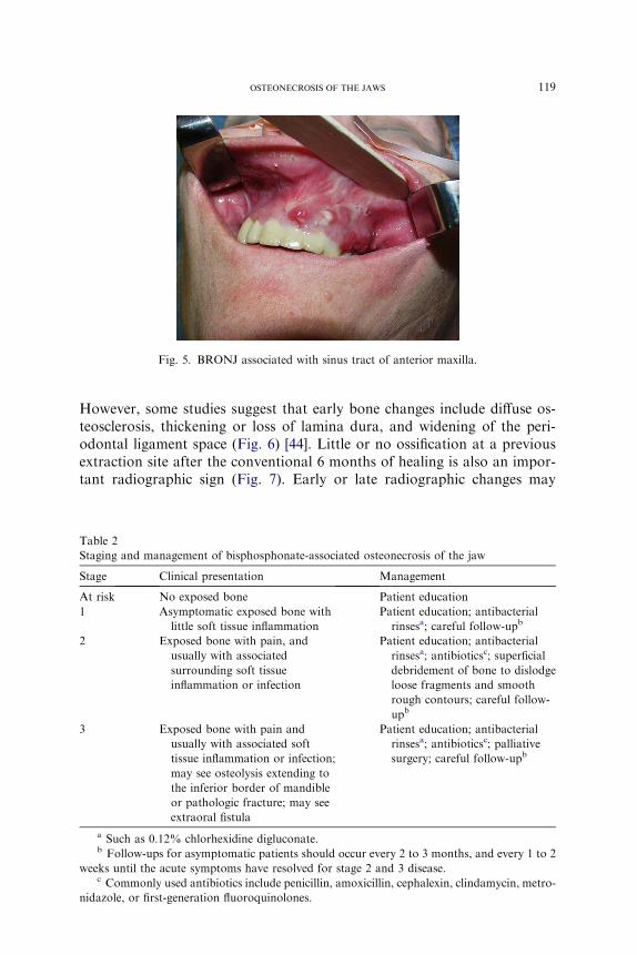
extraction site to exposure and necrosis of the entire jaw (Fig. 4). The area ofexposed bone may be surrounded by inflamed erythematous soft tissue.When infected, purulent discharge may be seen at the site of the exposedbone and intra- and extraoral fistulae (Fig. 5). The AAOMS has proposeda clinical staging system based on the presence of symptoms and extent ofdisease (Table 2) [4,44].
Radiographic findings
Conventional radiographs will not show a change until the bone is dem-ineralized 30% to 50%. Therefore, and also partly because of the two-dimensional nature of these films, panoramic and periapical radiographsmay not show significant changes in the early stages of osteonecrosis.
Fig. 3. BRONJ in the mylohyoid area.
Fig. 4. Extensive BRONJ seen in this surgical specimen of the mandible.
119OSTEONECROSIS OF THE JAWS
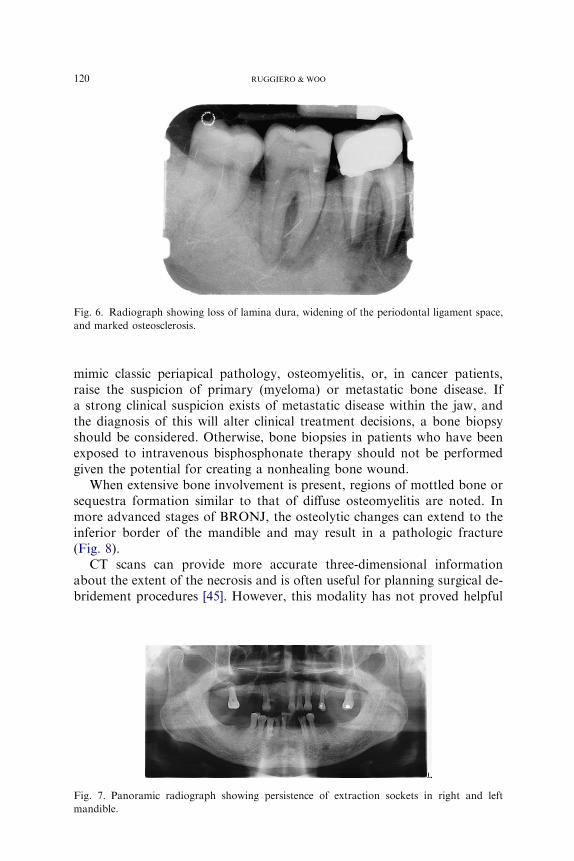
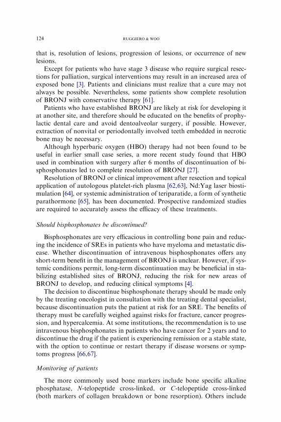
However, some studies suggest that early bone changes include diffuse os-teosclerosis, thickening or loss of lamina dura, and widening of the peri-odontal ligament space (Fig. 6) [44]. Little or no ossification at a previousextraction site after the conventional 6 months of healing is also an impor-tant radiographic sign (Fig. 7). Early or late radiographic changes may
Fig. 5. BRONJ associated with sinus tract of anterior maxilla.
Table 2
Staging and management of bisphosphonate-associated osteonecrosis of the jaw
Stage Clinical presentation Management
At risk No exposed bone Patient education
1 Asymptomatic exposed bone with
little soft tissue inflammation
Patient education; antibacterial
rinsesa; careful follow-upb
2 Exposed bone with pain, and
usually with associated
surrounding soft tissue
inflammation or infection
Patient education; antibacterial
rinsesa; antibioticsc; superficial
debridement of bone to dislodge
loose fragments and smooth
rough contours; careful follow-
upb
3 Exposed bone with pain and
usually with associated soft
tissue inflammation or infection;
may see osteolysis extending to
the inferior border of mandible
or pathologic fracture; may see
extraoral fistula
Patient education; antibacterial
rinsesa; antibioticsc; palliative
surgery; careful follow-upb
a Such as 0.12% chlorhexidine digluconate.b Follow-ups for asymptomatic patients should occur every 2 to 3 months, and every 1 to 2
weeks until the acute symptoms have resolved for stage 2 and 3 disease.c Commonly used antibiotics include penicillin, amoxicillin, cephalexin, clindamycin, metro-
nidazole, or first-generation fluoroquinolones.
120 RUGGIERO & WOO
mimic classic periapical pathology, osteomyelitis, or, in cancer patients,raise the suspicion of primary (myeloma) or metastatic bone disease. Ifa strong clinical suspicion exists of metastatic disease within the jaw, andthe diagnosis of this will alter clinical treatment decisions, a bone biopsyshould be considered. Otherwise, bone biopsies in patients who have beenexposed to intravenous bisphosphonate therapy should not be performedgiven the potential for creating a nonhealing bone wound.
When extensive bone involvement is present, regions of mottled bone orsequestra formation similar to that of diffuse osteomyelitis are noted. Inmore advanced stages of BRONJ, the osteolytic changes can extend to theinferior border of the mandible and may result in a pathologic fracture(Fig. 8).
CT scans can provide more accurate three-dimensional informationabout the extent of the necrosis and is often useful for planning surgical de-bridement procedures [45]. However, this modality has not proved helpful
Fig. 6. Radiograph showing loss of lamina dura, widening of the periodontal ligament space,
and marked osteosclerosis.
Fig. 7. Panoramic radiograph showing persistence of extraction sockets in right and left
mandible.

121OSTEONECROSIS OF THE JAWS
for early identification of BRONJ in asymptomatic individuals. MRI candetect marrow edema, which may be an early sign of bone ischemia and ne-crosis, such as is seen in avascular necrosis of the hip secondary to long-termuse of steroids. However, whether this modality is helpful for detecting earlylesions of BRONJ is unclear, although established lesions are detectable asexpected [45]. Technetium 99m sestamibi is not taken up in areas of BRONJ[18], although fluorodeoxyglucose–positron emission tomography (FDG-PET) integrated with CT may show focal uptake [46]. All imaging modali-ties have proved helpful in determining the extent of the existing necroticprocess, especially in advanced lesions, but have not shown any efficacy inassessing patients at risk for BRONJ. New avenues of research includethe use of dental cone tomography, which uses only approximately 10%of the radiation of routine CT scans [47,48].
Histopathology and microbiology
Microscopic examination of debrided specimens of exposed bone typi-cally will show necrotic bone with associated bacterial debris and granula-tion tissue [26,49,50]. Microbial cultures from areas of exposed bone willusually isolate normal oral microbes and are therefore not always helpful[26]. However, when extensive soft tissue involvement is present, microbialculture data may help define comorbid oral infections and facilitate the se-lection of an appropriate antibiotic regimen.
The presence of actinomycetes in the culture and within biopsy specimensmust be interpreted with caution. The presence of actinomycetes alone doesnot indicate a diagnosis of actinomycosis, because they are a common en-dogenous pathogen in dental plaque. The diagnosis of actinomycosis ismade if actinomycotic organisms are present and associated with suppura-tion within the necrotic bone fragments, and actinomycetes was culturedfrom a sterile area within the bone and not from a swab of necrotic boneexposed to the oral cavity. The presence of pain, suppuration, and/or drain-ing sinus tracts together with both diagnostic criteria also helps to confirmthis diagnosis [51].
Fig. 8. Panoramic radiograph showing extensive involvement of the mandible with pathologic
fracture of anterior portion.

122 RUGGIERO & WOO
Differential diagnosis
Several other conditions may also lead to necrosis of the bone with seques-tration. Odontogenic infections, left untreated, may result in osteomyelitis, al-though this does not usually present with clinical exposure of the dead bone.
Postradiation osteonecrosis of the jaws is in many ways similar toBRONJ and occurs after a patient has received high doses of radiation tothe jawbones, usually for the treatment of primary head and neck malignan-cies. However, these conditions have a marked difference in presentation, inthat more than 95% of osteoradionecrosis occurs in the mandible, becauseone of the primary mechanisms is reduced vascularity and sclerosis of ves-sels as a result of the high doses of radiation. The mandible has a limitedblood supply compared with the maxilla (which is supplied by many collat-eral vessels) and is therefore more frequently involved [52].
Benign sequestration of the lingual plate differs from BRONJ in that thelesions are much smaller (usually several millimeters only), the patients areyoung to middle-aged adults who have not been exposed to bisphospho-nates, the lesions last only a few days before the piece of bone is either spon-taneously exfoliated or removed by a dental care provider, and no sequelaeare present [30].
Necrotizing periodontitis is usually seen in patients who have suppressedimmune systems, particularly those who have HIV infection or severe mal-nutrition [53,54]. It is caused by a polymicrobial infection of the periodon-tium that cannot be adequately contained by the patient’s poor immunefunction, leading to destruction and exposure of bone. This conditionmay spread from the bone to involve the adjacent soft tissues, a conditionknown as noma or necrotizing stomatitis. It is endemic within Africannations that have high rates of malnutrition. Herpes zoster infections ofthe maxilla and mandible have been reported to lead to necrosis of thesoft tissues and underlying bone. The putative mechanism is one of vascularcompromise [55,56].
Management
The goal of management for patients at risk for developing BRONJ orwho have active disease is to preserve the quality of life through controllingpain, managing infection, and preventing the development of new areas ofnecrosis. The BRONJ treatment algorithms that have been published are ei-ther a consensus of expert opinions or based on case series data [4,25,44,57].These management strategies have varied according to the risk for develop-ing BRONJ or the stage of disease.
Prevention strategies (oral bisphosphonate therapy)
Low-dose oral bisphosphonate therapy is primarily used for patients whohave osteoporosis, and is rarely used for treating myeloma or metastatic

123OSTEONECROSIS OF THE JAWS
carcinoma. The incidence of BRONJ in the osteoporosis population hasbeen cited as 1 in 100,000 patient years [58], although a more recent studyshows that it may be as high as 1 in 2260 to 8470 patients [35]. Patientswho have myeloma are often treated with clodronate (an oral non–nitro-gen-containing bisphosphonate) in European countries [59]; clodronate isnot approved for human use in the United States but may be associatedwith a lower incidence of BRONJ.
The risk is associated with cumulative dose/number of years of use. Dif-ferent periods deemed ‘‘safe’’, such as 3 or 5 years, are based on anecdotaldata [4]. Patients should undergo routine dental care with regular radio-graphic examinations, be educated about the risk for developing BRONJ,and provide informed consent for surgical procedures.
Current evidence does not contraindicate the placement of dental im-plants. However, the studies are small and do not include the longer periodsof bisphosphonate exposure [60].
Prevention strategies (intravenous bisphosphonate therapy)
For the most part, patients who have cancer undergoing bisphosphonatetherapy should be managed with preventive measures. The main emphasis isto minimize the risk for occurrence of BRONJ, which translates into mini-mizing the need for dentoalveolar surgery. Therefore, optimizing dentalhealth is the main objective in managing these patients. The best way to op-timize dental health is to educate all patients about their risk for developingBRONJ and evaluate each patient fully for odontogenic infections using fullmouth intraoral and panoramic radiographs. All carious teeth should beidentified and restored. All nonvital teeth should be either endodonticallytreated or extracted, especially if they have not started bisphosphonate ther-apy. If periapical lesions are present and require apical surgery, this proce-dure should be weighed against an outright extraction. Discussion of therisks and benefits of all surgical procedures, including periodontal surgery,must be undertaken and this should be reflected on the consent form.Patients should be followed until all surgical sites are completely healed.Routine dental care, such as restorations and scaling, and prophylaxisshould be performed regularly and patients should be encouraged to keeproutine follow-up appointments and be educated on the importance ofmaintaining excellent oral hygiene.
Staging and management of bisphosphonate-associated osteonecrosisof the jaws
The clinical staging system was developed to more accurately categorizepatients who have BRONJ, direct rational treatment guidelines, and collectdata to assess the prognosis in patients who have used either intravenous ororal bisphosphonates (Table 2) [4,44]. Data are being collected on how dif-ferent management strategies related to staging correlate with outcome;
124 RUGGIERO & WOO
that is, resolution of lesions, progression of lesions, or occurrence of newlesions.
Except for patients who have stage 3 disease who require surgical resec-tions for palliation, surgical interventions may result in an increased area ofexposed bone [3]. Patients and clinicians must realize that a cure may notalways be possible. Nevertheless, some patients show complete resolutionof BRONJ with conservative therapy [61].
Patients who have established BRONJ are likely at risk for developing itat another site, and therefore should be educated on the benefits of prophy-lactic dental care and avoid dentoalveolar surgery, if possible. However,extraction of nonvital or periodontally involved teeth embedded in necroticbone may be necessary.
Although hyperbaric oxygen (HBO) therapy had not been found to beuseful in earlier small case series, a more recent study found that HBOused in combination with surgery after 6 months of discontinuation of bi-sphosphonates led to complete resolution of BRONJ [27].
Resolution of BRONJ or clinical improvement after resection and topicalapplication of autologous platelet-rich plasma [62,63], Nd:Yag laser biosti-mulation [64], or systemic administration of teriparatide, a form of syntheticparathormone [65], has been documented. Prospective randomized studiesare required to accurately assess the efficacy of these treatments.
Should bisphosphonates be discontinued?
Bisphosphonates are very efficacious in controlling bone pain and reduc-ing the incidence of SREs in patients who have myeloma and metastatic dis-ease. Whether discontinuation of intravenous bisphosphonates offers anyshort-term benefit in the management of BRONJ is unclear. However, if sys-temic conditions permit, long-term discontinuation may be beneficial in sta-bilizing established sites of BRONJ, reducing the risk for new areas ofBRONJ to develop, and reducing clinical symptoms [4].
The decision to discontinue bisphosphonate therapy should be made onlyby the treating oncologist in consultation with the treating dental specialist,because discontinuation puts the patient at risk for an SRE. The benefits oftherapy must be carefully weighed against risks for fracture, cancer progres-sion, and hypercalcemia. At some institutions, the recommendation is to useintravenous bisphosphonates in patients who have cancer for 2 years and todiscontinue the drug if the patient is experiencing remission or a stable state,with the option to continue or restart therapy if disease worsens or symp-toms progress [66,67].
Monitoring of patients
The more commonly used bone markers include bone specific alkalinephosphatase, N-telopeptide cross-linked, or C-telopeptide cross-linked(both markers of collagen breakdown or bone resorption). Others include

125OSTEONECROSIS OF THE JAWS
osteocalcin, pyridinoline and deoxypyridinoline. Many of these have beenstudied in clinical trials for osteoporosis [68].
Although no controlled studies have shown that these markers for boneturnover are useful for monitoring the progress of BRONJ, these are beingused [69]. Alkaline phosphatase and N-telopeptide cross-linked levels areclearly high in patients who have bone metastases and myeloma, and higherlevels clearly correlate with imminent risk for a SRE, cancer progression, ordeath [70]. Disease causes the levels of these markers to rise, and the use ofbisphosphonates will cause these markers to fall. Studies are underway tomonitor levels of other markers of bone metabolism, such as receptor acti-vator for nuclear factor kappa B ligand (RANKL) and osteoprotegerin.
Summary
Bisphosphonates are very effective drugs for treating osteoporosis, Pa-get’s disease of bone, and other metabolic bone diseases, multiple myeloma,and metastatic cancer to the bones. In placebo-controlled trials, the use ofbisphosphonates significantly reduced SREs in patients who had cancer. Bi-sphosphonates also reduces vertebral and nonvertebral fractures in patientswho have osteoporosis by 25% to 77% [71,72]. The development of BRONJas an adverse reaction does not diminish its importance in health care.
Dental professionals should be aware of this condition and be sure totake a careful medication and health history for all patients. Patients takinga bisphosphonate should not be denied regular dental care, but should beeducated about this condition and encouraged to maintain an excellent levelof dental hygiene and care.
References
[1] Migliorati CA. Bisphosphonates and oral cavity avascular bone necrosis. J Clin Oncol 2003;
21(22):4253–4.
[2] Marx RE. Pamidronate (Aredia) and zoledronate (Zometa) induced avascular necrosis of
the jaws: a growing epidemic. J Oral Maxillofac Surg 2003;61(9):1115–7.
[3] Ruggiero SL,Mehrotra B, Rosenberg TJ, et al. Osteonecrosis of the jaws associated with the
use of bisphosphonates: a review of 63 cases. J Oral Maxillofac Surg 2004;62(5):527–34.
[4] American Association of Oral and Maxillofacial Surgeons position paper on bisphospho-
nate-related osteonecrosis of the jaws. J Oral Maxillofac Surg 2007;65(3):369–76.
[5] Fleisch H. Bisphosphonates: mechanisms of action. Endocr Rev 1998;19(1):80–100.
[6] RogersMJ. New insights into the molecular mechanisms of action of bisphosphonates. Curr
Pharm Des 2003;9(32):2643–58.
[7] Russell RG. Bisphosphonates: mode of action and pharmacology. Pediatrics 2007;
119(Suppl 2):S150–62.
[8] Gunsolley JC. A meta-analysis of six-month studies of antiplaque and antigingivitis agents.
J Am Dent Assoc 2006;137(12):1649–57.
[9] Roelofs AJ, Thompson K, Gordon S, et al. Molecular mechanisms of action of bisphosph-
onates: current status. Clin Cancer Res 2006;12(20 Pt 2):6222s–30s.
[10] Santini D, Vespasiani Gentilucci U, Vincenzi B, et al. The antineoplastic role of bisphosph-
onates: from basic research to clinical evidence. Ann Oncol 2003;14(10):1468–76.

126 RUGGIERO & WOO
[11] WhyteMP,Wenkert D, Clements KL, et al. Bisphosphonate-induced osteopetrosis. N Engl
J Med 2003;349(5):457–63.
[12] Clezardin P, Ebetino FH, Fournier PG. Bisphosphonates and cancer-induced bone disease:
beyond their antiresorptive activity. Cancer Res 2005;65(12):4971–4.
[13] van der PluijmG, Que I, Sijmons B, et al. Interference with the microenvironmental support
impairs the de novo formation of bone metastases in vivo. Cancer Res 2005;65(17):7682–90.
[14] Kunzmann V, Bauer E, Feurle J, et al. Stimulation of gammadelta T cells by aminobis-
phosphonates and induction of antiplasma cell activity in multiple myeloma. Blood 2000;
96(2):384–92.
[15] Wood J, BonjeanK, Ruetz S, et al. Novel antiangiogenic effects of the bisphosphonate com-
pound zoledronic acid. J Pharmacol Exp Ther 2002;302(3):1055–61.
[16] Santini D, Vincenzi B, Dicuonzo G, et al. Zoledronic acid induces significant and long-
lasting modifications of circulating angiogenic factors in cancer patients. Clin Cancer Res
2003;9(8):2893–7.
[17] Vincenzi B, SantiniD,DicuonzoG, et al. Zoledronic acid-related angiogenesismodifications
and survival in advanced breast cancer patients. J Interferon Cytokine Res 2005;25(3):
144–51.
[18] Berenson JR, Rosen LS, Howell A, et al. Zoledronic acid reduces skeletal-related events in
patients with osteolytic metastases. Cancer 2001;91(7):1191–200.
[19] Berenson JR. Recommendations for zoledronic acid treatment of patients with bone metas-
tases. Oncologist 2005;10(1):52–62.
[20] Richardson PG, Barlogie B, Berenson J, et al. A phase 2 study of bortezomib in relapsed,
refractory myeloma. N Engl J Med 2003;348(26):2609–17.
[21] Richardson PG, Hideshima T, Anderson KC. Bortezomib (PS-341): a novel, first-in-class
proteasome inhibitor for the treatment of multiple myeloma and other cancers. Cancer Con-
trol 2003;10(5):361–9.
[22] Lipton A. Bisphosphonates and metastatic breast carcinoma. Cancer 2003;97(Suppl 3):
848–53.
[23] Saad F. Clinical benefit of zoledronic acid for the prevention of skeletal complications in
advanced prostate cancer. Clin Prostate Cancer 2005;4(1):31–7.
[24] Woo SB, Hande K, Richardson PG. Osteonecrosis of the jaw and bisphosphonates. N Engl
J Med 2005;353(1):99–102 [discussion: 99–102].
[25] Marx RE, Sawatari Y, Fortin M, et al. Bisphosphonate-induced exposed bone (osteonecro-
sis/osteopetrosis) of the jaws: risk factors, recognition, prevention, and treatment. J Oral
Maxillofac Surg 2005;63(11):1567–75.
[26] Badros A,Weikel D, SalamaA, et al. Osteonecrosis of the jaw inmultiple myeloma patients:
clinical features and risk factors. J Clin Oncol 2006;24(6):945–52.
[27] Magopoulos C, Karakinaris G, Telioudis Z, et al. Osteonecrosis of the jaws due to
bisphosphonate use. A review of 60 cases and treatment proposals. Am J Otolaryngol
2007;28(3):158–63.
[28] Polizzotto MN, Cousins V, Schwarer AP. Bisphosphonate-associated osteonecrosis of the
auditory canal. Br J Haematol 2006;132(1):114.
[29] Huja SS, Fernandez SA, Hill KJ, et al. Remodeling dynamics in the alveolar process in skel-
etally mature dogs. Anat Rec A Discov Mol Cell Evol Biol 2006;288(12):1243–9.
[30] Peters E, Lovas GL, Wysocki GP. Lingual mandibular sequestration and ulceration. Oral
Surg Oral Med Oral Pathol 1993;75(6):739–43.
[31] DimopoulosMA,Kastritis E, Anagnostopoulos A, et al. Osteonecrosis of the jaw in patients
with multiple myeloma treated with bisphosphonates: evidence of increased risk after treat-
ment with zoledronic acid. Haematologica 2006;91(7):968–71.
[32] ZervasK, Verrou E, Teleioudis Z, et al. Incidence, risk factors andmanagement of osteonec-
rosis of the jaw in patients with multiple myeloma: a single-centre experience in 303 patients.
Br J Haematol 2006;134(6):620–3.

127OSTEONECROSIS OF THE JAWS
[33] Montazeri AH, Erskine JG, McQuaker IG. Oral sodium clodronate induced osteonecrosis
of the jaw in a patient with myeloma. Eur J Haematol 2007;79(1):69–71.
[34] Pozzi S, Marcheselli R, Sacchi S, et al. Bisphosphonate-associated osteonecrosis of the jaw:
a review of 35 cases and an evaluation of its frequency in multiple myeloma patients. Leuk
Lymphoma 2007;48(1):56–64.
[35] Mavrokokki T, Cheng A, Stein B, et al. Nature and frequency of bisphosphonate-associated
osteonecrosis of the jaws in Australia. J Oral Maxillofac Surg 2007;65(3):415–23.
[36] Maerevoet M, Martin C, Duck L. Osteonecrosis of the jaw and bisphosphonates. N Engl
J Med 2005;353(1):99–102 [discussion: 99–102].
[37] Katz H. Endodontic implications of bisphosphonate-associated osteonecrosis of the jaws:
a report of three cases. J Endod 2005;31(11):831–4.
[38] Sarathy AP, Bourgeois SL Jr, Goodell GG. Bisphosphonate-associated osteonecrosis of the
jaws and endodontic treatment: two case reports. J Endod 2005;31(10):759–63.
[39] KhamaisiM,RegevE,YaromN, et al. Possible association between diabetes and bisphosph-
onate-related jaw osteonecrosis. J Clin Endocrinol Metab 2007;92(3):1172–5.
[40] Ardine M, Generali D, DonadioM, et al. Could the long-term persistence of low serum cal-
cium levels and high serum parathyroid hormone levels during bisphosphonate treatment
predisposemetastatic breast cancer patients to undergo osteonecrosis of the jaw? AnnOncol
2006;17(8):1336–7.
[41] Bamias A, Kastritis E, Bamia C, et al. Osteonecrosis of the jaw in cancer after treatment with
bisphosphonates: incidence and risk factors. J Clin Oncol 2005;23(34):8580–7.
[42] Durie BG,KatzM, Crowley J. Osteonecrosis of the jaw and bisphosphonates. N Engl JMed
2005;353(1):99–102 [discussion: 99–102].
[43] Khosla S, BurrD,Cauley J, et al. Bisphosphonate-associated osteonecrosis of the jaw: report
of a task force of the American Society for bone and mineral research. J Bone Miner Res
2007;22(10):1479–91.
[44] Ruggiero SL, Fantasia J, Carlson E. Bisphosphonate-related osteonecrosis of the jaw: back-
ground and guidelines for diagnosis, staging and management. Oral Surg Oral Med Oral
Pathol Oral Radiol Endod 2006;102(4):433–41.
[45] Chiandussi S, BiasottoM,Dore F, et al. Clinical and diagnostic imaging of bisphosphonate-
associated osteonecrosis of the jaws. Dentomaxillofac Radiol 2006;35(4):236–43.
[46] Catalano L, Del Vecchio S, Petruzziello F, et al. Sestamibi and FDG-PET scans to support
diagnosis of jaw osteonecrosis. Ann Hematol 2007;86(6):415–23.
[47] Danforth RA, Dus I, Mah J. 3-D volume imaging for dentistry: a new dimension. J Calif
Dent Assoc 2003;31(11):817–23.
[48] Hashimoto K, Arai Y, Iwai K, et al. A comparison of a new limited cone beam computed
tomography machine for dental use with a multidetector row helical CT machine. Oral
Surg Oral Med Oral Pathol Oral Radiol Endod 2003;95(3):371–7.
[49] Hansen T, Kunkel M, Weber A, et al. Osteonecrosis of the jaws in patients treated with
bisphosphonates - histomorphologic analysis in comparison with infected osteoradionecro-
sis. J Oral Pathol Med 2006;35(3):155–60.
[50] Merigo E, Manfredi M, Meleti M, et al. Jaw bone necrosis without previous dental extrac-
tions associated with the use of bisphosphonates (pamidronate and zoledronate): a four-case
report. J Oral Pathol Med 2005;34(10):613–7.
[51] Russo TA. Agents of actinomycosis. In: Mandell GL, Bennett JE, Dolin R, editors. Prin-
ciples and practice of infectious diseases. 6th edition. Philadelphia: Elsevier Churchill Liv-
ingston; 2005. p. 2924–34.
[52] MarxRE. Osteoradionecrosis: a new concept of its pathophysiology. J OralMaxillofac Surg
1983;41(5):283–8.
[53] Folayan MO. The epidemiology, etiology, and pathophysiology of acute necrotizing ulcer-
ative gingivitis associated with malnutrition. J Contemp Dent Pract 2004;5(3):28–41.
[54] Enwonwu CO. Noma–the ulcer of extreme poverty. N Engl J Med 2006;354(3):221–4.
128 RUGGIERO & WOO
[55] Pogrel MA, Miller CE. A case of maxillary necrosis. J Oral Maxillofac Surg 2003;61(4):
489–93.
[56] Mendieta C, Miranda J, Brunet LI, et al. Alveolar bone necrosis and tooth exfoliation fol-
lowing herpes zoster infection: a review of the literature and case report. J Periodontol
2005;76(1):148–53.
[57] Dental management of patients receiving oral bisphosphonate therapy: expert panel recom-
mendations. J Am Dent Assoc 2006;137(8):1144–50.
[58] Merck. Available at: http://www.merck.com/newsroom/press_releases/product/fosamax_
statement.html. Accessed June 2007.
[59] McCloskey EV, Dunn JA, Kanis JA, et al. Long-term follow-up of a prospective, double-
blind, placebo-controlled randomized trial of clodronate in multiple myeloma. Br J Haema-
tol 2001;113(4):1035–43.
[60] JeffcoatMK. Safety of oral bisphosphonates: controlled studies on alveolar bone. Int J Oral
Maxillofac Implants 2006;21(3):349–53.
[61] Treister N, Woo SB. Images in clinical medicine. Bisphosphonate-associated osteonecrosis
of the jaw. N Engl J Med 2006;355(22):2348.
[62] Curi MM, Cossolin GS, Koga DH, et al. Treatment of avascular osteonecrosis of the man-
dible in cancer patients with a history of bisphosphonate therapy by combining bone resec-
tion and autologous platelet-rich plasma: Report of 3 cases. J Oral Maxillofac Surg 2007;
65(2):349–55.
[63] Adornato MC, Morcos I, Rozanski J. The treatment of bisphosphonate-associated osteo-
necrosis of the jaws with bone resection and autologous platelet-derived growth factors.
J Am Dent Assoc 2007;138(7):971–7.
[64] Vescovi P, Merigo E, Meleti M, et al. Nd:YAG laser biostimulation of bisphosphonate-
associated necrosis of the jawbonewith andwithout surgical treatment. Br J OralMaxillofac
Surg 2007 [epub ahead of print].
[65] Harper RP, Fung E. Resolution of bisphosphonate-associated osteonecrosis of the mandi-
ble: possible application for intermittent low-dose parathyroid hormone [rhPTH(1-34)].
J Oral Maxillofac Surg 2007;65(3):573–80.
[66] Lacy MQ, Dispenzieri A, Gertz MA, et al. Mayo clinic consensus statement for the use of
bisphosphonates in multiple myeloma. Mayo Clin Proc 2006;81(8):1047–53.
[67] Kyle RA, Yee GC, Somerfield MR, et al. American Society of Clinical Oncology 2007 clin-
ical practice guideline update on the role of bisphosphonates in multiple myeloma. J Clin
Oncol 2007;25(17):2464–72.
[68] Garnero P, ShihWJ,Gineyts E, et al. Comparison of new biochemicalmarkers of bone turn-
over in late postmenopausal osteoporotic women in response to alendronate treatment.
J Clin Endocrinol Metab 1994;79(6):1693–700.
[69] Marx RE. Oral and intravenous bisphosphonate-induced osteonecrosis of the jaws. 1st
edition. Chicago: Quintessence Publishing Co. Inc; 2007.
[70] Coleman RE, Major P, Lipton A, et al. Predictive value of bone resorption and formation
markers in cancer patients with bone metastases receiving the bisphosphonate zoledronic
acid. J Clin Oncol 2005;23(22):4925–35.
[71] Liberman UA, Weiss SR, Broll J, et al. Effect of oral alendronate on bone mineral density
and the incidence of fractures in postmenopausal osteoporosis. The Alendronate Phase III
Osteoporosis Treatment Study Group. N Engl J Med 1995;333(22):1437–43.
[72] Black DM, Delmas PD, Eastell R, et al. Once-yearly zoledronic acid for treatment of post-
menopausal osteoporosis. N Engl J Med 2007;356(18):1809–22.