Bioorganic & Medicinal Chemistry LettersPyrazol-4-yl)-3H... · y Current address: SBA School of...
7
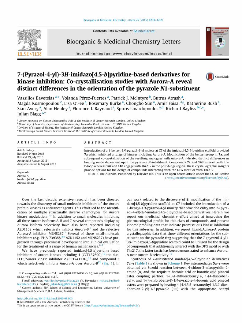
7-(Pyrazol-4-yl)-3H-imidazo[4,5-b]pyridine-based derivatives for kinase inhibition: Co-crystallisation studies with Aurora-A reveal distinct differences in the orientation of the pyrazole N1-substituent Vassilios Bavetsias a,⇑ , Yolanda Pérez-Fuertes a , Patrick J. McIntyre b , Butrus Atrash a , Magda Kosmopoulou c , Lisa O’Fee a , Rosemary Burke a , Chongbo Sun a , Amir Faisal a,y , Katherine Bush a , Sian Avery a , Alan Henley a , Florence I. Raynaud a , Spiros Linardopoulos a,d , Richard Bayliss b,c,⇑ , Julian Blagg a,⇑ a Cancer Research UK Cancer Therapeutics Unit at The Institute of Cancer Research, London, United Kingdom b University of Leicester, Department of Biochemistry, Lancaster Road, Leicester LE1 9HN, United Kingdom c Division of Structural Biology, The Institute of Cancer Research, London, United Kingdom d Breakthrough Breast Cancer Research Centre at The Institute of Cancer Research, London, United Kingdom article info Article history: Received 9 June 2015 Revised 29 July 2015 Accepted 3 August 2015 Available online 6 August 2015 Keywords: Aurora-A Imidazo[4,5-b]pyridine Aurora kinase abstract Introduction of a 1-benzyl-1H-pyrazol-4-yl moiety at C7 of the imidazo[4,5-b]pyridine scaffold provided 7a which inhibited a range of kinases including Aurora-A. Modification of the benzyl group in 7a, and subsequent co-crystallisation of the resulting analogues with Aurora-A indicated distinct differences in binding mode dependent upon the pyrazole N-substituent. Compounds 7a and 14d interact with the P-loop whereas 14a and 14b engage with Thr217 in the post-hinge region. These crystallographic insights provide options for the design of compounds interacting with the DFG motif or with Thr217. Ó 2015 The Authors. Published by Elsevier Ltd. This is an open access article under the CC BY license (http://creativecommons.org/licenses/by/4.0/). Over the last decade, extensive research has been directed towards the discovery of small molecule inhibitors of the Aurora protein kinases as anticancer agents. This effort led to the identifi- cation of multiple structurally diverse chemotypes for Aurora kinase modulation. 1–5 In addition to small molecules inhibiting all three Aurora isoforms A, B and C, several compounds displaying Aurora isoform selectivity have also been reported including AZD1152 which selectively inhibits Aurora-B, 6 and the selective Aurora-A inhibitor MLN8237. 7 Several of these small-molecule inhibitors (e.g., PHA-739358, 8,9 AZD1152 and MLN8237) have pro- gressed through preclinical development into clinical evaluation for the treatment of a range of human malignancies. 1–5 We have previously reported imidazo[4,5-b]pyridine-based inhibitors of Aurora kinases including 1 (CCT137690), 10 the dual FLT3/Aurora kinase inhibitor 2 (CCT241736), 11 and compound 3 which selectively inhibits Aurora-A over Aurora-B 12 (Fig. 1). In our work related to the discovery of 3, modification of the imi- dazo[4,5-b]pyridine scaffold at C7 included the introduction of a 1-benzyl-1H-pyrazol-4-yl moiety that provided entry into 7-(pyra- zol-4-yl)-3H-imidazo[4,5-b]pyridine-based derivatives. Herein, we report our medicinal chemistry effort aimed at improving the pharmacological profile for this class of compounds, and present kinome profiling data that indicate promiscuous kinase inhibition for this subseries. In addition, we report ligand/Aurora-A protein crystallographic data that show different orientations for the sub- stituent on the pyrazole ring suggesting that the 7-(pyrazol-4-yl)- 3H-imidazo[4,5-b]pyridine scaffold could be utilised for the design of compounds that additionally interact with the DFG motif or with Thr217, the latter tactic has been demonstrated to enhance Aurora- A over Aurora-B selectivity. 12 Synthesis of 7-substituted imidazo[4,5-b]pyridine derivatives 7a–e (Table 1) is shown in Scheme 1. Key intermediates 5a–e were obtained via Suzuki reaction between 4-chloro-3-nitropyridin-2- amine (4) and the requisite boronic acid or boronic acid pinacol ester coupling partner. 1-(3,4-Difluorobenzyl)-, 1-(4-fluoroben- zyl)-, and 1-(4-chlorobenzyl)-1H-pyrazole-4-boronic acid pinacol esters were prepared by heating 4-(4,4,5,5-tetramethyl-1,3,2-diox- aborolan-2-yl)-1H-pyrazole (11) with the appropriate benzyl http://dx.doi.org/10.1016/j.bmcl.2015.08.003 0960-894X/Ó 2015 The Authors. Published by Elsevier Ltd. This is an open access article under the CC BY license (http://creativecommons.org/licenses/by/4.0/). ⇑ Corresponding authors. Tel.: +44 (0)20 87224158 (V.B.), +44 (0)116 2297100 (R.B.), +44 (0)20 87224051 (J.B.). E-mail addresses: [email protected] (V. Bavetsias), richard.bayliss@ leicester.ac.uk (R. Bayliss), [email protected] (J. Blagg). y Current address: SBA School of Science and Engineering, Lahore University of Management Sciences, D.H.A., Lahore, Pakistan. Bioorganic & Medicinal Chemistry Letters 25 (2015) 4203–4209 Contents lists available at ScienceDirect Bioorganic & Medicinal Chemistry Letters journal homepage: www.elsevier.com/locate/bmcl
Transcript of Bioorganic & Medicinal Chemistry LettersPyrazol-4-yl)-3H... · y Current address: SBA School of...
Bioorganic & Medicinal Chemistry Letters 25 (2015) 4203–4209
Contents lists available at ScienceDirect
Bioorganic & Medicinal Chemistry Letters
journal homepage: www.elsevier .com/ locate/bmcl
7-(Pyrazol-4-yl)-3H-imidazo[4,5-b]pyridine-based derivatives forkinase inhibition: Co-crystallisation studies with Aurora-A revealdistinct differences in the orientation of the pyrazole N1-substituent
http://dx.doi.org/10.1016/j.bmcl.2015.08.0030960-894X/� 2015 The Authors. Published by Elsevier Ltd.This is an open access article under the CC BY license (http://creativecommons.org/licenses/by/4.0/).
⇑ Corresponding authors. Tel.: +44 (0)20 87224158 (V.B.), +44 (0)116 2297100(R.B.), +44 (0)20 87224051 (J.B.).
E-mail addresses: [email protected] (V. Bavetsias), [email protected] (R. Bayliss), [email protected] (J. Blagg).
y Current address: SBA School of Science and Engineering, Lahore University ofManagement Sciences, D.H.A., Lahore, Pakistan.
Vassilios Bavetsias a,⇑, Yolanda Pérez-Fuertes a, Patrick J. McIntyre b, Butrus Atrash a,Magda Kosmopoulou c, Lisa O’Fee a, Rosemary Burke a, Chongbo Sun a, Amir Faisal a,y, Katherine Bush a,Sian Avery a, Alan Henley a, Florence I. Raynaud a, Spiros Linardopoulos a,d, Richard Bayliss b,c,⇑,Julian Blagg a,⇑aCancer Research UK Cancer Therapeutics Unit at The Institute of Cancer Research, London, United KingdombUniversity of Leicester, Department of Biochemistry, Lancaster Road, Leicester LE1 9HN, United KingdomcDivision of Structural Biology, The Institute of Cancer Research, London, United KingdomdBreakthrough Breast Cancer Research Centre at The Institute of Cancer Research, London, United Kingdom
a r t i c l e i n f o
Article history:Received 9 June 2015Revised 29 July 2015Accepted 3 August 2015Available online 6 August 2015
Keywords:Aurora-AImidazo[4,5-b]pyridineAurora kinase
a b s t r a c t
Introduction of a 1-benzyl-1H-pyrazol-4-yl moiety at C7 of the imidazo[4,5-b]pyridine scaffold provided7a which inhibited a range of kinases including Aurora-A. Modification of the benzyl group in 7a, andsubsequent co-crystallisation of the resulting analogues with Aurora-A indicated distinct differences inbinding mode dependent upon the pyrazole N-substituent. Compounds 7a and 14d interact with theP-loop whereas 14a and 14b engage with Thr217 in the post-hinge region. These crystallographic insightsprovide options for the design of compounds interacting with the DFG motif or with Thr217.
� 2015 The Authors. Published by Elsevier Ltd. This is an open access article under the CC BY license(http://creativecommons.org/licenses/by/4.0/).
Over the last decade, extensive research has been directedtowards the discovery of small molecule inhibitors of the Auroraprotein kinases as anticancer agents. This effort led to the identifi-cation of multiple structurally diverse chemotypes for Aurorakinase modulation.1–5 In addition to small molecules inhibitingall three Aurora isoforms A, B and C, several compounds displayingAurora isoform selectivity have also been reported includingAZD1152 which selectively inhibits Aurora-B,6 and the selectiveAurora-A inhibitor MLN8237.7 Several of these small-moleculeinhibitors (e.g., PHA-739358,8,9 AZD1152 and MLN8237) have pro-gressed through preclinical development into clinical evaluationfor the treatment of a range of human malignancies.1–5
We have previously reported imidazo[4,5-b]pyridine-basedinhibitors of Aurora kinases including 1 (CCT137690),10 the dualFLT3/Aurora kinase inhibitor 2 (CCT241736),11 and compound 3which selectively inhibits Aurora-A over Aurora-B12 (Fig. 1). In
our work related to the discovery of 3, modification of the imi-dazo[4,5-b]pyridine scaffold at C7 included the introduction of a1-benzyl-1H-pyrazol-4-yl moiety that provided entry into 7-(pyra-zol-4-yl)-3H-imidazo[4,5-b]pyridine-based derivatives. Herein, wereport our medicinal chemistry effort aimed at improving thepharmacological profile for this class of compounds, and presentkinome profiling data that indicate promiscuous kinase inhibitionfor this subseries. In addition, we report ligand/Aurora-A proteincrystallographic data that show different orientations for the sub-stituent on the pyrazole ring suggesting that the 7-(pyrazol-4-yl)-3H-imidazo[4,5-b]pyridine scaffold could be utilised for the designof compounds that additionally interact with the DFGmotif or withThr217, the latter tactic has been demonstrated to enhance Aurora-A over Aurora-B selectivity.12
Synthesis of 7-substituted imidazo[4,5-b]pyridine derivatives7a–e (Table 1) is shown in Scheme 1. Key intermediates 5a–e wereobtained via Suzuki reaction between 4-chloro-3-nitropyridin-2-amine (4) and the requisite boronic acid or boronic acid pinacolester coupling partner. 1-(3,4-Difluorobenzyl)-, 1-(4-fluoroben-zyl)-, and 1-(4-chlorobenzyl)-1H-pyrazole-4-boronic acid pinacolesters were prepared by heating 4-(4,4,5,5-tetramethyl-1,3,2-diox-aborolan-2-yl)-1H-pyrazole (11) with the appropriate benzyl

Figure 1. Imidazo[4,5-b]pyridine-based inhibitors of Aurora kinases.
Table 1C7-Pyrazole modifications
Compd R Aurora-AIC50 (lM)
Aurora-B IC50
(lM)MLM/HLM%met/30 min
7a 0.212 ± 0.107 0.461 ± 0.147 40/43
7b 0.182 0.347 52/39
7c 0.322 0.458 31/28
7d 0.190 0.271 37/16
7e >10 >10 31/20
IC50s are mean values of two independent determinations or mean (±SD) for n > 2.17
MLM/HLM: percentage of compound metabolised after a 30 min incubation.18
4204 V. Bavetsias et al. / Bioorg. Med. Chem. Lett. 25 (2015) 4203–4209
bromide at 80 �C for 2–3 h in acetonitrile in the presence of Cs2CO3,reaction conditions previously reported for the synthesis of 1-ben-zyl-3-heterocyclic pyrazoles.13 (1-Benzyl-1H-pyrazol-4-yl)boronicacid and (1-(4-fluorophenyl)-1H-pyrazol-4-yl)boronic acid,required for the synthesis of 5a and 5e respectively, were commer-cially available. N-chlorosuccinimide-mediated C5-pyridinechlorination was followed by reaction with 1,3-dimethyl-1H-pyra-zole-4-carbaldehyde in the presence of Na2S2O4, as previouslydescribed,11,12,14 to afford the imidazo[4,5-b]pyridine derivatives7a–e (Scheme 1, Table 1).
Scheme 1. Reagents and conditions: (a) boronic acid or boronic acid pinacol ester, THF/H150 �C, microwave irradiation, 15 min (for the introduction of 1-(4-fluorophenyl)-1H-pyr1H-pyrazole-4-carbaldehyde, 1 M aqueous Na2S2O4, 80 �C, 16–20 h.
The 2-amino-3-nitro-pyridine derivative 9 (Scheme 2), keyintermediate for the synthesis of 14a and 14b (Table 2), wasobtained from 4-chloro-3-nitropyridin-2-amine (4) by a Suzukicross-coupling reaction to (1-(3-(methoxycarbonyl)benzyl)-1H-pyrazol-4-yl)boronic acid pinacol ester, prepared by reacting 11with methyl 3-(bromomethyl)benzoate13, followed by C5-pyridinechlorination with N-chlorosuccinimide (Scheme 2). An attempt toprepare 10a directly from the methyl ester intermediate 9 upontreatment with dimethylamine under microwave irradiationresulted in formation of the corresponding carboxylic acid.Coupling of this acid with dimethylamine via HATU carboxyl acti-vation provided 10a (Scheme 2). Starting from the methyl esterintermediate 9, access to 10b was achieved by alkaline esterhydrolysis followed by HATU-mediated coupling with1-methylpiperazine (Scheme 2). Imidazo[4,5-b]pyridine ringformation to afford 14a and 14b (Table 2) was effected by reacting10a and 10b with 1,3-dimethyl-1H-pyrazole-4-carbaldehyde inthe presence of Na2S2O4 as previously described.11,12,14 The ortho-substituted derivative 14c (Table 2) was prepared by a procedureanalogous to that described for its meta-isomer 14b (Scheme 2,Table 2).
Access to 2-amino-3-nitro-pyridine derivative 13 (Scheme 3),key intermediate for the preparation of 14e–g, was achieved byalkylation of 4-(4,4,5,5-tetramethyl-1,3,2-dioxaborolan-2-yl)-1H-pyrazole (11) with 3-(bromomethyl)benzaldehyde13 followed bya Suzuki cross-coupling reaction with 4-chloro-3-nitropyridin-2-amine (4) (Scheme 3). Reductive amination of the benzaldehydemoiety in 13 was accomplished under standard conditions (NaBH(OAc)3/AcOH with methylamine or pyrrolidine, and NaBH3CN withdimethylamine). The synthesis of 14e–g (Table 2) was finalised bythe C5-pyridine chlorination of the requisite intermediate followedby imidazo[4,5-b]pyridine ring formation as described for thepreparation of 7a–e (Scheme 1). Finally, the isoxazole derivative14d (Table 2) was prepared from 4 by a route analogous to thatdescribed for the synthesis of 7a–e (Scheme 1). For this syntheticsequence, (1-((5-methylisoxazol-3-yl)methyl)-1H-pyrazol-4-yl)boronic acid pinacol ester, required for the Suzuki cross-couplingreaction, was prepared by alkylation of 4-(4,4,5,5-tetramethyl-
2O, Pd(dppf)Cl2, Na2CO3, 80 �C, 3.5–26 h or DME, Pd(dppf)Cl2, 1 M aqueous Na2CO3,azol-4-yl); (b) N-chlorosuccinimide, CH3CN, reflux, 3.5–7 h; (c) EtOH, 1,3-dimethyl-
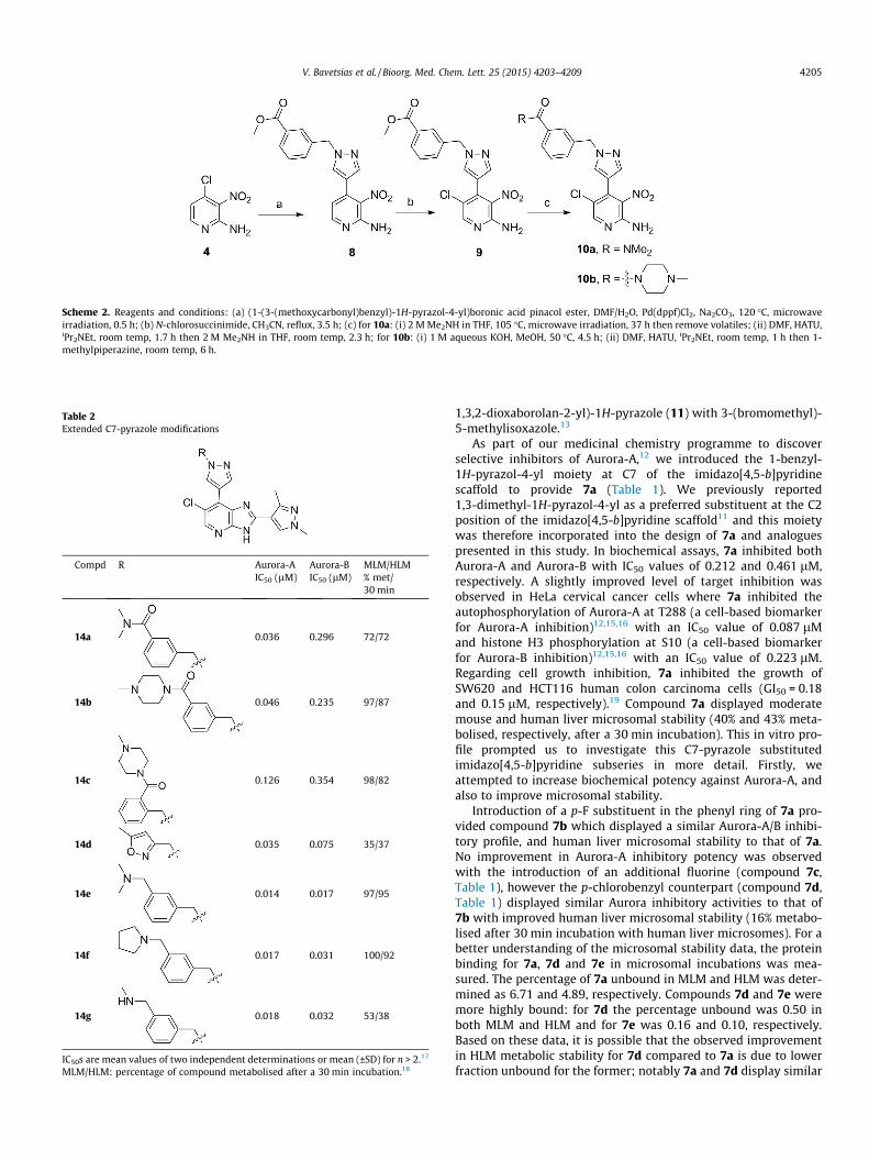
Table 2Extended C7-pyrazole modifications
Compd R Aurora-AIC50 (lM)
Aurora-BIC50 (lM)
MLM/HLM% met/30 min
14a 0.036 0.296 72/72
14b 0.046 0.235 97/87
14c 0.126 0.354 98/82
14d 0.035 0.075 35/37
14e 0.014 0.017 97/95
14f 0.017 0.031 100/92
14g 0.018 0.032 53/38
IC50s are mean values of two independent determinations or mean (±SD) for n > 2.17
MLM/HLM: percentage of compound metabolised after a 30 min incubation.18
Scheme 2. Reagents and conditions: (a) (1-(3-(methoxycarbonyl)benzyl)-1H-pyrazol-4-yl)boronic acid pinacol ester, DMF/H2O, Pd(dppf)Cl2, Na2CO3, 120 �C, microwaveirradiation, 0.5 h; (b) N-chlorosuccinimide, CH3CN, reflux, 3.5 h; (c) for 10a: (i) 2 M Me2NH in THF, 105 �C, microwave irradiation, 37 h then remove volatiles; (ii) DMF, HATU,iPr2NEt, room temp, 1.7 h then 2 M Me2NH in THF, room temp, 2.3 h; for 10b: (i) 1 M aqueous KOH, MeOH, 50 �C, 4.5 h; (ii) DMF, HATU, iPr2NEt, room temp, 1 h then 1-methylpiperazine, room temp, 6 h.
V. Bavetsias et al. / Bioorg. Med. Chem. Lett. 25 (2015) 4203–4209 4205
1,3,2-dioxaborolan-2-yl)-1H-pyrazole (11) with 3-(bromomethyl)-5-methylisoxazole.13
As part of our medicinal chemistry programme to discoverselective inhibitors of Aurora-A,12 we introduced the 1-benzyl-1H-pyrazol-4-yl moiety at C7 of the imidazo[4,5-b]pyridinescaffold to provide 7a (Table 1). We previously reported1,3-dimethyl-1H-pyrazol-4-yl as a preferred substituent at the C2position of the imidazo[4,5-b]pyridine scaffold11 and this moietywas therefore incorporated into the design of 7a and analoguespresented in this study. In biochemical assays, 7a inhibited bothAurora-A and Aurora-B with IC50 values of 0.212 and 0.461 lM,respectively. A slightly improved level of target inhibition wasobserved in HeLa cervical cancer cells where 7a inhibited theautophosphorylation of Aurora-A at T288 (a cell-based biomarkerfor Aurora-A inhibition)12,15,16 with an IC50 value of 0.087 lMand histone H3 phosphorylation at S10 (a cell-based biomarkerfor Aurora-B inhibition)12,15,16 with an IC50 value of 0.223 lM.Regarding cell growth inhibition, 7a inhibited the growth ofSW620 and HCT116 human colon carcinoma cells (GI50 = 0.18and 0.15 lM, respectively).19 Compound 7a displayed moderatemouse and human liver microsomal stability (40% and 43% meta-bolised, respectively, after a 30 min incubation). This in vitro pro-file prompted us to investigate this C7-pyrazole substitutedimidazo[4,5-b]pyridine subseries in more detail. Firstly, weattempted to increase biochemical potency against Aurora-A, andalso to improve microsomal stability.
Introduction of a p-F substituent in the phenyl ring of 7a pro-vided compound 7b which displayed a similar Aurora-A/B inhibi-tory profile, and human liver microsomal stability to that of 7a.No improvement in Aurora-A inhibitory potency was observedwith the introduction of an additional fluorine (compound 7c,Table 1), however the p-chlorobenzyl counterpart (compound 7d,Table 1) displayed similar Aurora inhibitory activities to that of7b with improved human liver microsomal stability (16% metabo-lised after 30 min incubation with human liver microsomes). For abetter understanding of the microsomal stability data, the proteinbinding for 7a, 7d and 7e in microsomal incubations was mea-sured. The percentage of 7a unbound in MLM and HLM was deter-mined as 6.71 and 4.89, respectively. Compounds 7d and 7e weremore highly bound: for 7d the percentage unbound was 0.50 inboth MLM and HLM and for 7e was 0.16 and 0.10, respectively.Based on these data, it is possible that the observed improvementin HLM metabolic stability for 7d compared to 7a is due to lowerfraction unbound for the former; notably 7a and 7d display similar

Scheme 3. Reagents and conditions: (a) 3-(bromomethyl)benzaldehyde, CH3CN, Cs2CO3, 80 �C, 2 h; (b) THF/H2O, Pd(dppf)Cl2, Na2CO3, 80 �C, 2 h.
Figure 2. Imidazo[4,5-b]pyridine-based inhibitors of Aurora kinases co-crystallisedwith Aurora-A: compound 15: PDB ID 2X6D;10 compound 16: PDB ID 4B0G.11
4206 V. Bavetsias et al. / Bioorg. Med. Chem. Lett. 25 (2015) 4203–4209
MLM stabilities despite the lower fraction unbound for 7d. In HeLacells, 7d inhibited both Aurora-A (p-T288 IC50 = 0.040 lM) andAurora-B (p-HH3 IC50 = 0.330 lM); potencies similar to thoseobserved with compound 7a. The direct attachment of a p-fluo-rophenyl group to the N1-pyrazole was detrimental to Aurora inhi-bition, with compound 7e inhibiting Aurora-A and Aurora-B withIC50 values greater than 10 lM (Table 1). Cell growth inhibitionstudies revealed that compounds 7b–d maintained the inhibitorypotencies seen with 7a; for example, 7d inhibited the growth ofSW620 and HCT116 cells (GI50 = 0.26 and 0.24 lM, respectively).Likewise, 7b and 7c showed potent HCT116 cell growth inhibition(GI50 = 0.20 and 0.13 lM, respectively). We postulated that potentcell growth inhibitory activity relative to biochemical Aurora-Aand Aurora-B modulation may be attributable to gain of off-targetkinase inhibition in this sub-series, although we acknowledge thattranslation to more potent cellular inhibition of Aurora-A versusAurora-A biochemical potency may also be a contributing factor.The potent cell-based activity prompted us to investigate kinomeselectivity profiles for this class of compound by screening 7aand 7d in a 102-kinase panel at a concentration of 1 lM.20
Indeed, both compounds inhibited a range of kinases includingERK8, GSK3b, MLK1, JAK2, TrkA and VEGFR greater than 80%(Table S1, Supplementary data) with Gini coefficients21 of 0.273for 7a and 0.364 for 7d. These Gini coefficient values are signifi-cantly lower to those of previously reported 7-(piperazin-1-yl)-3H-imidazo[4,5-b]pyridine- and 7-phenoxy-3H-imidazo[4,5-b]pyridine-based inhibitors of Aurora kinases.10,12 The Gini coeffi-cient for compound 3was reported as 0.719,12 and the correspond-ing value for compound 1 is 0.560. To understand the potential forfurther improvement, we obtained the crystal structure of 7abound to Aurora-A22 (Fig. 3, Table S3, Supplementary data). Thisstructure shows the ligand in the ATP binding site with the pyri-dine nitrogen atom hydrogen bonded to the backbone NH ofAla213 and the imidazole NH interacting with the carbonyl ofAla213, consistent with previous reports of the hinge bindingmode for the imidazo[4,5-b]pyridine scaffold.10–12 Similar to crys-tal structures of previous compounds 1, 15 and 16 (Figs. 1 and 2;compounds 51 and 40c in Ref. 10, and compound 21a in Ref. 11,respectively) the N-benzyl substituent on the C7-pyrazole is ori-ented towards the P-loop, making Van der Waals contacts withVal147 and Gly142 (Fig. 3).
In vivo mouse pharmacokinetic profiling of 7d revealed low oralbioavailability (16%) with moderate clearance (0.016 L/h, 13.3 mL/min/kg) and volume of distribution (0.02 L, 1.0 L/kg). Similar phar-macokinetic parameters were observed for 7a with low oralbioavailability (13%), moderate clearance (0.012 L/h, 10.0 mL/min/kg) and volume of distribution (0.01 L, 0.5 L/kg). The mouseplasma protein binding for 7a and 7d was determined as 99.48%and >99.9%, respectively. We determined low kinetic solubilityfor both 7a and 7d (<0.0001 mg/mL in phosphate buffer,pH = 6.8)23 which may be a contributing factor to the observed
low oral bioavailability. In an attempt to improve the aqueous sol-ubility, the Aurora inhibitory potency and kinase selectivity of thissub-series, we explored the introduction of basic substituents suchas 1-methylpiperazine and pyrrolidine as well as replacement ofthe phenyl ring with a more polar heterocycle. This approachwas guided by the ligand/protein interactions observed in the pro-tein crystal structure of 7a, which showed that the N-benzyl sub-stituent on the C7-pyrazole is oriented towards the P-loop(Fig. 4). It was anticipated that the introduction of small sub-stituents on the phenyl ring in 7a would be well tolerated withoutaltering its orientation in the kinase active site. Alternatively, weenvisaged that Thr217 in Aurora-A may be accessed via an appro-priate pyrazole N1-benzyl derivatisation such as a bulky amidosubstituent, which would form a favourable hydrogen bond inter-action with the side chain hydroxyl of Thr217. Similar approacheswere previously applied by us in the design of selective inhibitorsof Aurora-A.12,16
Replacement of the phenyl ring in 7a with 5-methylisoxazole(compound 14d) led to a significant improvement in Aurora inhibi-tion in our biochemical assays (Aurora-A IC50 = 0.035 lM, Aurora-BIC50 = 0.075 lM; Table 2) but promiscuous kinase inhibitionremained. In a panel of 105 kinases at a compound concentrationof 1 lM, 22 proteins including ERK8, MLK1, JAK2, TrkA andVEGFR were inhibited by P80% (Gini coefficient = 0.320,Table S2, Supplementary data). Compound 14d inhibited thegrowth of SW620 and HCT116 cells with GI50 values of 0.35 and0.34 lM respectively, similar to the cell growth inhibitionobserved with 7d. The crystal structure of 14d bound to Aurora-A22,24,25 (Fig. 5, Table S3, Supplementary data) shows a similarbinding mode to that adopted by 7a; however, the P-loop ofAurora-A adopts a different conformation, stabilised by interac-tions with the 5-methylisoxazole; this P-loop conformation is verysimilar to that previously observed for compound 1 (compound 51in Ref. 10).

Figure 3. Crystal structure of 7a bound to Aurora-A at 3.1 Å resolution. (A) The ATP binding site viewed from above. The P-loop has been removed to provide a clear view ofthe ligand, shown with carbon atoms coloured yellow, and the surrounding protein, shown with carbon atoms coloured teal. The final 2mFo-DFc electron density map isshown as a wire-mesh, contoured at 1 r. (B) Side view with the P-loop shown and key interacting residues labelled. (C) Crystal structure of 16 (compound 21a in Ref. 11;carbon atoms coloured white) bound to Aurora-A with the P-loop shown and key interacting residues labelled. Structure taken from the Protein Data Bank (PDB) ID 4B0G.
Figure 4. Comparison of crystal structure of Aurora-A bound to 7a and published crystal structures from this series. (A) Overlay of structures showing superposed proteinbackbones (all coloured teal) with 7a (carbon atoms coloured yellow), and published compounds 15, 1 and 16 taken from PDB IDs 2X6D, 2X6E and 4B0G, respectively. (B–E)Magnified view of the interactions between compounds 7a, 15, 1 and 16 respectively and the P-loop with the protein surface shown.
Figure 5. Crystal structure of 14d bound to Aurora-A at 3.1 Å resolution. (A) The ATP binding site viewed from above. The P-loop has been removed to provide a clear view ofthe ligand, shown with carbon atoms coloured orange and the surrounding protein, shown with carbon atoms coloured teal. The final 2mFo-DFc electron density map isshown as a wire-mesh, contoured at 1 r. (B) Side view with the P-loop shown and key interacting residues labelled. (C) Overlay of the structures of Aurora-A bound tocompound 14d (coloured as before) and compound 1 (compound and protein coloured grey).
V. Bavetsias et al. / Bioorg. Med. Chem. Lett. 25 (2015) 4203–4209 4207
The m-dimethylbenzamide derivative 14a (Table 2) was a morepotent inhibitor of Aurora-A compared to 7a but its human livermicrosomal stability was lower (72% metabolised after a 30 minincubation). A similar trend was observed with the m-1-methylpiperazine derivative 14b which was also a more potentinhibitor of Aurora-A compared to 7a but with poor metabolic
stability in both mouse and human liver microsomes (Table 2).Both 14a and 14b were more potent (by 8- and 5-fold, respec-tively) inhibitors of Aurora-A relative to Aurora-B (Table 2). Thecrystal structures of 14a and 14b bound to Aurora-A22,24,25 weredetermined (Table S3, Supplementary data; Figs. 6 and 7) andrevealed a consistent difference in the binding mode of these

Figure 6. Crystal structure of 14a bound to Aurora-A at 2.8 Å resolution. (A) The ATP binding site viewed from above. The P-loop has been removed to provide a clear view ofthe ligand, shown with carbon atoms coloured pink and the surrounding protein, shown with carbon atoms coloured teal. The final 2mFo-DFc electron density map is shownas a wire-mesh, contoured at 1 r. (B) Side view with the P-loop shown and Ala213, Thr217 labelled. (C) Modelling a glutamic acid in position 217 (Glu*), as found in Aurora-B,leads to steric clashes with the ligand, modelled clash shown as red dashes.
Figure 7. Crystal structure of 14b bound to Aurora-A at 2.9 Å resolution. (A) The ATP binding site viewed from above. The P-loop has been removed to provide a clear view ofthe ligand, shown with carbon atoms coloured brown and the surrounding protein, shown with carbon atoms coloured teal. The final 2mFo-DFc electron density map isshown as a wire-mesh, contoured at 1 r. (B) Side view with the P-loop shown and Ala213, Thr217 residues labelled. (C) Modelling a glutamic acid in position 217 (Glu*), asfound in Aurora-B, leads to steric clashes with the ligand, modelled clash shown as red dashes.
4208 V. Bavetsias et al. / Bioorg. Med. Chem. Lett. 25 (2015) 4203–4209
ligands when compared to 7a and 14d. Bulkier substituents on thepyrazole group no longer interact with the pocket formed by thetwo b-strands of the P-loop. Instead, these bulkier groups are posi-tioned between the b1 strand of the P-loop and the post-hingeregion. This orientation places them in close proximity to Thr217, with a minimum interaction distance of 3.8 Å, which mayexplain the selectivity of these compounds towards Aurora-A.Notably, Aurora-B and Aurora-C have a glutamic acid at the equiv-alent position to Thr217, and computational modelling of this Thrto Glu replacement suggests that the bulkier R-groups of 14a and14b may invoke a steric clash with a glutamic acid at position217 (Fig. 6C, Fig. 7C).12
The o-regioisomer of 14b (compound 14c) displayed a similarprofile to that of 14b although its potency against Aurora-A wasmarginally lower and selectivity over Aurora-B was eroded(Table 2). The introduction of the smaller m-dimethylamino basicsubstituent (compound 14e) led to potent inhibition of bothAurora-A and Aurora-B although mouse and human liver microso-mal stability was poor (Table 2). Likewise, the m-pyrrolidinederivative 14f potently inhibited both Aurora-A (IC50 = 0.017 lM)and Aurora-B (IC50 = 0.031 lM) but was also highly metabolised(Table 2). The potent Aurora-A and Aurora-B activity of these com-pounds suggests that they do not adopt a binding mode involvinginteraction with Thr217 in Aurora-A similar to 14a and 14b, butmight bind similarly to 7a and 14d. Unfortunately, we have beenunable to resolve the binding mode of compounds 14e and 14fby co-crystal structure determination to verify this hypothesis.The m-methylamino counterpart of 14e (compound 14g) also dis-played potent inhibition of both Aurora-A (IC50 = 0.018 lM) andAurora-B (IC50 = 0.032 lM) and demonstrated improved metabolicstability compared to 14e and 14f (Table 2). Compound 14g inhib-ited the growth of SW620 and HCT116 cells (GI50 = 0.34 and0.23 lM, respectively); however, profiling in a 105-kinase panel
at a concentration of 1 lM revealed 30 kinases including ERK8,GSK3b, MLK1, JAK2, TrkA and VEGFR that were inhibited byP80% (Gini coefficient = 0.267, Table S2, Supplementary data).Based on these findings, our interest in this 7-(pyrazol-4-yl)-3H-imidazo[4,5-b]pyridine-based sub-series was discontinued andour efforts focussed on compound 3 (Fig. 1) as a highly selectiveinhibitor of Aurora-A kinase.12 However, the kinase inhibitory pro-files of compounds 7a, 7d, 14d, and 14g suggest that the 7-(pyra-zol-4-yl)-3H-imidazo[4,5-b]pyridine scaffold could serve as thebasis for a multitargeted kinase inhibitor design. In addition, theligand/Aurora-A protein crystallographic data indicate distinct dif-ferences in the orientation of the pyrazole N-substituent. The phe-nyl group in 7a and the 5-methylisoxazole in 14d point towardsthe P-loop, whereas the bulkier benzamido substituted phenylrings in 14a and 14b position between the b1 strand of the P-loopin close proximity to Thr217 and also to the DFG motif. Theseligand/protein crystallographic insights could therefore be furtherexploited in the design of compounds interacting with the DFGmotif or in enhancing the selectivity for Aurora-A inhibition overAurora-B.
Acknowledgments
This work was supported by CRUK Grant numbers C309/A8274and C309/A11566. S.L. is also supported by Breakthrough BreastCancer. R.B. acknowledges the support of a Royal SocietyResearch Fellowship, Breakthrough Breast Cancer Project GrantAURA 05/06 and MRC CASE studentship MR/K016903/1. Weacknowledge NHS funding to the NIHR Biomedical ResearchCentre at The Institute of Cancer Research and the Royal MarsdenNHS Foundation Trust. We also thank Dr Amin Mirza, Mr MeirionRichards, Dr Maggie Liu for their assistance with NMR, mass spec-trometry and HPLC, and Dr Charlotte Dodson for Aurora-A protein

V. Bavetsias et al. / Bioorg. Med. Chem. Lett. 25 (2015) 4203–4209 4209
production. We thank Diamond Light Source for beamtime and thestaff of beamlines I03 and I04 for assistance with data collection.
Supplementary data
Supplementary data associated with this article can be found, inthe online version, at http://dx.doi.org/10.1016/j.bmcl.2015.08.003.
References and notes
1. Pollard, J. R.; Mortimore, M. J. Med. Chem. 2009, 52, 2629.2. Green, M. R.; Woolery, J. E.; Mahadevan, D. Expert Opin. Drug Discov. 2011, 6,
291.3. Cheung, C. H. A.; Coumar, M. S.; Chang, J.-Y.; Hsieh, H.-P. Expert Opin. Ther.
Patents 2011, 21, 857.4. Kollareddy, M.; Zheleva, D.; Dzubak, P.; Brahmkshatriya, P. S.; Lepsik, M.;
Hajduch, M. Invest New Drugs 2012, 30, 2411.5. Gautschi, O.; Heighway, J.; Mack, P. C.; Purnell, P. R.; Lara, P. N., Jr.; Gandara, D.
R. Clin. Cancer Res. 2008, 14, 1639.6. Mortlock, A. A.; Foote, K. M.; Heron, N. M.; Jung, F. H.; Pasquet, G.; Lohmann, J.-
J. M.; Warin, N.; Renaud, F.; De Savi, C.; Roberts, N. J.; Johnson, T.; Dousson, C.B.; Hill, G. B.; Perkins, D.; Hatter, G.; Wilkinson, R. W.; Wedge, S. R.; Heaton, S.P.; Odedra, R.; Keen, N. J.; Crafter, C.; Brown, E.; Thompson, K.; Brightwell, S.;Khatri, L.; Brady, M. C.; Kearney, S.; McKillop, D.; Rhead, S.; Parry, T.; Green, S. J.Med. Chem. 2007, 50, 2213.
7. Manfredi, M. G.; Ecsedy, J. A.; Chakravarty, A.; Silverman, L.; Zhang, M.; Hoar, K.M.; Stroud, S. G.; Chen, W.; Shinde, V.; Huck, J. J.; Wysong, D. R.; Janowick, D. A.;Hyer, M. L.; LeRoy, P. J.; Gershman, R. E.; Silva, M. D.; Germanos, M. S.; Bolen, J.B.; Claiborne, C. F.; Sells, T. B. Clin. Cancer Res. 2011, 17, 7614.
8. Fancelli, D.; Moll, J.; Varasi, M.; Bravo, R.; Artico, R.; Berta, D.; Bindi, S.;Cameron, A.; Candiani, I.; Cappella, P.; Carpinelli, P.; Croci, W.; Forte, B.;Giorgini, M. L.; Klapwijk, J.; Marsiglio, A.; Pesenti, E.; Rocchetti, M.; Roletto, F.;Severino, D.; Soncini, C.; Storici, P.; Tonani, R.; Zugnoni, P.; Vianello, P. J. Med.Chem. 2006, 49, 7247.
9. Caprinelli, P.; Ceruti, R.; Giorgini, M. L.; Cappella, P.; Gianellini, L.; Croci, V.;Degrassi, A.; Texido, G.; Rocchetti, M.; Vianello, P.; Rusconi, L.; Storici, P.;Zugnoni, P.; Arrigoni, C.; Soncini, C.; Alli, C.; Patton, V.; Marsiglio, A.; Ballinari,D.; Pesenti, E.; Fancelli, D.; Moll, J. Mol. Cancer Ther. 2007, 6, 3158.
10. Bavetsias, V.; Large, J. M.; Sun, C.; Bouloc, N.; Kosmopoulou, M.; Matteucci, M.;Wilsher, N. E.; Martins, V.; Reynisson, J.; Atrash, B.; Faisal, A.; Urban, F.; Valenti,M.; de Haven Brandon, A.; Box, G.; Raynaud, F. I.; Workman, P.; Eccles, S. A.;Bayliss, R.; Blagg, J.; Linardopoulos, S.;McDonald, E. J. Med. Chem. 2010, 53, 5213.
11. Bavetsias, V.; Crumpler, S.; Sun, C.; Avery, S.; Atrash, B.; Faisal, A.; Moore, A. S.;Kosmopoulou, M.; Brown, N.; Sheldrake, P. W.; Bush, K.; Henley, A.; Box, G.;
Valenti, M.; de Haven Brandon, A.; Raynaud, F. I.; Workman, P.; Eccles, S. A.;Bayliss, R.; Linardopoulos, S.; Blagg, J. J. Med. Chem. 2012, 55, 8721.
12. Bavetsias, V.; Faisal, A.; Crumpler, S.; Brown, N.; Kosmopoulou, M.; Joshi, A.;Atrash, B.; Pérez-Fuertes, Y.; Schmitt, J. A.; Boxall, K. J.; Burke, R.; Sun, C.; Avery,S.; Bush, K.; Henley, A.; Raynaud, F. I.; Workman, P.; Bayliss, R.; Linardopoulos,S.; Blagg, J. J. Med Chem. 2013, 56, 9122.
13. Curtis, M. P.; Sammons, M. F.; Piotrowski, D. W. Tetrahedron Lett. 2009, 50,5479.
14. Yang, D.; Fokas, D.; Li, J.; Yu, L.; Baldino, C. M. Synthesis 2005, 47.15. Manfredi, M. G.; Ecsedy, J. A.; Meetze, K. A.; Balani, S. K.; Burenkova, O.; Chen,
W.; Galvin, K. M.; Hoar, K. M.; Huck, J. J.; LeRoy, P. J.; Ray, E. T.; Sells, T. B.;Stringer, B.; Stroud, S. G.; Vos, T. J.; Weatherhead, G. S.; Wysong, D. R.; Zhang,M.; Bolen, J. B.; Claiborne, C. F. Proc. Natl. Acad. Sci. U.S.A. 2007, 104, 4106.
16. Bouloc, N.; Large, J. M.; Kosmopoulou, M.; Sun, C.; Faisal, A.; Matteucci, M.;Reynisson, J.; Brown, N.; Atrash, B.; Blagg, J.; McDonald, E.; Linardopoulos, S.;Bayliss, R.; Bavetsias, V. Bioorg. Med. Chem. Lett. 2010, 20, 5988.
17. Aurora-A and Aurora-B IC50 values were determined by using a microfluidicassay which monitors the separation of a phosphorylated product from itssubstrate, as described in Ref. 12.
18. MLM and HLM microsomal stabilities were determined as described in Ref. 12.19. GI50 values were determined after 72 h incubation as described in the
following reference: Chan, F.; Sun, C.; Perumal, M.; Nguyen, Q.-D.; Bavetsias,V.; McDonald, E.; Martins, V.; Wilsher, N.; Raynaud, F. I.; Valenti, M.; Eccles, S.;te Poele, R.; Workman, P.; Aboagye, E. O.; Linardopoulos, S. Mol. Cancer Ther.2007, 6, 3147.
20. Kinase selectivity profiling for compounds 7a, 7d, 14d and 14g was carried outat the International Centre for Protein Kinase Profiling, Division of SignalTransduction Therapy, University of Dundee.
21. Graczyk, P. P. J. Med. Chem. 2007, 50, 5773.22. The crystal structure of Aurora-A in complex with compound 7a was
determined as in Ref. 12. Other co-crystal structures used a mutant form ofAurora-A kinase domain (C290A, C393A), which has enhanced expression andgenerates superior crystals (Refs. 24,25). Hexagonal bipyramidal shaped crystalsof Aurora A C290A,C393A were grown at 18 �C by hanging drop vapourdiffusion in a 1 lL + 1 lL mixture of mother liquor (0.1 M Tris, pH 8.9; 0.2 Mlithium sulfate; 30% w/v PEG 400) and Aurora A (12 mg/mL, 1 mM inhibitorcompound, 5 mMMgCl2). Crystals were cryoprotected using 25% (v/v) ethyleneglycol in mother liquor before cryo-cooling in liquid nitrogen. The structurewas solved using molecular replacement with PDB:4CEG as the model andrefinement was carried out as described in Ref. 12. Coordinates and structurefactors have been deposited in the protein data bank with accession codes5AAD, 5AAE, 5AAF, 5AAG (corresponding to the Aurora-A co-crystal structuresof 7a, 14d, 14a and 14b, respectively).
23. Solubility measurements were performed by Pharmorphix� Solid StateServices, Member of the Sigma–Aldrich Group, Cambridge, UK.
24. Rowan, F. C.; Richards, M.; Bibby, R. A.; Thompson, A.; Bayliss, R.; Blagg, J. ACSChem. Biol. 2013, 8, 2184.
25. Burgess, S. G.; Bayliss, R. Acta Crystallogr. F Struct. Biol. Commun. 2015, 71, 315.