Biomineralized metal–organic framework nanoparticles ...
10
Biomineralized metal–organic framework nanoparticles enable a primer exchange reaction- based DNA machine to work in living cells for imaging and gene therapy† Juan Zhang, Mengyun He, Cunpeng Nie, Manman He, Qingshan Pan, Chang Liu, Yanlei Hu, Tingting Chen and Xia Chu * Sensitive tumor imaging and precise tumor therapy play critical roles in the cancer combat. Herein, we build a DNA machine based on a primer exchange reaction (PER) for mRNA imaging and gene therapy. By using zeolitic imidazolate framework-8 nanoparticles (ZIF-8 NPs) to co-deliver the components including a primer, hairpin and strand displacing polymerase to the living cells, the PER-based DNA machine can be initiated by intracellular survivin mRNA and continuously produce Bcl-2 antisense DNA (ASD), which enables the DNA machine not only to image survivin mRNA but also to implement gene therapy. The results demonstrate that ZIF-8 NPs can protect the polymerases and nucleic acid probes from protease attack and nuclease degradation. After internalization, pH-responsive ZIF-8 NPs can efficiently release cargos from endo-lysosomes due to the protonation effect. The intracellular PER-based DNA machine has been demonstrated to be able to sensitively image survivin mRNA expression levels and selectively kill the cancer cells and has no effect on the normal cells. The PER-based DNA machine may provide a promising platform for early stage tumor diagnosis and more precise tumor therapy. Introduction Antisense DNA (ASD) is a DNA single strand with a length of 18– 25 nucleotides. It can hybridize with the mRNA of the target gene and inhibit the translation of the target protein. 1,2 So, ASDs are a promising therapy as a gene drug. 3–5 However, free ASDs are negatively charged, which makes it difficult for them to cross the cell membrane, accordingly limiting their extensive clinical applications. 6,7 In recent years, many kinds of nano- carriers have been constructed for ASD delivery, such as DNA nanostructures, 8,9 liposomes 10–12 and inorganic nano- particles. 13–15 They can transport ASDs to the cytoplasm. However, these nanoparticles commonly tend to accumulate in the liver, kidneys and spleen when they target the tumor site, which results in the damage to healthy cells and causes serious side effects. Therefore, development of strategies that can deliver or produce ASDs exclusively in the cancer cells remains a great challenge. In addition, delivery or production of adequate amounts of ASDs in cancer cells is also signicantly important for the efficacy of the tumor therapy. A recent study reported a primer exchange reaction (PER) to program the autonomous synthesis of single-stranded DNA (ssDNA) from a short (7–9 nt) DNA primer in vitro. 16–18 Using a single PER hairpin as a catalytic template, the PER pathway starts with a prescribed short DNA primer, then repeatedly appends a nascent single-stranded sequence to the short primer with the aid of a strand displacing polymerase (for example, Klenow (exo-)). By the random walk process of three-way branch migration, the extended primer can spontaneously dissociate from the PER hairpin, and the hairpin becomes free and reacts with another primer to induce the next cycle of primer exchange. Inspired by this specic reaction, we want to employ the PER system to build a DNA machine, which is driven by a strand displacing polymerase and fueled by dNTPs, to continuously produce ASDs if the nucleotide sequences of the ASDs are programmed to design the PER hairpin. More importantly, if the region of the primer hybridization is rst blocked by a DNA strand, the machine can be designed to be triggered by the over-expressed target molecules in cancer cells, such as over-expressed mRNAs and miRNAs. Accordingly, the DNA machine triggered by specic molecules in cancer cells can selectively kill cancer cells and has no effect on normal cells. However, how to deliver efficiently the components of the DNA machine including the primer, hairpin and especially strand displacing polymerase to living cells is a major challenge we face. State Key Laboratory of Chemo/Bio-Sensing and Chemometrics, College of Chemistry and Chemical Engineering, Hunan University, Changsha 410082, P. R. China. E-mail: [email protected] † Electronic supplementary information (ESI) available. See DOI: 10.1039/d0sc00339e Cite this: Chem. Sci. , 2020, 11, 7092 All publication charges for this article have been paid for by the Royal Society of Chemistry Received 18th January 2020 Accepted 16th June 2020 DOI: 10.1039/d0sc00339e rsc.li/chemical-science 7092 | Chem. Sci., 2020, 11, 7092–7101 This journal is © The Royal Society of Chemistry 2020 Chemical Science EDGE ARTICLE Open Access Article. Published on 16 June 2020. Downloaded on 4/2/2022 2:51:03 PM. This article is licensed under a Creative Commons Attribution-NonCommercial 3.0 Unported Licence. View Article Online View Journal | View Issue
Transcript of Biomineralized metal–organic framework nanoparticles ...

ChemicalScience
EDGE ARTICLE
Ope
n A
cces
s A
rtic
le. P
ublis
hed
on 1
6 Ju
ne 2
020.
Dow
nloa
ded
on 4
/2/2
022
2:51
:03
PM.
Thi
s ar
ticle
is li
cens
ed u
nder
a C
reat
ive
Com
mon
s A
ttrib
utio
n-N
onC
omm
erci
al 3
.0 U
npor
ted
Lic
ence
.
View Article OnlineView Journal | View Issue
Biomineralized m
State Key Laboratory of Chemo/Bio-Sensing
and Chemical Engineering, Hunan Unive
E-mail: [email protected]
† Electronic supplementary informa10.1039/d0sc00339e
Cite this: Chem. Sci., 2020, 11, 7092
All publication charges for this articlehave been paid for by the Royal Societyof Chemistry
Received 18th January 2020Accepted 16th June 2020
DOI: 10.1039/d0sc00339e
rsc.li/chemical-science
7092 | Chem. Sci., 2020, 11, 7092–71
etal–organic frameworknanoparticles enable a primer exchange reaction-based DNA machine to work in living cells forimaging and gene therapy†
Juan Zhang, Mengyun He, Cunpeng Nie, Manman He, Qingshan Pan, Chang Liu,Yanlei Hu, Tingting Chen and Xia Chu *
Sensitive tumor imaging and precise tumor therapy play critical roles in the cancer combat. Herein, we build
a DNA machine based on a primer exchange reaction (PER) for mRNA imaging and gene therapy. By using
zeolitic imidazolate framework-8 nanoparticles (ZIF-8 NPs) to co-deliver the components including
a primer, hairpin and strand displacing polymerase to the living cells, the PER-based DNA machine can
be initiated by intracellular survivin mRNA and continuously produce Bcl-2 antisense DNA (ASD), which
enables the DNA machine not only to image survivin mRNA but also to implement gene therapy. The
results demonstrate that ZIF-8 NPs can protect the polymerases and nucleic acid probes from protease
attack and nuclease degradation. After internalization, pH-responsive ZIF-8 NPs can efficiently release
cargos from endo-lysosomes due to the protonation effect. The intracellular PER-based DNA machine
has been demonstrated to be able to sensitively image survivin mRNA expression levels and selectively
kill the cancer cells and has no effect on the normal cells. The PER-based DNA machine may provide
a promising platform for early stage tumor diagnosis and more precise tumor therapy.
Introduction
Antisense DNA (ASD) is a DNA single strand with a length of 18–25 nucleotides. It can hybridize with the mRNA of the targetgene and inhibit the translation of the target protein.1,2 So, ASDsare a promising therapy as a gene drug.3–5 However, free ASDsare negatively charged, which makes it difficult for them tocross the cell membrane, accordingly limiting their extensiveclinical applications.6,7 In recent years, many kinds of nano-carriers have been constructed for ASD delivery, such as DNAnanostructures,8,9 liposomes10–12 and inorganic nano-particles.13–15 They can transport ASDs to the cytoplasm.However, these nanoparticles commonly tend to accumulate inthe liver, kidneys and spleen when they target the tumor site,which results in the damage to healthy cells and causes seriousside effects. Therefore, development of strategies that candeliver or produce ASDs exclusively in the cancer cells remainsa great challenge. In addition, delivery or production ofadequate amounts of ASDs in cancer cells is also signicantlyimportant for the efficacy of the tumor therapy.
and Chemometrics, College of Chemistry
rsity, Changsha 410082, P. R. China.
tion (ESI) available. See DOI:
01
A recent study reported a primer exchange reaction (PER) toprogram the autonomous synthesis of single-stranded DNA(ssDNA) from a short (7–9 nt) DNA primer in vitro.16–18 Usinga single PER hairpin as a catalytic template, the PER pathwaystarts with a prescribed short DNA primer, then repeatedlyappends a nascent single-stranded sequence to the short primerwith the aid of a strand displacing polymerase (for example,Klenow (exo-)). By the random walk process of three-way branchmigration, the extended primer can spontaneously dissociatefrom the PER hairpin, and the hairpin becomes free and reactswith another primer to induce the next cycle of primerexchange. Inspired by this specic reaction, we want to employthe PER system to build a DNA machine, which is driven bya strand displacing polymerase and fueled by dNTPs, tocontinuously produce ASDs if the nucleotide sequences of theASDs are programmed to design the PER hairpin. Moreimportantly, if the region of the primer hybridization is rstblocked by a DNA strand, the machine can be designed to betriggered by the over-expressed target molecules in cancer cells,such as over-expressed mRNAs and miRNAs. Accordingly, theDNAmachine triggered by specic molecules in cancer cells canselectively kill cancer cells and has no effect on normal cells.However, how to deliver efficiently the components of the DNAmachine including the primer, hairpin and especially stranddisplacing polymerase to living cells is a major challenge weface.
This journal is © The Royal Society of Chemistry 2020

Scheme 1 Schematic illustration of a mRNA-triggered primerexchange reaction (PER)-based DNA machine. In step 1, a dumbbellshaped hairpin with two loops (PER hairpin) was first designed. Oneloop (domain c, blue) was programmed to be complementary to thesequences of the survivin mRNA, and could hybridize with the survivinmRNA and expose the complementary domain of the primera* (green). In step 2, primer a with a length of 8 nucleotides subse-quently bound with domain a* and was extended using domain b* asthe template such as in the PCR with the aid of Klenow (exo-)fragmentpolymerase to append a nascent single-stranded sequence b (termedthe copied b domain, cb) to the 30-end of the short primer until thestrand displacing elongation reaction was halted at the stop sequence,two pairs of synthetic nucleotides, iso-dG (20-O-methyl G) and iso-dC(20-O-methyl C), on the PER hairpin. In step 3, the b domain on thehairpin was then able to competewith the copied b domain (cb) via therandom walk process of three-way branch migration, and theb domain on the hairpin again hybridized with domain b*. In step 4, thecopied b domain was displaced and a part of the 8 nucleotide longprimer also was away from the PER hairpin due to the lower hybrid-ization temperature. The extended primer was spontaneously disso-ciated from the PER hairpin. The PER hairpin became free and boundwith another primer to induce the next cycle of the primer exchangereaction.
Edge Article Chemical Science
Ope
n A
cces
s A
rtic
le. P
ublis
hed
on 1
6 Ju
ne 2
020.
Dow
nloa
ded
on 4
/2/2
022
2:51
:03
PM.
Thi
s ar
ticle
is li
cens
ed u
nder
a C
reat
ive
Com
mon
s A
ttrib
utio
n-N
onC
omm
erci
al 3
.0 U
npor
ted
Lic
ence
.View Article Online
Recently, nanoscale metal–organic framework nanoparticles(MOF NPs) have been used for loading therapeutic or imagingagents due to their good biocompatibility, simple preparation,porous structure andmultifunctional features.19–23 Among theseMOF NPs, zeolitic imidazolate framework-8 nanoparticles (ZIF-8 NPs) constructed using zinc ions (Zn2+) and 2-methyl-imidazole (2-MIM) possess unique pH-sensitive biodegrad-ability, which makes them an ideal nanocarrier for the deliveryof various cargos into living cells including nucleic acidprobes24,25 and chemical drugs.26–29 In addition, the greenbiomimetic mineralization synthesis process in aqueous mediais favourable for the maintenance of the activities of bio-macromolecules and improvement of the bio-macromolecularstability against various denaturing conditions. To date, severalenzymes30,31 and antibodies32 have been loaded in the ZIF-8 NPsfor catalysis and disease treatment. Moreover, our group hasdemonstrated that ZIF-8 NPs can efficiently deliver and releasenative active proteins in living cells,33 and has realized a rollingcircle amplication (RCA) reaction in living cells by using ZIF-8NPs to deliver DNA polymerase and nucleic acids.34 So, thebiomineralized ZIF-8 NPs should be suited for the delivery ofcomponents including a primer, hairpin and strand displacingpolymerase to build a PER-based DNA machine working inliving cells.
Herein, we build a tumor-associated biomarker initiatedPER-based DNA machine for mRNA imaging and gene therapy.Using nanoscale ZIF-8 NPs to co-deliver Klenow (exo-)fragmentpolymerase (KFP) and nucleic acid probes into living cells, weimplement an intracellular PER-based polymerase reaction. Byprogramming the nucleotide sequences of the ASD to designthe PER hairpin, the machine can continuously produce Bcl-2ASD, which enables the DNA machine not only to image survi-vin mRNA but also to perform gene therapy. The resultsdemonstrate that ZIF-8 NPs can protect the polymerases andnucleic acid probes from protease attack and nuclease degra-dation. Aer internalization into cells, pH-responsive ZIF-8 NPsenable efficient release and escape of cargos from endo-lyso-somes due to the protonation effect. The intracellular PER-based DNA machine has been demonstrated to be able tosensitively image survivin mRNA expression levels and selec-tively kill the cancer cells and has no effect on the normal cells.The PER-based DNA machine may provide a potential platformfor early stage diagnosis, and more precise and efficient therapyof tumors.
Results and discussionMechanism and design of the PER-based DNA machine
The mechanism and design of the mRNA-triggered primerexchange reaction (PER)-based DNA machine are depicted inScheme 1. A dumbbell shaped hairpin with two loops was rstdesigned. One loop (domain c, blue) was programmed to becomplementary to the sequences of survivin mRNA and couldhybridize with survivin mRNA and expose the complementarydomain of the primer a*. Primer a with a length of 8 nucleotidesis subsequently bound with domain a* (step 1) and extendedwith the aid of a strand displacing polymerase (such as KFP) to
This journal is © The Royal Society of Chemistry 2020
append a nascent single-stranded sequence b (termed thecopied b domain) to the 30-end of the short primer until thestrand displacing elongation reaction was halted at the stopsequence on the PER hairpin (step 2). The b domain on thehairpin was then able to compete with the copied b domain viathe random walk process of three-way branch migration (step3).35 Finally, the extended primer was spontaneously dissociatedfrom the PER hairpin once the copied b domain was displaced(step 4). The PER hairpin became free and bound with anotherprimer to induce the next cycle of the primer exchange reaction.Therefore, a DNA single strand with an arbitrary sequence couldbe appended to the 30-end of the short primer through the PER-based machine. Importantly, the machine could be designed tobe triggered by a specic biomarker such as tumor-associatedmRNA or miRNA through programming domain c.
Here, we chose survivin mRNA, a mRNA over-expressed inmost cancers,36,37 as the model target to initiate the DNAmachine. The B-cell lymphoma 2 (Bcl-2) protein is an anti-apoptotic protein and is commonly associated with therapyresistance in various human cancers.38,39 So, Bcl-2 antisense
Chem. Sci., 2020, 11, 7092–7101 | 7093

Chemical Science Edge Article
Ope
n A
cces
s A
rtic
le. P
ublis
hed
on 1
6 Ju
ne 2
020.
Dow
nloa
ded
on 4
/2/2
022
2:51
:03
PM.
Thi
s ar
ticle
is li
cens
ed u
nder
a C
reat
ive
Com
mon
s A
ttrib
utio
n-N
onC
omm
erci
al 3
.0 U
npor
ted
Lic
ence
.View Article Online
DNA (Bcl-2 ASD) was chosen as the gene drug. A dumbbellshaped hairpin was rst designed, whose domain c (blue) wascomplementary to the survivin mRNA and domain b sequence(red) was the same as that of Bcl-2 ASD. The principle of theintracellular PER-based DNA machine for mRNA imaging andgene therapy is illustrated in Scheme 2. The ZIF-8 NPs encap-sulated BSA and KFP were rst prepared. Then, the nucleic acidincluding the hairpin, primer and molecular beacon (MB) wasadsorbed on the surface of ZIF-8 NPs by electrostatic interac-tion. Aer cellular uptake, the ZIF-8 NPs were biodegraded inacidic endo-lysosomes, releasing the KFP, hairpin, primer andMB. The intracellular survivin mRNA caused the DNA machineto produce Bcl-2 ASD continuously in the presence of KFP anddNTPs through the mechanism in Scheme 1. For survivinmRNA imaging, the MB complementary to Bcl-2 ASD wasdelivered into the cells, the produced Bcl-2 ASD hybridized withthe MB and resulted in the uorescence recovery of Cy5. WhentheMB was not delivered, the Bcl-2 ASD bound with a key regionof Bcl-2 mRNA, accordingly inhibiting the translation of the Bcl-2 protein and resulting in cell apoptosis. So, this PER-basedDNAmachine provided a promising strategy for sensitive tumorimaging and precise gene therapy.
Scheme 2 Illustration of the intracellular survivin mRNA-triggered primegene silencing. By using zeolitic imidazolate framework-8 nanoparticlesand strand displacing polymerase to the living cells, the PER-based DNAmproduce Bcl-2 ASD through the mechanism in Scheme 1, which enabimplement gene therapy.
7094 | Chem. Sci., 2020, 11, 7092–7101
Synthesis and characterization of ZIF-8 NPs
The ZIF-8 NPs encapsulated bovine serum albumin (BSA),a model protein, were rst prepared according to the methodpreviously reported by our group.32,33 The transmission electronmicroscopy (TEM) image showed that the monodisperseBSA@ZIF-8 NPs were obtained with an average size of 87 � 6.8nm, which is a desirable size for intracellular delivery (Fig. 1A).The nucleic acid probes were then adsorbed on the surface ofthe BSA@ZIF-8 NPs through electrostatic interaction. Thedynamic light scattering (DLS) analysis indicated that theaverage hydrodynamic diameters of the ZIF-8 NPs, BSA@ZIF-8NPs and BSA@ZIF-8/DNA NPs were 80.47� 19.25, 108.6� 18.59and 161.9 � 25.69 nm, respectively (Fig. S1†). Moreover, zetapotential measurements suggested that the surface charges ofZIF-8 NPs and BSA@ZIF-8 NPs were positive due to the abun-dant Zn2+ on the surface of ZIF-8 (Fig. 1B), which enablednegatively charged nucleic acid probes to adsorb on the surfaceof BSA@ZIF-8 NPs through electrostatic interaction. Therefore,the zeta potential of the BSA@ZIF-8 NPs changed from positiveto negative aer adsorption of nucleic acid probes (Fig. 1B). Thecrystalline form of BSA@ZIF-8 and BSA@ZIF-8/DNA NPs wasconrmed by powder X-ray diffraction (PXRD), which was the
r exchange reaction (PER)-based DNA machine for mRNA imaging and(ZIF-8 NPs) to co-deliver the components including the primer, hairpinachine can be initiated by intracellular survivinmRNA and continuouslyles the DNA machine not only to image survivin mRNA but also to
This journal is © The Royal Society of Chemistry 2020

Fig. 1 Characterization of the prepared nanoparticles. (A) TEM imageof the BSA@ZIF-8 NPs. (B) Zeta potential distribution of ZIF-8 NPs,BSA@ZIF-8 NPs, and BSA@ZIF-8/DNA NPs. (C) PXRD patterns ofsimulated ZIF-8 NPs, synthetic ZIF-8 NPs, BSA@ZIF-8 NPs, andBSA@ZIF-8/DNA NPs. (D) FTIR spectral characterization of the modelprotein (BSA), ZIF-8 NPs, BSA@ZIF-8 NPs, and BSA@ZIF-8/DNA NPs.(E) UV-vis absorption spectra of native BSA provided for preparation ofthe BSA@ZIF-8 NPs (red) and BSA released from BSA@ZIF-8 NPsthrough acidic decomposition (green). (F) The pH-responsive releasekinetics of the guest protein BSA from BSA@ZIF-8 NPs at pH 7.4 and5.5.
Edge Article Chemical Science
Ope
n A
cces
s A
rtic
le. P
ublis
hed
on 1
6 Ju
ne 2
020.
Dow
nloa
ded
on 4
/2/2
022
2:51
:03
PM.
Thi
s ar
ticle
is li
cens
ed u
nder
a C
reat
ive
Com
mon
s A
ttrib
utio
n-N
onC
omm
erci
al 3
.0 U
npor
ted
Lic
ence
.View Article Online
same as that of the ZIF-8 NPs (Fig. 1C) and showed that proteinsand nucleic acid probes did not affect the crystalline structureof ZIF-8 NPs. Fourier transform infrared spectroscopy (FTIR)analysis also demonstrated the successful incorporation of BSAand adsorption of nucleic acid probes. As seen in Fig. 1D, thecharacteristic absorption peaks of BSA corresponding to amideI in the range from 1640 to 1660 cm�1 were obtained in thespectrum of BSA@ZIF-8 and absorption peaks of DNA corre-sponding to the phosphodiester bond and stretching vibrationsof PO4
3� about 1236 cm�1 and 1040 cm�1 were observed in theFTIR spectrum of the BSA@ZIF-8/DNA NPs. However, the FTIRspectrum of the ZIF-8 NPs did not show these characteristicpeaks. All the above data showed that the protein was indeedencapsulated in the ZIF-8 NPs and nucleic acid probes were alsosuccessfully adsorbed on the surface of the BSA@ZIF-8 NPs.
The protein encapsulation efficiency was determined usingUV-vis absorption analysis as shown in Fig. 1E. The proteinencapsulation efficiency (the percentage of the protein encap-sulated inside BSA@ZIF-8 NPs over the protein amount initiallyadded in the reaction mixture) was calculated to be �93.2%,corresponding to an estimate of the loading capacity of �77.6mg BSA encapsulated in 1 mg BSA@ZIF-8 NPs. In addition, thenucleic acid probe's loading efficiency was also investigated byuorescence spectroscopy. As shown in Fig. S2,† the loading
This journal is © The Royal Society of Chemistry 2020
efficiencies of the FAM-labeled HP, the Cy3-labeled primer andthe Cy5-labeled MB were calculated to be 73.5 � 2.5%, 90.5 �3.5% and 92.4 � 4.8%, respectively, corresponding to theloading capacity of �2.46 � 10�10 mol FAM-HP, �3.02 � 10�9
mol Cy3-primer and �4.62 � 10�10 mol Cy5-MB probe per mgBSA@ZIF-8/DNA NPs.
Moreover, the ZIF-8 NPs undergo pH-sensitive biodegrada-tion for stimulating the release of guest cargoes. The releasekinetics of FITC-BSA from FITC-BSA@ZIF-8 NPs was investi-gated under acidic (pH 5.5) and physiological (pH 7.4) condi-tions. The FITC-BSA@ZIF-8 NPs were stable under physiologicalconditions (pH 7.4), whereas they rapidly degraded and releasedencapsulated FITC-BSA in 2 hours under acidic conditions (pH5.5) (Fig. 1F). The pH-sensitive biodegradation may be associ-ated with the fact that the metal–ligand bonds of 2-methyl-imidazole with Zn2+ were easily hydrolyzed to yield theprotonated ligand under acidic conditions. This unique prop-erty makes the ZIF-8 NPs especially suited for intracellulardelivery of cargoes because the ZIF-8 NPs can release thecargoes in acidic endosomes/lysosomes. In addition, the ZIF-8NPs could prevent the proteases from accessing the cagedproteins, thereby protecting the encapsulated proteins fromdegradation in the biological environment. As can be seen fromFig. S3,† the KFP released from BSA + KFP@ZIF-8 NPs withprotease pre-treatment still could trigger a primer exchangereaction (PER). However, the protease-treated KFP could notinitiate a PER, and a faint signal the same as that in the absenceof KFP was obtained. Similarly, ZIF-8 NPs can also protect theadsorbed nucleic acid probes from digestion by DNase I. As isseen in Fig. S4,† the uorescence signal of the MB labeled withCy5 and BHQ2 increased rapidly with time aer treatment withDNase I, whereas no obvious uorescence change was shownwhen the MB was adsorbed on the surface of BSA@ZIF-8 NPsaer incubation with the same concentration of DNase I. Asa result, the protein@ZIF-8/DNA NP represents a satisfactorynanocarrier for transporting proteins and nucleic acid probesinto the cytosol in living cells.
The biocompatibility and cytotoxicity of the BSA + KFP@ZIF-8 NPs were evaluated by using CCK-8 assays. The differentconcentrations of BSA + KFP@ZIF-8 NPs (60, 80 and 100 mgmL�1) in a culture medium were added to HeLa cells andincubated for different periods. As shown in Fig. S5A,† 60 mgmL�1 BSA + KFP@ZIF-8 NPs showed only low cytotoxicity, andmore than 85.1% of the cells remained alive aer incubation for24 h, whereas higher concentrations of BSA + KFP@ZIF-8 NPscaused substantial toxicity to the cells. Also, another cytotoxicityexperiment was performed by using CCK-8 assay. The HeLacells were treated with BSA + KFP@ZIF-8 NPs (60, 80 and 100 mgmL�1) at 37 �C for 3 h followed by replacement of another freshculture medium for 48 hours. As shown in Fig. S5B,† BSA +KFP@ZIF-8 NPs (60 mg mL�1) had lower cytotoxicity than otherconcentrations. In addition, comet assay (Fig. S6†) and ROSassay (Fig. S7†) were also carried out to investigate the effect ofthe BSA + KFP@ZIF-8 NPs on the biological events such as DNAdamage and ROS formation. The results indicated that the 60 mgmL�1 BSA + KFP@ZIF-8 working concentration did not induceobvious DNA damage and ROS formation.
Chem. Sci., 2020, 11, 7092–7101 | 7095

Chemical Science Edge Article
Ope
n A
cces
s A
rtic
le. P
ublis
hed
on 1
6 Ju
ne 2
020.
Dow
nloa
ded
on 4
/2/2
022
2:51
:03
PM.
Thi
s ar
ticle
is li
cens
ed u
nder
a C
reat
ive
Com
mon
s A
ttrib
utio
n-N
onC
omm
erci
al 3
.0 U
npor
ted
Lic
ence
.View Article Online
in vitro performance of the PER-based DNA machine
The in vitro performance of the survivin mRNA triggered PER-based DNA machine was rst investigated. We used in vitrosurvivin DNA, which had the same sequences as survivin mRNAexcept for T instead of U, to hybridize with the hairpin to initiatethe PER. The produced extended primer strand was monitoredusing uorescence recovery resulting from the hybridizationwith the molecular beacon (MB) labeled with Cy5 and BHQ2. Ascan be seen from Fig. 2A, a signicantly increased uorescencesignal could be obtained, indicating that the survivin DNAindeed could initiate a PER in the presence of the hairpin (HP),primer, dNTPs and KFP (curve e). In contrast, no obvious uo-rescence signal was observed when either the hairpin (curve a),primer (curve b), dNTPs (curve c) or KFP (curve d) was omitted.In addition, PAGE assay was also carried out. As can be seenfrom Fig. 2B, survivin DNA could hybridize with the hairpin toform a double-stranded complex (lane 4). However, in theabsence of the dNTPs or KFP, this double-stranded complexcould not initiate a PER even if the primer is present (lane 6 and7). An obvious new band corresponding to the extended primerstrand could be obtained in the presence of survivin DNA,hairpin, primer, KFP and dNTPs (lane 8), indicating that a PERwas performed. These results were consistent with those ob-tained by the uorescence experiment, strongly demonstratingthat the survivin DNA indeed could trigger a PER-based DNA
Fig. 2 (A) Fluorescence responses of the activable PER based-DNAmachine to target mRNA. (a) 200 nM primers + 20 nM survivin DNA +200 mM dNTPs + 25 U mL�1 KFP + 30 nM MB; (b) 20 nM HP + 20 nMsurvivin DNA + 200 mM dNTPs + 25 UmL�1 KFP + 30 nMMB; (c) 20 nMHP + 200 nM primers + 20 nM survivin DNA + 25 U mL�1 KFP + 30 nMMB; (d) 20 nM HP + 200 nM primers + 20 nM survivin DNA + 200 mMdNTPs + 30 nM MB; and (e) 20 nM HP + 200 nM primers + 20 nMsurvivin DNA + 200 mM dNTPs + 25 U mL�1 KFP + 30 nM MB. (B)Corresponding to polyacrylamide gel electrophoresis (PAGE) of thePER based-DNA machine as mentioned above. (C) Relationshipbetween the fluorescence intensities at 665 nm and the concentra-tions of the survivin DNA target from 0 to 50 nM. Insert: linearcorrelation between fluorescence intensities and survivin DNAconcentrations in the range of 0.005–20 nM. (D) Specificity of thesurvivin DNA initiated PER-based DNA machine.
7096 | Chem. Sci., 2020, 11, 7092–7101
machine and the latter could produce a great number ofextended primer strands.
Next, to obtain an optimal PER-based DNAmachine reactionefficiency, we optimized the experimental conditions includingthe KFP concentration and PER reaction time (Fig. S8†). Theeffect of the KFP concentration on the PER reaction was rstinvestigated. The result showed that the uorescence intensitygradually increased with the KFP concentration from 0 to 20 UmL�1, and reached saturation at about 25 U mL�1. So, 25 UmL�1 KFP was employed in the subsequent experiments. Theeffect of the PER reaction time was also studied. The uores-cence intensity increased rapidly with the reaction time andreached a maximal signal in 2 h. Therefore, 2 h was selected asthe optimal reaction time.
Under the optimized conditions, we investigated thechanges of uorescence with the target survivin DNA concen-trations. The uorescence signals increased gradually with theincrease of the survivin DNA concentration from 0 nM to 50 nM(Fig. S9†). The uorescence intensities at 665 nm were corre-lated linearly with the survivin DNA concentrations in the rangeof 5 pM to 20 nM (Fig. 2C), and the detection limit was calcu-lated to be 2.3 pM (in terms of the 3s/slope), indicating the highsensitivity of our strategy. This high sensitivity should beattributed to the powerful ability of the PER-based DNAmachine to produce extended primer strands continuously aerthe activation by the target survivin DNA.
To investigate the specicity of PER-based DNA machineactivated by the target survivin DNA, some other mRNAsincluding c-myc mRNA, GalNac-T mRNA, TK1 mRNA and threevariants including single base-mismatched (MM1), doublebase-mismatched (MM2), and three base-mismatched (MM3)survivin mRNA were examined instead of the survivin DNAtarget. As can be seen from Fig. S10† and 2D, only single base-mismatched (MM1) survivin mRNA showed relatively highuorescence signals, and the other ve mRNA molecules allgave negligible uorescence signals. The HP was replaced by theHPc-myc hybridized with the c-myc mRNA and the primer wasreplaced by the primerc-myc. When c-mycmRNA reacted with theHPc-myc in the presence of the primerc-myc, dNTPs and KFP, c-myc mRNA also gave relatively high uorescence signals. Thisresult indicated that the PER-based DNA machine could betriggered exclusively by the target mRNA and other mRNAscould not initiate the DNA machine to produce antisenseoligonucleotides.
Imaging of survivin mRNA in living cells
Having demonstrated the feasibility of the survivin mRNAinitiated PER-based DNAmachine in vitro, we then attempted tocarry out the PER in living cells by using BSA + KFP@ZIF-8/HP +primer + MB NPs to co-deliver the KFP and nucleic acid probes.Before survivin mRNA imaging, the time-dependent uores-cence imaging experiments were performed by incubating BSA+ KFP@ZIF-8/HP + primer + MB NPs with HeLa cells fordifferent time periods to monitor the PER-based DNA machinework. As can be seen from Fig. S11,† no apparent red uores-cence can be observed for 1 h. This was probably due to the
This journal is © The Royal Society of Chemistry 2020
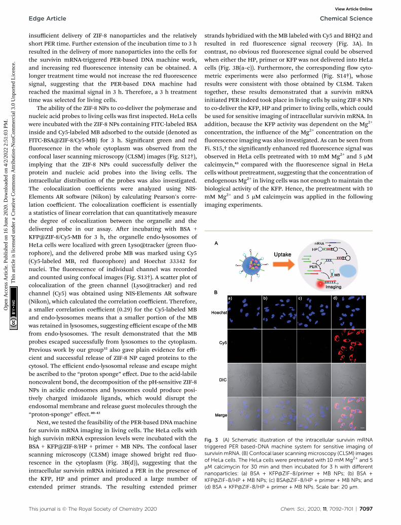
Fig. 3 (A) Schematic illustration of the intracellular survivin mRNAtriggered PER based-DNA machine system for sensitive imaging ofsurvivin mRNA. (B) Confocal laser scanningmicroscopy (CLSM) imagesof HeLa cells. The HeLa cells were pretreated with 10 mM Mg2+ and 5mM calcimycin for 30 min and then incubated for 3 h with differentnanoparticles: (a) BSA + KFP@ZIF-8/primer + MB NPs; (b) BSA +KFP@ZIF-8/HP + MB NPs; (c) BSA@ZIF-8/HP + primer + MB NPs; and(d) BSA + KFP@ZIF-8/HP + primer + MB NPs. Scale bar: 20 mm.
Edge Article Chemical Science
Ope
n A
cces
s A
rtic
le. P
ublis
hed
on 1
6 Ju
ne 2
020.
Dow
nloa
ded
on 4
/2/2
022
2:51
:03
PM.
Thi
s ar
ticle
is li
cens
ed u
nder
a C
reat
ive
Com
mon
s A
ttrib
utio
n-N
onC
omm
erci
al 3
.0 U
npor
ted
Lic
ence
.View Article Online
insufficient delivery of ZIF-8 nanoparticles and the relativelyshort PER time. Further extension of the incubation time to 3 hresulted in the delivery of more nanoparticles into the cells forthe survivin mRNA-triggered PER-based DNA machine work,and increasing red uorescence intensity can be obtained. Alonger treatment time would not increase the red uorescencesignal, suggesting that the PER-based DNA machine hadreached the maximal signal in 3 h. Therefore, a 3 h treatmenttime was selected for living cells.
The ability of the ZIF-8 NPs to co-deliver the polymerase andnucleic acid probes to living cells was rst inspected. HeLa cellswere incubated with the ZIF-8 NPs containing FITC-labeled BSAinside and Cy5-labeled MB adsorbed to the outside (denoted asFITC-BSA@ZIF-8/Cy5-MB) for 3 h. Signicant green and reduorescence in the whole cytoplasm was observed from theconfocal laser scanning microscopy (CLSM) images (Fig. S12†),implying that the ZIF-8 NPs could successfully deliver theprotein and nucleic acid probes into the living cells. Theintracellular distribution of the probes was also investigated.The colocalization coefficients were analyzed using NIS-Elements AR soware (Nikon) by calculating Pearson's corre-lation coefficient. The colocalization coefficient is essentiallya statistics of linear correlation that can quantitatively measurethe degree of colocalization between the organelle and thedelivered probe in our assay. Aer incubating with BSA +KFP@ZIF-8/Cy5-MB for 3 h, the organelle endo-lysosomes ofHeLa cells were localized with green Lyso@tracker (green uo-rophore), and the delivered probe MB was marked using Cy5(Cy5-labeled MB, red uorophore) and Hoechst 33342 fornuclei. The uorescence of individual channel was recordedand counted using confocal images (Fig. S13†). A scatter plot ofcolocalization of the green channel (Lyso@tracker) and redchannel (Cy5) was obtained using NIS-Elements AR soware(Nikon), which calculated the correlation coefficient. Therefore,a smaller correlation coefficient (0.29) for the Cy5-labeled MBand endo-lysosomes means that a smaller portion of the MBwas retained in lysosomes, suggesting efficient escape of theMBfrom endo-lysosomes. The result demonstrated that the MBprobes escaped successfully from lysosomes to the cytoplasm.Previous work by our group32 also gave plain evidence for effi-cient and successful release of ZIF-8 NP caged proteins to thecytosol. The efficient endo-lysosomal release and escape mightbe ascribed to the “proton sponge” effect. Due to the acid-labilenoncovalent bond, the decomposition of the pH-sensitive ZIF-8NPs in acidic endosomes and lysosomes could produce posi-tively charged imidazole ligands, which would disrupt theendosomal membrane and release guest molecules through the“proton-sponge” effect.40–42
Next, we tested the feasibility of the PER-based DNAmachinefor survivin mRNA imaging in living cells. The HeLa cells withhigh survivin mRNA expression levels were incubated with theBSA + KFP@ZIF-8/HP + primer + MB NPs. The confocal laserscanning microscopy (CLSM) image showed bright red uo-rescence in the cytoplasm (Fig. 3B(d)), suggesting that theintracellular survivin mRNA initiated a PER in the presence ofthe KFP, HP and primer and produced a large number ofextended primer strands. The resulting extended primer
This journal is © The Royal Society of Chemistry 2020
strands hybridized with the MB labeled with Cy5 and BHQ2 andresulted in red uorescence signal recovery (Fig. 3A). Incontrast, no obvious red uorescence signal could be observedwhen either the HP, primer or KFP was not delivered into HeLacells (Fig. 3B(a–c)). Furthermore, the corresponding ow cyto-metric experiments were also performed (Fig. S14†), whoseresults were consistent with those obtained by CLSM. Takentogether, these results demonstrated that a survivin mRNAinitiated PER indeed took place in living cells by using ZIF-8 NPsto co-deliver the KFP, HP and primer to living cells, which couldbe used for sensitive imaging of intracellular survivin mRNA. Inaddition, because the KFP activity was dependent on the Mg2+
concentration, the inuence of the Mg2+ concentration on theuorescence imaging was also investigated. As can be seen fromFi. S15,† the signicantly enhanced red uorescence signal wasobserved in HeLa cells pretreated with 10 mM Mg2+ and 5 mMcalcimycin,43 compared with the uorescence signal in HeLacells without pretreatment, suggesting that the concentration ofendogenous Mg2+ in living cells was not enough to maintain thebiological activity of the KFP. Hence, the pretreatment with 10mM Mg2+ and 5 mM calcimycin was applied in the followingimaging experiments.
Chem. Sci., 2020, 11, 7092–7101 | 7097
Chemical Science Edge Article
Ope
n A
cces
s A
rtic
le. P
ublis
hed
on 1
6 Ju
ne 2
020.
Dow
nloa
ded
on 4
/2/2
022
2:51
:03
PM.
Thi
s ar
ticle
is li
cens
ed u
nder
a C
reat
ive
Com
mon
s A
ttrib
utio
n-N
onC
omm
erci
al 3
.0 U
npor
ted
Lic
ence
.View Article Online
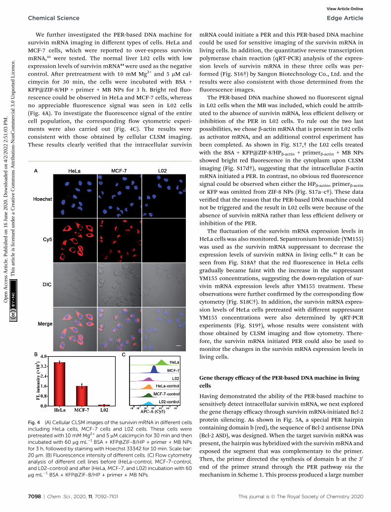
We further investigated the PER-based DNA machine forsurvivin mRNA imaging in different types of cells. HeLa andMCF-7 cells, which were reported to over-express survivinmRNA,35 were tested. The normal liver L02 cells with lowexpression levels of survivin mRNA44 were used as the negativecontrol. Aer pretreatment with 10 mM Mg2+ and 5 mM cal-cimycin for 30 min, the cells were incubated with BSA +KFP@ZIF-8/HP + primer + MB NPs for 3 h. Bright red uo-rescence could be observed in HeLa and MCF-7 cells, whereasno appreciable uorescence signal was seen in L02 cells(Fig. 4A). To investigate the uorescence signal of the entirecell population, the corresponding ow cytometric experi-ments were also carried out (Fig. 4C). The results wereconsistent with those obtained by cellular CLSM imaging.These results clearly veried that the intracellular survivin
Fig. 4 (A) Cellular CLSM images of the survivin mRNA in different cellsincluding HeLa cells, MCF-7 cells and L02 cells. These cells werepretreated with 10 mMMg2+ and 5 mM calcimycin for 30 min and thenincubated with 60 mg mL�1 BSA + KFP@ZIF-8/HP + primer + MB NPsfor 3 h, followed by staining with Hoechst 33342 for 10 min. Scale bar:20 mm. (B) Fluorescence intensity of different cells. (C) Flow cytometryanalysis of different cell lines before (HeLa-control, MCF-7-control,and L02-control) and after (HeLa, MCF-7, and L02) incubation with 60mg mL�1 BSA + KFP@ZIF-8/HP + primer + MB NPs.
7098 | Chem. Sci., 2020, 11, 7092–7101
mRNA could initiate a PER and this PER-based DNA machinecould be used for sensitive imaging of the survivin mRNA inliving cells. In addition, the quantitative reverse transcriptionpolymerase chain reaction (qRT-PCR) analysis of the expres-sion levels of survivin mRNA in these three cells was per-formed (Fig. S16†) by Sangon Biotechnology Co., Ltd. and theresults were also consistent with those determined from theuorescence images.
The PER-based DNA machine showed no uorescent signalin L02 cells when the MB was included, which could be attrib-uted to the absence of survivin mRNA, less efficient delivery orinhibition of the PER in L02 cells. To rule out the two lastpossibilities, we chose b-actin mRNA that is present in L02 cellsas activator mRNA, and an additional control experiment hasbeen completed. As shown in Fig. S17,† the L02 cells treatedwith the BSA + KFP@ZIF-8/HPb-actin + primerb-actin + MB NPsshowed bright red uorescence in the cytoplasm upon CLSMimaging (Fig. S17d†), suggesting that the intracellular b-actinmRNA initiated a PER. In contrast, no obvious red uorescencesignal could be observed when either the HPb-actin, primerb-actinor KFP was omitted from ZIF-8 NPs (Fig. S17a–c†). These dataveried that the reason that the PER-based DNA machine couldnot be triggered and the result in L02 cells were because of theabsence of survivin mRNA rather than less efficient delivery orinhibition of the PER.
The uctuation of the survivin mRNA expression levels inHeLa cells was also monitored. Sepantronium bromide (YM155)was used as the survivin mRNA suppressant to decrease theexpression levels of survivin mRNA in living cells.45 It can beseen from Fig. S18A† that the red uorescence in HeLa cellsgradually became faint with the increase in the suppressantYM155 concentrations, suggesting the down-regulation of sur-vivin mRNA expression levels aer YM155 treatment. Theseobservations were further conrmed by the corresponding owcytometry (Fig. S18C†). In addition, the survivin mRNA expres-sion levels of HeLa cells pretreated with different suppressantYM155 concentrations were also determined by qRT-PCRexperiments (Fig. S19†), whose results were consistent withthose obtained by CLSM imaging and ow cytometry. There-fore, the survivin mRNA initiated PER could also be used tomonitor the changes in the survivin mRNA expression levels inliving cells.
Gene therapy efficacy of the PER-based DNA machine in livingcells
Having demonstrated the ability of the PER-based machine tosensitively detect intracellular survivin mRNA, we next exploredthe gene therapy efficacy through survivin mRNA-initiated Bcl-2protein silencing. As shown in Fig. 5A, a special PER hairpincontaining domain b (red), the sequence of Bcl-2 antisense DNA(Bcl-2 ASD), was designed. When the target survivin mRNA waspresent, the hairpin was hybridized with the survivin mRNA andexposed the segment that was complementary to the primer.Then, the primer directed the synthesis of domain b at the 30
end of the primer strand through the PER pathway via themechanism in Scheme 1. This process produced a large number
This journal is © The Royal Society of Chemistry 2020

Fig. 5 Gene silencing efficacy of the survivin mRNA triggered PER-based DNAmachine in living cells. (A) Schematic illustration of gene silencingbased on the PER DNA machine triggered by survivin mRNA. (B) Reverse transcription quantitative PCR (qRT-PCR) analysis of the relative Bcl-2mRNA expression levels in total RNA extracted from HeLa cells and L02 cells treated with 60 mg mL�1 different nanoparticles. Western blotanalysis of the Bcl-2 protein and b-actin protein in cell lysates from HeLa cells (C) and L02 cells (D). (E) Confocal images of annexin V Alexa Fluor488 (AV 488)/PI stained apoptotic HeLa and L02 cells without treatment and after treatment with BSA + KFP@ZIF-8/HP + primer NPs. (F) Flowcytometry assay of apoptotic HeLa cells after treatment with the samples: BSA + KFP@ZIF-8 NPs; BSA@ZIF-8/HP + primer NPs; BSA + KFP@ZIF-8/primer NPs; BSA + KFP@ZIF-8/HP NPs; and BSA + KFP@ZIF-8/HP + primer NPs. (G) Cell viability of HeLa and L02 cells after incubation withdifferent nanoparticles. Cells without treatment served as the control. Scale bar: 20 mm.
Edge Article Chemical Science
Ope
n A
cces
s A
rtic
le. P
ublis
hed
on 1
6 Ju
ne 2
020.
Dow
nloa
ded
on 4
/2/2
022
2:51
:03
PM.
Thi
s ar
ticle
is li
cens
ed u
nder
a C
reat
ive
Com
mon
s A
ttrib
utio
n-N
onC
omm
erci
al 3
.0 U
npor
ted
Lic
ence
.View Article Online
of Bcl-2 ASDs, which bound to a key region of the Bcl-2 mRNAand inhibited the translation of the Bcl-2 protein, accordinglyresulting in cell apoptosis.
This journal is © The Royal Society of Chemistry 2020
The relative expression levels of Bcl-2 mRNA in HeLa cells wererst measured using quantitative reverse transcription-PCR (qRT-PCR) analysis (Fig. 5B). As can be seen, the Bcl-2mRNA expression
Chem. Sci., 2020, 11, 7092–7101 | 7099

Chemical Science Edge Article
Ope
n A
cces
s A
rtic
le. P
ublis
hed
on 1
6 Ju
ne 2
020.
Dow
nloa
ded
on 4
/2/2
022
2:51
:03
PM.
Thi
s ar
ticle
is li
cens
ed u
nder
a C
reat
ive
Com
mon
s A
ttrib
utio
n-N
onC
omm
erci
al 3
.0 U
npor
ted
Lic
ence
.View Article Online
levels of HeLa cells treated with BSA + KFP@ZIF-8/HP + primerNPs for 3 h followed by replacement with another fresh culturemedium for 48 h decreased by approximately 50% compared withthose of untreated cells, whereas no obvious changes in the Bcl-2mRNA expression levels could be observed for the cells treatedwith other nanoparticles, in which either the KFP, HP or primerwas omitted. This result indicated that the Bcl-2 ASD could beproduced in the HeLa cells using the PER-based machine onlywhen the KFP, HP and primer were present and no Bcl-2 ASDcould be produced when either the KFP, HP and primer or absent,further demonstrating the reliability of the PER-based machine. Itwas noteworthy that the Bcl-2 mRNA expression levels in L02 cellsshowed no signicant change even when the cells were treatedwith BSA + KFP@ZIF-8/HP + primer NPs. This was due to the lackof survivin mRNA in the L02 cells, leading to the failure to initiatethe PER-based machine. This result implied that the PER-basedmachine had the potential to selectively kill cancer cells and hasno effect on normal cells. The Bcl-2 protein expression levels werealso studied by western blotting analysis. It could be clearly seenfrom Fig. 5C that Bcl-2 protein expression levels decreasedremarkably in HeLa cells treated with BSA + KFP@ZIF-8/HP +primer NPs compared with those of untreated cells or the cellstreated with other nanoparticles, in which either the KFP, HP orprimer was omitted. In contrast, no appreciable decrease in theBcl-2 protein expression levels could be observed in the L02 cellstreated with BSA + KFP@ZIF-8/HP + primer NPs (Fig. 5D), sug-gesting that the PER-basedmachine could be triggered only by thesurvivin mRNA in the HeLa cells. In addition, the Bcl-2 proteinexpression levels were also investigated by immunouorescenceassays. HeLa cells treated with BSA + KFP@ZIF-8/HP + primer NPsshowed weaker green uorescence in the cytoplasm than theuntreated cells or the cells treated with other nanoparticles(Fig. S20†). Likewise, L02 cells treatedwith BSA + KFP@ZIF-8/HP +primer NPs still exhibited bright green uorescence (Fig. S21†).The results conrmed strongly that the intracellular survivinmRNA indeed could initiate the PER-based machine to produceBcl-2 ASD to decrease the Bcl-2 protein expression levels,providing a powerful gene-silencing approach.
To investigate the gene therapy efficacy, CLSM imaging aerannexin V/PI co-staining was performed. As is seen in Fig. S22,†the Bcl-2 ASD bound with the a key region of Bcl-2 mRNA,accordingly inhibiting the translation of the Bcl-2 protein andresulting in cell apoptosis through Bcl-2 anti-apoptosis-relatedsignaling pathway. Phosphatidylserine (PS), a lipid, is normallyrestricted to the inner leaet of the plasma membrane and is onlyexposed to the cell cytoplasm. However, PS will be exposed on theouter leaet of the plasma membrane during apoptosis. AnnexinV, a calcium-binding protein, can bind to PS. Therefore, uo-rescently labeled annexin V is used to detect PS that is exposed onthe outside of apoptotic cells. In addition, apoptotic cells can bedistinguished by co-staining with propidium iodide (PI) because PIenters apoptotic cells but is excluded from living cells. So, brightgreen and red uorescence was observed in HeLa cells incubatedwith BSA + KFP@ZIF-8/HP + primer NPs (Fig. 5E and S23†), indi-cating that the PER-based system could serve as an efficient genedrug machine for continuous production of Bcl-2 ASD to inducecell apoptosis. In contrast, the L02 cells gave no obvious green and
7100 | Chem. Sci., 2020, 11, 7092–7101
red uorescence aer incubation with BSA + KFP@ZIF-8/HP +primer NPs (Fig. 5E and S24†), indicating that the survivin mRNAinitiated PER-based machine provided an efficient strategy toselectively kill the cancer cells. In addition, the ow cytometryanalysis of annexin V/PI co-staining was also carried out (Fig. 5F).The total apoptotic cell percent (including early and late apoptosis)induced by BSA + KFP@ZIF-8 NPs, BSA@ZIF-8/HP + primer NPs,BSA + KFP@ZIF-8/primer NPs, BSA + KFP@ZIF-8/HP NPs and BSA+ KFP@ZIF-8/HP + primer NPs was determined to be 3.6%, 2.3%,3.1%, 3.0% and 63.6%, respectively. Nevertheless, no signicantincrease in the total apoptotic cell percent was obtained in L02cells evenwhen the cells were incubatedwith BSA +KFP@ZIF-8/HP+ primer NPs (Fig. S25†). This result was consistent with that ob-tained by the CLSM imaging. Finally, the cell viability of HeLa cells,L02 cells (Fig. 5G and S26†) and MCF-7 cells (Fig. S27†) was eval-uated aer incubation with different nanoparticles. The cellviability of HeLa cells and MCF-7 incubated with BSA + KFP@ZIF-8/HP + primer NPs signicantly decreased compared with that ofuntreated cells or the cells treated with other nanoparticles. Incontrast, the cell viability of L02 cells was nearly unchanged evenwhen the cells were treated with BSA + KFP@ZIF-8/HP + primerNPs. Otherwise, these results strongly demonstrated that the sur-vivinmRNA initiated PER-basedmachine could efficiently producea gene drug to induce cell apoptosis and provide a promisingstrategy to selectively kill the cancer cells.
Conclusions
In summary, we successfully built an intracellular survivin mRNAinitiated PER-based DNA machine by using ZIF-8 NPs to co-deliver the components including the hairpin, primer and KFP toliving cells. The machine was driven by the strand displacingpolymerase KFP and fueled by dNTPs and could continuouslyproduce Bcl-2 ASD aer being triggered by the intracellular sur-vivin mRNA. The results demonstrated that the machine couldsensitively image survivin mRNA expression levels and selectivelykill the cancer cells and has no effect on the normal cells. ThePER-based DNA machine holds great potential for early stagetumor diagnosis and precise tumor therapy. In addition, byreprogramming the hairpin stem domain and loop domain, theDNA machine could be retrotted to produce the desired DNAsingle strand with arbitrary sequences aer being triggered byother biomarkers such as mRNA, miRNA, small molecules andeven proteins (when reprogramming the loop domain with theaptamer). Moreover, the ability of ZIF-8 NPs to co-deliver proteinsand nucleic acids also provides an excellent platform to designvarious protein enzyme-based nucleic acid amplication reac-tions in living cells for disease diagnosis and therapy.
Conflicts of interest
The authors declare no conict of interest.
Acknowledgements
This work was supported by the National Natural ScienceFoundation of China (No. 21525522, 21991080 and 21705039),
This journal is © The Royal Society of Chemistry 2020

Edge Article Chemical Science
Ope
n A
cces
s A
rtic
le. P
ublis
hed
on 1
6 Ju
ne 2
020.
Dow
nloa
ded
on 4
/2/2
022
2:51
:03
PM.
Thi
s ar
ticle
is li
cens
ed u
nder
a C
reat
ive
Com
mon
s A
ttrib
utio
n-N
onC
omm
erci
al 3
.0 U
npor
ted
Lic
ence
.View Article Online
Hunan Provincial Science and Technology Department (No.2019RS3011), the Foundation for Innovative Research Groups ofNSFC (Grant 21521063), and the Fundamental Research Fundsfor the Central University.
Notes and references
1 O. Nakagawa, X. Ming, L. Huang and R. L. Juliano, J. Am.Chem. Soc., 2010, 132, 8848–8849.
2 Q. B. Mou, Y. Ma, F. Ding, X. H. Gao, D. Y. Yan, X. Y. Zhu andC. Zhang, J. Am. Chem. Soc., 2019, 141, 6955–6966.
3 D. R. Corey, Nat. Neurosci., 2017, 20, 497–499.4 M. A. Havens and M. L. Hastings, Nucleic Acids Res., 2016, 44,6549–6563.
5 X. J. Ren, R. J. Deng, L. D. Wang, K. X. Zhang and J. H. Li,Chem. Sci., 2017, 8, 5692–5698.
6 C. A. Stein and Y. C. Cheng, Science, 1993, 261, 1004–1012.7 A. Astriab-Fisher, D. Sergueev, M. Fisher, B. R. Shaw andR. L. Juliano, Pharm. Res., 2002, 19, 744–754.
8 J. J. Yang, Q. Jiang, L. He, P. F. Zhen, Q. Liu, S. L. Liu,M. F. Fu, J. B. Liu, C. Li and B. Q. Ding, ACS Appl. Mater.Interfaces, 2018, 10, 23693–23699.
9 J. B. Liu, T. T. Wu, X. H. Lu, X. H. Wu, S. L. Liu, S. Zhao,X. H. Xu and B. Q. Ding, J. Am. Chem. Soc., 2019, 141,19032–19037.
10 Y. Wang, L. Miao, A. Satterlee and L. Huang, Adv. DrugDelivery Rev., 2015, 87, 68–80.
11 O. B. Garbuzenko, M. Saad, V. P. Pozharov, K. R. Reuhl,G. Mainelis and T. Minko, Proc. Natl. Acad. Sci. U. S. A.,2010, 107, 10737–10742.
12 O. Nakagawa, X. Ming, L. Huang and R. L. Juliano, J. Am.Chem. Soc., 2010, 132, 8848–8849.
13 Z. J. Wang, Y. Fu, Z. Z. Kang, X. G. Liu, N. Chen, Q. Wang,Y. Q. Tu, L. H. Wang, S. P. Song, D. S. Ling, H. Y. Song,X. Q. Kong and C. H. Fan, J. Am. Chem. Soc., 2017, 139,15784–15791.
14 R. Huschka, A. Barhoumi, Q. Liu, J. A. Roth, L. Ji andN. J. Halas, ACS Nano, 2012, 6, 7681–7691.
15 M. Z. Alyami, S. K. Alsaiari, Y. Y. Li, S. S. Qutub, F. A. Aleisa,R. Sougrat, J. S. Merzaban and N. M. Khashab, J. Am. Chem.Soc., 2020, 142, 1715–1720.
16 T. E. Schaus, S. W. Woo, F. Xuan, X. Chen and P. Yin, Nat.Commun., 2017, 8, 696–704.
17 J. Y. Kishi, T. E. Schaus, N. Gopalkrishnan, F. Xuan andP. Yin, Nat. Chem., 2018, 10, 155–164.
18 J. Y. Kishi, B. J. Beliveau, S. W. Lapan, E. R. West, A. Zhu,H. M. Sasaki, S. K. Saka, Y. Wang, C. L. Cepko and P. Yin,Nat. Methods, 2019, 16, 533–544.
19 J. T. Liu, T. R. Liu, P. Du, L. Zhang and J. P. Lei, Angew.Chem., Int. Ed., 2019, 131, 7890–7894.
20 D. W. Zhang, Q. Lei, J. Y. Zhu, J. X. Fan, C. X. Li, C. Li, Z. S. Xu,S. X. Cheng and X. Z. Zhang, Nano Lett., 2017, 17, 284–291.
21 X. T. Yang, Q. Tang, Y. Jiang, M. N. Zhang, M. Wang andL. Q. Mao, J. Am. Chem. Soc., 2019, 141, 3782–3786.
22 C. Liu, J. Xing, O. U. Akakuru, L. J. Luo, S. Sun, R. F. Zou,Z. S. Yu, Q. L. Fang and A. G. Wu, Nano Lett., 2019, 19,5674–5682.
This journal is © The Royal Society of Chemistry 2020
23 F. J. Lyu, Y. F. Zhang, R. N. Zare, J. Ge and Z. Liu, Nano Lett.,2014, 14, 5761–5765.
24 J. T. Yi, T. T. Chen, J. Huo and X. Chu, Anal. Chem., 2017, 89,12351–12359.
25 H. M. Wang, Y. Q. Chen, H. Wang, X. Q. Liu, X. Zhou andF. N. Wang, Angew. Chem., Int. Ed., 2019, 58, 1–6.
26 M. Zheng, S. Liu, X. G. Guan and Z. G. Xie, ACS Appl. Mater.Interfaces, 2015, 7, 22181–22187.
27 C. Y. Sun, C. Qin, X. L. Wang, G. S. Yang, K. Z. Shao,Y. Q. Lan, Z. M. Su, P. Huang, C. G. Wang and E. B. Wang,Dalton Trans., 2012, 41, 6906–6909.
28 H. Y. Zhang, Q. Li, R. L. Liu, X. K. Zhang, Z. H. Li andY. X. Luan, Adv. Funct. Mater., 2018, 28, 1802830–1802840.
29 Y. Zhang, F. M. Wang, E. G. Ju, Z. Liu, Z. W. Chen, J. S. Renand X. G. Qu, Adv. Funct. Mater., 2016, 26, 6454–6461.
30 S. K. Alsaiari, S. Patil, M. Alyami, K. O. Alamoudi, F. A. Aleisa,J. S. Merzaban, M. Li and N. M. Khashab, J. Am. Chem. Soc.,2018, 140, 143–146.
31 G. Cheng, W. Q. Li, L. Ha, X. H. Han, S. J. Hao, Y. Wan,Z. G. Wang, F. P. Dong, X. Zou, Y. W. Mao and S. Zheng, J.Am. Chem. Soc., 2018, 140, 7282–7291.
32 Y. F. Feng, H. R. Wang, S. N. Zhang, Y. Zhao, J. Gao,Y. Y. Zheng, P. Zhao, Z. J. Zhang, M. J. Zaworotko,P. Cheng, S. Q. Ma and Y. Chen, Adv. Mater., 2018, 31,1805148–1805154.
33 T. T. Chen, J. T. Yi, Y. Y. Zhao and X. Chu, J. Am. Chem. Soc.,2018, 140, 9912–9920.
34 J. Zhang, M. Y. He, C. P. Nie, M. M. He, Q. S. Pan, C. Liu,Y. L. Hu, J. T. Yi, T. T. Chen and X. Chu, Anal. Chem.,2019, 91, 9049–9057.
35 C. S. Lee, R. W. Davis and N. Davidson, J. Mol. Biol., 1970, 48,1–22.
36 Z. Wu, G. Q. Liu, X. L. Yang and J. H. Jiang, J. Am. Chem. Soc.,2015, 137, 6829–6836.
37 W. Pan, T. T. Zhang, H. J. Yang, W. Diao, N. Li and B. Tang,Anal. Chem., 2013, 85, 10581–10588.
38 A. Webb, D. Cunningham, F. Cotter, P. A. Clarke, F. Stefano,P. Ross, M. Corbo and Z. Dziewanowska, Lancet, 1997, 349,1137–1141.
39 X. J. Yang, C. G. Koh, S. J. Liu, X. G. Pan, R. Santhanam,B. Yu, Y. Peng, J. X. Pang, S. Golan, Y. Talmon, Y. Jin,N. Muthusamy, J. C. Byrd, K. K. Chan, L. J. Lee,G. Marcucci and R. J. Lee, Mol. Pharm., 2008, 6, 221–230.
40 Y. W. Li, N. Xu, W. H. Zhu, L. Wang, B. Liu, J. X. Zhang andZ. G. Xie, ACS Appl. Mater. Interfaces, 2018, 10, 22974–22984.
41 H. Ren, L. Zhang, J. An, T. Wang, L. Li, X. Si, L. He, X. Wu,C. Wang and Z. Su, Chem. Commun., 2014, 50, 1000–1002.
42 C. Moreira, H. Oliveira, L. R. Pires, S. Simoes, M. A. Barbosaand A. P. Pego, Acta Biomater., 2009, 5, 2995–3006.
43 Y. J. Yang, J. Huang, X. H. Yang, K. Quan, L. Wang, N. L. Xie,M. Ou and K. M. Wang, Anal. Chem., 2016, 88, 5981–5987.
44 D. G. He, X. He, X. Yang and H. W. Li, Chem. Sci., 2017, 8,2832–2840.
45 L. Liu, J. W. Liu, H. Wu, X. N. Wang, R. Q. Yu and J. H. Jiang,Anal. Chem., 2018, 90, 1502–1505.
Chem. Sci., 2020, 11, 7092–7101 | 7101