Biomedical Engineering in Diagnostic and Treatment ...mimls.org/uploads/bsm2017/Dr Kuryati Kipli -...
39
Biomedical Engineering in Diagnostic and Treatment: Experience Sharing in Sarawak Dr Kuryati Kipli, Senior Lecturer, Department of Electrical & Electronic , Faculty of Engineering, UNIMAS.
Transcript of Biomedical Engineering in Diagnostic and Treatment ...mimls.org/uploads/bsm2017/Dr Kuryati Kipli -...
Biomedical Engineering in Diagnostic
and Treatment:
Experience Sharing in Sarawak
Dr Kuryati Kipli, Senior Lecturer, Department of Electrical & Electronic ,
Faculty of Engineering, UNIMAS.

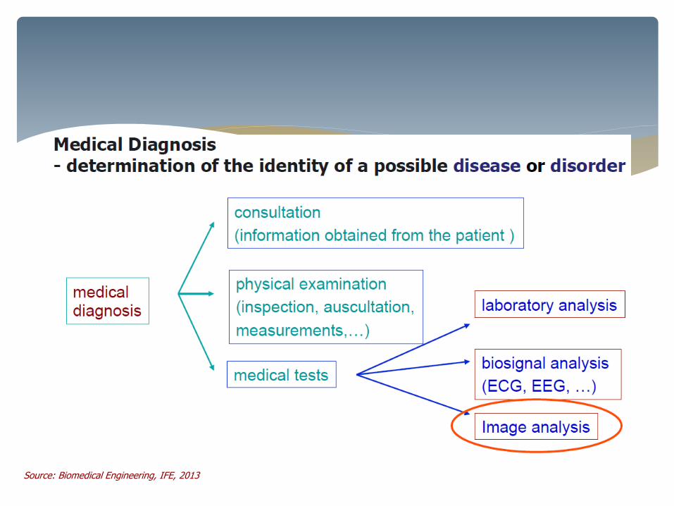
Brief Intro on BME & BMET
Imaging for Biomedical Applications
Research on Biomedical Image Processing
& Analysis
Challenges
Content
Biomedical Engineering (BME) and Biomedical Engineering Technology (BMET) field in Malaysia is at its early stage.
Research and development in those areas offer a great potential for improvement of health care and health services.
The range of focus areas includes biomechanics, medical devices, medical imaging, biomaterials and physiological signal processing.
BME/BMET in Malaysia

Bachelor of BMET (hons)
BEng(Hons) Biomedical Engineering
Bachelor of Biomedical Engineering
Bachelor of Biomedical Engineering (Prosthetics and Orthotics)
Bachelor of Engineering (Bio-Medical)
Bachelor of Engineering (Hons.) Electronics Majoring in Bio-Instrumentation
Bachelor of Electronic Engineering with Honours (BEJ) (Biomedical)
BME Degree offered in Msia
Bachelor of Electronic Engineering with Honours (BEJ) (Biomedical)

Investigation of complex medical problems and development of
engineering methods to solve them
Combine engineering principles with medical & biological sciences to design and create equipment, devices, computer system and software used in healthcare.
Engineering framework including biomaterials, biomechanics, and
bioelectricity
Career at manufacturing facilities, company, universities, hospitals,
research facilities and government.
Biomedical Engineering (BME)
BME & BMET area
Manage and support the manufacture and use of existing medical devices and
technology in patient care
Services : installation and maintenance of biomedical equipment.
Medical device systems, electrical engineering technology, patient safety
and regulations, organizational leadership and supervision, clinical
communication
Career at biomedical instrumentation industries, hospitals, medical equipment
manufacturers.
Biomedical Engineering
Technologist (BMET)
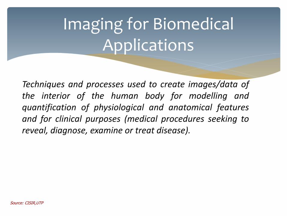
Imaging for Biomedical Applications
Techniques and processes used to create images/data of the interior of the human body for modelling and quantification of physiological and anatomical features and for clinical purposes (medical procedures seeking to reveal, diagnose, examine or treat disease).
Source: CISIR,UTP

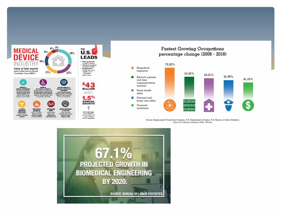
Imaging for Biomedical Applications On an average, 2 out of every 5 visits to hospital involve an imaging procedure in US (SG-2) More than 1B imaging procedures/year worldwide (Medtech Insight) Growth driven by new applications, increasing accuracy and increasing speed
Medtech Insight, CRIS
Source: Biomedical Engineering, IFE, 2013

Imaging Technology

Medical Imaging – Biomedical Engineering
radiography
(J. Hall-Edwards, 1896)
angiography
(E. Moniz, 1927) ultrasonography
(I. Edler, C. Hertz, 1953)
CT tomography
(A. Cormack, G. Hounsfield, 1972)
PET tomography
(M. Ter-Pogossian et.al., 1973)
Endoscopic capsule
(Given Imaging, 2001)
MRI tomography
(P. Lauterbur, P. Mansfield, 1973
since 80ties)
endoscopy
(B. Hirschowitz, since 70ties)
The History of Medical Imaging
Termography
(since 60thies, XX c.)
19
Introduction to Medical Imaging
CT+MRI, PET+MRI
(Gen. Electric, 2010)
Source: Biomedical Engineering, IFE, 2013

Source: Biomedical Engineering, IFE, 2013
• Each has it pro & con; cost, capability to reveal certain ROI, execution time

Medical Imaging – Biomedical Engineering
x y
f(x,y)
!
x, y( ) ² f x, y( )
y
Monochrome image as a 2D function
7
Introduction to Medical Imaging Source: Biomedical Engineering, IFE, 2013

Medical Imaging – Biomedical Engineering
0 50 100 150 200 250 30060
80
100
120
140
160
180
200
220
Distance along profile
Image brightness profile
8
Introduction to Medical Imaging Source: Biomedical Engineering, IFE, 2013
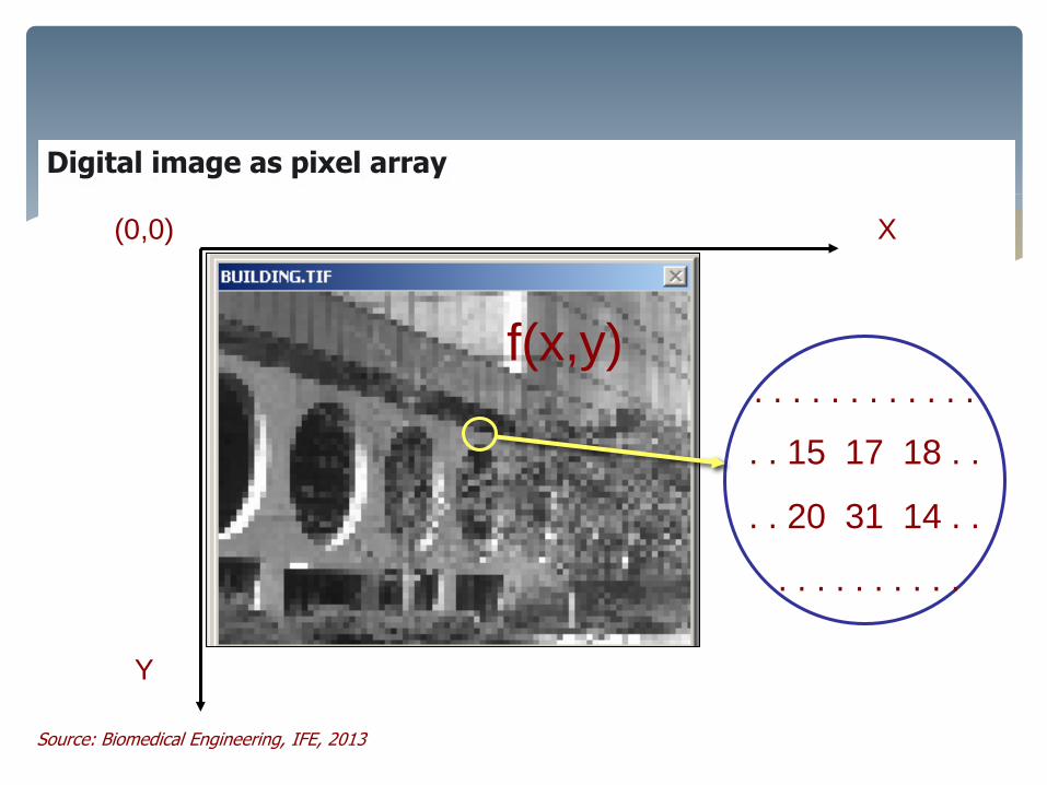
Medical Imaging – Biomedical Engineering
X
Y
(0,0)
. . . . . . . . . . . .
. . 15 17 18 . .
. . 20 31 14 . .
. . . . . . . . . .
f(x,y)
Digital image as pixel array
12
Introduction to Medical Imaging Source: Biomedical Engineering, IFE, 2013
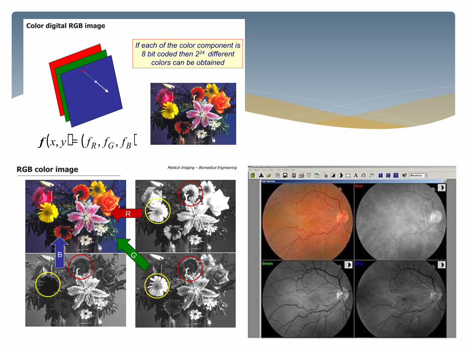
Medical Imaging – Biomedical Engineering
R
B
RGB color image
9
Introduction to Medical Imaging
Medical Imaging – Biomedical Engineering
( ) ( )BGR fffyx ,,, =f
If each of the color component is
8 bit coded then 224 different
colors can be obtained
Color digital RGB image
14
Introduction to Medical Imaging
Medical Imaging – Biomedical Engineering
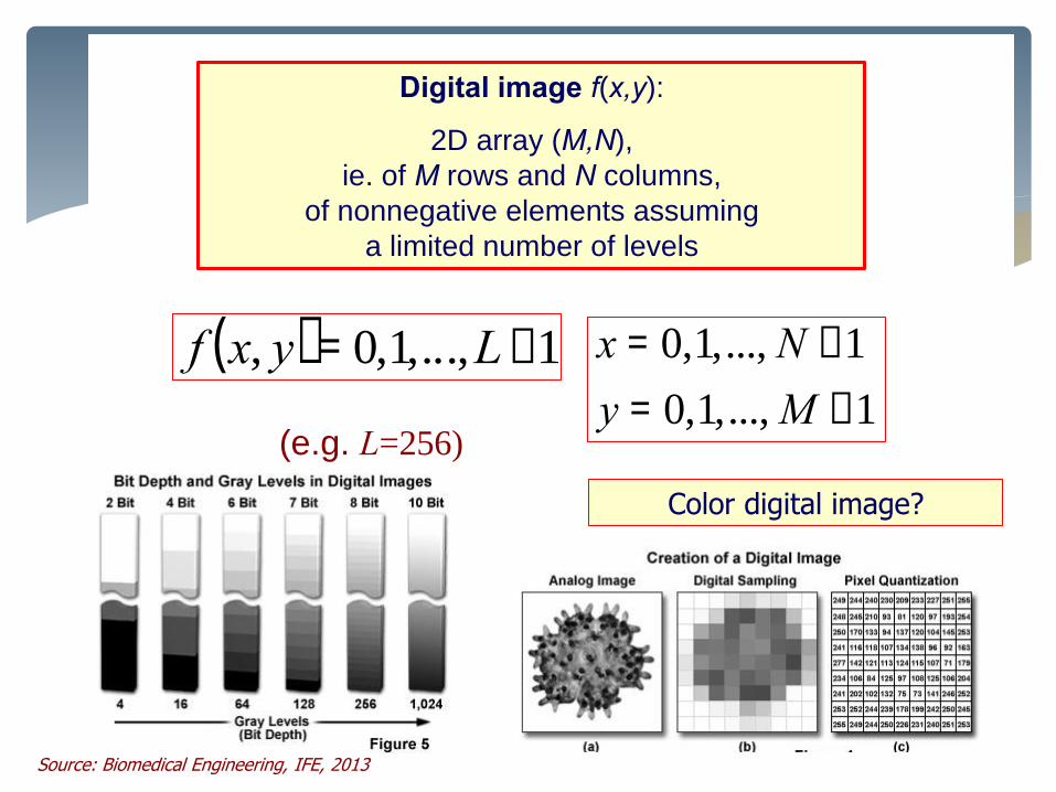
Digital image f(x,y):
2D array (M,N),
ie. of M rows and N columns,
of nonnegative elements assuming
a limited number of levels
Color digital image?
1...,,1,0
1...,,1,0
-=
-=
My
Nx( ) 1...,,1,0, -= Lyxf
(e.g. L=256)
Digital image as pixel array
13
Introduction to Medical Imaging
Source: Biomedical Engineering, IFE, 2013
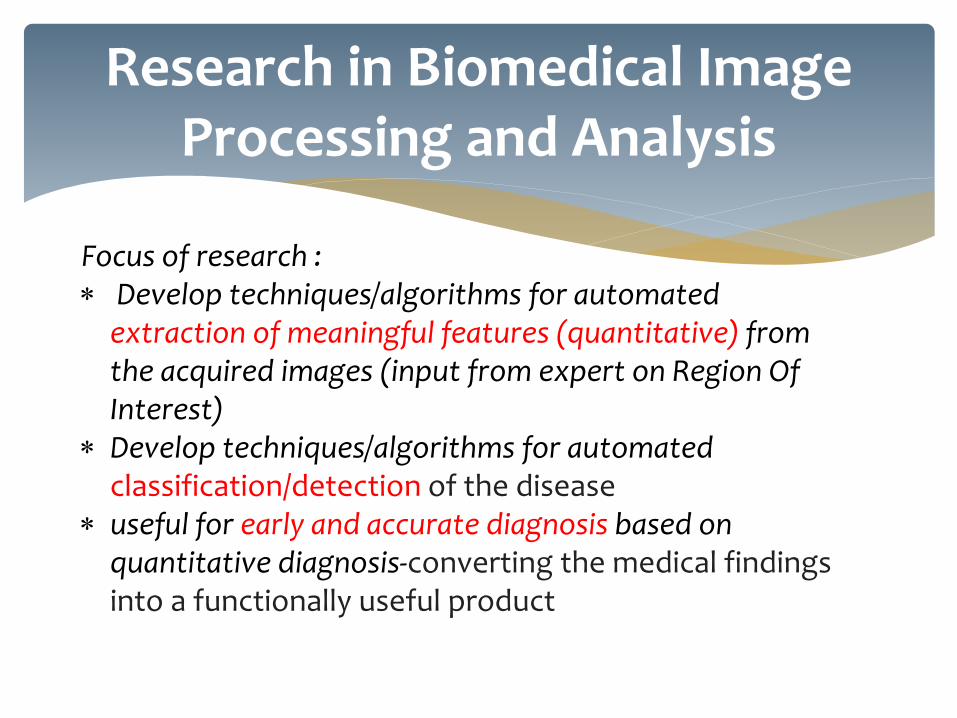
Focus of research : Develop techniques/algorithms for automated
extraction of meaningful features (quantitative) from the acquired images (input from expert on Region Of Interest)
Develop techniques/algorithms for automated classification/detection of the disease
useful for early and accurate diagnosis based on quantitative diagnosis-converting the medical findings into a functionally useful product
Research in Biomedical Image Processing and Analysis
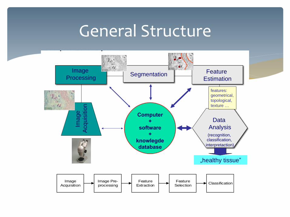
General Structure Medical Imaging – Biomedical Engineering
Introduction to Medical Imaging
18
Computer vision system
Image
Processing Feature
Estimation Segmentation
Data
Analysis
(recognition, classification,
interpretaction)
Computer +
software +
knowlegde database
Ima
ge
Acq
uis
itio
n
„healthy tissue”
features: geometrical,
topological,
texture …
Image Acquisition
Image Pre-processing
Feature Selection
Feature Extraction
Classification

Towards Automated Detection of Depression from sMRI
• Architecture of brain sMRI
based depression detection system
• Brain sMRI volumetric features
• DoC feature selection algorithm
• Classification model

Journal Publications
[1] Kipli, Kuryati and Kouzani, Abbas Z. (2014), “Degree of Contribution (DoC) Feature Selection Algorithm for Structural Brain MRI Volumetric Features in Depression Detection”, International Journal of Computer Assisted Radiology and Surgery (IJCARS),Springer, In Press. DOI: 10.1007/s11548-014-1130-9.
[2] Kipli, Kuryati, Kouzani, Abbas Z. and Williams, Lana J. (2013), “Towards automated detection of depression from brain structural magnetic resonance images”, Neuroradiology, pp. 1-18, Springer, Berlin, Germany.
[3] Kipli, Kuryati, Kouzani, Abbas Z. and A. Hamid, Isredza R. (2013), "Investigating Machine Learning Techniques for Detection of Depression Using Structural MRI Volumetric Features," International Journal of Bioscience, Biochemistry and Bioinformatics vol. 3, no. 5, pp. 444-448.
Conference Publications
[1] K. Kipli, A. Z. Kouzani, and Y. Xiang, (2013), "An Empirical Comparison of Classification Algorithms for Diagnosis of Depression from Brain SMRI Scans," in International Conference on Advanced Computer Science Applications and Technologies (ACSAT), Kuching, Malaysia pp. 333-336.
[2] Kipli, Kuryati, Kouzani, Abbas Z., and A. Hamid, Isredza R. (2013), "Investigating Machine Learning Techniques for Detection of Depression Using Structural MRI Volumetric Features," in 2nd International Conference on Bioinformatics and Biomedical Science (ICBBS), Kuala Lumpur, Malaysia.
(Selected for publication in the International Journal of Bioscience, Biochemistry and Bioinformatics (IJBBB, ISSN: 2010-3638))
[3] Kipli, Kuryati, Kouzani, Abbas Z. and Joordens, Matthew (2013), "Evaluation of Feature Selection Algorithms for Detection of Depression from Brain sMRI Scans," in IEEE International Conference on Complex Medical Engineering (CME 2013), Beijing, China.
[4] Kipli, Kuryati and Kouzani, Abbas Z. (2013), "An Algorithm for Determination of Rank and Degree of Contribution of sMRI Volumetric Features in Depression Detection," in the 35th Annual International Conference of the IEEE Engineering in Medicine and Biology Society (EMBC’13), Osaka, Japan.
[5] Kipli, Kuryati, Kouzani, Abbas Z. and Joordens, Matthew (2012) Computer-aided detection of depression from magnetic resonance images, in CME 2012 : Proceedings of the 2012 IEEE/ICME International Conference on Complex Medical Engineering, pp. 500-505, IEEE Computer Society, Los Alamitos, Calif.
[6] K. Kipli, (2012), “Performing Brain Structural MRI Volumetric Attributes Feature Evaluations and Classifications for Detection of Depression” in Institute for Frontier Materials (IFM), Annual Research Conference 2012, Deakin University on 5th & 6th Nov 2012, The Pier, 10 Western Beach Foreshore Road, Waterfront Geelong.
[7] Kipli, Kuryati, Kouzani, Abbas Z., Xiang, Yong and Joordens, Matthew (2011) Evaluation of segmentation algorithms for extraction of RNFL in OCT images, in ICIS 2011 : Proceedings of the IEEE International Conference on Intelligent Computing and Intelligent Systems, pp. 447-451, IEEE, Piscataway, N.J.
List of Publications


Develop automated screening tool for early disease detection
Ways to identify these diseases is by observing the abnormalities of vascular pattern
Many features in retinal-useful for diseases diagnosing
Automated Retinal Image Analysis
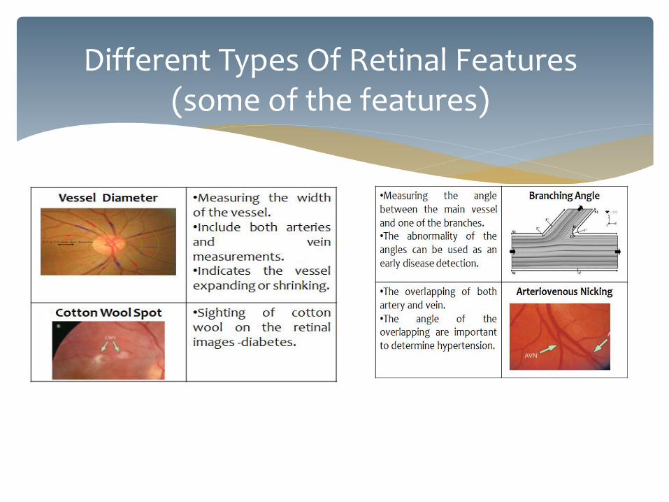
Different Types Of Retinal Features (some of the features)
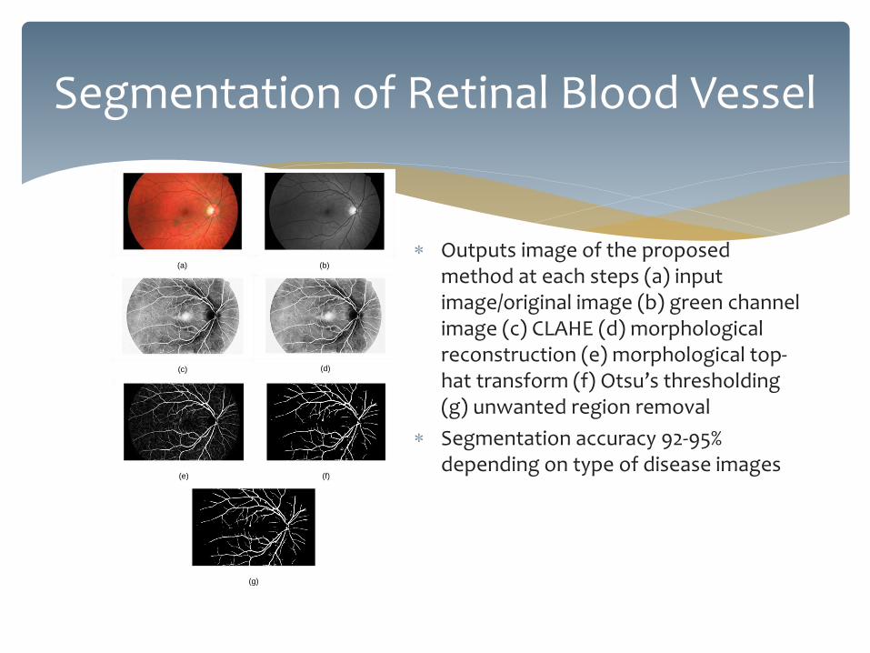
Outputs image of the proposed method at each steps (a) input image/original image (b) green channel image (c) CLAHE (d) morphological reconstruction (e) morphological top-hat transform (f) Otsu’s thresholding (g) unwanted region removal
Segmentation accuracy 92-95% depending on type of disease images
Segmentation of Retinal Blood Vessel
(a) (b)
(c) (d)
(e) (f)
(g)
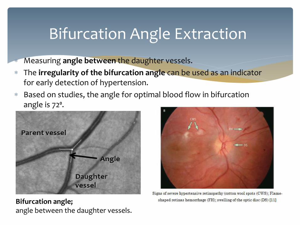
Bifurcation angle; angle between the daughter vessels.
Bifurcation Angle Extraction
Measuring angle between the daughter vessels.
The irregularity of the bifurcation angle can be used as an indicator for early detection of hypertension.
Based on studies, the angle for optimal blood flow in bifurcation angle is 72⁰.

Images : VICAVR Database-retinal image with marked artery and vein vessels by three different experts.
Checking 8 neighborhood of every positive pixel
When there are three vessels coming out from one point, they are marked as bifurcation.
Bifurcation Localization

Start
Image Acquisition
Bifurcation Localization
Measuring angle
End
Output: Algorithm for angle identification & measurement

shape of a feature, surface analysis, volumetric, size, area, angle, length, intensity, peak value, texture
Extracted features/pattern

Classification
Medical Imaging – Biomedical Engineering
Introduction to Medical Imaging
24
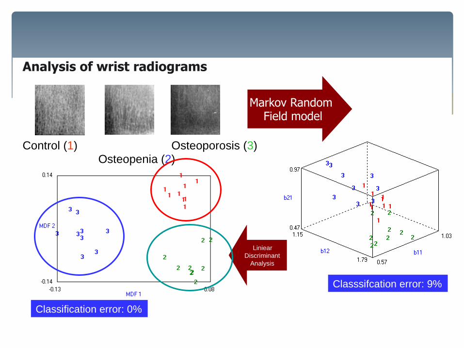
Analysis of wrist radiograms
Control (1) Osteopenia (2)
Osteoporosis (3)
Markov Random Field model
Classsifcation error: 9%
Liniear
Discriminant
Analysis
Classification error: 0%
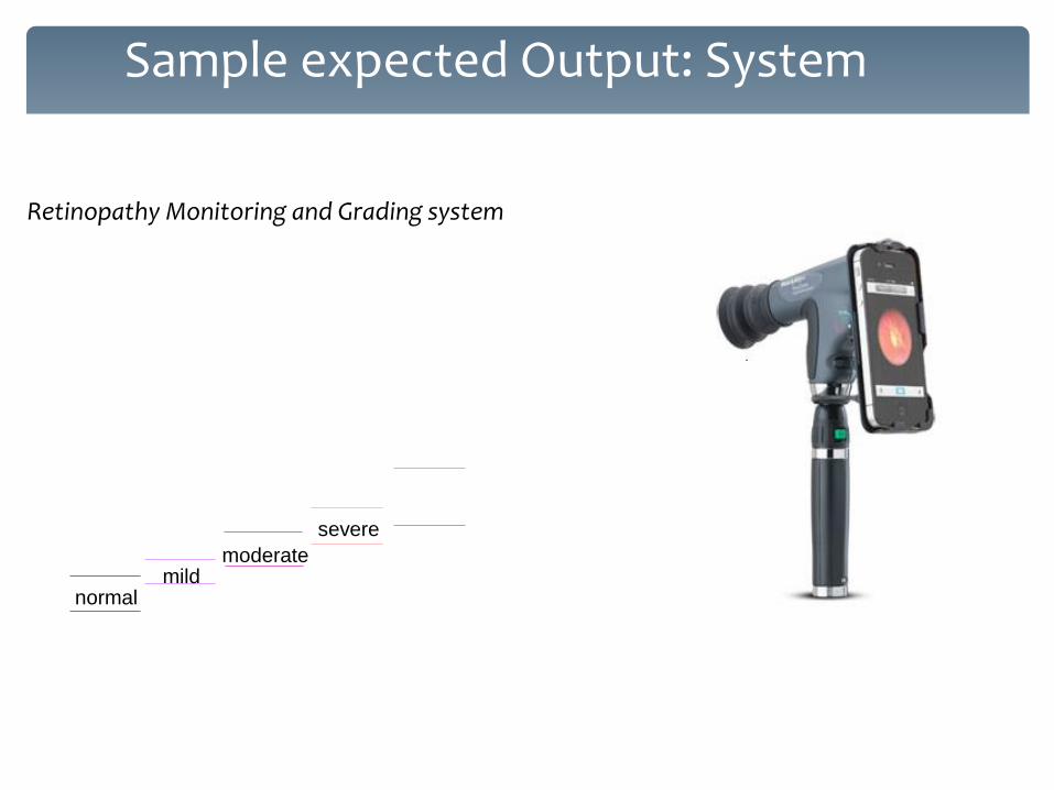
Sample expected Output: System
FAZ
FA
Z a
rea (
pix
els
)
normal mild
moderate
severe
PDR
normal mild
moderate
severe
PDR
normal mild
moderate
severe
FAZ FAZ
FA
Z a
rea (
pix
els
)
Retinopathy Monitoring and Grading system

Other Related Research

Nearfield Electromagnetic Imaging Technique for Brain Tumour Detection
Microwave imaging is a well-established technique which is being employed to build low- cost and portable medical diagnostic tools
IMAGE RECONSTRUCTION: BRAIN IMAGING
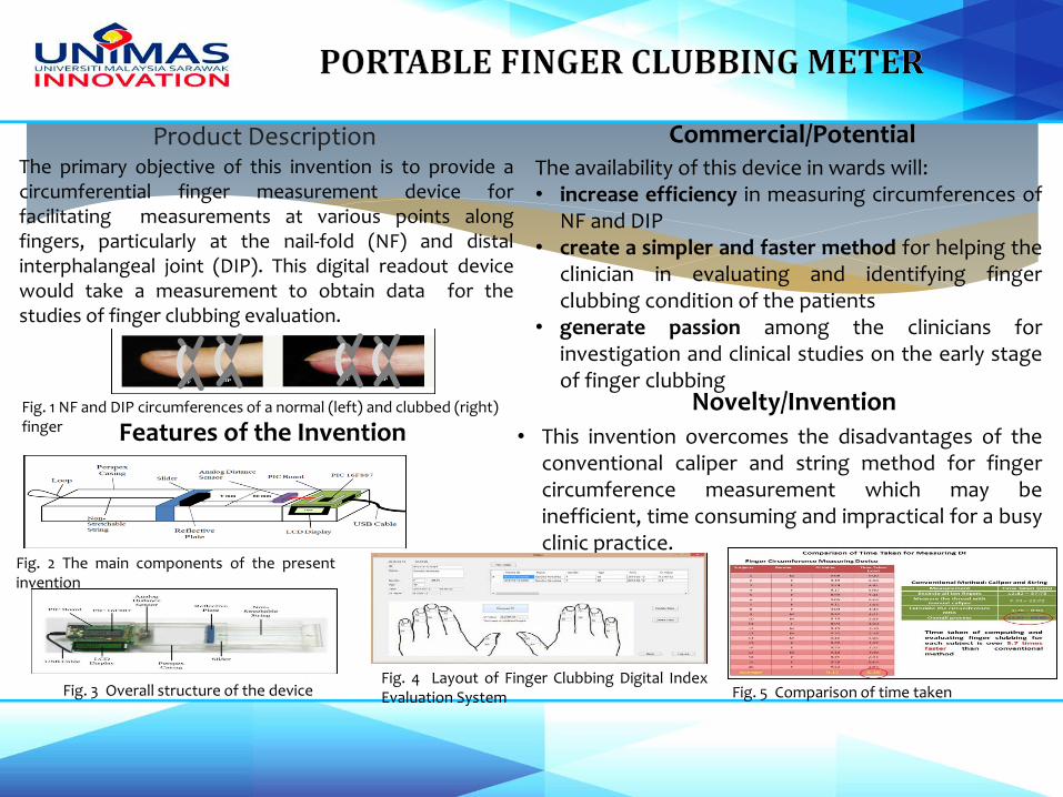
Product Description
Novelty/Invention
Commercial/Potential
• This invention overcomes the disadvantages of the conventional caliper and string method for finger circumference measurement which may be inefficient, time consuming and impractical for a busy clinic practice.
The primary objective of this invention is to provide a circumferential finger measurement device for facilitating measurements at various points along fingers, particularly at the nail-fold (NF) and distal interphalangeal joint (DIP). This digital readout device would take a measurement to obtain data for the studies of finger clubbing evaluation.
The availability of this device in wards will: • increase efficiency in measuring circumferences of
NF and DIP • create a simpler and faster method for helping the
clinician in evaluating and identifying finger clubbing condition of the patients
• generate passion among the clinicians for investigation and clinical studies on the early stage of finger clubbing
Features of the Invention
Fig. 2 The main components of the present invention
Fig. 3 Overall structure of the device Fig. 4 Layout of Finger Clubbing Digital Index Evaluation System
Fig. 1 NF and DIP circumferences of a normal (left) and clubbed (right) finger
Fig. 5 Comparison of time taken
Product Description
Novelty/Invention
Commercial/Potential
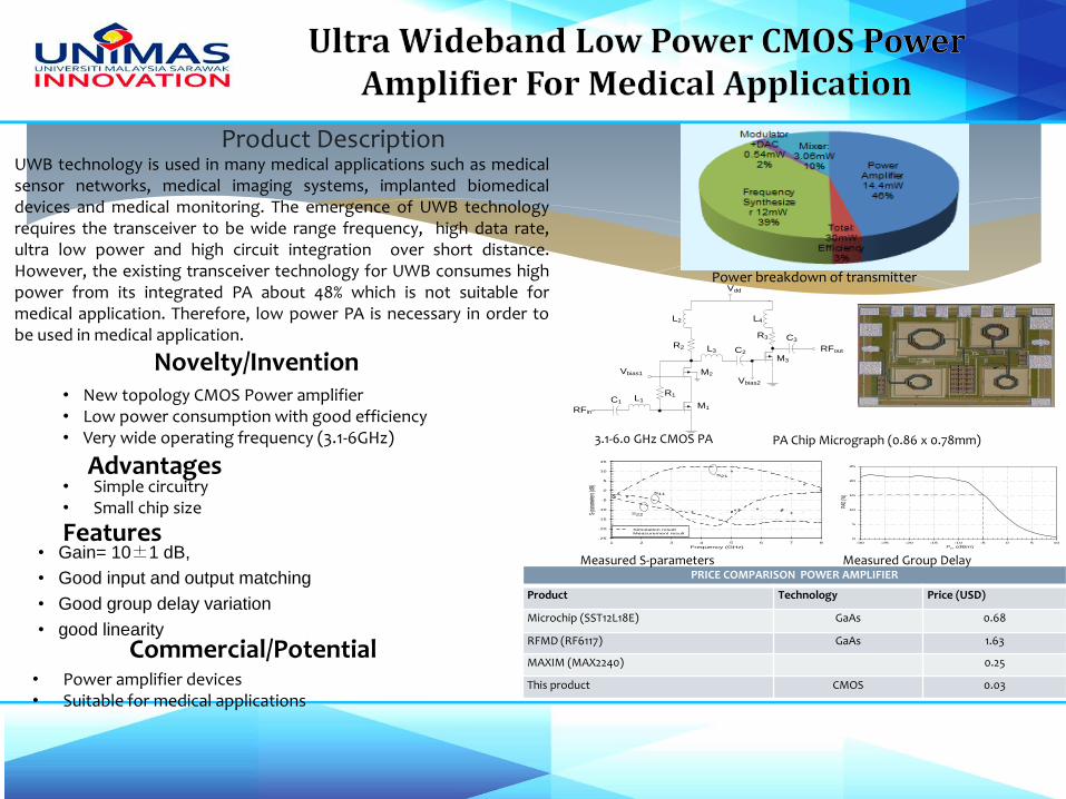
• Simple circuitry • Small chip size
• Power amplifier devices • Suitable for medical applications
Pin (dBm)
-30 -25 -20 -15 -10 -5 0 5 10
PAE
(%)
0
5
10
15
20
25
Frequency (GHz)
1 2 3 4 5 6 7 8
S-pa
ramete
rs (dB
)
-25
-20
-15
-10
-5
0
5
10
15
Simulation result
Measurement result
S21
S11
S22
Vdd
C1L1
L3 C2
L2
R1
L4
C3
M1
M2
M3
RFin
RFout
Vbias1
Vbias2
R2
R3
• Gain= 10±1 dB,
• Good input and output matching
• Good group delay variation
• good linearity
Features
Advantages
UWB technology is used in many medical applications such as medical sensor networks, medical imaging systems, implanted biomedical devices and medical monitoring. The emergence of UWB technology requires the transceiver to be wide range frequency, high data rate, ultra low power and high circuit integration over short distance. However, the existing transceiver technology for UWB consumes high power from its integrated PA about 48% which is not suitable for medical application. Therefore, low power PA is necessary in order to be used in medical application.
• New topology CMOS Power amplifier • Low power consumption with good efficiency • Very wide operating frequency (3.1-6GHz)
PRICE COMPARISON POWER AMPLIFIER
Product Technology Price (USD)
Microchip (SST12L18E) GaAs 0.68
RFMD (RF6117) GaAs 1.63
MAXIM (MAX2240) 0.25
This product CMOS 0.03
Power breakdown of transmitter
PA Chip Micrograph (0.86 x 0.78mm) 3.1-6.0 GHz CMOS PA
Measured S-parameters Measured Group Delay


Automated, Accurate
Portable, simple
Access to data
Collaboration from both parties
Challenges