Biomechanical Measures During Landing.pdf
11
Biomechanical Measures During Landing and Postural Stability Predict Second Anterior Cruciate Ligament Injury After Anterior Cruciate Ligament Reconstruction and Return to Sport Mark V. Paterno,* yz§||{ PT, MS, SCS, ATC, Laura C. Schmitt, yz§# PT, PhD, Kevin R. Ford, yz|| PhD, FACSM, Mitchell J. Rauh, { PT, PhD, MPH, FACSM, Gregory D. Myer, yza MS, CSCS, Bin Huang, yb PhD, and Timothy E. Hewett, yz||c PhD, FACSM From y Cincinnati Children’s Hospital Medical Center, Cincinnati, Ohio, z Sports Medicine Biodynamics Center and Human Performance Laboratory, Cincinnati, Ohio, § Division of Occupational Therapy and Physical Therapy, Cincinnati, Ohio, || Department of Pediatrics, College of Medicine, University of Cincinnati, Cincinnati, Ohio, { Graduate Program in Orthopaedic and Sports Sciences, Rocky Mountain University of Health Professions, Provo, Utah, # Department of Physical Therapy, Ohio State University, Columbus, Ohio, a Graduate Program in Athletic Training, Rocky Mountain University of Health Professions, Provo, Utah, b Department of Epidemiology and Biostatistics, Cincinnati, Ohio, and c Departments of Orthopaedic Surgery, College of Medicine and the Department of Biomedical Engineering, University of Cincinnati, Cincinnati, Ohio and The Ohio State University Background: Athletes who return to sport participation after anterior cruciate ligament reconstruction (ACLR) have a higher risk of a second anterior cruciate ligament injury (either reinjury or contralateral injury) compared with non–anterior cruciate ligament– injured athletes. Hypotheses: Prospective measures of neuromuscular control and postural stability after ACLR will predict relative increased risk for a second anterior cruciate ligament injury. Study Design: Cohort study (prognosis); Level of evidence, 2. Methods: Fifty-six athletes underwent a prospective biomechanical screening after ACLR using 3-dimensional motion analysis during a drop vertical jump maneuver and postural stability assessment before return to pivoting and cutting sports. After the initial test session, each subject was followed for 12 months for occurrence of a second anterior cruciate ligament injury. Lower extremity joint kinematics, kinetics, and postural stability were assessed and analyzed. Analysis of variance and logistic regres- sion were used to identify predictors of a second anterior cruciate ligament injury. Results: Thirteen athletes suffered a subsequent second anterior cruciate ligament injury. Transverse plane hip kinetics and fron- tal plane knee kinematics during landing, sagittal plane knee moments at landing, and deficits in postural stability predicted a sec- ond injury in this population (C statistic 5 0.94) with excellent sensitivity (0.92) and specificity (0.88). Specific predictive parameters included an increase in total frontal plane (valgus) movement, greater asymmetry in internal knee extensor moment at initial contact, and a deficit in single-leg postural stability of the involved limb, as measured by the Biodex stability system. Hip rotation moment independently predicted second anterior cruciate ligament injury (C 5 0.81) with high sensitivity (0.77) and spec- ificity (0.81). Conclusion: Altered neuromuscular control of the hip and knee during a dynamic landing task and postural stability deficits after ACLR are predictors of a second anterior cruciate ligament injury after an athlete is released to return to sport. Keywords: anterior cruciate ligament reconstruction; kinetics; kinematics; postural stability Anterior cruciate ligament (ACL) rupture is a devastating injury that is linked to short-term functional deficits and significant long-term morbidity, including premature development of osteoarthritis and significant progressive The American Journal of Sports Medicine, Vol. XX, No. X DOI: 10.1177/0363546510376053 Ó 2010 The Author(s) 1 AJSM PreView, published on August 11, 2010 as doi:10.1177/0363546510376053
Transcript of Biomechanical Measures During Landing.pdf

Biomechanical Measures During Landingand Postural Stability Predict SecondAnterior Cruciate Ligament Injury AfterAnterior Cruciate Ligament Reconstructionand Return to SportMark V. Paterno,*yz§||{ PT, MS, SCS, ATC, Laura C. Schmitt,yz§# PT, PhD, Kevin R. Ford,yz|| PhD,FACSM, Mitchell J. Rauh,{ PT, PhD, MPH, FACSM, Gregory D. Myer,yza MS, CSCS,Bin Huang,yb PhD, and Timothy E. Hewett,yz||c PhD, FACSMFrom yCincinnati Children’s Hospital Medical Center, Cincinnati, Ohio, zSports MedicineBiodynamics Center and Human Performance Laboratory, Cincinnati, Ohio, §Division ofOccupational Therapy and Physical Therapy, Cincinnati, Ohio, ||Department of Pediatrics,College of Medicine, University of Cincinnati, Cincinnati, Ohio, {Graduate Program inOrthopaedic and Sports Sciences, Rocky Mountain University of Health Professions, Provo,Utah, #Department of Physical Therapy, Ohio State University, Columbus, Ohio, aGraduateProgram in Athletic Training, Rocky Mountain University of Health Professions, Provo, Utah,bDepartment of Epidemiology and Biostatistics, Cincinnati, Ohio, and cDepartments ofOrthopaedic Surgery, College of Medicine and the Department of Biomedical Engineering,University of Cincinnati, Cincinnati, Ohio and The Ohio State University
Background: Athletes who return to sport participation after anterior cruciate ligament reconstruction (ACLR) have a higher risk ofa second anterior cruciate ligament injury (either reinjury or contralateral injury) compared with non–anterior cruciate ligament–injured athletes.
Hypotheses: Prospective measures of neuromuscular control and postural stability after ACLR will predict relative increased riskfor a second anterior cruciate ligament injury.
Study Design: Cohort study (prognosis); Level of evidence, 2.
Methods: Fifty-six athletes underwent a prospective biomechanical screening after ACLR using 3-dimensional motion analysisduring a drop vertical jump maneuver and postural stability assessment before return to pivoting and cutting sports. After theinitial test session, each subject was followed for 12 months for occurrence of a second anterior cruciate ligament injury. Lowerextremity joint kinematics, kinetics, and postural stability were assessed and analyzed. Analysis of variance and logistic regres-sion were used to identify predictors of a second anterior cruciate ligament injury.
Results: Thirteen athletes suffered a subsequent second anterior cruciate ligament injury. Transverse plane hip kinetics and fron-tal plane knee kinematics during landing, sagittal plane knee moments at landing, and deficits in postural stability predicted a sec-ond injury in this population (C statistic 5 0.94) with excellent sensitivity (0.92) and specificity (0.88). Specific predictiveparameters included an increase in total frontal plane (valgus) movement, greater asymmetry in internal knee extensor momentat initial contact, and a deficit in single-leg postural stability of the involved limb, as measured by the Biodex stability system. Hiprotation moment independently predicted second anterior cruciate ligament injury (C 5 0.81) with high sensitivity (0.77) and spec-ificity (0.81).
Conclusion: Altered neuromuscular control of the hip and knee during a dynamic landing task and postural stability deficits afterACLR are predictors of a second anterior cruciate ligament injury after an athlete is released to return to sport.
Keywords: anterior cruciate ligament reconstruction; kinetics; kinematics; postural stability
Anterior cruciate ligament (ACL) rupture is a devastatinginjury that is linked to short-term functional deficits andsignificant long-term morbidity, including prematuredevelopment of osteoarthritis and significant progressive
The American Journal of Sports Medicine, Vol. XX, No. XDOI: 10.1177/0363546510376053� 2010 The Author(s)
1
AJSM PreView, published on August 11, 2010 as doi:10.1177/0363546510376053
disability, despite surgical or nonsurgical interven-tion.27,40,41,47,54 With respect to short-term outcomes, esti-mates of the overall likelihood that an athlete will incura subsequent ACL injury (either reinjury or contralateral)after return to sport participation after ACL reconstruc-tion (ACLR) range between 1 in 4 (25%) to 1 in 17 (6%)athletes.47,49,58
The risk of subsequent ACL injury is significantly highercompared with risk of initial ACL injury,47,49,51,58 especiallyin young, active individuals.28,48,51,58 A prospective, multi-center study from the Multicenter Orthopaedic OutcomeNetwork (MOON) group58 reported a rate of 1 of 17 (6%) sec-ond ACL injuries within the short timespan of 2 years afterinitial injury. This represents a substantially higher injuryrate compared with initial ACL injuries, reported to occur ata risk of 1 in 60 to 100,17,36 even in a ‘‘high-risk’’ populationof young, female athletes. In studies with longer termfollow-up, the rates of second ACL injuries are even higher.In a retrospective case series, Salmon et al49 found that 12%of their sample sustained a second ACL injury within 5years after primary ACLR. In a 10-year follow-up of thiscohort, the rate of injury was greater, as 1 in 3.7 individualshad suffered a second ACL injury.47 Shelbourne et al51 alsoreported that younger patients were at higher risk, as 17%of patients under the age of 18 years sustained a secondACL injury, while only 4% over the age of 25 years sus-tained a second injury.
These findings underscore the significantly increasedrisk for a second ACL injury, especially when resumingsport participation after initial ACLR. Despite the signifi-cantly higher risk of second ACL injury, informationregarding risk factors associated with second ACL injuriesis scarce. In the few studies that have reported such infor-mation,7,49 investigations have been limited to clinicalassessments and have only evaluated unmodifiable riskfactors, such as subjective ratings, demographics, and con-comitant injuries. Risk of second ACL injury is greatest inindividuals with a history of an initial ACL injury causedby contact with another player and with return to cuttingand pivoting sport activities.49 Identification of modifiablefactors predictive of second ACL injury is necessary toeffectively reduce this high risk of second injury and subse-quent sequelae.
Modifiable risk factors potentially predictive of initialACL injuries have been identified in female athletes. Ina prospective, controlled cohort study, Hewett et al21 inves-tigated the effect of biomechanical and neuromuscularmeasures on risk of ACL injury in uninjured female ath-letes. Of the 205 female athletes evaluated and followedduring the sport season, 9 athletes (4.4%) suffered anACL injury. Comparison of the prospectively measuredvariables of the injured and noninjured athletes generateda predictive model for first ACL injury. The most predictive
variable for ACL injury was lower extremity frontal planeloads at the knee, which were significantly correlated tohip adduction moments in athletes with ACL injury. Inaddition, athletes who went on to ACL injury also demon-strated significant side-to-side differences in lower extrem-ity biomechanics, as well as reduced relative lowerextremity flexor activation during landing, relative tothose who did not sustain ACL injury. Further, 4 of the 9athletes who sustained ACL injury identified during theoriginal study incurred a second ACL injury within a 5-year period. Although it is reasonable to assume that fac-tors associated with initial ACL injury may remain predic-tive of second ACL injury, this assumption has yet to bedefinitively supported. Prospective studies may help toidentify modifiable risk factors of subsequent ACL injuryin order to develop targeted rehabilitation interventionsto reduce this risk.
The purpose of the present study was to identify predic-tors of a second ACL injury after ACLR in a young, athleticsample. Our hypothesis was that after primary ACLR, pro-spectively measured deficits in neuromuscular control atthe hip and knee and in postural stability would predictsecond ACL injury. In addition, we hypothesized that thesemeasures would predict future injury with high sensitivityand specificity.
METHODS
Participants
A prospective, case-cohort design was used to identify thepredictors of a second ACL injury (ipsilateral or contralat-eral) after primary ACLR and return to sport. Fifty-sixyoung athletes (35 female, 21 male) who recently sustainedan ACL injury, underwent surgical reconstruction, com-pleted rehabilitation, and were released to return to theirprior level of activity were recruited to participate in thisstudy (Table 1). Twenty-five participants had an ACLRusing bone–patellar tendon–bone autograft tissue, 27 hadan ACLR using hamstring tendon graft, and 4 underwentan ACLR with allograft tissue. Inclusion criteria requiredthe participant to be between the ages of 10 and 25 years,have no history of ACL injury, and no history of bilaterallower extremity or lower back injury during the previous12 months. In addition, the participant was expected toreturn to a pivoting or cutting (level 1 or 2) sport at least50 hours per year. Daniel et al10 described a level 1 sportas jumping, pivoting, or hard cutting such as basketball,football, and soccer and a level 2 sport as one requiring lat-eral motion, but less jumping or hard cutting than level 1,such as baseball, racket sports, or skiing. Patients wereexcluded if they had elected not to return to pivoting or
*Address correspondence to Mark V. Paterno, PT, MS, SCS, ATC, Cincinnati Children’s Hospital, 3333 Burnet Avenue, MLC 10001, Cincinnati, OH45229 (e-mail: [email protected]).
The Cincinnati Children’s Hospital Medical Center Institutional Review Board and Rocky Mountain University of Health Professions approved this study.
One or more authors has declared a potential conflict of interest: The authors would like to acknowledge funding support from NFL Charities, the Uni-versity of Cincinnati University Research Council (URC), and the National Institutes of Health/NAIMS Grant R01-AR049735, R01-AR055563, R01-AR056259,F32 AR 055844 and the Cincinnati Children’s Hospital Institutional Clinical and Translational Science Award, NIH/NCRR Grant Number UL1RR026314.
2 Paterno et al The American Journal of Sports Medicine

cutting sports or were not released by both their physicianand physical therapist to return to their preinjury level ofparticipation. Involved and uninvolved limbs were opera-tionally identified with respect to their initial injury.
The study was approved by the Institutional ReviewBoards and informed consent was obtained from all partic-ipants and guardians (if applicable) before testing. Heightand weight and other demographic data were collectedfrom all participants. All participants then underwenta full 3-dimensional biomechanical analysis of movementduring a drop vertical jump (DVJ), an assessment of pos-tural stability, and an assessment of anterior-posterior(A-P) knee laxity. In the 12-month period after initial test-ing, 13 patients (11 females, 2 males) sustained a subse-quent ACL injury (10 contralateral, 3 ipsilateral).Characteristics of the study sample, by injury status, arepresented in Table 1 and details about their second injuryare outlined in Appendix 1 (see online Appendix for thisarticle at http://ajs.sagepub.com/supplemental/).
Testing Protocol
Drop Vertical Jump Maneuver. Each participant under-went 3-dimensional biomechanical motion analysis duringa DVJ. Trials were collected with EVaRT (Version 4,Motion Analysis Corporation, Santa Rosa, California)using a 10-camera motion analysis system (Eagle cameras,Motion Analysis Corporation) sampled at 240 Hz. Beforetesting, each participant was first instrumented with 37retroreflective markers (Figure 1). A static trial in a stand-ing, neutral position was used to align the participant withthe laboratory coordinate system and to serve as a refer-ence point for subsequent kinematic analysis. For theDVJ, the participant was positioned on top of a 31-cmbox and executed 3 trials. The participant dropped off thebox, landed with each foot onto separate force platforms(AMTI, Watertown, Massachusetts), and then immediatelyexecuted a maximal effort vertical jump toward an over-head target (Figure 2). Data from each force platformwere sampled at 1200 Hz and synchronized with themotion analysis system. These methods have been pub-lished previously,13 and we have demonstrated high reli-ability in obtaining variables of interest in individualsfollowing ACLR.45
Kinematic and Kinetic Analysis. Data analysis wasperformed in Visual3D (Version 4.0, C-Motion, Inc, Ger-mantown, Maryland) and MATLAB (Version 7, TheMathworks, Inc, Natick, Massachusetts). Initial contactof each limb was defined when the vertical ground-reac-tion force first exceeded 10 N. The landing phase wasdefined from initial contact to the lowest point of thebody’s center of mass. Lower extremity kinematic andkinetic variables were calculated during this phase andthe mean of the 3 landing trials was used for subsequentdata analysis.
Marker trajectories were filtered through a low-passButterworth digital filter at a cutoff frequency of 12 Hz.Hip joint centers were estimated based on the work ofBell et al,3 while the knee and ankle joint centers wereidentified as the midpoint between the medial and lateralknee and ankle joint markers, respectively. The joint cen-ters were used for 3-dimensional measures as well as anassessment of 2-dimensional frontal plane knee motion
TABLE 1Participant Demographic Dataa
Participants Enrolled Total (N 5 56) Initial ACLR (n 5 43) Second ACL Injury (n 5 13)
Mean 6 SD Mean 6 SD Mean 6 SD P Value
Age, y 16.41 2.97 16.60 3.29 15.77 1.36 .19Height, cm 167.26 11.67 167.02 12.37 168.08 9.40 .78Weight, kg 66.82 18.16 66.72 19.44 67.14 13.77 .94BMI 23.54 4.48 23.50 4.66 23.68 4.02 .90
aACLR, anterior cruciate ligament reconstruction; SD, standard deviation; P value, difference between initial ACLR group and secondACL injury group, independent t test; BMI, body mass index.
Figure 1. Picture of participant with the identification of the37 retroreflective marker locations used for 3-dimensionalmotion analysis.
Vol. XX, No. X, XXXX Predicting Second ACL Injury After ACL Reconstruction 3

during landing. All other kinematic analyses were calcu-lated using previously described methods21 with high reli-ability.12 Hip flexion, hip adduction, hip internal rotation,knee extension, knee adduction, and knee internal rotationkinematics were described as positive values.12
Inverse dynamics were used to calculate sagittal, fron-tal, and transverse plane knee and hip moments from thekinematic and force plate data.57 Vertical ground-reac-tion force data were filtered through a low-pass Butter-worth filter at a cutoff frequency of 12 Hz. Net internalmoments are described and represent the body’s reactionto the external load on each joint. Hip extensor, hipabductor, hip external rotator, knee flexor, knee abduc-tor, and knee external rotator moments were describedas positive values. Peak knee and hip moments were cal-culated for the landing phase of the DVJ. In addition, tobetter understand the neuromuscular contributions atthe onset of landing when injury often occurs,25 netmoments were calculated at initial contact and the netimpulse (area under the moment curve) during the first10% of the landing phase of the DVJ was also reported.All kinetic and power measures were normalized bybody weight (kg). Raw values for both the involved anduninvolved limbs in addition to side-to-side differences
in all kinematic and kinetic variables were calculatedfor data analysis.
Postural Stability Analysis. All participants had theirsingle limb, dynamic postural stability assessed on boththe involved and uninvolved limb using the Balance Sys-tem SD (Biodex, Shirley, New York). The participant waspositioned and balanced centrally on a single limb in thecenter of the dynamic, unstable platform (Figure 3). Theindividual stood with the test limb in slight flexion (lessthan 10�), the contralateral limb flexed, and both armscrossed with hands resting on the contralateral shoulder.The participant was instructed to maintain a stable pos-ture on the platform for 20 seconds while the stability sys-tem was set at a level 4 stability setting. The individualrepeated this 20-second trial 3 times on each limb. Limbtesting order was randomized. During each trial, the Bal-ance System recorded movement of the platform awayfrom a level position in degrees of deflection. Data weregenerated representing overall stability as well as devia-tions in the A-P and medial-lateral direction. Higher insta-bility values represent decreased ability to maintaina stable platform and more altered postural stability bythe participant. These methods have been previouslyreported with high reliability.46
Anterior-Posterior Knee Laxity. Participants had theirA-P knee laxity assessed with a CompuKT (MedMetricCorporation, San Diego, California). Individuals were posi-tioned supine with the tested knee resting on a standardwedge, flexed to 20�. The CompuKT was affixed to the dis-tal shank and A-P translation was assessed at 2 force lev-els (89 N and 134 N). All testing was executed by a singletester (M.V.P.), who has demonstrated high reliability inprior studies.37
Injury Surveillance
Participants were enrolled in the study between June2007 and October 2008. After the initial testing session,each individual was contacted monthly by e-mail or phonefor 12 months. At the time of each contact, the participantreported any knee injuries since their return to sports andthe number of athlete-exposures in which the athlete par-ticipated since the last date of contact. An athlete-exposurewas defined as participation in games and practice sessionsof a pivoting or cutting sport with their team where the ath-lete was at risk for ACL injury. All second ACL injuries rep-resented noncontact or indirect contact injuries (contact ata location other than the lower extremity),32 as there wasan absence of any direct contact to the athlete’s knee. Sec-ond ACL injury was confirmed with arthroscopy, MRI, orsignificant change (.3 mm) on the assessment of A-Pknee laxity on the CompuKT.
Statistical Analysis
Two group comparisons were conducted using the Studentt test or Kruskal-Wallis test for normal and nonnormallydistributed variables, respectively. Based on our hypothe-sis, and prior work identifying predictors of an initial
Figure 2. Diagrammatic skeletal representation of drop ver-tical jump maneuver used in this study.
4 Paterno et al The American Journal of Sports Medicine

ACL injury, specific biomechanical variables were consid-ered in these analyses. The variables selected were basedon the current clinical knowledge, literature reviews, andexpert opinions from biomechanical and sports medicineareas. These variables included kinematics, kinetics,power, and vertical ground-reaction force at the hip andknee at specific time points during the landing phase ofthe DVJ maneuver. Further, Z-scores corresponding tothe normalized variables were calculated using the stan-dardized deviation method. The standardized deviationmethod produces standard variables with a sample meanof 0 and standard deviation of 1. Differences betweeninvolved and uninvolved legs on their corresponding meas-ures were calculated, and used to assess for asymmetry
between the 2 legs. The standardized Z-score of the differ-ence measure was also computed.
Multivariable logistic regression, using variables with Pvalues of \.10 from the 2-group comparisons, was thenused to identify the most predictive variables of the secondACL injury. The P value of .10 was used so that potentiallyimportant predictors would not be excluded from this com-prehensive data set.24 The selected variables from the Stu-dent t test and Kruskal-Wallis analyses were grouped into2 different sets: 1 set including biomechanical measuresobtained from involved and uninvolved legs only, and theother set including differences between involved and unin-volved legs. Our rationale was to identify the most impor-tant predictors from each set, while avoiding the potentialproblem of excluding an important predictor because of itsstrong correlation with another predictive variable thathas already been included in the model. Only variablesthat were significant at P \ .05 remained in the finalmodel for each set. Finally, the 2 sets of predictive varia-bles remaining in the final models were combined andentered into a final multivariable stepwise logistic regres-sion analysis to determine a best predictive final model.Odds ratios (ORs) and corresponding 95% confidence inter-vals (CIs) from the last final model were reported. The cor-responding receiver operating curve (ROC) was plotted,and the area under the ROC statistics was reported.
RESULTS
Multivariable logistic regression identified 4 variables thatcombined to predict a second ACL injury after ACLR andreturn to sports with an area under the ROC of 0.94(Figure 4). These 4 variables included biomechanical meas-ures during the DVJ task and deficits of involved limb pos-tural stability. During landing, prediction variablesincluded the uninvolved limb transverse plane hip netmoment impulse, the 2-dimensional frontal plane kneejoint range of motion, and asymmetries in sagittal planeknee moments at initial contact. This integrated modelwas both highly sensitive (0.92) and specific (0.88) for pre-diction of second ACL injury risk.
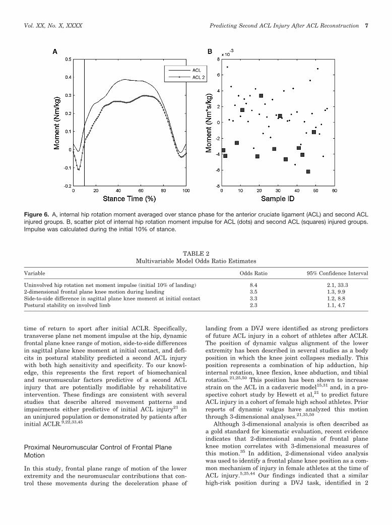
Univariate analyses indicated that the uninvolved limbtransverse plane hip net moment impulse during the ini-tial 10% of landing was associated with increased risk ofsecond ACL injury, with an area under the ROC of 0.81and both high sensitivity (0.77) and specificity (0.81). Inthe final multivariable logistic regression model, this vari-able was identified as the strongest predictor of secondACL injury (Figure 5). Participants who sustained a secondACL injury demonstrated a significantly different hiptransverse plane moment impulse compared with thosewho did not incur a second ACL injury (P\ .001; Figure 6).The impulse during the initial 10% of stance was represen-tative of a net hip internal rotator moment in the partici-pants who sustained a second ACL injury (22.4 3 1023
Nms/kg) compared with a net external rotator momenton the uninvolved limb of participants who did not incura second ACL injury (1.1 3 1023 Nms/kg; Figure 6).Greater hip external rotator moments may act to restrain
Figure 3. Participant during the assessment of postural sta-bility using the Biodex SD Stability System. The individual isin a unilateral stance position with the contralateral limbflexed and both arms crossed at the chest. The participantis tested with eyes open, but receives no visual feedbackon performance during the testing session.
Vol. XX, No. X, XXXX Predicting Second ACL Injury After ACL Reconstruction 5

hip internal rotation motion during this phase of landing.Specifically, participants with less hip external rotatormoment during early landing were over 8 times (OR 5
8.4; 95% CI 5 2.1, 33.3) more likely to sustain a secondACL injury than those with greater hip external rotationmoment (Table 2).
Two-dimensional frontal plane knee joint range ofmotion during the landing phase of the involved limb, inaddition to hip kinetics, demonstrated significantly greaterangular displacement in the group that suffered a secondACL injury compared with the ACLR patients who didnot suffer a second injury (P 5.03; Figure 7). The secondinjury group demonstrated an average of 16.2� of totalfrontal plane (valgus) movement, while the remainingpatients translated 12.1� in the frontal plane. Participantswith an increase in frontal plane motion were over 3 timesas likely (OR 5 3.5; 95% CI 5 1.3, 9.9) to incur a secondACL injury than those with reduced frontal plane motion.
Side-to-side differences in sagittal plane knee momentat initial contact were significantly different betweengroups (P 5 .03; Figure 5). The second injury group demon-strated a 4.1-fold greater asymmetry in internal kneeextensor moment at initial contact when compared withthe cohort of patients who did not suffer additional injury.Repeated-measure 2 3 2 analysis of variance showed a sig-nificant side (initial involved vs initial uninvolved) 3 group(initial ACLR vs second ACL injury) interaction (P 5 .03)of sagittal plane knee moment at initial contact of landing.Post hoc analysis demonstrated that the internal kneeextensor moment of the uninvolved limb at the point of ini-tial contact in the second injury group (22.8 3 1022 Nm/kg)was significantly lower than the involved limb of the secondinjury group (9.5 3 1022 Nm/kg) and both involved (11.4 3
1022 Nm/kg) and uninvolved (8.4 3 1022 Nm/kg) limbs inthe first injury group, which demonstrated a net knee flexormoment at the point of initial contact (Figure 8). Partici-pants who demonstrated asymmetry in this variable were
over 3 times as likely (OR 5 3.3; 95% CI 5 1.2, 8.8) to incura second ACL injury than those who demonstrated symme-try in sagittal plane knee moments at the point of initialcontact.
Participants with deficits in single-leg postural stabilityof the involved limb, as measured by the Biodex stabilitysystem, were twice as likely (OR 5 2.3; 95% CI 5 1.1, 4.7)to incur a second injury as compared with those who didnot demonstrate single-leg stability of the involved limb.The involved limb of the second injury group demonstrateda mean degree of deflection on the overall stability score of4.07� 6 2.06�, while the stability score on the involved limbof the first injury group was 3.63� 6 1.58�. Additional vari-ables, including graft type and A-P knee laxity, were evalu-ated in the statistical analysis; however, they were notpredictive of a second ACL injury in this cohort.
DISCUSSION
The findings of the current study support the testedhypothesis that altered neuromuscular control patternsduring landing and deficits in postural stability predictsubsequent ACL injuries in a sample of athletes at the
Sen
sitiv
ity
0.0
0.1
0.2
0.3
0.4
0.5
0.6
0.7
0.8
0.9
1.0
1 - Specificity
0.0 0.1 0.2 0.3 0.4 0.5
ROC Curve
0.6 0.7 0.8 0.9 1.0
Figure 4. The corresponding receiver operating curve (ROC)plotted for the overall model. Multivariable logistic regressionidentified 4 variables that predicted a second anterior cruci-ate ligament injury after anterior cruciate ligament recon-struction and return to sports. The area under the ROCstatistic is shown with an area under the ROC of 0.94.
Figure 5. Example of second anterior cruciate ligament(ACL) injury predictors in a patient with history of a rightACL injury and reconstruction. A, participants who sustaineda second ACL injury had a contralateral hip internal rotationmoment during the first 10% of the landing cycle comparedwith an external rotation moment in the group of patients whodid not suffer a second injury. B, participants who sustaineda second ACL injury had a side-to-side difference in sagittalplane knee moment at initial contact with a knee extensormoment on the contralateral limb and a knee flexor momentin the involved limb. All moments are described as internalmoments.
6 Paterno et al The American Journal of Sports Medicine
time of return to sport after initial ACLR. Specifically,transverse plane net moment impulse at the hip, dynamicfrontal plane knee range of motion, side-to-side differencesin sagittal plane knee moment at initial contact, and defi-cits in postural stability predicted a second ACL injurywith both high sensitivity and specificity. To our knowl-edge, this represents the first report of biomechanicaland neuromuscular factors predictive of a second ACLinjury that are potentially modifiable by rehabilitativeintervention. These findings are consistent with severalstudies that describe altered movement patterns andimpairments either predictive of initial ACL injury21 inan uninjured population or demonstrated by patients afterinitial ACLR.9,22,33,45
Proximal Neuromuscular Control of Frontal PlaneMotion
In this study, frontal plane range of motion of the lowerextremity and the neuromuscular contributions that con-trol these movements during the deceleration phase of
landing from a DVJ were identified as strong predictorsof future ACL injury in a cohort of athletes after ACLR.The position of dynamic valgus alignment of the lowerextremity has been described in several studies as a bodyposition in which the knee joint collapses medially. Thisposition represents a combination of hip adduction, hipinternal rotation, knee flexion, knee abduction, and tibialrotation.21,25,50 This position has been shown to increasestrain on the ACL in a cadaveric model15,31 and, in a pro-spective cohort study by Hewett et al,21 to predict futureACL injury in a cohort of female high school athletes. Priorreports of dynamic valgus have analyzed this motionthrough 3-dimensional analyses.21,35,50
Although 3-dimensional analysis is often described asa gold standard for kinematic evaluation, recent evidenceindicates that 2-dimensional analysis of frontal planeknee motion correlates with 3-dimensional measures ofthis motion.35 In addition, 2-dimensional video analysiswas used to identify a frontal plane knee position as a com-mon mechanism of injury in female athletes at the time ofACL injury.5,25,44 Our findings indicated that a similarhigh-risk position during a DVJ task, identified in 2
Figure 6. A, internal hip rotation moment averaged over stance phase for the anterior cruciate ligament (ACL) and second ACLinjured groups. B, scatter plot of internal hip rotation moment impulse for ACL (dots) and second ACL (squares) injured groups.Impulse was calculated during the initial 10% of stance.
TABLE 2Multivariable Model Odds Ratio Estimates
Variable Odds Ratio 95% Confidence Interval
Uninvolved hip rotation net moment impulse (initial 10% of landing) 8.4 2.1, 33.32-dimensional frontal plane knee motion during landing 3.5 1.3, 9.9Side-to-side difference in sagittal plane knee moment at initial contact 3.3 1.2, 8.8Postural stability on involved limb 2.3 1.1, 4.7
Vol. XX, No. X, XXXX Predicting Second ACL Injury After ACL Reconstruction 7

dimensions, predicted a second ACL injury. It is likely that2-dimensional assessment of frontal plane motion is a rep-resentation of contributions of hip abduction, hip rotation,and knee abduction, which collectively increase risk forACL injury. There is high clinical utility of a 2-dimensionalanalysis of frontal plane motion that could identify a riskfactor for future ACL injury after ACLR. Current criteriautilized to assess an athlete’s readiness to return to sportsafter ACLR may include clinical measures of range ofmotion, strength, and success on functional performancetests.39,55 Video analysis of frontal plane motion duringan athletic maneuver, such as the DVJ, appears to be anappropriate tool to include in an assessment of an athlete’sreadiness to safely return to sports.
Frontal plane motion of the knee during the landingphase of a DVJ is potentially controlled by neuromuscularcontribution at the hip, knee, and ankle. Several studieshave investigated the relationship of hip strength andhip kinematics during dynamic tasks such as landing, run-ning, and stair climbing,6,52,53,56 particularly in the pres-ence of patellofemoral injury. Souza and Powers52
assessed hip kinematics during the DVJ task in patientswith and without patellofemoral abnormality and reported
increased hip internal rotation motion during the DVJ anda coinciding deficit in hip extensor strength as measuredon an isokinetic dynamometer in patellofemoral patientscompared with controls. McLean et al34 noted an associa-tion between hip internal rotation kinematics and kneevalgus moment during athletic tasks, a position known toincrease strain on the ACL.15 Data from the current studyindicated that a deficit in net hip muscle external rotationtorque during the early phase of landing was a strong pre-dictor of future ACL injury, either when examined in thepresence of other potential risk factors or when examinedindependently, when it predicted ACL injury risk with anarea under the ROC curve of 0.81.
The clinical implications of these findings of hip muscleexternal rotation torque deficits are significant. The cur-rent results indicate that interventions targeted to addressimpaired hip strength have the potential to reduce secondACL injury after ACLR. Functional anatomical analysesindicate that the gluteus maximus is a strong hip extenderand external rotator43 while in a flexed hip posture.42 Dur-ing the DVJ, the average hip flexion angle starts at approx-imately 25� to 30� of flexion at initial contact and increasesto 55� of peak flexion.12 Considering gluteus maximusfunction, and the findings of Souza and Powers,53 whichindicate a relationship between hip extensor muscle per-formance and hip internal rotation kinematics with theDVJ task, an intervention focused on hip external rotationand hip extension strength and recruitment may assist inthe reduction of dynamic frontal plane knee motion.
Figure 7. Example of second anterior cruciate ligament(ACL) injury predictors in a patient with history of a rightACL injury and reconstruction. Participants who sustaineda second ACL injury had increased 2-dimensional peak fron-tal plane knee motion during the landing phase of the dropvertical jump.
P = 0.03
0.150
0.100
0.050
0.000
Ave
rag
e N
m/k
g
Sagittal Plane Knee Moment at Initial Contact
−0.050
−0.100
InjuryEpisode
Error bars: +/− 1SE
First ACL Injury
legendInvolved.KneeUninvolved.Knee
Second ACL Injury
Figure 8. Sagittal plane knee moments at initial contact forthe involved and uninvolved limb of the initial anterior cruci-ate ligament (ACL) injury group and the second ACL injurygroup.
8 Paterno et al The American Journal of Sports Medicine

Essentially, gluteus maximus strengthening and enhancedrecruitment after ACLR may have the potential to reducefuture ACL injury in this population. In addition toincreased hip strength, neuromuscular training focusedon plyometrics and landing techniques, such as squatjumps, scissor jumps, and tuck jumps, has the potentialto improve gluteal muscle recruitment and correct abnor-mal frontal plane mechanics during the DVJ.14 Previously,altered frontal plane motion has been shown to predictACL injury in a healthy population.21 Our findings nowindicate that altered frontal plane motion may predicta second ACL injury after ACLR. Current evidence indi-cates that neuromuscular interventions have the potentialto improve jumping mechanics and, in turn, reduce theACL injury rate in a healthy population.16,18-20,30 There-fore, implementation of these interventions at the endstages of rehabilitation after ACLR may have the potentialto also reduce second ACL injury rates.
Asymmetries in Sagittal Plane MomentsDuring Landing
Side-to-side differences in neuromuscular control of theknee during dynamic, athletic tasks was theorized to bea potential risk factor for ACL injury in a healthy popula-tion.22 Similarly, these results indicate that side-to-sideasymmetries in neuromuscular variables persist followingACLR, which is consistent with our earlier work, whichfound side-to-side differences in vertical ground-reactionforce and loading rate that persisted for up to 2 years afterACLR.45 Our current findings indicate that limb asymme-tries identified after ACLR are predictive of future ACLinjury, specifically asymmetries in sagittal plane kneemoments. Injuries to the ACL often occur with the kneeat low knee flexion angles.4,29 In addition, Lloyd26 sug-gested that cocontraction of the quadriceps and hamstringsassists in providing up to 80% of the resistance to frontalplane movement at the knee. Therefore, the ability of theknee to symmetrically activate the sagittal plane muscula-ture to assist in frontal plane stability during a bipedaltask, at a time when the knee is in a high-risk position oflow flexion, is critical. The asymmetry in sagittal planeknee moments at initial contact, as demonstrated by theparticipants who subsequently suffered a second ACLinjury, indicates that an imbalance in force absorptionstrategies during the onset of the DVH task may increasefuture injury risk.38
Deficits in Postural Stability
Balance or postural stability is defined as an individual’sability to maintain the center of mass over the base of sup-port. The deviation of the center of mass away from thebase of support during dynamic athletic activities hasrecently been identified by several authors as a mechanismthat increases valgus load on the ACL.23,25,44 This increasein frontal plane load through a poorly controlled trunk isa potential risk factor for ACL injury in a healthy
population of female athletes. Zazulak et al59,60 identifiedcore proprioception and kinesthetic awareness as a predic-tor of knee and ACL injury in a population of previouslyuninjured collegiate female athletes. In these studies,female athletes with decreased core proprioception andkinesthetic awareness were more likely to suffer a kneeinjury. Dynamic postural stability assessment is a methodto objectively quantify deviations in balance from a neutralpoint and may be a simple method to quantify an individ-ual’s ability to control movement of the center of mass inan objective and dynamic fashion. Prior studies have dem-onstrated deficits in postural stability after ACLinjury,1,2,22 an improvement in postural stability andknee kinematics with perturbation training after ACLinjury,8,11 and an improvement in postural stability withneuromuscular training after ACLR.22,61 The current find-ings of the association of postural deficits with increasedrisk of second ACL injury indicate that the presence ofaltered involved limb single-leg balance after ACLR maylimit the ability of the athlete to control the movement ofthe center of mass over the base of support during dynamicathletic activities and subsequently increase the risk ofknee injury. Intervention strategies throughout the post-operative course that target improved postural stabilityand control of the center of mass may help minimize subse-quent ACL injury risk after ACLR. These can include dou-ble- and single-leg balance activities on stable and unstablesurfaces, with and without perturbations.
Potential Study Limitations
Although the predictive model of second ACL injury afterACLR presented has high sensitivity and specificity, werecognize the potential limitations that should be consid-ered relative to the interpretation of the current studyresults. A relatively small sample cohort of 56 young, ath-letic individuals was prospectively enrolled in our study,which could potentially limit the generalizability of theresults to other populations. However, the 13 second ACLinjuries observed in our study, which exceeded our a prioripower analysis of 7.7 ACL injuries needed to meet suffi-cient statistical power, increased our confidence that theseresults may be applicable to the young, athletic population,despite the relatively small initial enrollment of 56patients. Another potential limitation of the study is thepotential for inequitable distribution of graft type amongthe sample. Specifically, there were very few participantswith allograft tissue ACLR compared to the number of par-ticipants who had ACLR using autograft patellar tendonand hamstrings graft material. Although it was not the pri-mary aim of the study to identify graft source as a potentialrisk factor for future ACL injury and graft type did notinfluence our predictive model, prior evidence7 indicatesthat allograft tissue may be a predictor of ipsilateral retearof an ACLR graft. As a result of our low number of partic-ipants with an allograft reconstruction, we cannot statewith confidence that allograft tissue was not a predictorof a second injury. Finally, the aims of this study werefocused on biomechanical variables in patients after
Vol. XX, No. X, XXXX Predicting Second ACL Injury After ACL Reconstruction 9

ACLR in an attempt to create a predictive model of whowould suffer a second ACL injury. An interesting analysiswould include a comparison of these groups to a healthygroup who never suffered a lower extremity injury.
SUMMARY AND CONCLUSION
Biomechanical Measures and Postural Control DeficitsPredict Second ACL Injury
Current evidence indicates a high incidence of second ACLinjury after ACLR, especially in populations who return tosport. However, potential mechanisms underlying thisincreased risk have not been identified in a prospectivecohort study. Our study prospectively evaluated biome-chanical and neuromuscular variables during the landingphase of a DVJ, in addition to assessing dynamic posturalstability at the time the athlete was released to return tosport, to determine if deficits in these variables were pre-dictive of a second ACL injury. The study findings indicatethat net hip rotation moment impulse, frontal plane kneerange of motion during landing, asymmetries in sagittalplane knee moments at initial contact, and postural stabil-ity are collectively a strong predictor of a second ACLinjury after ACLR with high sensitivity (0.92) and specific-ity (0.88). In addition, net hip rotation moment impulse, inisolation, is also a good predictor of second ACL injury withmoderate to high sensitivity (0.78) and specificity (0.81).
The identification of high-risk variables that predictfuture ACL injury, at the time of patient discharge to returnto sports after ACLR, is the first step in the development ofmore efficacious interventions, and more appropriate dis-charge criteria, for the determination of when an athleteshould return to sport after ACL injury rehabilitation.With these key identified variables that predict athletes athigh risk for future ACL injury, more valid criteria cannow be developed and implemented, as necessary criteriato be released to return to sports or activity. Ultimately, itfollows logically to pursue testing of specific rehabilitationinterventions to determine if targeted interventions couldalso be modified to address these specific impairments inhip external rotator muscle strength and recruitment andthus decrease the likelihood of future ACL injury.
ACKNOWLEDGMENT
We would like to thank Dr. Robert Heidt and the staff atWellington Orthopaedic and Sports Medicine Center, Dr.Keith Kenter and the staff of the University of CincinnatiSports Medicine, Dr. Eric Wall and the staff of and Cincin-nati Children’s Hospital Division of Orthopaedics, ThomasPalmer and the athletes within the Northern KentuckyUniversity Athletic Department and Theresa Behan andthe athletes within the Thomas More College AthleticDepartment for their participation in this study. We wouldalso like to thank Rebecca Reder, Michael Allen and theOrtho/Sports Physical Therapy Team at the CincinnatiChildren’s Hospital Medical Center.
REFERENCES
1. Beard DJ, Dodd CA, Trundle HR, Simpson AH. Proprioception
enhancement for anterior cruciate ligament deficiency: a prospective
randomised trial of two physiotherapy regimes. J Bone Joint Surg Br.
1994;76(4):654-659.
2. Beard DJ, Kyberd PJ, Dodd CA, Simpson AH, O’Connor JJ. Propri-
oception in the knee. J Bone Joint Surg Br. 1994;76(6):992-993.
3. Bell AL, Pedersen DR, Brand RA. A comparison of the accuracy of
several hip center location prediction methods. J Biomech.
1990;23(6):617-621.
4. Boden BP, Dean GS, Feagin JA Jr, Garrett WE Jr. Mechanisms of
anterior cruciate ligament injury. Orthopedics. 2000;23(6):573-578.
5. Boden BP, Torg JS, Knowles SB, Hewett TE. Video analysis of ante-
rior cruciate ligament injury: abnormalities in hip and ankle kinemat-
ics. Am J Sports Med. 2009;37(2):252-259.
6. Bolgla LA, Malone TR, Umberger BR, Uhl TL. Hip strength and hip
and knee kinematics during stair descent in females with and without
patellofemoral pain syndrome. J Orthop Sports Phys Ther. 2008;
38(1):12-18.
7. Borchers JR, Pedroza A, Kaeding C. Activity level and graft type as
risk factors for anterior cruciate ligament graft failure: a case-control
study. Am J Sports Med. 2009;37(12):2362-2367.
8. Chmielewski TL, Rudolph KS, Snyder-Mackler L. Development of
dynamic knee stability after acute ACL injury. J Electromyogr Kine-
siol. 2002;12(4):267-274.
9. Colby SM, Hintermeister RA, Torry MR, Steadman JR. Lower limb
stability with ACL impairment. J Orthop Sports Phys Ther.
1999;29(8):444-451; discussion 452-454.
10. Daniel DM, Stone ML, Dobson BE, Fithian DC, Rossman DJ, Kauf-
man KR. Fate of the ACL-injured patient: a prospective outcome
study. Am J Sports Med. 1994;22(5):632-644.
11. Fitzgerald GK, Axe MJ, Snyder-Mackler L. The efficacy of perturba-
tion training in nonoperative anterior cruciate ligament rehabilitation
programs for physically active individuals. Phys Ther. 2000;80(2):
128-140.
12. Ford KR, Myer GD, Hewett TE. Reliability of landing 3D motion anal-
ysis: implications for longitudinal analyses. Med Sci Sports Exerc.
2007;39(11):2021-2028.
13. Ford KR, Myer GD, Hewett TE. Valgus knee motion during landing in
high school female and male basketball players. Med Sci Sports
Exerc. 2003;35(10):1745-1750.
14. Ford KR, Myer GD, Smith RL, Byrnes RN, Dopirak SE, Hewett TE.
Use of an overhead goal alters vertical jump performance and biome-
chanics. J Strength Cond Res. 2005;19(2):394-399.
15. Fung DT, Zhang LQ. Modeling of ACL impingement against the inter-
condylar notch. Clin Biomech (Bristol, Avon). 2003;18(10):933-941.
16. Gilchrist J, Mandelbaum BR, Melancon H, et al. A randomized controlled
trial to prevent noncontact anterior cruciate ligament injury in female col-
legiate soccer players. Am J Sports Med. 2008;36(8):1476-1483.
17. Gomez E, DeLee JC, Farney WC. Incidence of injury in Texas girls’
high school basketball. Am J Sports Med. 1996;24(5):684-687.
18. Hewett TE, Ford KR, Myer GD. Anterior cruciate ligament injuries in
female athletes: part 2, a meta-analysis of neuromuscular interven-
tions aimed at injury prevention. Am J Sports Med. 2006;34(3):
490-498.
19. Hewett TE, Lindenfeld TN, Riccobene JV, Noyes FR. The effect of
neuromuscular training on the incidence of knee injury in female ath-
letes: a prospective study. Am J Sports Med. 1999;27(6):699-706.
20. Hewett TE, Myer GD, Ford KR. Anterior cruciate ligament injuries in
female athletes: part 1, mechanisms and risk factors. Am J Sports
Med. 2006;34(2):299-311.
21. Hewett TE, Myer GD, Ford KR, et al. Biomechanical measures of
neuromuscular control and valgus loading of the knee predict ante-
rior cruciate ligament injury risk in female athletes: a prospective
study. Am J Sports Med. 2005;33(4):492-501.
22. Hewett TE, Paterno MV, Myer GD. Strategies for enhancing proprio-
ception and neuromuscular control of the knee. Clin Orthop Relat
Res. 2002;402:76-94.
10 Paterno et al The American Journal of Sports Medicine

23. Hewett TE, Torg JS, Boden BP. Video analysis of trunk and knee
motion during non-contact anterior cruciate ligament injury in female
athletes: lateral trunk and knee abduction motion are combined com-
ponents of the injury mechanism. Br J Sports Med. 2009;43(6):417-422.
24. Hosmer D, Lemeshow S. Applied Logistic Regression. New York, NY:
John Wliey and Sons Inc; 1989.
25. Krosshaug T, Nakamae A, Boden BP, et al. Mechanisms of anterior
cruciate ligament injury in basketball: video analysis of 39 cases.
Am J Sports Med. 2007;35(3):359-367.
26. Lloyd DG. Rationale for training programs to reduce anterior cruciate
ligament injuries in Australian football. J Orthop Sports Phys Ther.
2001;31(11):645-654; discussion 661.
27. Lohmander LS, Ostenberg A, Englund M, Roos H. High prevalence of
knee osteoarthritis, pain, and functional limitations in female soccer
players twelve years after anterior cruciate ligament injury. Arthritis
Rheum. 2004;50(10):3145-3152.
28. Lyman S, Koulouvaris P, Sherman S, Do H, Mandl LA, Marx RG. Epi-
demiology of anterior cruciate ligament reconstruction: trends, read-
missions, and subsequent knee surgery. J Bone Joint Surg Am.
2009;91(10):2321-2328.
29. Malinzak RA, Colby SM, Kirkendall DT, Yu B, Garrett Jr WE. A compar-
ison of knee joint motion patterns between men and women in selected
athletic tasks. Clin Biomech (Bristol, Avon). 2001;16(5):438-445.
30. Mandelbaum BR, Silvers HJ, Watanabe D, et al. Effectiveness of
a neuromuscular and proprioceptive training program in preventing
the incidence of ACL injuries in female athletes: two-year follow up.
Am J Sports Med. 2005;33(7):1003-1010.
31. Markolf KL, Burchfield DM, Shapiro MM, Shepard MF, Finerman GA,
Slauterbeck JL. Combined knee loading states that generate high
anterior cruciate ligament forces. J Orthop Res. 1995;13(6):930-935.
32. Marshall SW, Padua D, McGrath M. Incidence of ACL injury. In:
Hewett TE, Schultz SJ, Griffin LY, eds. Understanding and Preventing
Noncontact ACL Injuries. Champaign, IL: Human Kinetics; 2007:5-30.
33. Mattacola CG, Perrin DH, Gansneder BM, Gieck JH, Saliba EN,
McCue FC 3rd. Strength, functional outcome, and postural stability
after anterior cruciate ligament reconstruction. J Athl Train.
2002;37(3):262-268.
34. McLean SG, Huang X, van den Bogert AJ. Association between
lower extremity posture at contact and peak knee valgus moment
during sidestepping: implications for ACL injury. Clin Biomech (Bris-
tol, Avon). 2005;20(8):863-870.
35. McLean SG, Walker K, Ford KR, Myer GD, Hewett TE, van den
Bogert AJ. Evaluation of a two dimensional analysis method as
a screening and evaluation tool for anterior cruciate ligament injury.
Br J Sports Med. 2005;39(6):355-362.
36. Messina DF, Farney WC, DeLee JC. The incidence of injury in Texas
high school basketball: a prospective study among male and female
athletes. Am J Sports Med. 1999;27(3):294-299.
37. Myer GD, Ford KR, Paterno MV, Nick TG, Hewett TE. The effects of
generalized joint laxity on risk of anterior cruciate ligament injury in
young female athletes. Am J Sports Med. 2008;36(6):1073-1080.
38. Myer GD, Paterno MV, Ford KR, Hewett TE. Neuromuscular training
techniques to target deficits before return to sport after anterior
cruciate ligament reconstruction. J Strength Cond Res. 2008;22(3):
987-1014.
39. Myer GD, Paterno MV, Ford KR, Quatman CE, Hewett TE. Rehabili-
tation after anterior cruciate ligament reconstruction: criteria based
progression through the return to sport phase. J Orthop Sports
Phys Ther. 2006;36(6):385-402.
40. Myklebust G, Bahr R. Return to play guidelines after anterior cruciate
ligament surgery. Br J Sports Med. 2005;39(3):127-131.
41. Myklebust G, Holm I, Maehlum S, Engebretsen L, Bahr R. Clinical,
functional, and radiologic outcome in team handball players 6 to 11
years after anterior cruciate ligament injury: a follow-up study. Am
J Sports Med. 2003;31(6):981-989.
42. Neumann DA. Kinesiology of the hip: a focus on muscular actions. J
Orthop Sports Phys Ther. 2010;40(2):82-94.
43. Newman D. Kinesiology of the Musculoskeletal System: Foundations
for Physical Rehabilitation. St Louis, MO: Mosby; 2002.
44. Olsen OE, Myklebust G, Engebretsen L, Bahr R. Injury mechanisms
for anterior cruciate ligament injuries in team handball: a systematic
video analysis. Am J Sports Med. 2004;32(4):1002-1012.
45. Paterno MV, Ford KR, Myer GD, Heyl R, Hewett TE. Limb asymme-
tries in landing and jumping 2 years following anterior cruciate liga-
ment reconstruction. Clin J Sport Med. 2007;17(4):258-262.
46. Paterno MV, Myer GD, Ford KR, Hewett TE. Neuromuscular training
improves single-limb stability in young female athletes. J Orthop
Sports Phys Ther. 2004;34(6):305-316.
47. Pinczewski LA, Lyman J, Salmon LJ, Russell VJ, Roe J, Linklater J. A
10-year comparison of anterior cruciate ligament reconstructions
with hamstring tendon and patellar tendon autograft: a controlled,
prospective trial. Am J Sports Med. 2007;35(4):564-574.
48. Rauh MJ, Macera CA, Ji M, Wiksten DL. Subsequent injury patterns
in girls’ high school sports. J Athl Train. 2007;42(4):486-494.
49. Salmon L, Russell V, Musgrove T, Pinczewski L, Refshauge K. Inci-
dence and risk factors for graft rupture and contralateral rupture after
anterior cruciate ligament reconstruction. Arthroscopy. 2005;21(8):
948-957.
50. Schmitz RJ, Shultz SJ, Nguyen AD. Dynamic valgus alignment and
functional strength in males and females during maturation. J Athl
Train. 2009;44(1):26-32.
51. Shelbourne KD, Gray T, Haro M. Incidence of subsequent injury to
either knee within 5 years after anterior cruciate ligament reconstruc-
tion with patellar tendon autograft. Am J Sports Med. 2009;37(2):
246-251.
52. Souza RB, Powers CM. Differences in hip kinematics, muscle
strength, and muscle activation between subjects with and without
patellofemoral pain. J Orthop Sports Phys Ther. 2009;39(1):12-19.
53. Souza RB, Powers CM. Predictors of hip internal rotation during run-
ning: an evaluation of hip strength and femoral structure in women
with and without patellofemoral pain. Am J Sports Med. 2009;
37(3):579-587.
54. von Porat A, Roos EM, Roos H. High prevalence of osteoarthritis 14
years after an anterior cruciate ligament tear in male soccer players:
a study of radiographic and patient relevant outcomes. Ann Rheum
Dis. 2004;63(3):269-273.
55. Wilk KE, Arrigo C, Andrews JR, Clancy WG. Rehabilitation after ante-
rior cruciate ligament reconstruction in the female athlete. J Athl
Train. 1999;34(2):177-193.
56. Willson JD, Ireland ML, Davis I. Core strength and lower extremity
alignment during single leg squats. Med Sci Sports Exerc. 2006;
38(5):945-952.
57. Winter D. Biomechanics and Motor Control of Human Movement.
New York, NY: John Wiley; 1990.
58. Wright RW, Dunn WR, Amendola A, et al. Risk of tearing the intact
anterior cruciate ligament in the contralateral knee and rupturing
the anterior cruciate ligament graft during the first 2 years after ante-
rior cruciate ligament reconstruction: a prospective MOON cohort
study. Am J Sports Med. 2007;35(7):1131-1134.
59. Zazulak BT, Hewett TE, Reeves NP, Goldberg B, Cholewicki J. Def-
icits in neuromuscular control of the trunk predict knee injury risk:
a prospective biomechanical-epidemiologic study. Am J Sports
Med. 2007;35(7):1123-1130.
60. Zazulak BT, Hewett TE, Reeves NP, Goldberg B, Cholewicki J.
The effects of core proprioception on knee injury: a prospective
biomechanical-epidemiogical study. Am J Sports Med. 2007;35(3):
368-373.
61. Zech A, Hubscher M, Vogt L, Banzer W, Hansel F, Pfeifer K. Neuro-
muscular training for rehabilitation of sports injuries: a systematic
review. Med Sci Sports Exerc. 2009 Sep 2 [Epub ahead of print].
For reprints and permission queries, please visit SAGE’s Web site at http://www.sagepub.com/journalsPermissions.nav
Vol. XX, No. X, XXXX Predicting Second ACL Injury After ACL Reconstruction 11