BIOMECHANICAL AND PHYSIOLOGICAL INFLUENCES rio_et_al... · PDF fileLaboratório de...
10
403 Rev. bras. paleontol. 18(3):403-412, Setembro/Dezembro 2015 © 2015 by the Sociedade Brasileira de Paleontologia doi: 10.4072/rbp.2015.3.06 BIOMECHANICAL AND PHYSIOLOGICAL INFLUENCES ON THE OSTEOHISTOLOGICAL DEPOSITION OF ANHANGUERIA (PTEROSAURIA, PTERODACTYLOIDEA) LÚCIA HELENA DE SOUZA ELEUTÉRIO, RENAN ALFREDO MACHADO BANTIM, FLAVIANA JORGE DE LIMA, RAFAEL CÉSAR LIMA PEDROSO DE ANDRADE Laboratório de Biodiversidade do Nordeste, Centro Acadêmico de Vitória, UFPE, Alto do Reservatório, s/n, 55608-680, Vitória de Santo Antão, Pernambuco, Brazil. [email protected], [email protected], fl[email protected], [email protected] ANTÔNIO ÁLAMO FEITOSA SARAIVA Laboratório de Paleontologia, URCA, Coronel Luiz Antonio, 1161, 63100-000, Crato, CE, Brazil. [email protected] ALEXANDER WILHELM ARMIN KELLNER Laboratório de Sistemática e Tafonomia de Vertebrados Fósseis, Departamento de Geologia e Paleontologia, Museu Nacional/UFRJ, Quinta da Boa Vista, Rio de Janeiro, RJ, Brazil. [email protected] JULIANA MANSO SAYÃO Laboratório de Biodiversidade do Nordeste, Centro Acadêmico de Vitória, UFPE, Alto do Reservatório Street, s/n, 55608-680, Vitória de Santo Antão, Pernambuco, Brazil. [email protected] ABSTRACT – The study of bone microstructure preserved in fossils provides substantial information about physiology, growth gradients and strategies, and some ecological considerations. Paleohistology is a useful tool for understanding the biological mechanisms of extinct animals. Presented here is the microstructure characterization of two Anhangueria pterosaurs. Thin sections of the first wing phalanx and metacarpal IV of MN 7060-V have been confectioned, as have sections of the radius, ulna and first wing phalanx of MPSC R2090. The histological analysis of bones of MN 7060-V revealed fibrolamellar tissue, few osteocytes and vascular canals. Bones of MPSC R2090 showed a mixed plexiform-fibrolamellar tissue and histovariability, showing that bones from the same individual grew in different patterns. The vascularization was high in the phalanx, intermediate in the ulna and absent in the radius. The absence of canals in the radius may be related to biomechanical issues, due to torsion resistance during flight. The histology and the absence of fused bones suggests that the specimens are not adults. Two distinct moments of growth were established. MN 7060-V is a subadult, with presence of bone porosity and MPSC R2090 is a young animal as determined by the high number of canals and plexiform-fibrolamellar tissues, which indicates fast growth. In this work, we concluded that in the Anhangueria clade, the growth of bones is not compatible with the ontogenetic stage. Young animals may present large proportions, whereas there were older individuals of smaller sizes in the same clade. Key words: paleohistology, Pterosauria, Anhangueria, Romualdo Formation, Araripe Basin. RESUMO – O estudo da microestrutura óssea preservada em organismos fósseis traz informações substanciais sobre a fisiologia, gradientes e estratégias de crescimento e uma série de considerações ecológicas. Para o entendimento dos mecanismos biológicos de animais extintos utiliza-se a paleohistologia. Apresentamos a caracterização microestrutural de dois pterossauros do clado Anhangueria. Foram confeccionadas lâminas da primeira falange alar e IV metacarpal de MN 7060-V, além da ulna, rádio e primeira falange alar de MPSC R2090. A análise histológica de ambos os ossos de MN 7060-V exibiu tecido fibrolamelar, poucos osteócitos e canais. Já os ossos de MPSC R2090 apresentaram tecido fibrolamelar plexiforme e histovariabilidade, sugerindo diferentes taxas de crescimento para os ossos de um mesmo indivíduo. A vascularização na falange foi elevada, intermediária na ulna e ausente no rádio. Essa ausência de canais pode estar relacionada a pressões biomecânicas, devido a resistência às cargas de torção exercidas durante o voo. A histologia e a ausência de fusão dos ossos sugerem indivíduos não adultos. Dois momentos diferentes de crescimento foram então estabelecidos. MN 7060-V é um subadulto, com presença de porosidade óssea e MPSC R2090 um jovem, devido à alta vascularização e tecido fibrolamelar plexiforme, indicativo de crescimento rápido. Foi concluído neste trabalho que, no clado Anhangueria, o crescimento ósseo não acompanha o estágio ontogenético dos indivíduos. Animais jovens podem apresentar grandes proporções, ao passo que indivíduos mais velhos tamanhos menores dentro de um mesmo clado. Palavras-chave: paleohistologia, Pterosauria, Anhangueria, Formação Romualdo, bacia do Araripe.
Transcript of BIOMECHANICAL AND PHYSIOLOGICAL INFLUENCES rio_et_al... · PDF fileLaboratório de...

403
Rev. bras. paleontol. 18(3):403-412, Setembro/Dezembro 2015© 2015 by the Sociedade Brasileira de Paleontologiadoi: 10.4072/rbp.2015.3.06
BIOMECHANICAL AND PHYSIOLOGICAL INFLUENCES ON THE OSTEOHISTOLOGICAL DEPOSITION OF ANHANGUERIA
(PTEROSAURIA, PTERODACTYLOIDEA)
LÚCIA HELENA DE SOUZA ELEUTÉRIO, RENAN ALFREDO MACHADO BANTIM, FLAVIANA JORGE DE LIMA, RAFAEL CÉSAR LIMA PEDROSO DE ANDRADE
Laboratório de Biodiversidade do Nordeste, Centro Acadêmico de Vitória, UFPE, Alto do Reservatório, s/n, 55608-680, Vitória de Santo Antão, Pernambuco, Brazil. [email protected], [email protected],
fl [email protected], [email protected]
ANTÔNIO ÁLAMO FEITOSA SARAIVALaboratório de Paleontologia, URCA, Coronel Luiz Antonio, 1161, 63100-000, Crato, CE, Brazil.
ALEXANDER WILHELM ARMIN KELLNERLaboratório de Sistemática e Tafonomia de Vertebrados Fósseis, Departamento de Geologia e Paleontologia,
Museu Nacional/UFRJ, Quinta da Boa Vista, Rio de Janeiro, RJ, Brazil. [email protected]
JULIANA MANSO SAYÃOLaboratório de Biodiversidade do Nordeste, Centro Acadêmico de Vitória, UFPE, Alto do Reservatório Street, s/n,
55608-680, Vitória de Santo Antão, Pernambuco, Brazil. [email protected]
ABSTRACT – The study of bone microstructure preserved in fossils provides substantial information about physiology, growth gradients and strategies, and some ecological considerations. Paleohistology is a useful tool for understanding the biological mechanisms of extinct animals. Presented here is the microstructure characterization of two Anhangueria pterosaurs. Thin sections of the fi rst wing phalanx and metacarpal IV of MN 7060-V have been confectioned, as have sections of the radius, ulna and fi rst wing phalanx of MPSC R2090. The histological analysis of bones of MN 7060-V revealed fi brolamellar tissue, few osteocytes and vascular canals. Bones of MPSC R2090 showed a mixed plexiform-fi brolamellar tissue and histovariability, showing that bones from the same individual grew in different patterns. The vascularization was high in the phalanx, intermediate in the ulna and absent in the radius. The absence of canals in the radius may be related to biomechanical issues, due to torsion resistance during fl ight. The histology and the absence of fused bones suggests that the specimens are not adults. Two distinct moments of growth were established. MN 7060-V is a subadult, with presence of bone porosity and MPSC R2090 is a young animal as determined by the high number of canals and plexiform-fi brolamellar tissues, which indicates fast growth. In this work, we concluded that in the Anhangueria clade, the growth of bones is not compatible with the ontogenetic stage. Young animals may present large proportions, whereas there were older individuals of smaller sizes in the same clade.
Key words: paleohistology, Pterosauria, Anhangueria, Romualdo Formation, Araripe Basin.
RESUMO – O estudo da microestrutura óssea preservada em organismos fósseis traz informações substanciais sobre a fi siologia, gradientes e estratégias de crescimento e uma série de considerações ecológicas. Para o entendimento dos mecanismos biológicos de animais extintos utiliza-se a paleohistologia. Apresentamos a caracterização microestrutural de dois pterossauros do clado Anhangueria. Foram confeccionadas lâminas da primeira falange alar e IV metacarpal de MN 7060-V, além da ulna, rádio e primeira falange alar de MPSC R2090. A análise histológica de ambos os ossos de MN 7060-V exibiu tecido fi brolamelar, poucos osteócitos e canais. Já os ossos de MPSC R2090 apresentaram tecido fi brolamelar plexiforme e histovariabilidade, sugerindo diferentes taxas de crescimento para os ossos de um mesmo indivíduo. A vascularização na falange foi elevada, intermediária na ulna e ausente no rádio. Essa ausência de canais pode estar relacionada a pressões biomecânicas, devido a resistência às cargas de torção exercidas durante o voo. A histologia e a ausência de fusão dos ossos sugerem indivíduos não adultos. Dois momentos diferentes de crescimento foram então estabelecidos. MN 7060-V é um subadulto, com presença de porosidade óssea e MPSC R2090 um jovem, devido à alta vascularização e tecido fi brolamelar plexiforme, indicativo de crescimento rápido. Foi concluído neste trabalho que, no clado Anhangueria, o crescimento ósseo não acompanha o estágio ontogenético dos indivíduos. Animais jovens podem apresentar grandes proporções, ao passo que indivíduos mais velhos tamanhos menores dentro de um mesmo clado.
Palavras-chave: paleohistologia, Pterosauria, Anhangueria, Formação Romualdo, bacia do Araripe.

404 REVISTA BRASILEIRA DE PALEONTOLOGIA, 18(3), 2015
INTRODUCTION
Extremely thin bony walls, resulting in a large medullary cavity, along with the presence of trabeculae, are some of the skeletal adaptations developed by Pterodactyloidea to allow fl ight (e.g. de Ricqlès et al., 2000; Witton & Habib, 2010; Kellner et al., 2013). In some cases, bone formation can be thinner than that presented by the birds (Owen, 1870; Seeley, 1870; Padian, 1985; Wellnhofer, 1991). Pneumaticity, which is a feature of this winged-archosaurs group, constitutes a major feature for specimen recognition (Kellner, 2006).
The maintenance of a pneumatic medullary cavity implies, among other physiological strategies, that the periosteal deposition is balanced by endosteal reabsorption to maintain the thin-walled tubular structure. However, endosteal structures, such as trabecular, support the bone for biomechanical reasons (Steel, 2008). To understand this mechanism and other related paleobiological features of extinct organisms, paleohistology is used (e.g. Sander, 2000; Steel, 2008; Andrade & Sayão, 2014).
Bone is composed of mineralized tissue (bone tissue) produced by deposition of hydroxyapatite, crystalline calcium phosphate and osteocytes, numerous canals, blood vessels and lymphatic cells (Padian & Lamm, 2013). Because of this composition, its structure presents excellent histological preservation when fossilized. The organic components present in the bones, including cells and blood vessels, decompose after their death as the inorganic part eventually becomes fossilized, maintaining their microstructure and preserving the shape of the decomposed structures (Chinsamy & Dodson, 1995; Sander, 2000; Steel, 2008).
The study of bone microstructure preserved in fossil organisms reveals substantial information about the physiology, gradients and growth strategies, indicating its ontogenetic stage and allowing some ecological considerations to be made (Bennett, 1993a; Chinsamy & Dodson, 1995; Horner et al., 2000; Kellner et al., 2013; Andrade & Sayão, 2014). The number of vascular canals, LAGs and tissue patterns are some of the features analyzed by paleohistology to reconstruct the past life story of extinct animals (Chinsamy et al., 2009). This work presents the histological characterization of fi ve bone elements belonging to two pterosaurs specimens of the Anhangueria clade from Romualdo Formation (Aptian/Albian), Araripe Basin.
The results of this study are important to elucidate questions about the ontogenetic stages of pterosaurs specimens of the Anhangueria clade. A histological comparison was conducted with their external morphology to test the validity of the morphological information in the ontogenetic determination. Biomechanical comparisons are also presented to justify the bone microstructure pattern found in one of the specimens analyzed. Thus, this work will contribute to the understanding of biomechanical and physiological infl uences in the osteohistological deposition of Anhangueria appendicular bones.
Paleohistological research history of pterosaurThe fi rst study on the histology of pterosaur bone tissue
was conducted experimentally by Bowerbank (1848), who described rudimentary bone fragments of the Cretaceous
of England (see Steel, 2008 for a review). Later, Quekett (1849a,b, 1855) published a set of paleohistological descriptions that were more informative. In Quekett (1849a,b), the structures present in the thin cross-sections were described from a humerus diaphysis whose taxonomic affi nity has not been set. He also published a catalog containing approximately 15 pterosaur bone slides (Quekett, 1855). In these sections, vascular canals, lacunae, canaliculae and trabeculae were found. The author also indicated possible “Haversian canals” (= secondary osteons), although without concentric rings in the secondary lamellar bone, they were subsequently reinterpreted as a single primary canal (Steel, 2003). Phillips (1871) recognized similarities between the histology of pterosaurs with other non-avian and avian reptiles, highlighting its vascular system and the presence of trabecular over the medullary cavity.
In the 20th century, some work was performed comparing pterosaur tissues with the tissues of other groups of extant and extinct animals. Seitz (1907) described details of a phalanx of Rhamphocephalus bucklandi von Meyer, 1832, highlighting the microanatomic and histological similarities between fl ying avian and non-avian reptiles. Seitz also found the phalanx of Pteranodon Marsh, 1876 characteristics to be similar to those of large, fl ightless birds. Thus, the author has recognized the importance of developing a reticular network pattern of bone structures in marrow cavity (trabeculae) and endosteal secondary structures.
Steel (2008) made a historical overview of bone histology studies in pterosaurs by 2004. In addition, fragments and whole bones of pterosaurs acquired by museums and university collections were analyzed. It was observed that the bones are structurally and histologically similar to those of birds because they have thin walls and fi brolamellar constitution. However, these bones had some specialized features not seen in birds, such as the “plywood-like” tissues described by some authors (see de Ricqlès et al., 2000 for a defi nition of this tissue). Due to the large amount of information about pterosaurs, paleohistological studies were summarized by Steel (2008). Here, only the most recent publications about this subject will be examined.
Kellner et al. (2006) analyzed the proximal portion of a high proportion ulna belonging to a pterodactyloid pterosaur of Argentina. Histological sections of the ulna revealed a growing young animal. Comparisons with more complete specimens suggested that this ulna belonged to a pterosaur with a six-meter wingspan, which would make him the largest fl ying reptile known in Argentina, so far. The cortex was composed of primary tissue with a fi brolamellar pattern and many osteocytes lacunae. Vascular canals were numerous and presented small diameter with longitudinal orientation and irregular anastomosis. No evidence of bone erosion and reconstruction, as secondary osteons or LAGs were observed in this specimen.
Pterodaustro Bonaparte, 1969, one of the few pterosaurs taxa that presents ontogenetic sequences, was used to obtain information about the skeletal element changes throughout development (Chinsamy et al., 2008). Long bones (femur,

405ELEUTÉRIO ET AL. – OSTEOHISTOLOGICAL DEPOSITION OF ANHANGUERIA
humerus, tibia, ulna and radius) were analyzed from skeletons of several individuals. Chinsamy et al. (2008) observed that after hatching, the juveniles grew rapidly for approximately two years until they reach approximately 53% of their adult size and reached sexual maturity. Subsequently, growth would continue for at least 3 to 4 years at relatively slower rates until the adult body size was achieved. It was concluded that Pterodaustro have a determined growth strategy.
Still, in Pterodaustro, in a continuation of previous work, Chinsamy et al. (2009) observed some changes in the bone microstructure of this pterosaur. Sections were sampled from 15 skeletal elements (including the femur, humerus, tibia, coracoid, vertebrae, etc.) to analyze the variations in the different bones. It was possible to infer all of the growing dynamics of this taxon from this study, providing information about the histological variability of the individuals. This study also documented the presence of what appears to be a remaining medullary tissue deposited during a previous reproductive phase in a large Pterodaustro femur. Thus, the same reproductive strategy was suggested for these animals as for birds.
Eleven bones belonging to five specimens of Rhamphorhynchus Meyer, 1846 were sampled in Prondvai et al. (2012) to describe the growth strategies from ontogenetic series of this pterosaur. A high initial growth rate was proposed during the early and juvenile ontogenetic phases, followed by a decrease in the bone deposition rate after this phase. The beginning of a powered fl ight phase would be the factor responsible for causing a slowdown in development, redirecting the metabolic energy to locomotive function. An initial phase of rapid growth, characterized by fi brolamellar tissue was suggested, followed by a long slow growth, indicated by bone deposition of parallel fi bers and lines of arrested growth (LAGs) in the cortex. This transition was also observed in Pterodaustro. In the same year, along with the publication of Prejanopterus curvirostris Vidarte & Calvo, 2010, a brief description of the histology of their fi rst phalanx wing was presented (Pereda-Suberbiola et al., 2012). Highly vascularized plexiform tissue and External Fundamental System (EFS) deposition covering the perimedullary region indicating the end of the expansion of the medullary cavity were found.
The largest flying Gondwana reptile, Tropeognathus Wellnhofer, 1987 cf. T. mesembrinus Wellnhofer, 1987, and another large pterosaur in the Romualdo Formation were studied by Kellner et al. (2013). In this study, paleohistological description of the distal portion of the fi rst phalanx of the fourth digit, which showed EFS deposition (= external fundamental system second Horner et al., 1999) and three LAGs present in the cortex, were performed. In addition, high bone remodeling with secondary osteons and three resorption zones was examined. This information determined that the specimen was an adult in a late developmental stage (Kellner et al., 2013).
Chinsamy et al. (2014), described the histology of Gargantuavis philoinos Buffetaut & Le Loeuff, 1998, made a comparison of the histological patterns of the bird Megalapteryx didinus Owen, 1883 with the theropod dinosaur Coelophysis rhodesiensis Raath, 1969 and the pterosaur
Pterodaustro guiñazui Bonaparte, 1969. Although there is no formal description of Pterodaustro histology in this work, fi brolamellar bone tissue and LAGs were reported.
In a paleohistological study, Prondvai et al. (2014) analyzed a single complete jaw and mandibular symphysis of 56 fragments of various sizes from an assembly previously regarded to be monospecifi c, which are often found in azdarchids Bakonydraco galaczi Ősi, Weishampel & Jianu, 2005. The morphometric data, combined with the symphysis histological characteristics, suggested that the smaller matched to an adult stage, which was not expected. The substantial difference between the size of the individual compared to that of other adults, along with the distinct micro anatomic composition, resulted in the identifi cation of at least two different pterosaur taxa in this assembly. The authors suggested that the size and morphology of mandibular symphysis would not be good indicators of skeletal maturity in pterosaurs. A second study published subsequently addressed another aspect of the same assembly, Prondvai & Stein (2014) studied the association validity of medullary tissue deposition with reproduction in specimens of B. galaczi.
Comparisons with birds revealed that these animals deposit endosteal tissue in the medullary cavity that can be a store for calcium during eggshell formation. This tissue has also been identifi ed in non-avian dinosaurs, in which its presence is considered to be a reliable indicator of female sexual maturity. Prondvai & Stein (2014) have suggested, however, a non-reproductive meaning for the medullary tissue pterosaurs, indicating that their presence may be associated with other mechanisms, such as physiological regulation.
Brief survey of Romualdo Formation fossilsThe Romualdo Formation, dating from the Aptian/Albian,
presents a rock pattern ranging from fossiliferous shale and marl, limestone levels of interbedded sandstones and friable gypsum (Cavalcanti & Viana, 1990; Neumann & Cabrera, 1999). Associated with these layers, calcareous concretions containing fossils are generally not compressed and are extremely well preserved (Kellner, 2002); these fossils are mainly fi sh and rarely tetrapod bones belonging to various groups such as Crocodylomorpha (Price, 1959; Maisey, 1991), Testudines (Hirayama, 1998; Oliveira & Romano, 2007), Pterosaurs (Kellner & Tomida., 2000; Vila Nova et al., 2011; Bantim et al., 2014b) and Dinosaurs (Kellner & Campos, 1996; Kellner, 1999;. Naish et al., 2004). In addition, fossils of invertebrates (Beurlen, 1964, 1966; Coimbra et al., 2002) and vegetables (Lima et al., 2012) are found, and there are possible occurrences of amber (Castro et al., 1970).
Bones analyzed here are three-dimensionally preserved in limestone concretions in the Romualdo Formation, unlike in the Crato Formation, where the fossils are usually compressed (Lima et al., 2014). The MN 7060-V specimen was collected during a controlled excavation of the Romualdo Site, Mount Alvernia District, Crato, Ceará (CE) (Vila Nova et al., 2011), the fi rst specimen of pterosaur that is georeferenced. The exact place of the MPSC R2090 collection is unknown, but the collection information report that this specimen was collected
406 REVISTA BRASILEIRA DE PALEONTOLOGIA, 18(3), 2015
on an outcrop at the Romualdo Formation, in the municipality of Santana do Cariri, CE.
MATERIAL AND METHODS
In this work, we used fi ve bones from two specimens from Museu Nacional do Rio de Janeiro, paleovertebrate collection (MN 7060-V) and the collection of the Museu de Paleontologia de Santana do Cariri (MPSC R2090). For this analysis, the following elements were used: fi rst phalanx of the wing digit and metacarpal IV (MN 7060-V, Figure 1) radius, ulna, and the fi rst phalanx of wing digit (MPSC R2090, Figure 2).
Prior to sampling, both bones had been mechanically prepared with the use of air scribes and manual tools. To preserve the external morphological information of the specimens, molds in silicon rubber (RTV CAL/N-ULTRALUB QUÍMICA LTDA, São Paulo, Brazil) and resin casts (RESAPOL T-208 catalyzed with BUTANOX M50-IBEX QUIMICOS E COMPÓSITOS, Recife, Brazil) were produced. The bones were subsequently measured and photographed according to the protocol proposed by Lamm (2013).
A complete osteological study involves the description of a signifi cant amount of samples so that it is possible to determine the variations present in the bone tissue (Ray et al., 2004). However, bone sectioning for histological analysis is quite destructive. Therefore, when possible, it is preferable that the cuts be made in the midshaft of long bones because it is considered to be the location that has low bone remodeling (Chinsamy & Dodson, 1995; Sander, 2000). All bone elements used here were sectioned in the midshaft, allowing a better understanding of the bone microstructure of these individuals. To prepare the histological slides, a 0.5 cm sample was obtained from each specimen.
Thin sections were produced using standard fossil histology techniques (Chinsamy & Raath, 1992; Lamm, 2013). The samples were embedded in epoxy clear resin RESAPOL T-208 catalyzed with BUTANOX M50 and cut with a diamond-tipped blade on a saw (multiple brands). Next, the mounting-side of the sections were wet-ground using a metallographic polishing machine (AROPOL-E, Arotec LTDA) with Arotec abrasive papers of increasing grit size (60/P60, 120/P120, 320/P400, 1200/P2500) until a fi nal thickness of 30-60 microns was reached. To observe the histological structures, an optical microscope in transmitted light mode with parallel/crossed nicols and fl uorescence fi lters was used to enhance birefringence. Representative histological images were taken using an AxioCam digital sight camera (Zeiss Inc., Barcelona, Spain) mounted to an Axio Imager.M2 transmitted light microscope (Zeiss Inc. Barcelona, Spain).
RESULTS
Fossil MN 7060-V The specimen MN 7060-V, fi rst phalanx wing and left
metacarpal IV, was described by Vila Nova et al. (2011) and identifi ed as belonging to Anhangueridae. MN 7060-V has its fi rst phalanx wing that is two times smaller than metacarpal IV,
Figure 2. Appendicular bones of MPSC R2090. A, ulna; B, radius; C, first and second wing phalanx. Scale bar = 10 cm.
Figure 1. Appendicular bones of MN 7060-V. A, first wing phalanx; B, IV metacarpal. Scale bar = 5 cm.
A
A B C
B

407ELEUTÉRIO ET AL. – OSTEOHISTOLOGICAL DEPOSITION OF ANHANGUERIA
a common feature in Pterodactyloidea (Wang et al., 2008). Due to the presence of anhanguerids in the Romualdo Formation, for bone proportions similar to those of MN-7060 V (metacarpal IV/ fi rst phalanx wing <0.6), we assign the specimen to a possible representative from Anhangueria, a clade that includes all pteranodontoids that are more closely related to Anhanguera blittersdorffi Campos & Kellner, 1985 than Istiodactylus latidens Seeley, 1901 and Cimoliopterus curvieri Bowerbank, 1851 (Rodrigues & Kellner, 2013). Metacarpal IV is preserved in three dimensions, without a signal of deformation, and presents external local erosion signals and several cracks, indicating the possibility that some outermost layers have been lost. The cortex has an average thickness of 563 μm, and the free medullary cavity has an average thickness of 985 μm. Histologically, the cortex consists of fi brolamellar tissue, as observed in most pterosaurs. The bone microstructure shows dispersed osteocytes in the cortex and three thick layers of non-anastomosed vascular canals, ranging from 14 to 18 μm and radially arranged with few osteocytes around them (Figure 3A). However, the EFS deposition due to the specimen preservation has not been identifi ed. We cannot attribute this fact to physiological or ontogenetic issues and this could be related to taphonomic processes or even the way the fossil has been prepared. The fi rst wing phalanx presents signals of preservation that are similar to those of metacarpal IV, showing a deformation in the longitudinal direction, which modifi es its natural curvature. The epiphysis shows some degree of porosity in bone surface (Figures 4A,B), which has been previously proposed to be an indication of an older animal (Bennett, 2001), while the extensor process of the phalanx is absent (Figure 4C). The cortex has an average thickness of 1,163 μm and a free medullary cavity of 800 μm. Histologically, the cortex showed the same general features of metacarpal IV, with widespread osteocytes in the bone matrix (Figure 3B). As in metacarpal IV, no LAGs, annulii deposition, EFS or areas of resorption or remodeling were observed.
Fossil MPSC R2090This specimen consists of an incomplete right forelimb
preserved in three dimensions comprising a full ulna, the radius distal portion of the carpal bones and two of the fourth digit phalanx, and indeterminate phalanx fragments. Three slides were confectioned with the ulna, radius and fi rst wing phalanx samples. MPSC R2090 shows a radius diameter (18 cm) that is smaller than half of the diameter of the ulna (40 cm) according to Kellner (2003). This is a feature shared by an unnamed clade, composed of Istiodactylus Howse, Milner & Martill, 2001, Ornithocheirus compressirostris Owen, 1851 and the Anhangueridae, and further defined by Rodrigues & Kellner (2013) as Anhangueria. Thus, we attribute MPSC R2090 to this clade. The ulna diaphysis is 35 mm in diameter at the cut site (Figure 5A). The cortex has an average thickness of 957 μm and a free medullary cavity of 2.900 μm. Histology of the ulna is composed of plexiform fi brolamellar tissue containing vascular canals arranged in blocks and a few anastomosed. In comparison with the other bones of the individual, vascularization was intermediate
Figure 4. First wing phalanx of MN 7060-V. A, posterior dorsal view with arrow pointing to porosity; B, posterior ventral view with arrow indicating the porosity; C, anterior dorsal view showing absence the extensor process of the phalanx. Scale bars = 5 cm.
Figure 3. Histological analysis of MN 7060-V. A, first wing phalanx with black arrows indicating three layers of thick, non-anastomosed vascular canals, radially arranged, with few osteocytes around them. The white arrows show the anastomosed canals; B, IV metacarpal with black arrows indicating radial vascular canals surrounded by few osteocytes, and white arrows indicating anastomosed vascular canals. Scale bars: A = 200 μm; B = 100 μm.
considering the number of canals, with many fractures along the bone. A line near the medullary cavity indicates that there was some pause in bone deposition, without forming a true line of arrested growth (LAG) because it does not follow the entire circumference of the bone (Figure 5B). The radius is 15 mm in the midshaft (Figure 5C). The cortex has an average thickness of 234 μm and a free medullary cavity of 1.159 μm. The radius is composed of avascular
A
B
A B C
408 REVISTA BRASILEIRA DE PALEONTOLOGIA, 18(3), 2015
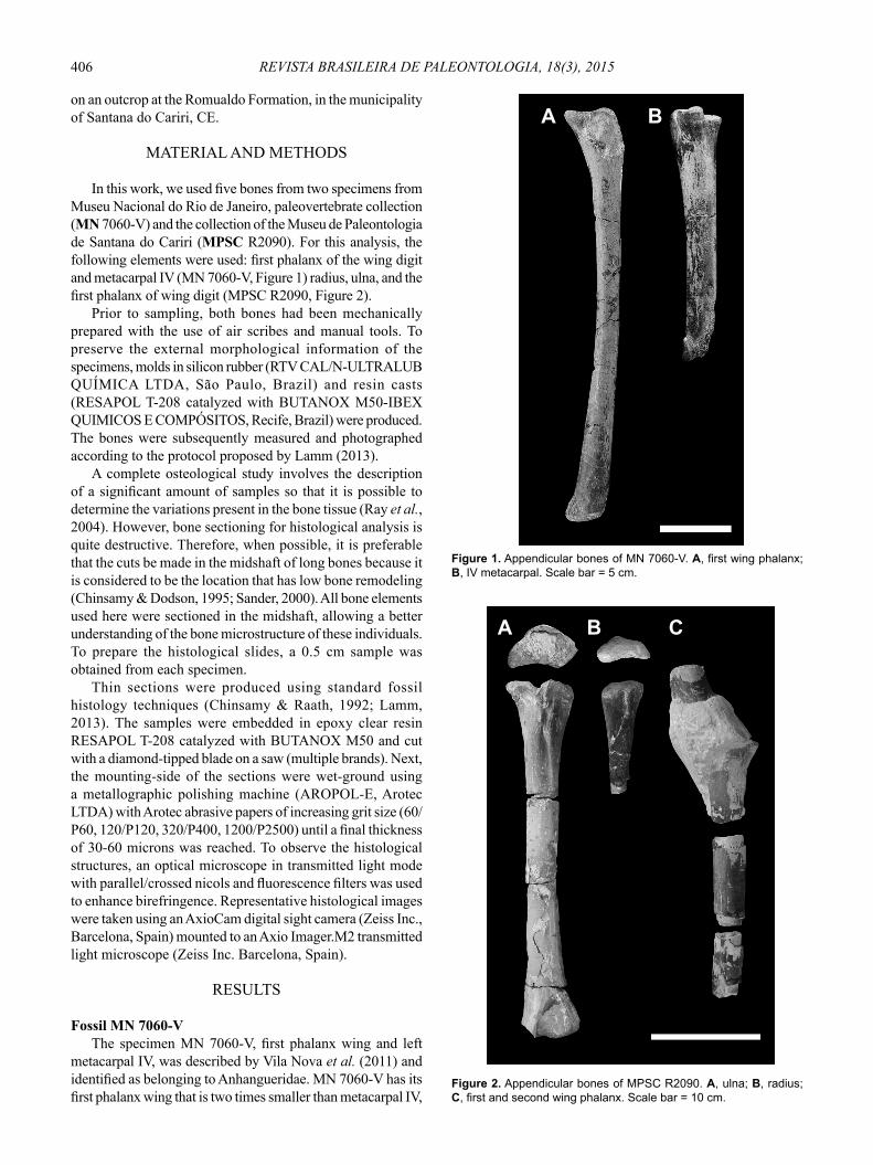
fi brolamellar cortex. Furthermore, the section presented only osteocytes lacunae directed by collagen fi bers and does not form osteons (Figure 5D). This composition demonstrates a pattern that has never been reported for pterosaurs. The observed pattern may be related to biomechanical factors, such as pressure suffered by bone during fl ight. In this case, ulna and phalanges have to be more robust to withstand fl ight pressures, whereas the radius, being supported by the ulna, could present a slower deposition. The fi rst wing phalanx diaphysis is 20 mm in diameter at the transect (Figure 5E). The fi rst wing phalanx presented in the cortex averaged a thickness of 889 μm with a free medullary cavity of 1.869 μm. The cortex consists of plexiform fi brolamellar tissue with few fractures along the bone. The vascular canals are arranged radially, mostly anastomosed. These canals are surrounded by osteocytes contributing to the formation of primary osteons (Figure 5F).
DISCUSSION
A considerable number of previous studies with many types of vertebrate fossils noted that the microstructure of bone throughout the skeleton varies according to the bone and its location in the skeleton (Enlow, 1969; Sayão, 2003; Chinsamy et al., 2009; Werning, 2012; Andrade & Sayão, 2014). Furthermore, the general morphology and biomechanical function of a particular bone element affect the type of bone that will be present (Francillon-Vieillot et al., 1990; Chinsamy-Turan, 2005; Werning, 2012). In pterosaurs, it was previously established that at least in Rhamphorhynchus, Pterodaustro and Anhangueria, the histological pattern can vary over the bones of the same individual (Sayão, 2003; Chinsamy et al., 2009; Prondvai et al., 2012).
Analysis that used many elements of the same skeleton are rare not only in the paleohistological study of pterosaurs (Steel, 2008; Chinsamy et al., 2009) but also in fossil archosaurs generally due to the scarcity of material available for this analysis (Andrade & Sayão, 2014). In both specimens studied here, basically the same microstructures previously described for anhanguerid pterosaurs (Sayão, 2003; Steel, 2008) were observed here, except for the absence of vascularization reported here for the radius MPSC R2090 (Figure 5D). This condition differs from that observed in the radius of Azhdarchoidea from the Crato Formation, which showed a high vascularization (Sayão, 2003).
The presence of vascularization is a key condition for bone growth. A decrease in the amount of vascular canals would result in a decrease in the deposition rate (Padian & Lamm, 2013). It is highly improbable that the radius of MPSC R2090 did not present any vascularization. However, it is possible that the bones present a considerable decrease in the number of canals, similar to that observed in the radius of basal pterosaurs non-pterodactyloid of Rhamphorhynchus. It was observed that the radius of the analyzed specimen had no vascular canals, showing a simple bone tissue, unlike the high vascularization which is found in the fi brolamellar complex (Prondvai et al., 2012). In both Rhamphorhynchus
and anhanguerids, thin radius bone walls are presented and positioned on a robust ulna, suggesting that biomechanical factors may be determining the osteohistological constitution of this bone.
The bone tissue vascularization was defi ned as the ratio between the areas of the circular canals for the total vascular area (de Riqclès et al., 1991). The context provided by de Margerie et al. (2005) on the biomechanics of long bones of birds suggests that different fl ight modes and different bending loads and inferred torsion, would have an impact on the organization of bone microstructure and its degree of vascularization. De Magerie (2002) found that the resistance to bending and torsion are maximal in the radius, which has the smallest vascularization of all examined bones. Thus, a histological optimization is found in bones with large twists and with high vascularization, as an adaptation at the time of fl ight. Thus, considering osteohistological composition
Figure 5. Histological section of MPSC R2090. A, ulna section; B, histology of ulna with a possible growth mark near the medullary cavity which does not form a true line of arrested growth (white arrows). Few scattered anastomosed vascular canals are indicated by black arrows; C, radius section; D, histology of radius showing osteocytes lacunae oriented by collagen fibers without osteons formation; E, first wing phalanx section; F, histology of the first wing phalanx composed of vascular canals mostly radially arranged, indicated by the white arrows and sometimes anastomosed (black arrows). The bone has few fractures and is composed of many anastomosed canals. Scale bars = 200 μm.
A B
C
E
D
F

409ELEUTÉRIO ET AL. – OSTEOHISTOLOGICAL DEPOSITION OF ANHANGUERIA
of the radius of MPSC R2090, it is possible to identify a relationship of biomechanics in determining the avascular pattern of the radius.
The slow growth rate observed in crocodiles, which is already known for other groups of living reptiles, indicates that the default slow bone growth may be a baseline characteristic for amniote (Padian et al., 2004) and possibly for archosaurian. In basal Pseudosuchia as Phytosauria and Aetosauria (Padian et al., 2001; de Ricqlès et al., 2003), a slow growth pattern was observed. This aspect also remains in Thalattosuchia (Hua & de Buffrènil, 1996) and Dyrosauridae (Andrade & Sayão, 2014), which represent more derived crocodylomorphs. It is speculated that the growth rate and physiology of pterosaurs would be more similar to that of living birds than “typical reptiles” (Bennett, 1993; de Ricqlès et al., 2000) and that pterosaurs would reach their ontogenic maturity in up to three or four years (Chinsamy et al., 2008). The histological pattern that characterizes a high metabolic rate and rapid growth strategy is consistent with the variation between the sampled specimens, which have cortices with different densities of osteocytes and vascular canals.
The presence of cyclical growth marks is continuous and partly caused by endogenous physiological processes. These rhythms are synchronized and amplifi ed by seasonal environmental variations such as temperature, light, humidity, food availability, etc. (Castanet et al., 1977; de Buffrénil, 1980). These factors are indicators of the periodicity of circannual growth cycles (Castanet et al., 1977; de Buffrénil, 1980; Hutton, 1986). However, many studies have tried to confi rm the annual cyclicity of LAGs in archosaurs. Although the LAGs are common in vertebrates, these marks are hardly found in pterosaurs that have been recorded, for example, in a Cretaceous Anhaguerid (Sayão, 2003) and Jurassic pterosaur (Padian et al., 2004). The reason these lines appear in the samples remains controversial. This feature is not found in MPSC R2090, which has histological and morphological characteristics of a young animal and a not-true LAG, whereas MN 7060-V presents histology consistent with a subadult animal in the absence of LAGs in the analyzed thin sections. The cortex is thin; the LAGs are probably quickly reabsorbed by not allowing their remains in the cortical region for long periods.
As for the vascularization of the bones, MPSC R2090 presented histovariability, as previously reported by Sayão (2003). The fi rst wing phalanx is highly vascularized in a semireticular network, the radius is avascular and the ulna presents an intermediate vascularization between the phalanx and the radius. The presence of histovariability among bones of the same individual is not common within Archosauria (Sayão, 2003), being observed within this clade in just a few dinosaur groups (Horner et al., 1999; Sander, 2000; Padian et al., 2001). Pterosaurs could present closer histological structures to those of other archosaurs, based on phylogenetic position, or closer histological structures to those of birds, due to fl ight adaptations.
The number and size of the vessels and the number of osteocytes are related to bone growth rate (Padian & Lamm,
2013). According to the “Amprino rule”, the number, size and orientation of the vessels are modified throughout development (Amprino, 1947). Due to the vascular canals that are responsible for much of the blood supply and nutrients to a bone, with greater vascularization, there are higher growth rates (Padian & Lamm, 2013). Comparing the sampled bones, the fi rst phalanx of MPSC R2090 should reach its asymptotic growth faster than the other bones, followed by the ulna and then the radius. The idea of growth at different speeds in the pterosaurs limbs is not new; morphometric studies indicated allometric growth for the appendicular skeleton (Wellnhofer, 1970, 1975; Mateer, 1976; Brower & Veinus, 1981; Vila Nova & Sayão, 2012; Bantim et al., 2014a), whose proportions vary among taxa (Kellner, 2003; Codorniú, 2007; Tomkins et al., 2010; Elgin & Frey, 2012; Vila Nova & Sayão, 2012).
The understanding of the growth and life patterns of pterosaurs has been limited because there are few representatives of different ontogenetic stages of the same taxon (Unwin, 2006). Morphological studies conducted by Bennett (1993a) showed that Nyctosaurus Marsh, 1876, Ornithocheirus Seeley, 1869 and Anhanguera Campos & Kellner, 1985 /Santanadactylus De Buisonjé, 1980 showed no signifi cant differences between the sizes of adults and immature specimens. These studies also indicated that the smaller individual had approximately 70% of the size of the largest representative and that the body size did not constitute a reliable indicator to differentiate the ontogenetic stage. The degree of skeletal fusion would be the best way to distinguish sub-adult and adult individuals. In a study of Rhamphorhynchus, Bennett (1995) found that the skeletons of smaller individuals were not fused and that many of the bones were not completely ossifi ed, whereas middle-age individuals showed some signal of fusion and higher porosity. By analyzing the morphology of MN-7060 V, (small-sized) there is a greater degree of porosity on the epiphysis, suggesting that this is an older individual than MPSC R2090. Regarding the degree of fusion of the bones, it was observed that the phalanx extensor process MN7060-V and carpal was not fused in MPSC R2090, which are characteristics of younger animals. Thus, osteohistological analysis used in this study showed that MN 7060-V was a sub-adult, whereas MPSC R2090 was a younger animal.
According to de Ricqlès et al. (2000), the presence of an external fundamental system (EFS) indicates that the animal would have more or less reached its full size at the time of its death, having completed its asymptotic growth. This microstructure has been identifi ed in Anhangueria, particularly in appendicular bones of an adult individual by Sayão (2003); this structure was fi rst described by Cormack (1987) in mammals. In a histological analysis of the distal phalanx of a Tropeognathus cf. T. mesembrinus from the Romualdo Formation recently made by Kellner et al. (2013), the external fundamental system (EFS) was also observed, indicating the presence of an adult animal. Histological analysis of samples MPSC R2090 and MN 7060-V did not present EFS, indicating that none of these specimens reached the asymptotic size, characterizing non-adults animals.

410 REVISTA BRASILEIRA DE PALEONTOLOGIA, 18(3), 2015
CONCLUSION
Based on the analysis of histological structures, two different growth times may be established: a subadult (MN-7060 V) and a young individual (MPSC R2090). These moments were established based on the number of vascular canals in the sampled bones, indicative of rapid growth. MN 7060-V has a smaller wingspan than MPSC R2090, based on the size of the bones. This study showed that in Anhangueria, bone growth does not follow the ontogenetic stages of the individuals. Young animals may present large proportions, whereas older individuals may have smaller sizes within the same clade.
Regarding external morphology, it is possible to use paleohistology to corroborate that the presence of not-fused bones is consistent with the ontogenetic stages of animal. Juveniles and sub-adults did not show the fusion of carpal bones or the extensor process of the phalanx, which would indicate achieving asymptotic size. Thus, the presence of fused bones can be a reference to the identifi cation of animals, because such bones do not occur in young and subadult animals.
It was also concluded that the appendicular bones of the same individual present histovariability and differences in their microstructure that resist torsional loads and bending during fl ight. Thus, paleohistology can provide parameters to analyze the different growth rates of the bones in the same individual according to their function, and may be an important tool to address issues of biomechanics of the appendicular bones in pterosaurs.
ACKNOWLEDGMENTS
The authors thank P.C. Nuvens (Museu de Paleontologia da Universidade Regional do Cariri) for the access to specimen MPSC R2090. We thank C.A. Chagas (Centro Acadêmico de Vitória - Universidade Federal de Pernambuco) for microscope access and imaging of the thin sections. We also thank the reviewers F. Pinheiro (Universidade Federal do Pampa) and an anonymous reviewer for comments that improved the paper. This study was supported by CNPQ (Proc.nº458164/2014-3 – to JMS) and Fundação Cearense de Apoio ao Desenvolvimento Científi co e Tecnológico – FUNCAP (Grant to AAFS). Additional funding was provided by Conselho Nacional de Desenvolvimento Científi co e Tecnológico – CNPq (fellowships to RAMB, RCLPA and FJL) and Fundação de Amparo a Ciência e Tecnologia de Pernambuco – FACEPE (fellowship to LHSE).
REFERÊNCIAS
Amprino, R. 1947. La structure du tissue osseux envisage comme expression de différences dans la vitesse de l’accroissement. Archives de Biologie, 58:315-330.
Andrade, R.C.L.P. & Sayão, J.M. 2014. Paleohistology and lifestyle inferences of a Dyrosaurid (Archosauria: Crocodylomorpha) from Paraíba Basin (Northeastern Brazil). PLOS ONE, 7:e102189. doi:10.1371/journal.pone.0102189
Bantim, R.A.M.; Saraiva, A.A.F.; Oliveira, G.R. & Sayão, J.M. 2014b. A new toothed pterosaur (Pterodactyloidea: Anhangueridae) from the Early Cretaceous Romualdo Formation, NE Brazil. Zootaxa, 3869:201-223. doi:10.11646/zootaxa.3869.3.1
Bantim, R.A.M.; Saraiva, A.A.F. & Sayão, J.M. 2014a. Skull variation and the shape of the sagittal premaxillary crest in anhanguerid pterosaurs (Pterosauria, Pterodactyloidea) from the Araripe Basin, Northeast Brazil. Historical Biology, 27: 656-664. doi:10.1080/08912963.2014.921818
Bennett, S.C. 1993. The ontogeny of Pteranodon and other pterosaurs. Paleobiology, 19:92-106.
Bennett, S.C. 1995. A statistical study of Rhamphorhynchus from the Solnhofen limestone of Germany: year classes of a single large species. Journal of Paleontology, 69:569-580.
Bennett, S.C. 2001. The osteology and functional morphology of the Late Cretaceous pterosaur Pteranodon. Part I. General description of osteology. Palaeontographica A, 260:1-153.
Beurlen, K. 1964. As espécies dos Cassiopinae, nova subfamília dos Turritellidae, no Cretáceo do Brasil. Arquivos de Geologia, 5:1-44.
Beurlen, K. 1966. Novos equinóides no Cretáceo do Nordeste do Brasil. Anais da Academia Brasileira de Ciências, 38:455-464.
Bowerbank, J.S. 1848. Microscopical observations on the structure of the bones of Pterodactylus giganteus and other fossil animals. Quarterly Journal of the Geological Society, 4:2-10. doi:10.1144/gsl.jgs.1848.004.01-02.07
Brower, J.C. & Venius, J. 1981. Allometry in pterosaurs. The University of Kansas Paleontological Contributions, 105:1-32.
Castanet, J.F.J.; Meunier, A. & de Ricqlès, K. 1977. L’enregistrement de la croissance cyclique par les tissue osseux chez les vertébrés poikilothermes: donnés comparative et essai de synthese. Bulletin biologique de la France et de la Belgique, 3:183-202.
Castro, C.; Menor, E.A. & Campantra, V.A. 1970. Descoberta de resinas fósseis na Chapada do Araripe Município de Porteiras, Ceará. Recife, Universidade Federal de Pernambuco, Instituto de Geociências, p. 1-11 (Notas Preliminares 1).
Cavalcanti, V. & Viana, M. 1990. Faciologia dos sedimentos não-lacustres da Formação Santana (Cretáceo Inferior da Bacia do Araripe, nordeste do Brasil). In: SIMPÓSIO SOBRE A BACIA DO ARARIPE E BACIAS INTERIORES DO NORDESTE, 1, 1990. Atas, Crato, p. 193-202.
Chinsamy, A. 2005. The microstructure of dinosaur bone: deciphering biology with fi ne-scale techniques. Baltimore, Johns Hopkins University Press, 216 p.
Chinsamy, A.; Buffetaut, E.; Canoville, A. & Angst, D. 2014. Insight into the growth dynamics and systematic affi nities of the Late Cretaceous Gargantuavis from bone microstructure. Naturwissenschaften, 101:447-452. doi:10.1007/s00114-014-1170-6
Chinsamy, A.; Codorniú, L. & Chiappe, L.M. 2008. Developmental growth patterns of the fi lter-feeder pterosaur, Pterodaustro guiñazui. Biology Letters, 4:282-285. doi:10.1098/rsbl.2008.0004
Chinsamy, A.; Codorniú, L. & Chiappe, L.M. 2009. Palaeobiological implications of the bone histology of Pterodaustro guiñazui. The Anatomical Record, 292:1462-77. doi:10.1002/ar.20990
Chinsamy, A. & Dodson, P. 1995. Inside a dinosaur bone. American Scientist, 83:174-180.
Chinsamy, A. & Raath, M.A. 1992. Preparation of fossil bone for histological examination. Paleontologia Africana, 29:39-44.
Codorniú, L. 2007. Evidencias de cambios alométricos en las cervicales de Pterodaustro guinazui (Pterosauria, Pterodactyloidea). Ameghiniana, 44:10.

411ELEUTÉRIO ET AL. – OSTEOHISTOLOGICAL DEPOSITION OF ANHANGUERIA
Coimbra, J.C.; Arai, M. & Carreño, A.L. 2002. Biostratigraphy of Lower Cretaceous microfossils from the Araripe Basin, northeastern Brazil. Geobios, 35:687-698. doi:10.1016/s0016-6995(02)00082-7
Cormack, D. 1987. Ham’s histology. New York, Lippincott, 732 p.de Buffrénil, V. 1980. Mise em évidence de l’incidence dês
conditions de milliu sur la croissance de Crocodylus siamensis (Schneider, 1801) et valeurdes marques de croissance squelettiques pour l’evaluation de l’âge individuel. Archives de Zoologie Expérimentale et Général, 121:63-76.
de Margerie, E. 2002. Laminar bone as an adaptation to torsional loads in fl apping fl ight. Journal of Anatomy, 201:521-526. doi:10.1046/j.1469-7580.2002.00118.x
de Margerie, E.; Sanchez, S.; Cubo, J. & Castanet, J. 2005. Torsional resistance as a principal component of the structural design of long bones: comparative multivariate evidence in birds. The Anatomical Record, 282A:49-66. doi: 10.1002/ar.a.20141
de Ricqlès, A.; Meunier, F.J.; Castanet, J. & Francillon-Vieillot, H. 1991. Comparative microstructure of bone. In: B.K. Hall (eds.) Bone matrix and bone specifi c products, CRC Press, p. 1-78.
de Ricqlès, A.; Padian, K.; Horner, J.R. & Francillon-Vieillot, H. 2000. Palaeohistology of the bones of pterosaurs (Reptilia: Archosauria): anatomy, ontogeny, and biomechanical implications. Zoological Journal of the Linnean Society, 129:349-385. doi:10.1111/j.1096-3642.2000.tb00016.x
de Ricqlès, A.; Padian, K.; Horner, J.R.; Lamm, E-T. & Myhrvold, N. 2003. Osteohistology of Confuciusornis sanctus (Theropoda: Aves). Journal of Vertebrate Paleontology, 23:373-386. doi:10.1671/0272-4634(2003)023[0373:oocsta]2.0.co;2
Elgin, R.A. & Frey, E. 2012. A nearly complete ornithocheirid pterosaur from the Aptian (Early Cretaceous) Crato Formation of NE Brazil. Acta Palaeontontologica Polonica, 57:101-110. doi:10.4202/app.2010.0079
Enlow, D.H. 1969. The bone of reptiles. In: C. Gans; A.D. Bellairs & T.S. Parsons (eds.) Biology of the Reptilia, Morphology, A. Academic Press, p. 45-80.
Francillon-Vieillot, H.J.; Arntzen, W. & Geraudie, J. 1990. Age, growth and longevity of sympatric Triturus cristatus, Triturus marmora-tus and their hybrids (Amphibia, Urodela): a skeletochronological comparison. Journal of Herpetology, 24:13-22. doi:10.2307/1564284
Hirayama, R. 1998. Oldest known sea turtle. Nature, 392:705-708. doi: 10.1038/33669
Horner, J.R.; de Ricqlès, A. & Padian, K. 1999. Variation in dinosaur skeletochronology indicators: implications for age assessment and physiology. Paleobiology, 25:295-304.
Horner, J.R.; de Ricqlès, A. & Padian, K. 2000. Long bone histology of the hadrosaurid dinosaur Maiasaura peeblesorum: growth dinamics and physiology based on an ontogenetic series of skeletal elements. Journal of Vertebrate Paleontology, 20:115-129. doi:10.1671/0272-4634(2000)020[0115:lbhoth]2.0.co;2
Hua, S. & de Buffrénil, V. 1996. Bone histology as a clue in the interpretation of functional adaptations in the Thalattosuchia (Reptilia, Crocodylia). Journal of Vertebrate Palaeontology, 16:703-717. doi:10.1080/02724634.1996.10011359
Hutton, J.M. 1986. Age determination of living Nile crocodiles from the cortical stratifi cation of bone. Copeia, 1986:332-341. doi:10.2307/1444994
Kellner, A.W.A. 1999. Short note on a new dinosaur (Theropoda, Coelurosauria) from the Santana Formation (Romualdo Member, Albian), northeastern Brazil. Boletim do Museu Nacional, 49:1-8.
Kellner, A.W.A. 2002. Membro Romualdo da Formação Santana, Chapada do Araripe, CE. Um dos mais importantes depósitos fossilíferos do Cretáceo brasileiro. In: C. Schobbenhaus; D.A. Campos; E.T. Queiroz; M. Winge & M.L.C.B. Born (eds.) Sítios geológicos e paleontológicos do Brasil, DNPM/CPRM/SIGEP, p. 121-130.
Kellner, A.W.A. 2003. Pterosaur phylogeny and comments on the evolutionary history of the group. In: E. Buffetaut & J-M. Mazin (eds.) Evolution and palaeobiology of pterosaurs, Geological Society, p. 105-137 (Special Publication 217). doi:10.1144/GSL.SP.2003.217.01.10
Kellner, A.W.A. 2006. Pterossauros - os senhores do céu do Brasil. Rio de Janeiro, Vieira & Lent, 176 p.
Kellner, A.W.A.; Calvo, J.; Sayão, J.M. & Porfiri, J.D. 2006. Pterosaur bones from the Portezuelo Formation (Cretaceous), Neuquén Group, Patagonia, Argentina. Arquivos do Museu Nacional, 64:369-375.
Kellner, A.W.A. & Campos, D.A. 1996. First Early Cretaceous theropod dinosaur from Brazil. Neues Jahrbuch für Geologie und Paläontologie Abhandlungen, 199:151-166.
Kellner, A.W.A.; Campos, D.A.; Sayão, J.M.; Saraiva, A.A.F.; Rodrigues, T.; Oliveira, G.; Cruz, L.A.; Costa, F.R.; Silva, H.P. & Ferreira, J.S. 2013. The largest fl ying reptile from Gondwana: a new specimen of Tropeognathus cf. T. mesembrinus Wellnhofer, 1987 (Pterodactyloidea, Anhangueridae) and other large pterosaurs from the Romualdo Formation, Lower Cretaceous, Brazil. Anais da Academia Brasileira de Ciências, 85:113-135. doi:10.1590/S0001-37652013000100009
Kellner, A.W.A. & Tomida, Y. 2000. Description of a new species of Anhangueridae (Pterodactyloidea) with comments on the pterosaur fauna from the Santana Formation (Aptian-Albian), northeastern Brazil. London, National Science Museum, p. 1-135 (Monographs 17).
Lamm, E-T. 2013. Bone histology of fossil tetrapods. In: K. Padian & E-T. Lamm (eds.) Preparation and sectioning of specimens, University of California Press, p. 55-160.
Lima, F.J.; Saraiva, A.A.F. & Sayão, J.M. 2012. Revisão da paleofl ora das formações Missão Velha, Crato e Romualdo, Bacia do Araripe, Nordeste do Brasil. Estudos Geológicos, 22:99-115. doi: 10.18190/1980-8208/estudosgeologicos.v22n1p99-115
Lima, F.J.; Saraiva, A.A.F.; Silva, M.A.P.; Bantim, R.A.M. & Sayão, J.M. 2014. A new angiosperm from the Crato Formation (Araripe Basin, Brazil) and comments on the Early Cretaceous monocotyledons. Anais da Academia Brasileira de Ciências, 86:1657-1672. doi:10.1590/0001-3765201420140339
Maisey, J.G. 1991. Santana fossils: an illustrated atlas. Neptune, T.F.C. Publications, 459 p.
Mateer, N.J. 1976. A study of Pteranodon. Bulletin of the Geological Institution of the University of Uppsala, 6:23-33.
Naish, D.; Martill, D.M. & Frey, E. (2004). Ecology, systematics and biogeographical relationships of dinosaurs, including a new theropod, from the Santana Formation (?Albian, Early Cretaceous) of Brazil. Historical Biology, 16:1-14. doi:10.1080/08912960410001674200
Neumann, V.H.M.L. & Cabrera, L. 1999. Una nueva propuesta estratigrafi ca para la tectono secuencia post-rifte de la cuenca de Araripe, nordeste de Brasil. In: SIMPÓSIO SOBRE O CRETÁCEO DO BRASIL, 5, 1999. Boletim de Resumos, Serra Negra, p. 279-285.
Oliveira, G.R. & Romano, P.S.R. 2007. Histórico dos achados de tartarugas fósseis do Brasil. Arquivos do Museu Nacional, 65:113-133.

412 REVISTA BRASILEIRA DE PALEONTOLOGIA, 18(3), 2015
Owen, R. 1870. A monograph of the fossil Reptilia of the Liassic Formations. Part III. Londres, Paleontographical Society, p. 41-81.
Padian, K. 1985. The origins and aerodynamics of fl ight in extinct vertebrates. Palaeontology, 28:423-433.
Padian, K.; de Ricqlès, A. & Horner, J.R. 2001. Dinosaurian growth rates and bird origins. Nature, 412:405-408. doi:10.1038/35086500
Padian, K.; Horner, J.R. & de Ricqlès, A.J. 2004. Growth in small dinosaurs and pterosaurs: the evolution of archosaurian growth strategies. Paleontology, 24:555-571. doi:10.1671/0272-4634(2004)024[0555:gisdap]2.0.co;2
Padian, K. & Lamm, E-T. 2013. Bone histology of fossil tetrapods: advancing methods, analysis, and interpretation. Oakland, University of California Press, 285 p.
Pereda-Suberbiola, X.; Knoll, F.; Ruiz-Omenaca, J.I.; Company, J. & Torcida Fernandez-Baldor, D. 2012. Reassessment of Prejanopterus curvirostris, a basal pterodactyloid pterosaur from the Early Cretaceous of Spain. Acta Geological Sinica, 86:1389-1401. doi:10.1111/1755-6724.12008
Phillips, J. 1871. Geology of Oxford and the Valley of the Thames. Oxford, Clarendon Press, 523 p.
Price, L.I. 1959. Sobre um crocodilídeo notossuquio do Cretáceo brasileiro. Rio de Janeiro, Departamento Nacional da Produção Mineral, Divisão de Geologia e Mineralogia, 56 p. (Boletim 188).
Prondvai, E.; Bodor, E.R. & Ősi, A. 2014. Does morphology refl ect osteohistology-based ontogeny? A case study of Late Cretaceous pterosaur jaw symphyses from Hungary reveals hidden taxonomic diversity. Paleobiology, 40:288-321. doi:10.1666/13030
Prondvai, E. & Stein, K. 2014. Medullary bone-like tissue in the mandibular symphyses of a pterosaur suggests non-reproductive signifi cance. Scientifi c Reports, 4:6253. doi:10.1038/srep06253
Prondvai, E.; Stein, K.; Ösi, A. & Sander, M.P. 2012. Life history of Rhamphorhynchus inferred from bone histology and the diversity of Pterosaurian growth strategies. PLOS ONE, 7:e31392. doi:10.1371/journal.pone.0031392
Quekett, J.T. 1849a. On the intimate structure of bone, as composing the skeleton in the four great classes of animals, viz., mammals, birds, reptiles and fi shes, with some remarks on the great value of the knowledge of such structure in determining the affi nities of minute fragments of organic remains. Transactions of the Microscopical Society of London, 2:46-58.
Quekett, J.T. 1849b. Additional observations on the intimate structure of bone. Transactions of the Microscopical Society of London, 2:59-64.
Quekett, J.T. 1855. Descriptive and illustrated catalogue of the histological series contained in the Museum of the Royal College of Surgeons of England. London, R. and J.E. Taylor, p. 122-129.
Ray, S.; Botha, J. & Chinsamy, A. 2004. Bone histology and growth patterns of some non-mammalian therapsids. Journal of Vertebrate Palaeontology, 24:634-648. doi:10.1671/0272-4634(2004)024[0634:bhagpo]2.0.co;2
Rodrigues, T. & Kellner, A.W.A. 2013. Taxonomic review of the Ornitocheirus complex (Pterosauria) from the Cretaceous of England. Zookeys, 308:1-112. doi:10.3897/zookeys.308.5559
Sander, M.P. 2000. Longbone histology of the Tendaguru sauropods: implications for growth and biology. Paleobiology, 26:466-488. doi: 10.1666/0094-8373(2000)026<0466:lhotts>2.0.CO;2
Sayão, J.M. 2003. Histovariability in bones of two pterodactyloid pterosaurs from the Santana Formation, Araripe Basin, Brazil: preliminary results. In: E. Buffetaut & J.M. Mazin (eds) Evolution and palaeobiology of pterosaurs, Geological Society, p. 335-342 (Special Publication 217). doi:10.1144/GSL.SP.2003.217.01.21
Seeley, H.G. 1870. On Ornithopsis, a gigant animal of the pterodactyl kind from the Wealden. Annals and Magazine of Natural History, 5:279-283.
Seitz, A.L. 1907. Vergleichende studien über den mikroskopischen knochenbau fossiler und rezenter reptilien und dessen bedeutung fur das wachstum und umbildung des knochengewebes im allgemeinen. Nova Acta Abhandlungen der Kaiserlichen Leopoldinisch-Carolinischen Deutschen Akademie der Naturforscher, 87:229-370.
Steel, L. 2003. The John Quekett sections and the earliest pterosaur histological studies. In: E. Buffetaut & J.-M. Mazin (eds.) Evolution and palaeobiology of pterosaurs, Geological Society, p. 325-334 (Special Publication 217). doi:10.1144/GSL.SP.2003.217.01.20
Steel, L. 2008. The palaeohistology of pterosaur bone: an overview. Zitteliana, B28:109-125.
Tomkins, J.L.; Lebas, N.R.; Witton, M.P.; Martill, D.M. & Humphries, S. 2010. Positive allometry and the prehistory of sexual selection. The American Naturalist, 176:141-148. doi:10.1086/653001
Unwin, D.M. 2006. The Pterosaurs from deep time. London, Pi Press, 352 p.
Vila Nova, B.C.; Saraiva, A.A.F.; Moreira, J.K.R. & Sayão, J.M. 2011. Controlled excavations in the Romualdo Formation Lagerstätte (Araripe Basin, Brazil) and pterosaur diversity: remarks based on new findings. Palaios, 26:173-179. doi:10.2110/palo.2010.p10-072r
Vila Nova, B.C. & Sayão, J.M. 2012. On wing disparity and morphological variation of the Santana Group pterosaurs. Historical Biology, 24:567-574. doi:10.1080/08912963.2012.658386
Wang, X.; Kellner, A.W.A.; Zhou, Z. & Campos, D.A. 2008. Discovery of a rare arboreal forest-dwelling flying reptile (Pterosauria, Pterodactyloidea) from China. Proceedings of the National Academy of Sciences of the United States of America, 105:1983-1987. doi:10.1073/pnas.0707728105
Wellnhofer, P. 1970. Die Pterodactyloidea (Pterosauria) der Oberjura-Plattenkalke Süddeutschlands. Bayerische Akademie der Wissenschaften Mathematisch-Naturwissenschaftliche Klasse Abhandlungen, 141:1-133.
Wellnhofer, P. 1975. Die Rhamphorhynchoidea (Pterosauria) der Oberjura-plattenkalke Stiddeutschlands. Palaeontographica A, 148:1-33.
Wellnhofer, P. 1991. The illustrated encyclopedia of pterosaurs. Londres, Salamander Books, 192 p.
Werning, S. 2012. The ontogenetic osteohistology of Tenontosaurus tilletti. PLOS ONE, 7:1-25. doi:10.1371/journal.pone.0033539
Witton, M.P. & Habib M.B. 2010. On the size and fl ight diversity of giant pterosaurs, the use of birds as pterosaur analogues and comments on pterosaur fl ightlessness. PLOS ONE, 5:e33539. doi:10.1371/journal.pone.0013982
Received in January, 2015; accepted in August, 2015.


![The CaFe Experiment: Short-Range Pairing Mechanisms in ...than modified, Urca cooling of neutron stars, even below threshold [25]. Since direct Urca cooling is about 10 6 times faster](https://static.fdocuments.in/doc/165x107/60aa113ee6285219e1240d74/the-cafe-experiment-short-range-pairing-mechanisms-in-than-modiied-urca.jpg)