View Article Online Biomaterials Science€¦ · 1. + −, 2-. Biomaterials Science

© 2016 WILEY-VCH Verlag GmbH & Co. KGaA, Weinheim wileyonlinelibrary.com 1
REV
IEW
Biomaterials in siRNA Delivery: A Comprehensive Review
William Ho, Xue-Qing Zhang, and Xiaoyang Xu*
W. Ho, Dr. X.-Q. Zhang, Prof. X. XuDepartment of Chemical, Biological and Pharmaceutical EngineeringNewark School of EngineeringNew Jersey Institute of TechnologyNewark, NJ 07102, USAE-mail: [email protected]
DOI: 10.1002/adhm.201600418
long. In the cytosol, siRNA is incorporated into the RNA induced silencing com-plex (RISC), which selectively degrades mRNA complementary to the bound anti-sense strand.[2,9,10] To evade the interferon response from introducing lengthy dsRNA strands and to help simplify this process, siRNA which is synthesized in the labora-tory can be directly introduced into cells.
Several advanced technologies such as shRNA, siRNA and miRNA have been developed and shown to induce RNAi response.[11] This review will focus on siRNA; the challenges it faces in its clinical application, the vast improve-ments made in siRNA delivery and future directions in its application. Synthetic siRNAs are chemically synthesized short dsRNAs, approximately 20 nucleotides in length with short (≈2 nucleotide) 3′ over-hangs. Their mode of action is identical to the natural siRNA products formed by
the enzymatic cleavage of longer dsRNA strands by Dicer.[1] The ability of siRNA to silence genes without integration into host DNA does not modify the genome itself, which removes a sig-nificant barrier in its FDA-approval process.[8]
The fantastic potential of siRNA to silence important genes in disease pathways comes with noteworthy challenges and bar-riers in its delivery. The therapeutic use of siRNA is hindered by the lack of effective in vivo delivery methods to ensure proper integration into the RNAi machinery. Ideally, a therapeutic injected intravenously will travel through the bloodstream, cross the endothelium of the vessel tissue, reach the intended target cells and act in a dose-dependent manner. Additionally, proper siRNA function usually requires endocytic uptake and subsequent escape from the endosome itself in order to func-tion in the RISC machinery of the cytosol.[1,12] However, there are barriers which offset siRNA delivery from this ideal picture. When administered intravenously, naked siRNA is susceptible to degradation by serum endonucleases and causes an innate immune response.[1,13–15] Furthermore, the negative charge of siRNA hinders the effectiveness of uptake by the similarly charged cellular membrane. Other unfavorable features such as large size and unstable tertiary structure in physiological conditions all prevent efficient internalization into the cell and subsequent RISC formation for gene silencing does not reliably occur.[16] This great potential of interfering RNAs is therefore stymied by factors intrinsic to the therapeutic itself, and calls out for a delivery vector which is both safe and effective.[1,8] The ideal siRNA delivery system must contain the following attrib-utes: (1) maintain structural integrity in the in vivo environ-ment at human physiological pH and temperature and protect
With the dearth of effective treatment options for prominent diseases including Ebola and cancer, RNA interference (RNAi), a sequence-specific mechanism for genetic regulation that can silence nearly any gene, holds the promise of unlimited potential in treating illness ever since its discovery in 1999. Given the large size, unstable tertiary structure in physiological condi-tions and negative charge of small interfering RNAs (siRNAs), the develop-ment of safe and effective delivery vehicles is of critical importance in order to drive the widespread use of RNAi therapeutics into clinical settings. Immense amounts of time and billions of dollars have been devoted into the design of novel and diverse delivery strategies, and there are a handful of delivery sys-tems that have been successfully translated into clinic. This review provides an introduction to the in vivo barriers that need to be addressed by siRNA delivery systems. We also discuss the progress up to the most effective and clinically advanced siRNA delivery systems including liposomal, polymeric and siRNA conjugate delivery systems, as well as their design to overcome the challenges.
1. Introduction
RNA interference (RNAi) is a tremendous innovation in the universal therapeutic treatment of disease. In this remarkable process which is ubiquitous to eukaryotic cells, a short strand of RNA is able to induce the degradation of a complementary sequence of mRNA. This gives RNAi great potential in its ability to modulate and silence virtually any gene in the tran-scriptome.[1–4] To this end, much research has been focused on treating diseases from viral infections to cancer in the hopes of delivering specific gene-based cures with minimal side effects.[2,5–7]
The prominence and subsequent significant interest in RNAi as a potential therapeutic dates to 1998 when Andrew Fire and Craig Mello identified in Caenorhabditis elegans the biological process in which double-stranded RNA (dsRNA) is used to silence and/or regulate gene expression.[2,8] The RNAi pathway is initiated by the presence of dsRNA in the cytosol. From there, the Dicer enzyme cleaves the dsRNA into fragments known as short interfering RNA (siRNA), which are 21–23 nucleotides
Adv. Healthcare Mater. 2016, DOI: 10.1002/adhm.201600418
www.advhealthmat.dewww.MaterialsViews.com

2 © 2016 WILEY-VCH Verlag GmbH & Co. KGaA, Weinheimwileyonlinelibrary.com
REV
IEW the siRNA from degradation, (2) cross endothelia to reach
target tissues (3) be capable of controlled release of siRNA ther-apeutic over an extended period of time, (4) possess the ability for efficient internalization into the cell via endocytosis and (5) once within the endosome, there must be effective endosomal escape and exposure of the siRNA to the cytosol for RISC appa-ratus formation and subsequent action. As a result, the asso-ciation of siRNAs with delivery systems presents a desirable protective approach to facilitate the crossing of natural bar-riers. Numerous efforts have been made over the past decade to develop safe and effective vector carriers for siRNA delivery.[17] Viral vectors are among the first vehicles that have been investi-gated for siRNA delivery. However, there are several limitations associated with these vectors, including carcinogenesis and immunogenicity.[18,19] Recent advances in biomaterial sciences have led to the development of new synthetic lipids and poly-mers which have the potential to address many of the limita-tions associated with viral vectors and offer alternative methods for siRNA delivery. These synthetic lipids and polymers, when combined with the latest developments in nanotechnology may provide powerful tools in designing clinically suitable, safe and effective delivery systems for siRNA. Furthermore, the rapid progress in nucleic acid chemistry which seeks to chemically modify the siRNA molecule itself offers exciting opportunities to confer advantages both in stability and delivery.[1]
This review will succinctly summarize the recent progress in biomaterials and nanotechnology currently being used for siRNA therapeutic delivery with an emphasis on clinical and preclinical trials. The promising characteristics of carrier plat-forms needed to bypass the obstacles of the systemic siRNA delivery will also be highlighted. In addition, perspectives and opportunities in delivering a combination of siRNA and other therapeutics will be discussed. The biomaterials mentioned are designed to assess the challenges in siRNA delivery and are aimed to deliver a combination of siRNA therapeutics in an effective and controlled manner to specific tissues in the body. Further challenges and unresolved issues still remain in this burgeoning field, but there have been major strides in the refinement of a more clinically effective siRNA therapeutic delivery system in recent years. If successful, the introduc-tion of safe, specific siRNA delivery systems will have a pow-erful impact on how clinicians approach therapeutic treatment for many years to come. We hope to give an inclusive insight toward this next generation of biomaterials and nanotech-nology, and facilitate the promotion of effective gene therapeu-tics into clinical use.
2. Non-Viral Delivery Systems for siRNA
Nucleic acid delivery, a field which includes siRNA delivery, has undergone numerous exploratory ventures in the aims of discovering the most potent vector for successful cell inter-nalization, endosomal escape, and in the case of RNAi delivery, activating the RISC complex for gene silencing. In recent years there has been a gradual shift in focus from viral-based vector delivery to synthetic vectors, such as lipids and polymers, due to perceived complications in the former and advantages in the latter regarding delivery into patients.[2]
2.1. Lipid-Based Delivery Systems
Unilamellar and multilamellar liposomes are formed through self-assembly of a lipid bilayer by amphipathic materials. Liposomes are commonly used as delivery vehicles for a broad spectrum of therapeutics including siRNA. The notable fea-tures of a lipid bilayer include two sets of hydrophilic polar head groups each separately pointed towards the outer and inner surfaces of the particle, with the component lipids available to be functionalized and modified. Lipids may self-assemble into spherical or amorphous structures, with lipids and nucleic acids interspersed throughout the bilayer. Cationic lipids have been used for the majority of liposomal gene delivery methods, as the encapsulation of negatively charged siRNA is improved with their use, as well neutral lipids are more frequently being used in conjunction to facilitate stability and transfection effi-ciency.[1] pKa values of ionizable cationic lipids can be modu-lated for efficient siRNA encapsulation and in vivo activity. It has been shown that lipids with pKa values under 7 are able to interact with negatively charged nucleic acids in environments where pH is below the pKa of ionizable lipids, due to the proto-nation of their amino groups.[20] When the environmental pH is above lipid pKa (e.g. in a physiological environment), liposomal
William Ho is a Master’s candidate for Biomedical Engineering at the University of Ottawa, Canada. He attained a B. Sc. in Biology with a minor in Economics at the University of Waterloo and has research experience at the Harvard Medical School under the supervision of Dr. Xiaoyang Xu. His current research interests include nanoparticles
for drug delivery, biomaterials science as well as surface nanotopography for improving implant design.
Xiaoyang Xu is an assistant professor of the Department of Chemical, Biological and Pharmaceutical Engineering at New Jersey Institute of Technology. He received his Ph.D. in Chemistry from Northwestern University (with Chad Mirkin) and completed his joint NIH postdoctoral training at MIT (with Robert Langer) and Harvard Medical
School (with Omid Farokhzad). His research focus is the development of novel biomaterials and nanotechnologies for medical applications including diagnosis, bioimaging, drug delivery, and regenerative medicine.
Adv. Healthcare Mater. 2016, DOI: 10.1002/adhm.201600418
www.MaterialsViews.comwww.advhealthmat.de

3© 2016 WILEY-VCH Verlag GmbH & Co. KGaA, Weinheim wileyonlinelibrary.com
REV
IEW
surface charge is neutral, which facilitates circulation around the body. Following intracellular internalization, the amino group of the ionizable lipid is positively charged in the acidic endosomal environment. This property aids in siRNA escape from the acidic endosome, as the protonated amino group associates with anionic endosomal lipids, leading to release of siRNA to the cytosol through the destabilization of endosomal integrity.[20,21]
Liposomes have been utilized as efficient delivery vectors for siRNA for almost 30 years since the successful use of lipofec-tion in 1987 to transfer nucleic acids into animal and human cells by Felgner et al.[22,23] This initial foray into the delivery of nucleic acids, which made use of a synthetic cationic lipid N-[1-(2,3-dioleyloxy)propylJ-N,N,N-trimethylammonium chlo-ride (DOTMA), was a major achievement in the history of lipo-somal vector development, as it proved possible the efficient transfection of DNA and RNA into human cells.[22] These early liposomes were unilamellar, with the incorporation of DOTMA functioning as a means to form lipid-nucleic acid complexes which facilitate cellular internalization.[22] More recently, a class of lipid nanoparticles (LNPs) termed the stable nucleic-acid-lipid particle (SNALP) has been developed, emerging as one of the leading lipid-based siRNA formulations under clin-ical development (Figure 1). In the SNALP delivery platform, cationic and helper lipid molecules form lipid bilayer surfaces through self-assembly, which facilitates encapsulation and cel-lular internalization of the nucleic acid payload. SNALPs also contain a polyethylene glycol (PEG) surface modification for evading the mononuclear phagocyte system and enhancing sta-bility.[2,24] In 2005, Morrissey and co-authors published results for the first SNALP formulation encapsulating siRNA which targeted the hepatitis B virus (HBV). This SNALP formulation demonstrated effective delivery of siRNA in a mouse model of replicating HBV, resulting in persistent and dose-dependent
HBV inhibition.[24] The next year Zimmerman et al. reported the first study to demonstrate the ability of SNALPs to systemi-cally deliver apolipoprotein B (ApoB) specific siRNAs in non-human primates. ApoB is an essential protein for the assembly and secretion of very-low-density lipoprotein (VLDL) and low-density lipoprotein (LDL), thus inhibition of ApoB by SNALP-formulated siApoB reduced LDL cholesterol levels in hyper-cholesterolemia patients.[25] The composition of this SNALP formulation incorporates the PEG lipid PEG-C-DMA, the cationic lipid 1,2-dilinoleyloxy-N,N-dimethyl-3-aminopropane (DLinDMA), the helper lipid 1,2-distear-oyl-sn-glycero-3-phos-phocholine (DSPC), and cholesterol in a 2:40:10:48 molar percent ratio (Figure 1). A single injection of this siRNA-con-taining SNALP caused dose-dependent ApoB mRNA silencing in the liver of cynomolgus monkeys, 48 hours after administra-tion with maximal silencing of over 90%. This potent silencing was due to ApoB cleavage at precisely the site predicted for the RNAi mechanism and lasted for 11 days at the highest siRNA dose.
There are a number of SNALP formulations currently under clinical investigation (Table 1). Tekmira Pharmaceuticals Cor-poration has developed a SNALP formulation to systemically deliver ApoB-specific siRNA (PRO-040201), which has com-pleted Phase I human clinical trials assessing safety, tolerability, pharmacokinetics and pharmacodynamics in hypercholester-olemia patients.[26] The results showed that TKM-ApoB was well tolerated with no evidence of liver toxicity. Significant reduction of ApoB and LDL cholesterol was observed, however the Phase I trial was terminated as one patient at the highest dosage level reported flu-like symptoms, which is consistent with immu-nostimulation caused by siRNA. Tekmira is currently devel-oping the next generation of SNALP-siApoB which is more potent and aims to avoid immunostimulatory side effects. Alnylam Pharmaceuticals is also evaluating SNALP-formulated
Adv. Healthcare Mater. 2016, DOI: 10.1002/adhm.201600418
www.MaterialsViews.comwww.advhealthmat.de
Figure 1. A graphical illustration of a stable nucleic acid–lipid particle (SNALP). Cationic lipids such as DLinDMA (yellow), cholesterol (purple) and helper lipid “DSPC” (red) molecules form lipid bilayer surfaces through self-assembly, facilitating encapsulation and cellular internalization of the nucleic acid payload. The helper lipid usually plays an important role for endosomal escape of the payload. SNALPs also contain a neutral hydrophilic PEG exterior (blue) for evading the mononuclear phagocyte system and enhancing particle stability.
4 © 2016 WILEY-VCH Verlag GmbH & Co. KGaA, Weinheimwileyonlinelibrary.com
REV
IEW
siRNA therapeutics that target proprotein convertase subtilisin/kexin type 9 (PCSK9) for the treatment of hypercholester-olemia. PCSK9 is a key gene involved in the regulation of low density lipoprotein receptor (LDLR) protein level and function, and the suppression of this gene results in higher LDLR levels in the liver and subsequently lower LDL cholesterol levels in the bloodstream.[27] ALN-PCS02 is the latest SNALP-formu-lated siPCSK9 by Alnylam which showed persistent and dose-dependent silencing of plasma PCSK9 of up to 84% and LDL reductions of up to 57% compared to baseline and placebo, which lasted weeks after a single intravenous administration. This formulation was well tolerated with no serious adverse effects reported.[1,28]
Additional clinical studies utilizing SNALP formulations have been conducted to combat other diseases. Geisbert et al. treated guinea pigs and nonhuman primates with SNALPs containing siRNA targeting the RNA polymerase L protein of the lethal Zaire Ebola virus (ZEBOV). This promising method showed protection of guinea pigs and nonhuman primates against viraemia and death from ZEBOV challenge.[29] With the outbreak of Ebola in West Africa ongoing, there may be renewed interest in the treatment of this lethal disease using siRNA loaded SNALPs. Tekmira thereafter commenced Phase I clinical trials evaluating their SNALP-formulated siRNA (TKM-100201) in healthy volunteers. Another clinical trial testing a new version of TKM-Ebola, termed TKM-Ebola-Guinea started with Ebola patients in mid-March 2015, utilizing siRNA tar-geting a different strain of Ebola. Additionally, TKM-PLK1 (TKM-080301) is being developed as a treatment for patients with advanced solid tumors that are refractory to standard therapy or for whom there is no standard therapy. TKM-PLK1 is a SNALP lipid nanoparticle encapsulating siRNA that tar-gets the critical tumor proliferation protein polo-like kinase 1 (PLK1), whose suppression has been shown to induce apop-tosis of tumor cells and inhibit their growth in vitro.[30] The effective delivery of siRNA by SNALPS to highly vascular-ized tumors makes them an ideal candidate for anticancer therapy and shows great promise for success.[31] The clinical Phase I/II patient enrollment was completed in January 2015
and trials are currently ongoing. Another SNALP formulated siRNA therapeutic (ALN-VSP02, Alnylam Pharmaceuticals) is being evaluated for the treatment of hepatocellular carcinoma presently in clinical trials. This formulation incorporates two separate siRNAs which simultaneously target vascular endothe-lial growth factor (VEGF) and kinesin spindle protein (KSP) mRNAs, two genes crucial for the proliferation and survival of cancer cells. The reports, including the results from an exten-sion study of chronic bi-weekly dosing up to 1.0 mg kg–1, show that ALN-VSP02 is well tolerated and generally safe in most of the treated patients. Multiple patients achieved stable disease or better, and tumor biopsies collected from patients displayed an anti-VEGF effect post-treatment.[32] Alnylam has also developed ALN-TTR01 and ALN-TTR02, which are first- and second-generation SNALP formulations to deliver siRNA for the treatment of transthyretin-mediated amyloidosis. These for-mulations are composed of a mixture of neutral lipids, a PEG lipid, and an ionizable lipid. Both have similar physicochem-ical properties but ALN-TTR02 features a DLinDMA analogue which has demonstrated a tenfold higher efficacy in preclinical studies.[33,34] ALN-TTR02 has moved on to Phase III clinical trials, after Phase II trials presented promising TTR knock-down with varying single doses.[33,35]
Table 1 summarizes other notable LNP formulations other than SNALPs which are undergoing clinical trials. AtuPLEX is a siRNA-lipoplex formulation developed by Silence Therapeutics which is based on the complexation of cationic and fusogenic lipids with negatively charged siRNA. This LNP formulation of a novel cationic lipid (AtuFECT01), a helper lipid (DPhyPE) and a PEG–lipid (PEG–DSPE) was shown to internalize into mouse vascular endothelium and modulate expression levels of endothelia-specifically expressed genes after intravenous admin-istration.[36,37] AtuFECT01 has a higher nucleic acid binding capacity compared to standard lipids such as DOTAP, lowering the amount of lipid required for siRNA complexation and sub-sequently reducing lipid-associated toxicity. DPhyPE facilitates the endosomal release of the siRNAs after endocytotic uptake of the lipoplex, while the PEG layer improves particle stability and blood circulation. Atu027 is a AtuPLEX-based formulation
Adv. Healthcare Mater. 2016, DOI: 10.1002/adhm.201600418
www.MaterialsViews.comwww.advhealthmat.de
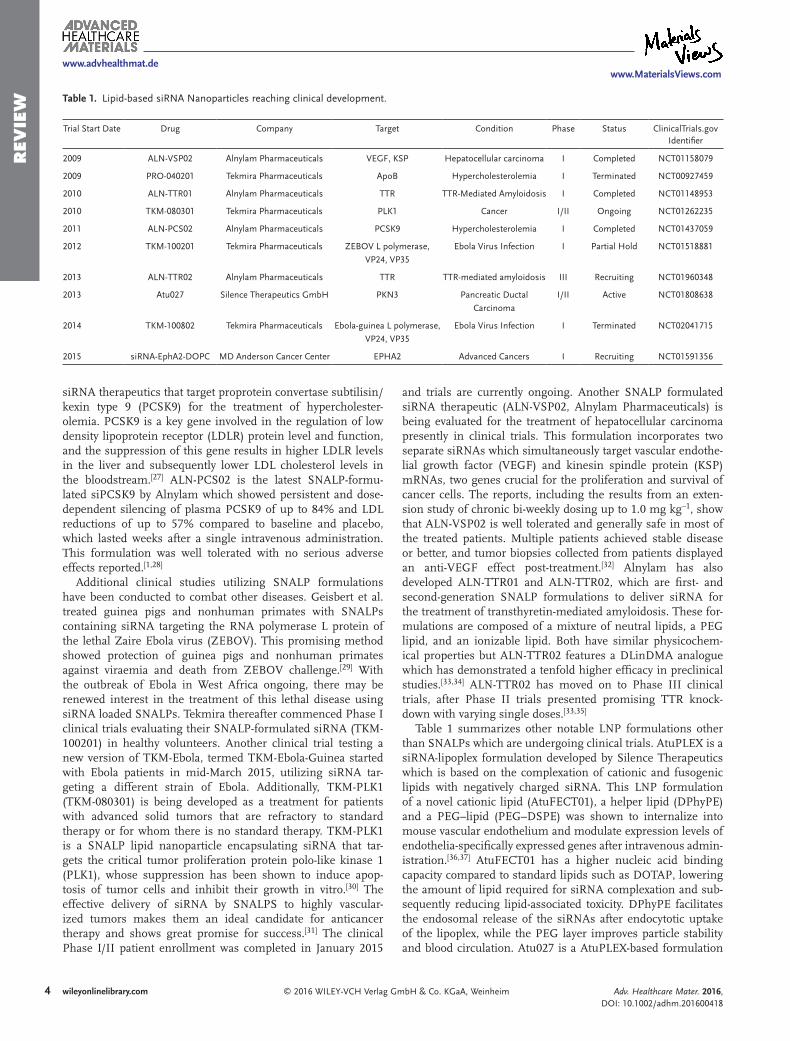
Table 1. Lipid-based siRNA Nanoparticles reaching clinical development.
Trial Start Date Drug Company Target Condition Phase Status ClinicalTrials.gov Identifier
2009 ALN-VSP02 Alnylam Pharmaceuticals VEGF, KSP Hepatocellular carcinoma I Completed NCT01158079
2009 PRO-040201 Tekmira Pharmaceuticals ApoB Hypercholesterolemia I Terminated NCT00927459
2010 ALN-TTR01 Alnylam Pharmaceuticals TTR TTR-Mediated Amyloidosis I Completed NCT01148953
2010 TKM-080301 Tekmira Pharmaceuticals PLK1 Cancer I/II Ongoing NCT01262235
2011 ALN-PCS02 Alnylam Pharmaceuticals PCSK9 Hypercholesterolemia I Completed NCT01437059
2012 TKM-100201 Tekmira Pharmaceuticals ZEBOV L polymerase,
VP24, VP35
Ebola Virus Infection I Partial Hold NCT01518881
2013 ALN-TTR02 Alnylam Pharmaceuticals TTR TTR-mediated amyloidosis III Recruiting NCT01960348
2013 Atu027 Silence Therapeutics GmbH PKN3 Pancreatic Ductal
Carcinoma
I/II Active NCT01808638
2014 TKM-100802 Tekmira Pharmaceuticals Ebola-guinea L polymerase,
VP24, VP35
Ebola Virus Infection I Terminated NCT02041715
2015 siRNA-EphA2-DOPC MD Anderson Cancer Center EPHA2 Advanced Cancers I Recruiting NCT01591356

5© 2016 WILEY-VCH Verlag GmbH & Co. KGaA, Weinheim wileyonlinelibrary.com
REV
IEW
with siRNA targeting protein kinase N3 (PKN3) transcript that has reached clinical trials for the treatment of patients with advanced solid cancer.[38] Atu027 was well tolerated in doses up to 0.336 mg kg–1 and stable disease was achieved in 41% of patients with advanced solid tumors after treatment (n = 34). These encouraging results also led to further clinical trials with sFLT1 (soluble variant of vascular endothelial growth factor receptor-1) being investigated as a potential biomarker, as this biomarker decreased from pretreatment levels in most patients after multiple doses. Another formulation from the M.D. Anderson Cancer Center incorporates siRNA that targets the oncoprotein Ephrin type-A receptor 2 precursor (EphA2) into the neutral liposome 1,2-dioleoyl-sn-glycero-3-phosphatidylcho-line (DOPC). The combination of siRNA-EphA2-DOPC with paclitaxel significantly reduced tumor growth in a mouse model of ovarian cancer compared with treatment with paclitaxel and nonsilencing RNA.[39] Human clinical trials started July 2015.
Although liposomes have shown great promise for delivering siRNA, there still remain concerns regarding lipid-associated toxicity and off-target siRNA silencing.[2] Cationic lipids may cause toxic effects in vitro and in vivo, including cell shrinking, reduced number of mitoses and vacuolization of the cyto-plasm.[40] Cationic lipids have also been reported to induce interferon response in mice.[41] Additionally, gene expression profiling studies have revealed that liposomal siRNA delivery systems can cause wide ranging gene changes in target cells which could potentially affect gene silencing activity and speci-ficity.[42] It appears that liposomes activate the immunostimu-latory response in a structure-dependent manner and the lipid-associated toxicity is also structure-specific, as both the head group and the hydrocarbon chain structure were found to have dramatic effects on toxicity levels.[40] Encouragingly, the concerns regarding siRNA-mediated immune stimulation and off-target gene silencing are being addressed with some success through the incorporation of modified nucleotide chemistries into siRNA sequences.[24,43–46] A few studies have also been published to decrease the liposomal carrier-induced inflammatory response. As an example, the co-administration of dexamethasone with siRNA-encapsulated liposomal formula-tion LNP201, comprised of a cationic lipid CLinDMA, choles-terol and the helper lipid PEG-dimyristoylglycerol at a molar ratio of 50:44:6, was able to inhibit carrier-induced inflamma-tory gene induction, cytokine release and mitogen-activated protein kinase (MAPK) phosphorylation in multiple tissues of mice.[43] PEG modification of liposomes confers a level of pro-tection from mononuclear phagocytic system (MPS) clearance, which improves circulation and passive cellular internalization of formulated siRNA, as well as lowering immunogenicity.[47] Notably, PEGylated liposomes encapsulating doxorubicin and amphotericin B have been approved by the FDA, which lends credence to the future clinical use of liposome-based therapies.[2] Other modifications to the liposomal surface aim to increase the specificity of lipid-based vectors by incorpo-rating multivalent targeting moieties, which upon successful action will optimize pharmacokinetics and facilitate homing of formulated siRNA to diseased sites and eliminate off-target adverse effects.[35,47] For example, an efficient tumor-specific siRNA delivery vehicle has been developed by incorporating an anti-transferrin receptor single chain antibody fragment to
a nanoscale liposome-based complex.[48–50] This antibody frag-ment was used as a targeting ligand for binding to transferrin receptors, which are typically upregulated on cancer cells and trigger cellular uptake via clathrin-coated pits. A pH-sensitive histidylated oligolysine peptide was incorporated in the com-plex to facilitate endosomal escape of siRNA.[51] This tumor-targeting liposome complex was able to efficiently and spe-cifically deliver siRNA to both primary and metastatic tumors after systemic administration, silence the target gene HER2, as well as significantly inhibit tumor growth in a mouse model of pancreatic cancer.[48,49] Other ligands including antibodies, engineered antibody fragments, proteins, peptides, small mole-cules, and aptamers can be utilized to decorate lipid-based car-riers to enhance targeting through ligand-receptor interactions, thus increasing the possibility of translating the potent effects of siRNA delivery systems into clinically useful therapeutics.
Recently, high-throughput combinatorial approaches have been reported in the development of novel lipid-like materials (termed lipidoids) to identify the most potent and safe lipid compounds for siRNA delivery applications.[52,53] Akinc et al. created a structurally diverse library of amino-alkyl-acrylate and -acrylamide lipidoids through Michael addition chem-istry.[52] These lipidoid selection processes solve problems in the conventional case-specific, time-consuming multi-step synthesis of lipids which limited the speed of development in previous generations of liposomes. The formulations of the leading candidate compound 98N12-5 screened from this study (Figure 2a), which consists of five 12-carbon alkyl-acrylamide chains attached to an amine core, were able to mediate potent
Adv. Healthcare Mater. 2016, DOI: 10.1002/adhm.201600418
www.MaterialsViews.comwww.advhealthmat.de
Figure 2. Chemical structures of a) Lipidoid 98N12-5, b) C12-200, and c) CKK-E12.

6 © 2016 WILEY-VCH Verlag GmbH & Co. KGaA, Weinheimwileyonlinelibrary.com
REV
IEW and persistent silencing in a variety of cellular targets and
animal species including mice, rats, and cynomolgus monkeys. In a cross-species study reported by Frank-Kamenetsky et al., 98N12-5 was utilized to deliver siRNA against PCSK9, resulting in a decrease of PCSK9 mRNA levels by 50–70% in mice and rats as well as a reduction of over 70% in human PCSK9 tran-script levels in transgenic mice.[27] In nonhuman primates, hepatic PCSK9 silencing persisted for up to 3 weeks after a single intravenous administration without measurable effects on either HDL cholesterol (HDLc) or triglycerides (TGs). This promising lipidoid-formulated anti-PCSK9 may represent a potential approach for the treatment of hypercholesterolaemia. Another lipidoid library was created via ring-opening of alkyl epoxides with a selection of amines, yielding amino alcohols consisting of polar amine-containing head groups and non-polar hydrocarbon tails.[53] Through high-throughput combi-natorial synthesis and screening, a leading compound C12-200 (chemical structure shown in Figure 2b) has been identified, which enables simultaneous silencing of five hepatic genes in mice at low doses of 0.2 mg kg–1 per siRNA after a single administration. In nonhuman primates, high levels of knock-down of the clinically relevant gene transthyretin were observed at doses as low as 0.03 mg kg–1. With an optimized SNALP-formulation of C12-200, Leuschner et al. reported efficient delivery of siRNA that targets the chemokine receptor CCR2 in mouse inflammatory monocytes.[54] This treatment led to effi-cient CCR2 silencing and subsequently demonstrated prom-ising therapeutic results in multiple disease models including cardiovascular disease, cancer and pancreatic islet transplanta-tion. More recently, Novobrantseva and colleagues reported the first demonstration of siRNA-mediated silencing in myeloid cell types of nonhuman primates using C12-200 LNPs encap-sulating siRNA targeting tumor necrosis factor α (TNFα).[55] Notably, Dong and colleagues reported another class of novel lipid-like materials by reacting either alkyl aldehydes, alkyl acrylates or alkyl epoxides with a selection of singular amino acids, lysine-based dipeptides and polypeptides.[56] This process yielded a library of compounds, termed lipopeptides, which were shown to be potent and selective siRNA delivery sys-tems for gene silencing in hepatocytes. An iterative screening approach led to the discovery of a lead material cKK-E12, which showed efficacious silencing in mice and rats in an Apolipopro-tein E (apoE) -dependent manner (Figure 2c). In nonhuman primates, more than 95% silencing of TTR mRNA levels were achieved at a very low dose of 0.3 mg kg–1. Additionally, cKK-E12 demonstrated potent selectivity toward liver parenchymal cells in vivo, with orders of magnitude lower doses required for silencing hepatocytes compared to cells in different organs.
Overall, lipid-based delivery systems show great promise and should be considered as one of the forerunners in the continuing search for siRNA vectors with optimally efficient transfection, safety and pharmacokinetics. The history of these lipid-based systems is long and varied, with many diverse evo-lutions in their development; it is encouragingly clear that each new iteration of liposomal systems, from lipofection to SNALPs to lipidoid and lipidoid-modular structures bring with them renewed vigor in their use and show improved charac-teristics. As understanding of the structure-function relation-ship grows, versatile lipid-based siRNA delivery systems with
multifunctional properties such as protection, targeting, mem-brane fusion, triggered siRNA release and low toxicity will be designed to overcome the existing barriers for siRNA delivery and improve therapeutic outcomes.
2.2. Polymer-Based Delivery Systems
Polymers have emerged as an alternative class of extensively investigated carriers for siRNA delivery. Many polymers have been thoroughly investigated as non-viral siRNA and plasmid vectors because of their well-characterized and diverse chem-istries and physical characteristics, and structure flexibilities, which allows for easy modification to fine-tune their physi-ochemical properties.[57] It is well-known that cationic polymers are capable of binding and condensing large nucleic acids by electrostatic interactions between positively charged regions of polymers and negatively charged phosphate groups of nucleic acids, resulting in the formation of polymer-nucleic acid poly-plexes.[58] These resulting polyplexes are capable of protecting siRNA against enzymatic degradation, significantly prolonging the half-life of siRNA.[57] In these polymers, the molecular weight, charge density, side chain structure, hydrophobicity as well as polymer-RNA ratio are able to be adjusted to opti-mize the delivery of nucleic acid to mammalian cells. Various chemical groups may also be attached to the polymeric carriers, changing their parameters as well as conferring new proper-ties to them.[58] Such cationic polymeric carriers have also been shown to undergo nonspecific endocytosis and exhibit endoso-molytic activity.[59] It has been proposed that polymers com-prising cationic amine groups have a strong buffering capacity when entrapped within the acidic endosomal environment. This property may lead to an accumulation of ions within the endo-some, resulting in an osmotic swelling that causes endosomal membrane rupture and release of its entrapped contents.[57]
An example of a cationic polymeric carrier is polyethyle-neimine (PEI), which is commonly used to deliver a wide range of nucleotide-based therapies, including DNA, siRNA and oli-gonucleotides.[60,61] In terms of their transfection activities, high-molecular weight PEIs deliver siRNAs more efficiently than low-molecular weight PEIs, as their high charge density creates a strong bond between PEI and siRNA which protects the payload against enzymatic degradation more efficiently.[60] The degree of branching in the polymer structure also impacts the efficiency and toxicity of PEI.[62] A branched PEI has shown enhanced efficacy of nucleic acid delivery compared to linear types.[63] In parallel, highly branched polymers such as poly-amidoamine dendrimers (PAMAM), polypropylenimines (PPI), poly(l-lysine) (PLL), and carbon-silane, have been developed for siRNA delivery.[64] However, there has been significant con-cern regarding the toxicity of PEI and other cationic polymers, which have been presumed to increase the permeability of the cell membrane by forming transient nanoscale pores that lead to cytotoxicity.[60,65] Although cationic polymers enhance siRNA encapsulation, their positive charge can form aggregates with complex protein mixtures upon systemic administration. Upon contact with biological fluids (e.g., blood, interstitial fluid or mucosal secretions), the proteins that adhere to their surface will greatly affect their circulation and biodistribution.[66–68]
Adv. Healthcare Mater. 2016, DOI: 10.1002/adhm.201600418
www.MaterialsViews.comwww.advhealthmat.de

7© 2016 WILEY-VCH Verlag GmbH & Co. KGaA, Weinheim wileyonlinelibrary.com
REV
IEW
Complement and immunoglobulin binding promotes par-ticle opsonization, leading to recognition by the mononuclear phagocyte system (MPS) and rapid clearance from the blood-stream.[66] PEG coating can shield the high charge density of the polyplexes, reducing toxicity and non-specific absorption; however this coating limits the cellular internalization as well as endosomal escape.[69] Kim et al. reported a mannosylated PEI-PEG conjugate in which one end of the PEG chain is con-jugated to mannose with the other end of the chain conjugated to the PEI backbone.[70] The yielded polymer showed reduced aggregation of siRNA-PEI complexes in serum containing medium and efficient cellular internalization as well as gene suppression which was likely due to ligand-receptor interac-tions. Additional new strategies are currently being explored to
bring about polymeric carriers which present low cytotoxicity and high transfection efficiency.
For example, Guo et al. conjugated PEG to amphiphilic poly-L-lysine-cholic acid (PLL-CA) via a pH-sensitive benzoic imine linker (Figure 3), which is stable at physiological pH but cleavable at lower pHs.[71] The polyplexes of PEG-PLL-CA-siRNA showed a slight cationic surface charge due to the masking effect of PEG, and significantly higher positive charge upon hydrolysis of the PEG linker at acidic pHs. In this way, amphiphilic carriers gain stealth capability in circulation, where the PEG shields the charge at physiological pH. When PEG deshields under acidic pH, toxic positive charges of PLL directly contact the acidic endosomal environment resulting in endosomal membrane disruption and siRNA release into the cytosol. Therefore, the selective hydrolysis of the PEG linker at endosomes/liposomes provide improved endosomal escape while simultaneously ensuring prolonged blood circulation. In addition, intravenous administration of these PEGylated polyplexes mediated significant tumor suppression and a simultaneous reduction in target mRNA levels in a mouse pros-tate carcinoma model with low toxicity and immunogenicity. Another example is polymerizable surfactants with pH-sensi-tive amphiphilic hemolytic activity, which facilitates endosomal membrane disruption at low pHs.[72] As further supporting evidence, nanoparticles containing (1-aminoethyl)iminobis[N-(oleicylcysteinylhistinyl-1-aminoethyl)propionamide] (EHCO) demonstrated endosomal release of siRNA to the cytoplasm.[73]
Felber et al. also reported a pH-sensitive polymer poly(ethylene glycol)-b-poly(propyl methacrylate-co-methacrylic acid) (PEG-b-P(PrMA-co-MAA) which can be complexed with PAMAM dendrimers and nucleic acids to form nanosized core-shell type polyion complex micelles (PICMs).[74] Upon cellular uptake, the acidic pH in the endosomal compartment pro-motes protonation of carboxylate groups of the MAA, resulting in the disassembly of PICM. The protonated endosomolytic MAA copolymer and the unshielded PAMAM-nucleic acid core may then aid in endosomal escape through interaction with the endosomal membrane and/or the proton sponge effect (Figure 4).
Another strategy that has been developed to promote endosomal escape involves the incorporation of molecules
Adv. Healthcare Mater. 2016, DOI: 10.1002/adhm.201600418
www.MaterialsViews.comwww.advhealthmat.de
Figure 3. This diagram shows the chemical structure of PEGylated PLL-cholic acid. PLLs are modified on one side with hydrophobic cholic acid, with successive PEG modification using benzoic imine, an acid sensitive linker. PEGylated PLL-cholic acid may form cationic micelles which are comprised of a hydrophobic core (allowing for encapsulation of water insoluble molecules), and a hydrophilic surface (allowing for electrostatic nucleic acid complexation).
Figure 4. This figure shows a schematic of the mechanism of PICM entry through receptor-mediated endocytosis and the PICM disassembly within acidic endosomal compartment. Reproduced with permission.[74] Copyright 2011, Elsevier.
8 © 2016 WILEY-VCH Verlag GmbH & Co. KGaA, Weinheimwileyonlinelibrary.com
REV
IEW
including polymers, lipids or peptides that can fuse with endo-somal membranes and disrupt the bilayer organization into polymeric delivery systems, resulting in pore formation and membrane disruption.[8,75] As an example, cationic lipid N, N-bis(2-hydroxyethyl)-N-methyl-N-(2-cholesteryloxycarbonyl aminoethyl) ammonium bromide (BHEM-Chol), has been reported to induce membrane perturbation, facilitating endo-somal escape upon its incorporation into the poly(ethylene glycol)-b-poly(d,l-lactide) (PEG-PLA) nanoparticles, which in turn led to remarkable and specific gene knockdown efficiency in cancer cells.[76,77] Miyata et al. reported poly(aspartamide) derivatives (PAsp(DET)) which displayed minimal membrane destabilization at physiological pH and significantly improved destabilization properties at the acidic endosomal environment through the conformational change of the 1,2-diaminoethane pH sensitive side chains, resulting in excellent in vitro and in vivo transfection activity with minimal cytotoxicity.[78]
Cyclodextrin polymers (CDP) have been developed as the first targeted siRNA delivery system to enter clinical trials for cancer.[79] A CDP system was first reported to deliver plasmid DNA in 1999, and years later this work transitioned into the
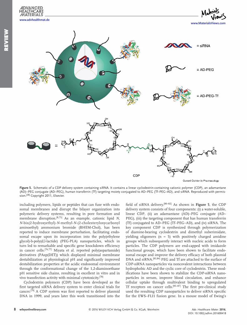
field of siRNA delivery.[80–82] As shown in Figure 5, the CDP delivery system consists of four components: (i) a water-soluble, linear CDP, (ii) an adamantane (AD)–PEG conjugate (AD–PEG), (iii) the targeting component that has human transferrin (Tf) conjugated to AD–PEG (Tf–PEG–AD), and (iv) siRNA. The key component CDP is synthesized through polymerization of diamine-bearing cyclodextrin and dimethyl suberimidate, yielding oligomers (n ≈ 5) with positively charged amidine groups which subsequently interact with nucleic acids to form particles. The CDP polymers are end-capped with imidazole functional groups, which have been shown to facilitate endo-somal escape and improve the delivery efficacy of both plasmid DNA and siRNA.[83,84] PEG and Tf are attached to the surface of CDP-siRNA nanoparticles via noncovalent interactions between hydrophobic AD and the cyclic core of cyclodextrin. These mod-ifications have been shown to stabilize the CDP-siRNA nano-particles in serum, improve blood circulation, and enhance cellular uptake through multivalent binding to upregulated Tf receptors on cancer cells.[85–87] The first pre-clinical study used the resulting CDP nanoparticles to deliver siRNA specific for the EWS–FLI1 fusion gene. In a mouse model of Ewing’s
Adv. Healthcare Mater. 2016, DOI: 10.1002/adhm.201600418
www.MaterialsViews.comwww.advhealthmat.de
Figure 5. Schematic of a CDP delivery system containing siRNA. It contains a linear cyclodextrin-containing cationic polymer (CDP), an adamantane (AD)–PEG conjugate (AD–PEG), human transferrin (Tf) targeting moiety conjugated to AD–PEG (Tf–PEG–AD), and siRNA. Reproduced with permis-sion.[28] Copyright 2011, Elsevier.

9© 2016 WILEY-VCH Verlag GmbH & Co. KGaA, Weinheim wileyonlinelibrary.com
REV
IEW
sarcoma, this targeted formulation was reported to inhibit target gene expression and tumor growth without any evidence of immune stimulation or toxicity.[82] Bartlett et al. then demon-strated the efficacy of the targeted CDP-siRNA nanoparticles in knocking down luciferase and ribonucleotide reductase genes in mice.[88,89] In a pilot safety study, targeted CDP-siRNA nan-oparticles were used to intravenously deliver escalating doses of an siRNA targeting the M2 subunit of ribonucleotide reduc-tase (RRM2) in cynomolgus monkeys.[90] The results showed that the nanoparticles were well tolerated up to 27 mg kg–1 of siRNA, and that multiple systemic doses of targeted nanopar-ticles containing nonchemically modified siRNA can safely be administered to non-human primates. Finally, Davis et al. reported the first human trial using this CDP- siRNA formula-tion (CALAA-01).[79,91] Tumor biopsies from melanoma patients obtained after treatment showed the presence of intracellu-larly localized nanoparticles in amounts that correlated with dose levels of the nanoparticles administered. Furthermore, a reduction was found in both the target messenger RNA and the protein levels, and the presence of the specific mRNA cleavage product provides evidence of an RNAi mechanism of action.
Polymeric carriers have also been employed to simultane-ously deliver a combination of chemotherapeutics and siRNA (two payloads with different physicochemical properties), emerging as a promising synergistic strategy for cancer treat-ment. Recently, Xu et al. developed a polymeric nanoparticle platform composed of an aqueous inner core, a cationic and hydrophobic PLGA layer, and a hydrophilic PEG corona to cir-cumvent chemoresistance in tumors by co-delivering a cisplatin prodrug and REV1/REV3L-specific siRNAs to the same tumor cells. This nanoparticle platform is able to simultaneously deliver DNA-damaging chemotherapeutic as well as suppress gene targets crucial to translesion DNA synthesis (TLS) path-ways in tumors.[35,92] Most mutations that result from cisplatin-induced DNA damage are the consequence of error-prone TLS, which could be responsible for the acquired resistance against DNA damaging agents by improving the capacity of tumor cells to either repair or tolerate DNA damage.[93,94] The versa-tile polymeric nanoparticles were shown to synergistically sup-press the target genes involved in TLS, resulting in tumor cell sensitization to chemotherapy and tumor inhibition in a mouse model in a more effective manner than cisplatin monotherapy. Another example delivery system that holds great potential for codelivery of siRNA and therapeutics is PLGA microparticles with surface-conjugated cationic PAMAM dendrimers which were able to mediate both efficient gene delivery by binding siRNA to the surface[95] and sustained intracellular release of encapsulated 17- β Estradiol.[96] Although the particle-medi-ated codelivery approach is still far from clinical evaluation, it presents a robust platform that not only screens and validates target pathways involved in drug resistance, but also achieves an efficacy that may not be possible with dual-drug or RNAi combinations alone.
2.3. siRNA Conjugate Delivery Systems
Liposomal and polymer-based delivery systems have been advanced the most for siRNA delivery, and have a vast
supporting body of literature due to their extensive previous development for the delivery of plasmid DNA, DNAzymes and antisense oligonucleotides.[28] Recently, siRNA conjugates have shown promise as delivery platforms, leading to the develop-ment of well-defined, single-component systems that optimize the usage of minimal amounts of delivery material. Rozema et al. have developed a polymer-siRNA conjugate delivery system termed Dynamic PolyConjugates (DPCs) for targeted delivery of siRNAs to hepatocytes.[97] This system is comprised of a membrane-disrupting polymer poly(butyl amino vinyl ether) (PBAVE) averaging 30 amino groups per polymer, and shielding PEG moieties and hepatic targeting ligand N-cetylga-lactosamine (GalNAc) that are covalently attached to the PBAVE backbone via a pH-sensitive carboxylated dimethyl maleic acid (CDM) chemistry (Figure 6a). In the acidic environment of the endosome, PEG and GalNAc moieties are released, exposing the membrane-disrupting polymer to promote endosomal release. The chemically modified siRNA is linked to the PBAVE backbone via a disulphide linkage in which it is reduced in the cytosol to release the siRNA. DPCs have been demonstrated to effectively knock down two mouse liver genes, namely apolipo-protein B (apoB) and peroxisome proliferator-activated receptor alpha (ppara), in a dose-dependent manner. The results showed that the siRNA DPC was nontoxic and well tolerated. In this novel modular platform, DPCs incorporate several key features, including hepatocyte specific targeting and reversibility of the PEG protection and polymer endosomolytic activity, which all contribute to the success of this study. To improve upon this DPC design, the use of different targeting ligands and the more stable attachment of the PEG shielding agent have been incorporated into the original platform by Arrowhead Research Corporation, yielding new generations of DPCs with longer cir-culation time and targeting of organs other than the liver.
Another siRNA conjugate delivery platform in development by Alnylam Pharmaceuticals contains chemically modified siRNA that is conjugated to a multivalent targeting ligand. In this system the chemically stabilized siRNA is attached at the 3’ terminus of the sense strand to three GalNAc molecules through a triantennary spacer molecule (Figure 6b). Based upon this delivery platform, the development of several drug candidates including ALN–TTRsc, ALN–PCS, ALN–AS1 and ALN–AT3 are being investigated for the treatment of tran-sthyretin amyloidosis, hypercholesterolemia, hepatic por-phyrias and haemophilia, respectively.[98] The multivalency of the GalNAc ligand is designed for high affinity binding to its receptor on hepatocytes. Variations of this triantennary structure and the spacing of the sugar moieties have also been studied to optimize the binding affinity and hepatocyte uptake.[99,100] ALN–TTRsc is designed to treat TTR-mediated amyloidosis through the silencing of disease-causing protein transthyretin (TTR), and is the most clinically advanced siRNA-GalNAc conjugates developed by Alnylam, with an ongoing Phase III trial underway. The Phase I clinical trial of ALN-TTRsc showed potent, rapid, dose-dependent, consistent, and durable knockdown of serum TTR, with multiple doses of 10 mg kg–1 (body weight) ALN-TTRsc achieving up to 94% TTR knockdown and single doses achieving up to 59.6% TTR knock-down.[1] This drug is generally safe and well-tolerated and con-firms human translation of siRNA-GalNAc platforms. Alnylam
Adv. Healthcare Mater. 2016, DOI: 10.1002/adhm.201600418
www.MaterialsViews.comwww.advhealthmat.de

10 © 2016 WILEY-VCH Verlag GmbH & Co. KGaA, Weinheimwileyonlinelibrary.com
REV
IEW
Adv. Healthcare Mater. 2016, DOI: 10.1002/adhm.201600418
www.MaterialsViews.comwww.advhealthmat.de
Figure 6. a) Schematic illustration of siRNA released from DPC conjugates following cellular uptake. During systemic circulation, the PBAVE polymer is shielded by PEG. After being internalized, DPC conjugates disassemble, shedding the PEG chains in response to the low pH environment of the endo-some. This exposes the membrane-disrupting PBAVE, which results in endosomal release of the siRNA conjugate into the cytosol where a reduction of the disulphide bond linking the siRNA to the polymer occurs, freeing it to begin the RNAi process. GalNAc is a hepatocyte targeting ligand which facilitates uptake. b) Chemical structure of a GalNAc-siRNA conjugate.

11© 2016 WILEY-VCH Verlag GmbH & Co. KGaA, Weinheim wileyonlinelibrary.com
REV
IEW
then announced positive initial Phase II data with ALN-TTRsc (revusiran) for the treatment of TTR cardiac amyloidosis, with clinical activity demonstrating up to a 98.2% knockdown of serum TTR. The other aforementioned drug candidates (ALN-PCS, ALN-AT3 and ALN-AS1) utilize the same siRNA-GalNAc conjugate platform, but deliver different siRNA sequences that target their specific hepatocellular proteins including PCSK9 antithrombin and aminolevulinate synthase 1. These drugs are under investigation to deliver siRNA and have shown prom-ising therapeutic potential for the treatment of their respective diseases.[98,101]
2.4. Inorganic Nanoparticle-Based Delivery Systems
Inorganic nanoparticles (NPs) are another important class of nanoparticle-based delivery vector, and in the past decade these NPs have been widely investigated as potential candidate siRNA delivery carriers. Compared to lipid and polymer based NPs, these inorganic NPs feature smaller dimensions and nar-rower size distributions, which facilitates preferential localiza-tion of NPs to disease areas and areas of interest through the enhanced permeability and retention (EPR) effect.[102] Tumors possess an abnormally dense and permeable vasculature and the tight junctions and basement membrane of tumor vascu-lature are poorly ordered, which allows matter 10–500 nm in size to accrue within the tumor interstitium.[103,104] Additionally, the lymphatic drainage system in tumors is impaired, which delays the clearance of the NPs.[105] To improve the accumu-lation of therapeutic NPs within disease sites, recent studies have reported the added benefit for the inclusion of targeting molecules to NP-based delivery systems.[106,107] Polyvalent deco-ration of NPs with a surface ligand provides many opportuni-ties, including improved spatial localization, controlled homing of nanoparticles to active diseased sites, and elimination of off-target adverse effects.[68] In section 2.4.1, the inorganic nanomaterials composed of gold, iron oxide, mesoporous silica, calcium phos-phate (CaP) and carbon-based NPs will be highlighted and the utilization of passive and active targeting toward their clinical transla-tion will be discussed.
In general, the incorporation of siRNA delivery functionalities into inorganic NPs with distinct properties facilitates the develop-ment of multifunctional nanoplatforms that display numerous beneficial effects simulta-neously, including molecular imaging capa-bility, high siRNA loading, gene silencing, biocompatibility and efficient payload release, and may provides solutions for the challenges currently faced in siRNA delivery.[108]
2.4.1. Gold Nanoparticles
Gold nanoparticles (AuNPs) are one of the most stable inorganic nanoparticles and they display wide-ranging electromagnetic as well
as optical properties that have evoked attention to their biomed-ical applications in biosensing and cancer therapy.[108,109]
Gold nanoparticles are relatively simple to synthesize, bio-compatible, non-immunogenic, and the conjugation/adsorp-tion of drugs or biomacromolecules to the nanoparticle surface is an uncomplicated procedure.[110] AuNP synthesis proceeds in a controllable fashion where aspects such as size, structure and surface ligand composition can be altered, therefore the sys-tematic analysis of each aspect may be evaluated for delineation of their individual effects on the particle’s pharmacokinetics.[111] For example, 5 kDa PEGylated gold nanorods (13 × 47 nm) have been reported with a half-life of up to 17 hours in nude mice.[112] Additional advantages such as scalable formulation, low size dispersity and multifunctional monolayers have driven research towards more efficient approaches for siRNA delivery.[113] Cur-rently, anchoring of siRNA to the surface of the particle can be divided into two categories, covalent conjugation and noncova-lent immobilization, each having their specific advantages.[113] Covalent AuNP conjugation is the attachment of compounds to the surface of AuNPs mainly through the metal-ligand interac-tion between gold and sulfur (from thiolated oligonucleotides). Covalent conjugation of siRNA to AuNPs is a useful tool for their delivery, however the method requires modification of the nucleic acid and thus careful consideration must be taken to ensure the proper effects of the siRNA remain after modi-fication as well as proper release from the AuNP. Noncovalent nucleic acid immobilization utilizes unmodified siRNA for gene therapy. Nucleic acids are negatively charged, which facili-tate the siRNA immobilization on the cationic nanoparticle surface through electrostatic interaction. Important aspects in effective formulation include AuNP-to-siRNA ratio, surface charge coverage, and hydrophobicity.[113]
One specific siRNA-AuNP conjugate particle is the spherical nucleic acid (SNA) nanoparticle, developed by the Mirkin Lab (Figure 7).[114] SNAs are comprised of closely packed siRNA
Adv. Healthcare Mater. 2016, DOI: 10.1002/adhm.201600418
www.MaterialsViews.comwww.advhealthmat.de
Figure 7. An illustrative overview of the synthesis of Gd(III)-functionalized SNAs. (i) Gd(III)-SNA conjugates were prepared from alkyne-modified T bases and azide-labeled Gd(III) complexes were subject to click chemistry to prepare Gd(III)-SNA conjugates. (ii) Next, the gold nanoparticle (Au-NP) surface was functionalized with Gd(III)-conjugated DNA, forming Gd(III)-SNA. Reproduced with permission.[114] Copyright 2011, Elsevier.

12 © 2016 WILEY-VCH Verlag GmbH & Co. KGaA, Weinheimwileyonlinelibrary.com
REV
IEW duplexes covalently functionalized to an AuNP core which
combine the silencing capability of siRNAs with stability and biocompatibility of AuNPs. There is present an ion cloud asso-ciated with the high-density oligonucleotide shell surrounding the particle, and steric inhibition at the particle surface. This unique micro-environment is able to protect nucleic acids from enzymatic degradation, resulting in increased stability of siRNAs and longer therapeutic lifetime.[114,115] SNAs targeting Bcl2L12 have been used to silence oncogene expression of glio-blastoma multifore in vitro. Notably, the SNAs were able to infiltrate the blood-brain barrier (BBB) and blood-tumor bar-rier to reduce tumor progression in a xenograft mouse model without causing any adverse effects. This development may prove effective for future treatment of diseases which require the traversing of the BBB, for which current chemotherapeutics generally have been inefficient in crossing.[116] SNAs may also be functionalized with magnetic resonance imaging contrast agents for the imaging and tracking of the agent in vivo. Gene silencing utilizing SNAs signifies a possible novel method for systemic RNAi therapy, with previously “undruggable” onco-genes as potential targets, as well as incorporating multiple siRNAs to target multiple gene targets.[114] However, there is still room for improvement in this novel approach, since the circulation time for these SNAs was short; in the first 5 minutes over 90% of the particle conjugates distributed to tissues, and the elimination half-life was only 8.5 hours.[114] For example, incorporating PEG or CD47 (a “self-marker” on cell mem-branes which was recently shown to increase circulation half-life in vivo[117]) onto the NP may be beneficial and potentially lead to development of SNAs with longer circulation times.
Additionally, Zheng and colleagues show that SNA-AuNPs completely penetrate keratinocytes in mouse and human epi-dermis within hours after application, offering an innovative method to circumvent the epidermal barrier, which has typically hindered the usage of gene-suppressing agents on the skin.[118] This is significant because AuNPs have now been shown to provide a simple siRNA transdermal delivery method without the use of disruption or transfection agents such as liposomes, peptides, or viruses, and thus avoid complications associated with these techniques. In mice, topical delivery of 1.5 µm epi-dermal growth factor receptor (EGFR) siRNA (50 nm SNA-NCs) for 3 weeks can silence EGFR mRNA up to 65% (almost com-pletely knocking down EGFR protein expression), suppress downstream ERK phosphorylation up to 74%, and reduce epi-dermal thickness by almost 40%.[118] There was no histological evidence of toxicity or cytokine activation in mice. Furthermore, after 3 weeks-post skin treatment, the internal organs of mice show near complete clearance of SNAs. This gold nanopar-ticle-based topical gene therapy approach may be beneficial in treating cutaneous tumors, skin inflammation, and dominant negative genetic skin disorders in a noninvasive manner.
Besides direct conjugation, the noncovalent immobiliza-tion of siRNA is an alternative delivery approach. For example, Wang et al. reported a biocompatible PEI-capped AuNP that mediates effective delivery into cells, resulting in gene silencing.[110,118] Here, PEI acts as both the reductant and stabi-lizer, which binds siRNA through electrostatic interactions and maintains consistent nanoparticle structure, size and function. Results showed that PEI-capped AuNPs/siRNA reduced the
expression of polo-like kinase 1 (PLK1) oncogene expression in tumor cells without increasing cytotoxicity levels as measured by MTT assay. Since AuNPs have already been utilized as tools for imaging, the data indicates their potential for therapeutic use as a theranostic system which utilizes both the imaging and therapeutic functionality of AuNPs complexed with siRNA.[110]
Some additional methods based on gold nanocarriers have also been developed, aiming at controlled gene delivery to cells to improve the gene transfection efficiency and specificity.[119] Examples include using photothermal effect of gold nanostruc-tures to elicit release of siRNA, remote temporal-spatial shape transformation of nanogold, and NIR laser-controlled drug release[119–123]
2.4.2. Mesoporous Silica Nanoparticles
Mesoporous silica nanoparticle (MSN) delivery systems present controllable porosity, which enables a large surface area avail-able for modification and drug encapsulation, making them ideal candidates for carrying siRNA to target disease sites.[108] Notable properties of silica such as biocompatibility, efficient in vivo elimination and Generally Recognized As Safe (GRAS) status by the FDA make it an attractive biomaterial, which if successful in clinical trials will face fewer hurdles in com-mercialization.[124,125] The mesoporous quality significantly increases surface area of the particles, and the structure of the mesopores can be controlled and modified with cationic mole-cules and other multifunctional moieties such as fluorescent molecules, active targeting ligands, therapeutic small mole-cule drugs or miRNA, which confers imaging, targeting and treatment functionality.[125,126] As a result, increased quantita-tive delivery over conventional NPs is possible and possibili-ties are being explored for combined imaging, diagnosis, and therapeutic functions. Zink, et al. used such a combinatorial approach to address cancer drug resistance by the endosomal delivery of chemotherapeutic agents and siRNA that targets the P-glycoprotein (Pgp) drug exporter. The MSNP surface was modified with PEI-PEG copolymer, facilitating electrostatic attachment of siRNA. Phosphonate-coated particle pores allow for electrostatic doxorubicin (Dox) attachment and subsequent release in an acidifying endosomal environment. Dox codeliv-ered with anti-Pgp siRNA by mesoporous silica nanoparticles (MSNPs) were shown to overcome Dox resistance in a multi-drug resistant (MDR) human breast cancer xenograft mouse model. Compared to free Dox or the carrier loaded with either drug or siRNA alone, the dual delivery system resulted in syn-ergistic inhibition of tumor growth in vivo and provides proof-of-principle testing of the use of a dual drug/siRNA MSNP to overcome Dox resistance in a xenograft mouse model.[127]
2.4.3. Iron Oxide Nanoparticles
Iron oxide nanoparticles are another class of inorganic nano-particles, which have been approved or under clinical inves-tigation for magnetic resonance imaging (MRI) due to their superparamagnetic properties and biocompatibility. Magnetic NPs are capable of remotely-controlled transfection through
Adv. Healthcare Mater. 2016, DOI: 10.1002/adhm.201600418
www.MaterialsViews.comwww.advhealthmat.de

13© 2016 WILEY-VCH Verlag GmbH & Co. KGaA, Weinheim wileyonlinelibrary.com
REV
IEW
the application of an external magnetic field.[124] This magnetic-based transfection is termed magnetofection, and has been recently reported to improve gene transfection efficiency. More recently, the incorporation of siRNA into iron oxide nanopar-ticle-based multifunctional theranostic nanoplatforms has ena-bled simultaneous molecular imaging and siRNA delivery.[108]
2.4.4. CaP Nanoparticles
CaP nanoparticles are another type of nanomaterial which has been developed for siRNA delivery and has shown successful transfection of a wide variety of mammalian cells with little tox-icity. CaP rapidly dissolves in the acidic pH of the endosomes, causing osmotic swelling that enables cytoplasmic release of encapsulated siRNA. This unique capability has been employed to develop CaP nanoparticles with improved transfection effi-ciency.[128] Huang et al. demonstrated increased Ca2+ concen-tration upon the cellular internalization of a lipid-coated CaP nanoparticle which releases more cargo to the cytoplasm than previously developed lipid-polycation-nucleic acid formulations. Furthermore, incorporation of a PEG-phospholipid conjugate with a targeting ligand anisamide to the lipid-coated CaP nano-particle resulted in a significant (≈40-fold in vitro and ≈fourfold in vivo) improvement in siRNA delivery compared to previous formulations.[128,129]
2.4.5. Carbon-Based siRNA Nanodelivery Systems
In the past decade, carbon-based materials have been emerging with potential applications in bioimaging, energy storage/con-version devices, sensors and drug delivery carriers. Their excel-lent physicochemical, optical, and electrical/thermal properties may prove great attributes for biomaterial applications and they present significantly increased surface area which makes functionalization easier as well as expanding payload carrying capacity. In particular, carbon-based nano-sized delivery sys-tems such as carbon nanotubes (CNTs), graphene nanosheets and nanodiamonds are showing promising ability for siRNA delivery applications.[130–132]
CNTs generally have a diameter of 1–2 nm, with length varying from 50 nm to 1 cm. CNTs have been reported to be viable platforms for delivering biologically active siRNA into cells both in vitro and in vivo.[133] Modified CNTs prepared through a functionalization method have been shown to pro-tect siRNA and facilitate its cellular uptake. For example, non-covalent functionalization of CNTs with a lipopolymer (DSPE-PEG) and PEI have been shown to effectively deliver siRNA to target cells.[134] Liver GAPDH siRNA uptake and gene silencing following intravenous injection was observed in a mouse model by Siu et al.[134] Alternatively, Wang and colleagues have chemically functionalized single-walled carbon nanotubes (SWNTs) with PEI. Subsequent binding of DSPE-PEG2000-Maleimide to the SWNTs facilitated conjugation with a tumor targeting peptide NGR (Cys-Asn-Gly-Arg-Cys-). Telomerase reverse transcriptase (hTERT) siRNA was loaded onto the tar-geted SWNT and the combination with photothermal therapy showed high antitumor activity without observed toxicity in a
tumor-bearing mouse model.[135] Another carbon-based mate-rial, graphene, exists as stable, functionalizable, biocompatible two-dimensional nanosized sheets.[124] Noncovalent adsorption for immobilizing drugs and nucleic acids is possible through π–π stacking, electrostatic, hydrophobic and other molecular interactions, which present it as a suitable candidate for siRNA transfection.[136] Additionally, SWNTs and graphene oxide with strong optical absorption in the broad-visible and near IR offer unique advantages for photothermal siRNA therapy.[119,137]
Carbon-derived nanodiamonds (NDs) have been extensively explored in biomedicine and shown to be a promising platform for imaging, drug and gene delivery. NDs, 2–8 nm diameter carbon carriers of truncated octahedral composition, possess several unique features including biocompatibility, functionali-zation versatility and unique surface electrostatics.[138] Recent in vivo studies have also alluded to the clinical potential of utilizing ND to deliver chemotherapeutic agents.[139] Fluorophore conju-gation and introduction of nitrogen defects have also provided a means of tracking NDs within biological samples.[140–142] Ho and co-workers reported an approach for siRNA delivery using ND-PEI (800Da) siRNA complexes. ND-PEI siRNA complexes showed high cell internalization and GFP knockdown, and more importantly, close to zero cytotoxicity.[143] The facile one-step production of ND-PEI makes for efficient scale-up and is an attractive quality for NDs as siRNA nanodelivery platforms.
Although great progress has been made to advance inorganic siRNA nanocarriers and numerous in vivo studies have shown their great potential for siRNA therapy,[131] their development is still at an early stage, with no siRNA-delivering inorganic or carbon-based delivery systems currently on clinical trial. There are still numerous issues with inorganic nanoparticles which need to be addressed. For example, a significant issue with carbon-based nanodelivery systems is their non-biodegrada-bility in vivo, which highlights a need for more studies evalu-ating their efficacy.[124] Another persistent problem with nano-particles in general is suboptimal biodistribution, with particles being trapped mainly in the liver and spleen due to reticu-loendothelial function, which may be ameliorated by “stealth” coating as well as active targeting. As time goes on, additional clinical and in vivo work by these nanodelivery systems will strengthen the credibility of their application. The continued investigation and refinement of their drug delivery properties as well as the mitigation of challenging factors such as toxicity and bioavailability will allow for accelerated developments in siRNA-mediated gene therapy via inorganic and carbon-based NPs towards the clinic.
3. Conclusion
In addition to lipid-, polymer-, siRNA conjugate- and inor-ganic systems there are other nano-and microsystems such as microhydrogels,[144] microneedles,[145] self-assembled siRNA oligonucleotide nanoparticles[146] and microsponges[147] which exemplify the myriad directions siRNA delivery is currently developing. Furthermore, controlled mixing processes such as microfluidic methods have been developed to achieve con-sistent quality and reproducibility of siRNA formulations with high yield and uniform size.[148–150] Advances in materials
Adv. Healthcare Mater. 2016, DOI: 10.1002/adhm.201600418
www.MaterialsViews.comwww.advhealthmat.de
14 © 2016 WILEY-VCH Verlag GmbH & Co. KGaA, Weinheimwileyonlinelibrary.com
REV
IEW science, nano- and micro-technology play an important role in
the development of a great multitude of siRNA delivery sys-tems with diverse size, shape, chemical properties, structures and functionalities in the past decade. The most clinically advanced delivery systems are nanoparticles formed by judi-cious assembly of lipids, polymers or lipidoids such as SNALP and CDP delivery systems. All these delivery systems contain a mixture of multiple molecular components, with each com-ponent specifically designed to overcome major delivery bar-riers including efficient siRNA binding/protection, endosomal escape, nanoparticle stability in circulation, or cell-specific tar-geting for improved bioavailability.
Significant progress has been made to drive RNAi-based medicine into clinical applications since the first demonstra-tion of gene knockdown in mammalian cells. However, the delivery of therapeutic siRNA to induce the potent and specific silencing of genetic targets in target cells remains one of the greatest challenges in RNAi therapy.
Despite the promising advancement of siRNA therapeutics through the different stages of clinical development, there are persistent delivery challenges which need to be addressed. For example, most of the clinically translated systems deliver siRNA to the liver and tumors, where the fenestrated or discontinuous endothelium facilitates the passage and retention of macro-molecular objects. Other tissues which do not display these characteristics are less accessible, and different tissues each pose particular challenges for delivery systems to overcome. Furthermore, considerable concern and attention must be paid to non-specific activation of TLRs by siRNA, which leads to inflammation and off-target effects.[151,152] Direct chemical modification of the siRNA sequence may improve the activity and delivery of siRNA therapeutics.[153] Overall, biological bar-riers and their effects on the siRNA delivery process must be thoroughly characterized and understood, and design of diverse delivery components based on this understanding should be implemented in the next generations of future delivery plat-forms for continued clinical success.
AcknowledgementsW.H. and X.-Q.Z. contributed equally to this work. The authors declare no conflict of interests in this work. The financial support from New Jersey Institute of Technology (NJIT) startup funding and NSF Innovation Corps (I-Corps) program is gratefully acknowledged.
Received: April 13, 2016Revised: June 7, 2016
Published online:
[1] H. Yin, R. L. Kanasty, A. A. Eltoukhy, A. J. Vegas, J. R. Dorkin, D. G. Anderson, Nat. Rev. Genet. 2014, 15, 541.
[2] K. A. Whitehead, R. Langer, D. G. Anderson, Nat. Rev. Drug Discov. 2009, 8, 129.
[3] A. Fire, S. Xu, M. K. Montgomery, S. A. Kostas, S. E. Driver, C. C. Mello, Nature 1998, 391, 806.
[4] K. Quon, P. D. Kassner, Expert Opin Ther Targets 2009, 13, 1027.[5] A. Ptasznik, Y. Nakata, A. Kalota, S. G. Emerson, A. M. Gewirtz,
Nat. Med. 2004, 10, 1187.
[6] A. Okumura, P. M. Pitha, R. N. Harty, Proc. Natl. Acad. Sci. USA 2008, 105, 3974.
[7] S. H. Kim, J. H. Jeong, S. H. Lee, S. W. Kim, T. G. Park, J. Controlled Release 2008, 129, 107.
[8] P. Resnier, T. Montier, V. Mathieu, J. P. Benoit, C. Passirani, Bioma-terials 2013, 34, 6429.
[9] G. Hutvagner, P. D. Zamore, Science 2002, 297, 2056.[10] E. Bernstein, A. A. Caudy, S. M. Hammond, G. J. Hannon, Nature
2001, 409, 363.[11] P. J. Paddison, A. A. Caudy, E. Bernstein, G. J. Hannon,
D. S. Conklin, Genes Dev. 2002, 16, 948.[12] S. C. Semple, A. Akinc, J. Chen, A. P. Sandhu, B. L. Mui,
C. K. Cho, D. W. Sah, D. Stebbing, E. J. Crosley, E. Yaworski, I. M. Hafez, J. R. Dorkin, J. Qin, K. Lam, K. G. Rajeev, K. F. Wong, L. B. Jeffs, L. Nechev, M. L. Eisenhardt, M. Jayaraman, M. Kazem, M. A. Maier, M. Srinivasulu, M. J. Weinstein, Q. Chen, R. Alvarez, S. A. Barros, S. De, S. K. Klimuk, T. Borland, V. Kosovrasti, W. L. Cantley, Y. K. Tam, M. Manoharan, M. A. Ciufolini, M. A. Tracy, A. de Fougerolles, I. MacLachlan, P. R. Cullis, T. D. Madden, M. J. Hope, Nat. Biotechnol. 2010, 28, 172.
[13] J. M. Layzer, A. P. McCaffrey, A. K. Tanner, Z. Huang, M. A. Kay, B. A. Sullenger, RNA 2004, 10, 766.
[14] A. L. Jackson, P. S. Linsley, Nat. Rev. Drug Discov. 2010, 9, 57
[15] D. N. Nguyen, K. P. Mahon, G. Chikh, P. Kim, H. Chung, A. P. Vicari, K. T. Love, M. Goldberg, S. Chen, A. M. Krieg, J. Chen, R. Langer, D. G. Anderson, Proc. Natl. Acad. Sci. USA 2012, 109, 797.
[16] T. Tokatlian, T. Segura, Wiley Interdiscip. Rev.: Nanomed. Nano-biotechnol. 2010, 2, 305.
[17] J. M. Lee, T. J. Yoon, Y. S. Cho, Biomed Res Int 2013, 2013, 782041.[18] N. Bessis, F. J. GarciaCozar, M. C. Boissier, Gene Ther. 2004, 11
Suppl 1, S10.[19] C. Baum, O. Kustikova, U. Modlich, Z. Li, B. Fehse, Hum. Gene
Ther. 2006, 17, 253.[20] Y. Y. Tam, S. Chen, P. R. Cullis, Pharmaceutics 2013, 5, 498.[21] S. C. Semple, S. K. Klimuk, T. O. Harasym, N. Dos Santos,
S. M. Ansell, K. F. Wong, N. Maurer, H. Stark, P. R. Cullis, M. J. Hope, P. Scherrer, Biochimica Et Biophysica Acta-Biomem-branes 2001, 1510, 152.
[22] P. L. Felgner, T. R. Gadek, M. Holm, R. Roman, H. W. Chan, M. Wenz, J. P. Northrop, G. M. Ringold, M. Danielsen, Proc. Natl. Acad. Sci. USA 1987, 84, 7413.
[23] R. W. Malone, P. L. Felgner, I. M. Verma, Proc. Natl. Acad. Sci. USA 1989, 86, 6077.
[24] D. V. Morrissey, J. A. Lockridge, L. Shaw, K. Blanchard, K. Jensen, W. Breen, K. Hartsough, L. Machemer, S. Radka, V. Jadhav, N. Vaish, S. Zinnen, C. Vargeese, K. Bowman, C. S. Shaffer, L. B. Jeffs, A. Judge, I. MacLachlan, B. Polisky, Nat. Biotechnol. 2005, 23, 1002.
[25] T. S. Zimmermann, A. C. Lee, A. Akinc, B. Bramlage, D. Bumcrot, M. N. Fedoruk, J. Harborth, J. A. Heyes, L. B. Jeffs, M. John, A. D. Judge, K. Lam, K. McClintock, L. V. Nechev, L. R. Palmer, T. Racie, I. Rohl, S. Seiffert, S. Shanmugam, V. Sood, J. Soutschek, I. Toudjarska, A. J. Wheat, E. Yaworski, W. Zedalis, V. Koteliansky, M. Manoharan, H. P. Vornlocher, I. MacLachlan, Nature 2006, 441, 111.
[26] J. C. Burnett, J. J. Rossi, K. Tiemann, Biotechnol. J. 2011, 6, 1130.
[27] M. Frank-Kamenetsky, A. Grefhorst, N. N. Anderson, T. S. Racie, B. Bramlage, A. Akinc, D. Butler, K. Charisse, R. Dorkin, Y. Fan, C. Gamba-Vitalo, P. Hadwiger, M. Jayaraman, M. John, K. N. Jayaprakash, M. Maier, L. Nechev, K. G. Rajeev, T. Read, I. Rohl, J. Soutschek, P. Tan, J. Wong, G. Wang, T. Zimmermann, A. de Fougerolles, H. P. Vornlocher, R. Langer, D. G. Anderson,
Adv. Healthcare Mater. 2016, DOI: 10.1002/adhm.201600418
www.MaterialsViews.comwww.advhealthmat.de

15© 2016 WILEY-VCH Verlag GmbH & Co. KGaA, Weinheim wileyonlinelibrary.com
REV
IEW
Adv. Healthcare Mater. 2016, DOI: 10.1002/adhm.201600418
www.MaterialsViews.comwww.advhealthmat.de
M. Manoharan, V. Koteliansky, J. D. Horton, K. Fitzgerald, Proc. Natl. Acad. Sci. USA 2008, 105, 11915.
[28] C. Alabi, A. Vegas, D. Anderson, Curr Opin Pharmacol 2012, 12, 427.
[29] T. W. Geisbert, A. C. H. Lee, M. Robbins, J. B. Geisbert, A. N. Honko, V. Sood, J. C. Johnson, S. de Jong, I. Tavakoli, A. Judge, L. E. Hensley, I. MacLachlan, Lancet 2010, 375, 1896.
[30] S. Reagan-Shaw, N. Ahmad, FASEB J. 2005, 19, 611.[31] L. Li, R. Wang, D. Wilcox, X. Zhao, J. Song, X. Lin,
W. M. Kohlbrenner, S. W. Fesik, Y. Shen, Gene Ther. 2012, 19, 775.[32] J. Tabernero, G. I. Shapiro, P. M. LoRusso, A. Cervantes,
G. K. Schwartz, G. J. Weiss, L. Paz-Ares, D. C. Cho, J. R. Infante, M. Alsina, M. M. Gounder, R. Falzone, J. Harrop, A. C. White, I. Toudjarska, D. Bumcrot, R. E. Meyers, G. Hinkle, N. Svrzikapa, R. M. Hutabarat, V. A. Clausen, J. Cehelsky, S. V. Nochur, C. Gamba-Vitalo, A. K. Vaishnaw, D. W. Sah, J. A. Gollob, H. A. Burris 3rd, Cancer Discovery 2013, 3, 406.
[33] T. Coelho, D. Adams, A. Silva, P. Lozeron, P. N. Hawkins, T. Mant, J. Perez, J. Chiesa, S. Warrington, E. Tranter, M. Munisamy, R. Falzone, J. Harrop, J. Cehelsky, B. R. Bettencourt, M. Geissler, J. S. Butler, A. Sehgal, R. E. Meyers, Q. Chen, T. Borland, R. M. Hutabarat, V. A. Clausen, R. Alvarez, K. Fitzgerald, C. Gamba-Vitalo, S. V. Nochur, A. K. Vaishnaw, D. W. Sah, J. A. Gollob, O. B. Suhr, N. Engl. J. Med. 2013, 369, 819.
[34] M. Jayaraman, S. M. Ansell, B. L. Mui, Y. K. Tam, J. Chen, X. Du, D. Butler, L. Eltepu, S. Matsuda, J. K. Narayanannair, K. G. Rajeev, I. M. Hafez, A. Akinc, M. A. Maier, M. A. Tracy, P. R. Cullis, T. D. Madden, M. Manoharan, M. J. Hope, Angew. Chem. Int. Ed. Engl. 2012, 51, 8529.
[35] X. Xu, W. Ho, X. Zhang, N. Bertrand, O. Farokhzad, Trends Mol. Med. 2015, 21, 223.
[36] A. Santel, M. Aleku, O. Keil, J. Endruschat, V. Esche, G. Fisch, S. Dames, K. Loffler, M. Fechtner, W. Arnold, K. Giese, A. Klippel, J. Kaufmann, Gene Ther. 2006, 13, 1222.
[37] A. Santel, M. Aleku, O. Keil, J. Endruschat, V. Esche, B. Durieux, K. Loffler, M. Fechtner, T. Rohl, G. Fisch, S. Dames, W. Arnold, K. Giese, A. Klippel, J. Kaufmann, Gene Ther. 2006, 13, 1360.
[38] B. Schultheis, D. Strumberg, A. Santel, C. Vank, F. Gebhardt, O. Keil, C. Lange, K. Giese, J. Kaufmann, M. Khan, J. Drevs, J. Clin. Oncol. 2014, 32, 4141.
[39] C. N. Landen, A. Chavez-Reyes, C. Bucana, R. Schmandt, M. T. Deavers, G. Lopez-Berestein, A. K. Sood, Cancer Res. 2005, 65, 6910.
[40] H. Lv, S. Zhang, B. Wang, S. Cui, J. Yan, J. Controlled Release 2006, 114, 100.
[41] Z. Ma, J. Li, F. He, A. Wilson, B. Pitt, S. Li, Biochem. Biophys. Res. Commun. 2005, 330, 755.
[42] S. Akhtar, I. Benter, Adv. Drug Delivery Rev. 2007, 59, 164.[43] C. Foged, Curr. Top. Med. Chem. 2012, 12, 97.[44] K. Kariko, M. Buckstein, H. Ni, D. Weissman, Immunity 2005, 23,
165.[45] F. Eberle, K. Giessler, C. Deck, K. Heeg, M. Peter, C. Richert,
A. H. Dalpke, J. Immunol. 2008, 180, 3229.[46] R. L. Kanasty, K. A. Whitehead, A. J. Vegas, D. G. Anderson, Mol.
Ther. 2012, 20, 513.[47] E. K. H. Chow, D. Ho, Sci. Transl. Med. 2013, 5.[48] K. F. Pirollo, G. Zon, A. Rait, Q. Zhou, W. Yu, R. Hogrefe,
E. H. Chang, Hum. Gene Ther. 2006, 17, 117.[49] K. F. Pirollo, A. Rait, Q. Zhou, S. H. Hwang, J. A. Dagata, G. Zon,
R. I. Hogrefe, G. Palchik, E. H. Chang, Cancer Res. 2007, 67, 2938.
[50] K. F. Pirollo, E. H. Chang, Cancer Res. 2008, 68, 1247.[51] W. Yu, K. F. Pirollo, B. Yu, A. Rait, L. Xiang, W. Huang, Q. Zhou,
G. Ertem, E. H. Chang, Nucleic Acids Res. 2004, 32, e48.
[52] A. Akinc, A. Zumbuehl, M. Goldberg, E. S. Leshchiner, V. Busini, N. Hossain, S. A. Bacallado, D. N. Nguyen, J. Fuller, R. Alvarez, A. Borodovsky, T. Borland, R. Constien, A. de Fougerolles, J. R. Dorkin, K. Narayanannair Jayaprakash, M. Jayaraman, M. John, V. Koteliansky, M. Manoharan, L. Nechev, J. Qin, T. Racie, D. Raitcheva, K. G. Rajeev, D. W. Sah, J. Soutschek, I. Toudjarska, H. P. Vornlocher, T. S. Zimmermann, R. Langer, D. G. Anderson, Nat. Biotechnol. 2008, 26, 561.
[53] K. T. Love, K. P. Mahon, C. G. Levins, K. A. Whitehead, W. Querbes, J. R. Dorkin, J. Qin, W. Cantley, L. L. Qin, T. Racie, M. Frank-Kamenetsky, K. N. Yip, R. Alvarez, D. W. Sah, A. de Fougerolles, K. Fitzgerald, V. Koteliansky, A. Akinc, R. Langer, D. G. Anderson, Proc. Natl. Acad. Sci. USA 2010, 107, 1864.
[54] F. Leuschner, P. Dutta, R. Gorbatov, T. I. Novobrantseva, J. S. Donahoe, G. Courties, K. M. Lee, J. I. Kim, J. F. Markmann, B. Marinelli, P. Panizzi, W. W. Lee, Y. Iwamoto, S. Milstein, H. Epstein-Barash, W. Cantley, J. Wong, V. Cortez-Retamozo, A. Newton, K. Love, P. Libby, M. J. Pittet, F. K. Swirski, V. Koteliansky, R. Langer, R. Weissleder, D. G. Anderson, M. Nahrendorf, Nat. Biotechnol. 2011, 29, 1005.
[55] T. I. Novobrantseva, A. Borodovsky, J. Wong, B. Klebanov, M. Zafari, K. Yucius, W. Querbes, P. Ge, V. M. Ruda, S. Milstein, L. Speciner, R. Duncan, S. Barros, G. Basha, P. Cullis, A. Akinc, J. S. Donahoe, K. Narayanannair Jayaprakash, M. Jayaraman, R. L. Bogorad, K. Love, K. Whitehead, C. Levins, M. Manoharan, F. K. Swirski, R. Weissleder, R. Langer, D. G. Anderson, A. de Fougerolles, M. Nahrendorf, V. Koteliansky, Mol. Ther. Nucleic Acids 2012, 1, e4.
[56] Y. Dong, K. T. Love, J. R. Dorkin, S. Sirirungruang, Y. Zhang, D. Chen, R. L. Bogorad, H. Yin, Y. Chen, A. J. Vegas, C. A. Alabi, G. Sahay, K. T. Olejnik, W. Wang, A. Schroeder, A. K. Lytton-Jean, D. J. Siegwart, A. Akinc, C. Barnes, S. A. Barros, M. Carioto, K. Fitzgerald, J. Hettinger, V. Kumar, T. I. Novobrantseva, J. Qin, W. Querbes, V. Koteliansky, R. Langer, D. G. Anderson, Proc. Natl. Acad. Sci. USA 2014, 111, 3955.
[57] Z. W. Wu, C. T. Chien, C. Y. Liu, J. Y. Yan, S. Y. Lin, J Drug Target 2012, 20, 551.
[58] N. A. Nikitenko, V. S. Prassolov, Acta Naturae 2013, 5, 35.[59] S. Akhtar, I. F. Benter, J. Clin. Invest. 2007, 117, 3623.[60] D. Jere, H. L. Jiang, R. Arote, Y. K. Kim, Y. J. Choi, M. H. Cho,
T. Akaike, C. S. Cho, Expert Opin. Drug Delivery 2009, 6, 827.[61] U. Lungwitz, M. Breunig, T. Blunk, A. Gopferich, Eur. J. Pharm.
Biopharm. 2005, 60, 247.[62] P. Muhonen, T. Tennila, E. Azhayeva, R. N. Parthasarathy,
A. J. Janckila, H. K. Vaananen, A. Azhayev, T. Laitala-Leinonen, Chem. Biodivers. 2007, 4, 858.
[63] L. Wightman, R. Kircheis, V. Rossler, S. Carotta, R. Ruzicka, M. Kursa, E. Wagner, J. Gene Med. 2001, 3, 362.
[64] Y. Wang, Z. Li, Y. Han, L. H. Liang, A. Ji, Curr. Drug Metab. 2010, 11, 182.
[65] S. Hong, P. R. Leroueil, E. K. Janus, J. L. Peters, M. M. Kober, M. T. Islam, B. G. Orr, J. R. Baker Jr., M. M. Banaszak Holl, Bio-conjugate Chem. 2006, 17, 728.
[66] D. E. Owens 3rd, N. A. Peppas, Int. J. Pharm. 2006, 307, 93.[67] N. Bertrand, J. C. Leroux, J. Controlled Release 2012, 161, 152.[68] X. Q. Zhang, X. Xu, N. Bertrand, E. Pridgen, A. Swami,
O. C. Farokhzad, Adv. Drug Delivery Rev. 2012, 64, 1363.[69] L. Wasungu, D. Hoekstra, J. Controlled Release 2006, 116, 255.[70] N. Kim, D. Jiang, A. M. Jacobi, K. A. Lennox, S. D. Rose,
M. A. Behlke, A. K. Salem, Int. J. Pharm. 2012, 427, 123.[71] J. Guo, W. P. Cheng, J. Gu, C. Ding, X. Qu, Z. Yang, C. O’Driscoll,
Eur. J. Pharm. Sci. 2012, 45, 521.[72] X. L. Wang, S. Ramusovic, T. Nguyen, Z. R. Lu, Bioconjugate Chem.
2007, 18, 2169.
16 © 2016 WILEY-VCH Verlag GmbH & Co. KGaA, Weinheimwileyonlinelibrary.com
REV
IEW
Adv. Healthcare Mater. 2016, DOI: 10.1002/adhm.201600418
www.MaterialsViews.comwww.advhealthmat.de
[73] X. L. Wang, R. Xu, X. Wu, D. Gillespie, R. Jensen, Z. R. Lu, Mol. Pharm. 2009, 6, 738.
[74] A. E. Felber, B. Castagner, M. Elsabahy, G. F. Deleavey, M. J. Damha, J. C. Leroux, J. Controlled Release 2011, 152, 159.
[75] M. Morille, C. Passirani, A. Vonarbourg, A. Clavreul, J. P. Benoit, Biomaterials 2008, 29, 3477.
[76] X. Z. Yang, S. Dou, T. M. Sun, C. Q. Mao, H. X. Wang, J. Wang, J. Controlled Release 2011, 156, 203.
[77] N. Fang, J. Wang, H.-Q. Mao, K. W. Leong, V. Chan, Colloids Surf. B. Biointerfaces 2003, 29, 233.
[78] K. Miyata, M. Oba, M. Nakanishi, S. Fukushima, Y. Yamasaki, H. Koyama, N. Nishiyama, K. Kataoka, J. Am. Chem. Soc. 2008, 130, 16287.
[79] M. E. Davis, Mol. Pharm. 2009, 6, 659.[80] H. Gonzalez, S. J. Hwang, M. E. Davis, Bioconjugate. Chem. 1999,
10, 1068.[81] M. E. Davis, S. H. Pun, N. C. Bellocq, T. M. Reineke,
S. R. Popielarski, S. Mishra, J. D. Heidel, Curr. Med. Chem. 2004, 11, 179.
[82] S. Hu-Lieskovan, J. D. Heidel, D. W. Bartlett, M. E. Davis, T. J. Triche, Cancer Res. 2005, 65, 8984.
[83] S. Mishra, J. D. Heidel, P. Webster, M. E. Davis, J. Controlled Release 2006, 116, 179.
[84] R. P. Kulkarni, S. Mishra, S. E. Fraser, M. E. Davis, Bioconjugate Chem. 2005, 16, 986.
[85] D. W. Bartlett, M. E. Davis, Bioconjugate Chem. 2007, 18, 456.[86] N. C. Bellocq, S. H. Pun, G. S. Jensen, M. E. Davis, Bioconjugate
Chem. 2003, 14, 1122.[87] S. Mishra, P. Webster, M. E. Davis, Eur. J. Cell Biol. 2004, 83, 97.[88] D. W. Bartlett, H. Su, I. J. Hildebrandt, W. A. Weber, M. E. Davis,
Proc. Natl. Acad. Sci. USA 2007, 104, 15549.[89] D. W. Bartlett, M. E. Davis, Biotechnol. Bioeng. 2008, 99, 975.[90] J. D. Heidel, Z. Yu, J. Y. Liu, S. M. Rele, Y. Liang, R. K. Zeidan,
D. J. Kornbrust, M. E. Davis, Proc. Natl. Acad. Sci. USA 2007, 104, 5715.
[91] M. E. Davis, J. E. Zuckerman, C. H. Choi, D. Seligson, A. Tolcher, C. A. Alabi, Y. Yen, J. D. Heidel, A. Ribas, Nature 2010, 464, 1067.
[92] X. Xu, K. Xie, X. Q. Zhang, E. M. Pridgen, G. Y. Park, D. S. Cui, J. Shi, J. Wu, P. W. Kantoff, S. J. Lippard, R. Langer, G. C. Walker, O. C. Farokhzad, Proc. Natl. Acad. Sci. USA 2013, 110, 18638.
[93] K. Xie, J. Doles, M. T. Hemann, G. C. Walker, Proc. Natl. Acad. Sci. USA 2010, 107, 20792.
[94] J. Doles, T. G. Oliver, E. R. Cameron, G. Hsu, T. Jacks, G. C. Walker, M. T. Hemann, Proc. Natl. Acad. Sci. USA 2010, 107, 20786.
[95] X. Q. Zhang, J. Intra, A. K. Salem, Bioconjugate Chem. 2007, 18, 2068.
[96] L. Hong, Y. Krishnamachari, D. Seabold, V. Joshi, G. Schneider, A. K. Salem, Tissue Eng. Part C 2011, 17, 319.
[97] D. B. Rozema, D. L. Lewis, D. H. Wakefield, S. C. Wong, J. J. Klein, P. L. Roesch, S. L. Bertin, T. W. Reppen, Q. Chu, A. V. Blokhin, J. E. Hagstrom, J. A. Wolff, Proc. Natl. Acad. Sci. USA 2007, 104, 12982.
[98] R. Kanasty, J. R. Dorkin, A. Vegas, D. Anderson, Nat. Mater. 2013, 12, 967.
[99] E. A. Biessen, D. M. Beuting, H. C. Roelen, G. A. van de Marel, J. H. van Boom, T. J. van Berkel, J. Med. Chem. 1995, 38, 1538.
[100] P. C. Rensen, S. H. van Leeuwen, L. A. Sliedregt, T. J. van Berkel, E. A. Biessen, J. Med. Chem. 2004, 47, 5798.
[101] M. Yasuda, L. Gan, B. Chen, S. Kadirvel, C. Yu, J. D. Phillips, M. I. New, A. Liebow, K. Fitzgerald, W. Querbes, R. J. Desnick, Proc. Natl. Acad. Sci. USA 2014, 111, 7777.
[102] O. C. Farokhzad, R. Langer, ACS Nano 2009, 3, 16.[103] V. Torchilin, Adv. Drug Delivery Rev. 2011, 63, 131.
[104] N. Bertrand, J. Wu, X. Xu, N. Kamaly, O. C. Farokhzad, Adv. Drug Delivery Rev. 2014, 66, 2.
[105] J. Fang, H. Nakamura, H. Maeda, Adv. Drug Delivery Rev. 2011, 63, 136.
[106] J. Hrkach, D. Von Hoff, M. Mukkaram Ali, E. Andrianova, J. Auer, T. Campbell, D. De Witt, M. Figa, M. Figueiredo, A. Horhota, S. Low, K. McDonnell, E. Peeke, B. Retnarajan, A. Sabnis, E. Schnipper, J. J. Song, Y. H. Song, J. Summa, D. Tompsett, G. Troiano, T. Van Geen Hoven, J. Wright, P. LoRusso, P. W. Kantoff, N. H. Bander, C. Sweeney, O. C. Farokhzad, R. Langer, S. Zale, Sci. Transl. Med. 2012, 4, 128ra139.
[107] F. Gu, L. Zhang, B. A. Teply, N. Mann, A. Wang, A. F. Radovic-Moreno, R. Langer, O. C. Farokhzad, Proc. Natl. Acad. Sci. USA 2008, 105, 2586.
[108] S. H. Ku, K. Kim, K. Choi, S. H. Kim, I. C. Kwon, Adv. Healthcare Mater. 2014, 3, 1182.
[109] M. C. Daniel, D. Astruc, Chem Rev 2004, 104, 293.[110] W. J. Song, J. Z. Du, T. M. Sun, P. Z. Zhang, J. Wang, Small 2010,
6, 239.[111] S. L. Harper, J. L. Carriere, J. M. Miller, J. E. Hutchison,
B. L. Maddux, R. L. Tanguay, ACS Nano 2011, 5, 4688.[112] E. C. Dreaden, L. A. Austin, M. A. Mackey, M. A. El-Sayed, Ther.
Deliv. 2012, 3, 457.[113] Y. Ding, Z. Jiang, K. Saha, C. S. Kim, S. T. Kim, R. F. Landis,
V. M. Rotello, Mol. Ther. 2014, 22, 1075.[114] S. A. Jensen, E. S. Day, C. H. Ko, L. A. Hurley, J. P. Luciano,
F. M. Kouri, T. J. Merkel, A. J. Luthi, P. C. Patel, J. I. Cutler, W. L. Daniel, A. W. Scott, M. W. Rotz, T. J. Meade, D. A. Giljohann, C. A. Mirkin, A. H. Stegh, Sci. Transl. Med. 2013, 5, 209ra152.
[115] J. I. Cutler, E. Auyeung, C. A. Mirkin, J. Am. Chem. Soc. 2012, 134, 1376.
[116] C. Zhan, W. Lu, Curr. Pharm. Biotechnol. 2012, 13, 2380.[117] P. L. Rodriguez, T. Harada, D. A. Christian, D. A. Pantano,
R. K. Tsai, D. E. Discher, Science 2013, 339, 971.[118] D. Zheng, D. A. Giljohann, D. L. Chen, M. D. Massich, X. Q. Wang,
H. Iordanov, C. A. Mirkin, A. S. Paller, Proc. Natl. Acad. Sci. USA 2012, 109, 11975.
[119] L. Feng, X. Yang, X. Shi, X. Tan, R. Peng, J. Wang, Z. Liu, Small 2013, 9, 1989.
[120] M. K. G. Jayakumar, N. M. Idris, Y. Zhang, Proc. Natl. Acad. Sci. USA 2012, 109, 8483.
[121] R. Huschka, A. Barhoumi, Q. Liu, J. A. Roth, L. Ji, N. J. Halas, ACS Nano 2012, 6, 7681.
[122] C. C. Chen, Y. P. Lin, C. W. Wang, H. C. Tzeng, C. H. Wu, Y. C. Chen, C. P. Chen, L. C. Chen, Y. C. Wu, J. Am. Chem. Soc. 2006, 128, 3709.
[123] G. B. Braun, A. Pallaoro, G. Wu, D. Missirlis, J. A. Zasadzinski, M. Tirrell, N. O. Reich, ACS Nano 2009, 3, 2007.
[124] M. S. Draz, B. A. Fang, P. Zhang, Z. Hu, S. Gu, K. C. Weng, J. W. Gray, F. F. Chen, Theranostics 2014, 4, 872.
[125] F. Tang, L. Li, D. Chen, Adv. Mater. 2012, 24, 1504.[126] M. Liong, J. Lu, M. Kovochich, T. Xia, S. G. Ruehm, A. E. Nel,
F. Tamanoi, J. I. Zink, ACS Nano 2008, 2, 889.[127] H. Meng, W. X. Mai, H. Y. Zhang, M. Xue, T. Xia, S. J. Lin, X. Wang,
Y. Zhao, Z. X. Ji, J. I. Zink, A. E. Nel, Acs Nano 2013, 7, 994.[128] J. Li, Y. C. Chen, Y. C. Tseng, S. Mozumdar, L. Huang, J. Controlled
Release 2010, 142, 416.[129] J. Li, Y. Yang, L. Huang, J. Controlled Release 2012, 158, 108.[130] X. Y. Yang, G. L. Niu, X. F. Cao, Y. K. Wen, R. Xiang, H. Q. Duan,
Y. S. Chen, J. Mater. Chem. 2012, 22, 6649.[131] V. Biju, Chem. Soc. Rev. 2014, 43, 744.[132] G. Hong, S. Diao, A. L. Antaris, H. Dai, Chem Rev 2015, 115,
10816.[133] K. Bates, K. Kostarelos, Adv. Drug Delivery Rev. 2013, 65, 2023.

17© 2016 WILEY-VCH Verlag GmbH & Co. KGaA, Weinheim wileyonlinelibrary.com
REV
IEW
Adv. Healthcare Mater. 2016, DOI: 10.1002/adhm.201600418
www.MaterialsViews.comwww.advhealthmat.de
[134] K. S. Siu, X. Zheng, Y. Liu, Y. Zhang, X. Zhang, D. Chen, K. Yuan, E. R. Gillies, J. Koropatnick, W. P. Min, Bioconjugate Chem. 2014, 25, 1744.
[135] L. Wang, J. Shi, H. Zhang, H. Li, Y. Gao, Z. Wang, H. Wang, L. Li, C. Zhang, C. Chen, Z. Zhang, Y. Zhang, Biomaterials 2013, 34, 262.
[136] L. M. Zhang, Z. X. Lu, Q. H. Zhao, J. Huang, H. Shen, Z. J. Zhang, Small 2011, 7, 460.
[137] N. Huang, H. Wang, J. Zhao, H. Lui, M. Korbelik, H. Zeng, Lasers Surg. Med. 2010, 42, 638.
[138] X. Q. Zhang, R. Lam, X. Xu, E. K. Chow, H. J. Kim, D. Ho, Adv. Mater. 2011, 23, 4770.
[139] E. K. Chow, X. Q. Zhang, M. Chen, R. Lam, E. Robinson, H. Huang, D. Schaffer, E. Osawa, A. Goga, D. Ho, Sci. Transl. Med. 2011, 3, 73ra21.
[140] F. Neugart, A. Zappe, F. Jelezko, C. Tietz, J. P. Boudou, A. Krueger, J. Wrachtrup, Nano Lett. 2007, 7, 3588.
[141] C. Bradac, T. Gaebel, N. Naidoo, M. J. Sellars, J. Twamley, L. J. Brown, A. S. Barnard, T. Plakhotnik, A. V. Zvyagin, J. R. Rabeau, Nat. Nanotechnol. 2010, 5, 345.
[142] Y. R. Chang, H. Y. Lee, K. Chen, C. C. Chang, D. S. Tsai, C. C. Fu, T. S. Lim, Y. K. Tzeng, C. Y. Fang, C. C. Han, H. C. Chang, W. Fann, Nat. Nanotechnol. 2008, 3, 284.
[143] M. Chen, X. Q. Zhang, H. B. Man, R. Lam, E. K. Chow, D. A. Ho, J. Phys. Chem. Lett. 2010, 1, 3167.
[144] C. A. Hong, S. H. Lee, J. S. Kim, J. W. Park, K. H. Bae, H. Mok, T. G. Park, H. Lee, J. Am. Chem. Soc. 2011, 133, 13914.
[145] S. Coulman, C. Allender, J. Birchall, 2006, 23, 205.[146] H. Lee, A. K. Lytton-Jean, Y. Chen, K. T. Love, A. I. Park,
E. D. Karagiannis, A. Sehgal, W. Querbes, C. S. Zurenko, M. Jayaraman, C. G. Peng, K. Charisse, A. Borodovsky, M. Manoharan, J. S. Donahoe, J. Truelove, M. Nahrendorf, R. Langer, D. G. Anderson, Nat. Nanotechnol. 2012, 7, 389.
[147] J. B. Lee, J. Hong, D. K. Bonner, Z. Poon, P. T. Hammond, Nat. Mater. 2012, 11, 316.
[148] P. M. Valencia, E. M. Pridgen, M. Rhee, R. Langer, O. C. Farokhzad, R. Karnik, ACS Nano 2013, 7, 10671.
[149] P. M. Valencia, O. C. Farokhzad, R. Karnik, R. Langer, Nat. Nano-technol. 2012, 7, 623.
[150] D. Chen, K. T. Love, Y. Chen, A. A. Eltoukhy, C. Kastrup, G. Sahay, A. Jeon, Y. Dong, K. A. Whitehead, D. G. Anderson, J. Am. Chem. Soc. 2012, 134, 6948.
[151] M. E. Kleinman, K. Yamada, A. Takeda, V. Chandrasekaran, M. Nozaki, J. Z. Baffi, R. J. Albuquerque, S. Yamasaki, M. Itaya, Y. Pan, B. Appukuttan, D. Gibbs, Z. Yang, K. Kariko, B. K. Ambati, T. A. Wilgus, L. A. DiPietro, E. Sakurai, K. Zhang, J. R. Smith, E. W. Taylor, J. Ambati, Nature 2008, 452, 591.
[152] M. Robbins, A. Judge, E. Ambegia, C. Choi, E. Yaworski, L. Palmer, K. McClintock, I. MacLachlan, Hum. Gene Ther. 2008, 19, 991.
[153] A. D. Judge, G. Bola, A. C. Lee, I. MacLachlan, Mol. Ther. 2006, 13, 494.