Biology Using Engineering Tools: The Negative Feedback ... · Biology Using Engineering Tools: The...
24
1 Biology Using Engineering Tools: The Negative Feedback Amplifier Marc R. Birtwistle 1,2 and Walter Kolch 1 1 Systems Biology Ireland, University College Dublin, Belfield, Dublin 4, Republic of Ireland 2 Corresponding author: Marc Birtwistle; [email protected] Key Words: negative feedback, signal transduction, quantitative modeling, mitogen activated protein kinase, spatiotemporal dynamics Running Title: Biological Negative Feedback Amplifiers List of Abbreviations: negative feedback amplifier (NFA), Extracellular-signal Regulated Kinase 1/2 (ERK1/2), Mitogen-activated protein kinase (MAPK), mitogen-activated protein kinase kinase kinases (MAP3K or MAPKKK), mitogen-activated protein kinase kinase (MAP2K or MAPKK), receptor tyrosine kinases (RTKs), Ras guanidine nucleotide exchange factors (RasGEFs), doubly phosphorylated, active ERK (ppERK), epidermal growth factor (EGF), EGF Receptor (EGFR/ErbB1), dual specificity phosphatase (DUSP), Raf-oestrogen receptor fusion protein (BXB- ER),
Transcript of Biology Using Engineering Tools: The Negative Feedback ... · Biology Using Engineering Tools: The...
1
Biology Using Engineering Tools: The Negative Feedback Amplifier
Marc R. Birtwistle1,2
and Walter Kolch1
1Systems Biology Ireland, University College Dublin, Belfield, Dublin 4, Republic of Ireland
2Corresponding author: Marc Birtwistle; [email protected]
Key Words: negative feedback, signal transduction, quantitative modeling, mitogen activated protein
kinase, spatiotemporal dynamics
Running Title: Biological Negative Feedback Amplifiers
List of Abbreviations: negative feedback amplifier (NFA), Extracellular-signal Regulated Kinase 1/2
(ERK1/2), Mitogen-activated protein kinase (MAPK), mitogen-activated protein kinase kinase
kinases (MAP3K or MAPKKK), mitogen-activated protein kinase kinase (MAP2K or MAPKK),
receptor tyrosine kinases (RTKs), Ras guanidine nucleotide exchange factors (RasGEFs), doubly
phosphorylated, active ERK (ppERK), epidermal growth factor (EGF), EGF Receptor
(EGFR/ErbB1), dual specificity phosphatase (DUSP), Raf-oestrogen receptor fusion protein (BXB-
ER),

2
Abstract
Negative feedback is an ubiquitous feature of biological networks. Recent work from Sturm
and colleagues1 presents experimental evidence that biological negative feedback can serve the same
function as it does for engineered systems: robustness to perturbations within the feedback loop. Such
behavior has important implications for how to attack deregulated signaling networks containing
negative feedback in diseases such as cancer.

3
Main Text
How does a cell in an organism differentiate signals from noise while being immersed in bath
of growth factors and hormones? This issue is one of the major challenges in understanding how the
high fidelity and specificity of biological responses are generated. Engineers have considered similar
problems for a long time. Can we borrow concepts from engineering to understand biology?
In the early 20th century, the reliable transmission of telephone signals became a growing
problem as lines became longer. To transmit signals over longer distances, amplification was needed,
but noise and distortion were added every time the signal was amplified. A solution to this problem
was the negative feedback amplifier (NFA), an invention made in 1927 by Harold Stephen Black, an
employee of Bell Laboratories, who proposed that the output of the amplifier be used to attenuate its
input, creating a negative feedback loop2. This proposal seemed so counterproductive that it took 9
years for a patent to be issued. Why would one want to decrease the magnitude of a signal that needs
to be amplified? There are actually two good reasons for doing this. First, consider the effects of
signal amplification on the dynamic range of the transduction system. With higher amplification
(gain), the output will be saturated at lower input magnitudes, reducing the dynamic range. However,
with negative feedback in place, the amplification is diminished, thus allowing the output to respond
to a greater range of input magnitudes, in a more linear fashion. Second, consider that during signal
transmission, there is a perturbation to the amplifier, such that its output magnitude is now suddenly
too high. With a negative feedback loop in place, this increased magnitude is passed back to the
amplifier input as increased negative feedback strength, consequently attenuating the input signal and
mitigating the effects of the perturbation. Thus, although the amplifier gain is reduced by the negative
feedback, the NFA affords increased resistance to perturbations within the amplifier and a greater
range of responsiveness to different input strengths. These properties seem very useful for biological
signaling networks that constantly have to deal with intrinsic and extrinsic noise while simultaneously
responding to a sea of varied-concentration growth factors.

4
Recent work demonstrates that the mammalian Extracellular-signal Regulated Kinase 1/2
(ERK1/2) cascade has properties similar to the NFA found in electronic systems, namely, it amplifies
input signals, makes input-output response curves more linear, and confers robustness to perturbations
within the feedback loop1. But how exactly does an enzymatic kinase cascade function as a NFA?
And what are the implications for biology and medicine?
MAPK cascades as signal amplifiers
Mitogen-activated protein kinase (MAPK) cascades are conserved from yeast to mammals.
Although originally discovered as kinases that are activated by mitogens, the acronym has become the
name for a large family of kinases that respond to different signals including hormones, stress,
radiation and mechanical cues3, 4
. They share a common topology with a three-tiered module of
kinases at the core. First, mitogen-activated protein kinase kinase kinases (MAP3K or MAPKKK)
activate a mitogen-activated protein kinase kinase (MAP2K or MAPKK) by phosphorylating two
serines in the MAP2K activation loop. The activated MAP2K in turn phosphorylates a threonine and
tyrosine in the MAPK activation loop thereby activating the MAPK3, 5, 6
. The kinase module in
general functions as a linear rather than a branched transducer, as MAP2Ks are highly selective, only
featuring MAPKs as substrates7. Although in stress activated MAPK cascades MAP2Ks can be more
promiscuous, they still only phosphorylate MAPKs. For instance, the MAP2Ks MKK4 and MKK7
cooperate to activate JNK MAPK, but while MKK7 selectively activates JNK, MKK4 can activate
both JNK and p38 MAPKs8. In contrast, MAPKs typically have many substrates, making signal
transduction promiscuous at the cascade output level. These various pathway outputs can be specified
by different MAPK activation dynamics, and are thought to mediate various biological responses4, 6, 9-
12.
What is the reason for having a linear cascade of three or even four kinases? Why would cells
favour a design encompassing several processes to accomplish a single communication task? One
reason is that the cascade design enables large peak signalling magnitudes combined with stable “off”
states13
. In addition, it offers more interfaces for independent regulation by feedback, cross-talk, and

5
scaffolding, which allows the integration of different inputs and the fine tuning of the activation
kinetics. In a pathway where output specificity is controlled by the MAPK activation dynamics, the
accuracy of the control mechanisms should increase the number of distinct responses.
Many of these theoretical considerations have been experimentally investigated in the
ERK1/2 pathway, which is activated by many receptor tyrosine kinases (RTKs) and features Ras as
the G-protein that activates the kinase module consisting of Raf-MEK-ERK (Fig. 1). Ras is activated
by RTKs via Ras guanidine nucleotide exchange factors (RasGEFs), such as SOS, which exchange
GDP for GTP. The ERK1/2 pathway regulates diverse cellular functions including proliferation,
survival, migration, and differentiation5, 6, 14
. Due to overexpression or mutations of RTKs and
mutations in Ras and B-Raf in many human cancers, the pathway has become a major target for
cancer drug development15, 16
. Although the amplifier function of the pathway has been suspected,
only quantitative experimentation could confirm it. On average, a cell possesses approximately 20,000
Ras molecules, but only 0.3-5% become activated in response to 10% foetal calf serum, and only 30-
50% in response to large mitogen doses17-19
. This is because only a percentage of total Ras is at the
plasma membrane, where it can be activated by receptors, and then only a fraction of the correctly
localised Ras will become activated. The number of ERK1/2 molecules that become activated
(ppERK) in response to Ras activation, however, is much higher. Approximately 50% of the total
60,000 to 100,000 ERK1/2 molecules per cell become active in response to typical EGF doses 20, 21
,
and even more than 50% in many instances as estimated from gel shift assays22, 23
. A main reason for
this signal amplification is that under many conditions, at each tier of the cascade the signal will be
amplified. Thus, in many situations the sensitivity of the final output (ppERK) to the initial input
(RasGTP) increases multiplicatively when a tier is added24, 25
. This sensitivity amplification was
demonstrated experimentally in Xenopus oocytes, where the Hill coefficient (which quantifies the
steepness of a sigmoidal input/output response, i.e. the sensitivity) is greater for a three-tiered cascade
than for a two-tiered cascade26
. The multi-site phosphorylation mechanism operating within the kinase
module adds further to this sensitivity amplification, if the phosphorylations occur in a distributive
manner, where each phosphorylation involves separate kinase and substrate binding and dissociation
events, rather than in a processive manner, where kinase and substrate do not dissociate after the first

6
phosphorylation event27, 28
. It has been shown in vitro that the activation of ERK by MEK29
and
deactivation of ERK by MAPK phosphatase 3 follow a distributive mechanism30
. Recent work shows
that both mono- and bi-phosphorylated forms of ERK are observable after Epo stimulation in primary
erythroid progenitor cells31
, implying that either ERK phosphorylation or dephosphorylation may be
distributed in vivo. However, it will be interesting to examine this issue in different cell types as the
organisation of the cascade by scaffolding proteins32, 33
may convert the distributive mechanism into
an apparent processive one.
While the cascade structure of MAPK pathways allows for amplification of signals, it comes
at an inevitable tradeoff: with higher amplification comes a shorter linear range of input doses to
which the system output will respond13,34
. Also, amplification, as discussed here, does not take into
account system dynamics, which are thought to be important for physiological effects elicited by
MAPK signalling pathways10, 11, 35, 36
. How do cells control MAPK dynamics and respond to large
ranges of input doses? One answer to this question is by negative feedback.
Negative feedback: modulating input/output sensitivity, tuning dynamics, and conferring robustness
to perturbations
On top of the core ERK1/2 pathway lays a plethora of negative feedback loops that span
multiple time scales (Fig. 2). The light blue lines in Fig. 2 denote short-term negative feedback loops,
which begin to act nearly immediately upon activation of ERK. Activated ERK leads to the
phosphorylation and inactivation of the RasGEF SOS37, 38
, and modelling suggests that several
phosphorylation sites on SOS all independently mediate strong negative feedback39
. This would
afford a simple mechanism for a dosage dependent negative feedback. Gab1, a scaffolding protein
involved in PI-3K and RasGEF recruitment to the plasma membrane, is also inhibited by ERK-
dependent phosphorylation40
. In addition, Raf-1 is phosphorylated and inhibited by ERK1, 41
, and this
is one of the strongest negative feedbacks in mouse fibroblasts42
. Although underappreciated in the
Ras-ERK pathway modelling literature, many studies show that activated ERK phosphorylates the
EGF Receptor (EGFR/ErbB1) on T66943
, decreasing receptor internalization and substrate

7
specificity44
, and mediating decreased EGFR kinase activity45
. The phosphorylation state of this site
also negatively correlates with ERK inhibition-mediated increases of EGFR activity and plasma
membrane retention46
. Moreover, these ERK inhibition-mediated effects on EGFR lead to increased
epithelial-to-mesenchymal transition and migration46
. Interestingly, this threonine site and the peptide
motif recognized by ERK (PLTP) is conserved on ErbB2 and ErbB4, but not on ErbB3, suggesting
that ERK also feeds back to the ErbB2 and ErbB4 receptors, but not to ErbB3. Modelling reveals,
however, that negative feedback to receptors can have competing effects, as receptors are responsible
for the recruitment of both positive and negative effectors of ERK signalling47
. Thus, the classification
of such feedback as strictly “negative” or “positive” is likely to be context-dependent.
Long-term negative feedback from ERK is depicted by the dark blue lines in Fig. 2. It only
begins to take effect approximately 30 minutes after ERK activation, and depends on new protein
synthesis. Active nuclear ERK leads to the transcription of multiple cytoplasmic and nuclear dual
specificity phosphatase (DUSP) isoforms, which upon translation, dephosphorylate and deactivate
ERK10, 48, 49
. In some instances, the stability and/or the phosphatase activity of these newly synthesized
DUSPs is also controlled by ERK activity, resulting in an positive feedforward loop embedded into
the negative feedback50, 51
. The Sprouty and Spred family of proteins, which inhibit RasGEF
recruitment and Raf activation, are also upregulated by Ras-ERK pathway activity52, 53
. Yet another
active ERK-mediated transcriptional negative feedback involves upregulation of Mig6/RALT, which
not only inhibits the activity of various RTKs54, 55
, but also leads to increased EGFR degradation in a
manner apparently independent from the traditional ligand-stimulated pathway.56
It is clear that a variety of negative feedback mechanisms have been uncovered, and likely
there are more yet to be discovered. Moreover, many of these feedbacks are general, operating in
many different cell lines. One obvious reason for many cell types having so many different negative
feedbacks is robustness. If one or more feedbacks are compromised then others can compensate and
the overall system behaviour would remain relatively unchanged. It may also be that different
scaffolds allow local regulation of negative feedback, which can result in different ERK signalling
patterns in different areas of the cell.

8
The collective effects of short-term negative feedbacks are typically very strong, with
inhibition of ERK1/2 signalling causing massive upregulation of upstream components35, 57, 58
.
Notably, as demonstrated by Sturm and co-workers1, when immediate strong negative feedback is
combined with the amplifier properties as described above, the ERK pathway adopts characteristics of
the well-known NFA that is widely employed in engineered systems, as first theoretically predicted
by Sauro and Kholodenko59
and eventually experimentally proven by Sturm et al1. The experimental
setup of Sturm and co-workers consisted of two different ERK1/2 cascade inputs: one endogenous,
being stimulated by growth factors and containing the ERK dependent negative feedbacks; and the
other exogenous, starting with a tamoxifen-inducible synthetic Raf-oestrogen receptor fusion protein
(BXB-ER) that is not subject to negative feedback. This setup allowed analysis of ERK activation in
the presence or absence of the immediate negative feedbacks. Negative feedback amplifiers as known
from engineering have several properties that were reproduced experimentally using this setup. First,
according to theory, when negative feedback is present, the relationship between system output and
input becomes more linear, countering the effects of multiple kinase tiers and multi-site
phosphorylation to amplify input/output sensitivity. Experimentally, when the ERK pathway was
activated by increasing serum levels, the ppERK levels increased smoothly, in a linear fashion.
However, when the ERK pathway was stimulated by increasing tamoxifen levels, ppERK levels
increased sharply, with bimodal cell population responses. Thus, negative feedback makes the ERK
response more linear with respect to increasing input magnitude. Second, again according to theory,
negative feedback should make the system robust to perturbations within the feedback loop, but not
against perturbations outside of it. Experimentally, when the ERK pathway was stimulated with EGF,
ppERK levels were resistant to the effects of MEK inhibition (using the specific chemical inhibitor
U0126), much more than when the pathway was stimulated with tamoxifen. These results show that
the negative feedback indeed confers robustness towards perturbations to the amplifying module.
However, inhibition of the EGFR (by 4557W) yielded a more linear, dose-dependent inhibition.
Interestingly, this linear inhibition occurred despite the negative feedback from ERK to the EGFR. As
the EGFR is outside of the amplifier module, these results suggest that the NFA effect depends on the
combination of an amplifier with negative feedback. Alternatively, this might also mean that the

9
strongest negative feedbacks lie downstream of the EGFR, as was indeed found to be the case in
mouse fibroblasts42
. In a more general sense these results indicate that the engineered NFA design
captures the essence of the biological design of the ERK pathway. This then predicts that other salient
properties of the engineered NFA should apply to the biological NFA.
A salient NFA property is that despite the reduced gain, it still will linearly amplify signals2.
Therefore, the ERK cascade should behave as a linear amplifier. In fact, linear increases in RasGTP
after stimulation with growth factors correspond to linear increases in ppERK in different cell lines19,
60, 61. However, the relationship between growth factor dose and ppERK is generally log-linear, with
ten-fold increases in growth factor dose leading to linear increases in ppERK10, 47, 60-62
. One possible
explanation for this is variable amplification of the growth factor signal into ppERK signals as a
function of growth factor dose 62
. Analysis of a network model for how ErbB receptors lead to
activation of ERK through Ras suggested that the control of this variable amplifier strength lies with
the amount of active Ras, the Raf-MEK association rate, and the MEK dephosphorylation rate62
.
However, if the relationship between RasGTP and ppERK levels is linear, which as mentioned above
has been observed in several cell lines, and is also a consequence of NFA-like behavior, then
amplification within the ERK cascade is constant, not variable. This suggests that the mechanisms
converting log increases in growth factor concentration to linear increases in RasGTP and ppERK
would lie upstream of the Ras-ERK cascade, rather than inside the cascade as implied by model
analysis. Thus, when RasGTP levels vary linearly with ppERK levels, it is unclear what gives rise to
this amazing biological phenomenon that such a wide, logarithmic range of growth factor
concentrations are able to induce linearly increasing yet appreciable changes in Ras-ERK cascade
activation.
One potential explanation for this log-linear behavior is negative cooperativity of ligand-
receptor binding. In general, one may write the following hill-type equation to relate ligand
concentration, L, to the levels of ligand-bound receptor (R*),
R*
Rmax*
L L50
n
1 L L50 n
. (1)

10
Here, L50 is the ligand dose eliciting half-maximal receptor activation responses, R*max is the
maximum level of ligand-bound (active) receptors, and n is the cooperativity coefficient (n<1 means
negative cooperativity). In some cases, there is a linear relationship between the levels of active
(tyrosine phosphorylated) receptor and ppERK levels47, 60
. In such cases, R* may be taken as a
surrogate for ppERK levels. It was reported that n=0.31 for EGF binding to EGFR63
, and plotting Eq.
1 with this value of n indeed gives a log-linear relationship between ligand dose and active receptor
levels spanning approximately five decades (Fig. 3). Amazingly, this corresponds quite closely to the
dose-response characteristics of EGF-stimulated ERK activation in several cell lines, where limit of
detection occurs between 10-3
to 10-2
nM EGF stimulation, and saturation occurs around 10 nM EGF,
with log-linear increases between [e.g. 47, 62
, MRB personal observations]. Thus, negative
cooperativity of ligand-receptor binding may provide a simple yet effective mechanism for making
cells respondent to a wide range of growth factor concentrations, which would work in synergy with
the downstream negative feedback amplifier system to transduce these signals reliably. If this is the
case, then one might expect similar behavior from other growth factor receptor systems. Indeed,
negative cooperativity in ligand-receptor binding is observed for platelet derived growth factor type
BB64
, insulin 65
66
, and insulin-like growth factor 66
.
Strong negative feedback combined with a time-delay and strong stimulation can lead to
oscillatory behaviour.67
The long-term negative feedbacks clearly have a significant time delay.
Although many of the short-term feedbacks result from direct feedback phosphorylation from ERK
and therefore have small time-delays, some are only dependent on ERK activity, and most likely are
mediated by downstream kinases such as the RSKs68
. Nevertheless, even the negative feedbacks
involving direct ERK phosphorylation take time to travel from the substrate and through the pathway
back to ERK, introducing some time-delay. A strong stimulation causes rapid and large activation of
ERK, which, with a time-delay, causes strong negative feedback. This not only causes ERK activity to
go back down but also the negative feedback, which then leads to increasing ERK activity, albeit not
to levels as high as the original stimulation, as there is now negative feedback present. Thus, in this
situation the amplitude of ERK activation in each activation/feedback cycle would be decreased

11
relative to the one previous, leading to so-called “damped oscillations”. Such damped oscillations
have been observed experimentally 57
. Do such oscillations have a physiological role? This question is
currently unanswered, but could lead a new line of studies about how the activation dynamics of the
ERK pathway can specify cell fate decisions. If the ERK cascade exhibits bistability, delayed negative
feedback mechanisms can cause sustained oscillations where ERK activity constantly switches
between the low and high activity steady-states with a defined frequency69
. A closely related
phenomenon was recently observed in human mammary epithelial cells stimulated with low EGF
doses, where total ERK2 nuclear levels (as observed by lowly expressed ERK2-GFP) oscillated with a
period of approximately 15 minutes after ligand stimulation70, 71
. As primarily only activated ppERK
is translocated to the nucleus, these results imply sustained oscillations in ppERK levels.
Another function of negative feedback is adaption, i.e. return of activity to near pre-stimulus
levels despite the persistence of stimulus72
. This is also referred to as “transient” signalling, which is
the topic of much study as whether ERK signalling is transient or sustained in response to growth
factors is thought to play a major role in cell-fate decisions11
. In general, the stronger the negative
feedback, the nearer Ras and ERK activity return to pre-stimulus levels. However, strong and fast
negative feedback also reduces the peak signalling amplitude before adaption is complete, so some
time delay is desirable. Since, as discussed above, too much time delay can lead to oscillations, there
is a fine balance the strength and dynamics of the negative feedback and the system behaviour. Which
of the negative feedback(s) are responsible for transient signalling and adaptation is still largely
unclear.
When ERK itself is responsible for direct negative feedback by phosphorylation, then the
system will not exhibit perfect adaption, where Ras and ERK activity return exactly to pre-stimulus
levels.72
. A perfect adaptive behaviour is characteristic of an engineering design termed “integral
negative feedback”, where the strength of the negative feedback is proportional to the time-integrated
forward activity. Recent modelling work suggests that the long-term, transcriptional negative
feedbacks might act as such integral negative feedback circuits, as mRNA responses of active ERK-
dependent gene transcription are proportional to the total time active ERK spends in the nucleus10
.
Thus, a distinguishing function of the long-term negative feedback versus the short-term negative
12
feedbacks may be perfect adaptation. Such feedbacks, however, can be saturated by oncogenic insults.
For instance, DUSP6 expression induced by constitutively active Ras is unable to bring about
adaptation of ppERK levels73
.
Part and parcel with adaption comes desensitization, or the ability of ERK pathway activity
not only to return to pre-stimulus levels (adaptation), but also to reduce its sensitivity to subsequent
changes in stimulus levels, despite the persistence of the stimulus. Expression of the various
transcriptional negative feedback regulators, DUSPs, Mig6 and Sprouty, may help to accomplish
desensitization, allowing ERK activity to respond similarly to a wide range of input strengths. This
also may be controlled at the receptor level by changes in receptor trafficking and cell surface
availability74
, and also, as discussed above, may be partly an inherent property of receptors with
negative cooperativity in ligand binding. However, very little MAPK modelling work to date has been
focused on desensitization, so it is unclear precisely how the various negative feedback mechanisms
coupled with receptor trafficking and ligand binding may play a role.
Outlook for medicine: fighting against the negative feedback amplifier
Given the multitude of strong negative feedbacks operating in many different cell types,
treatment strategies focused on inhibiting entities within such feedback loops is most likely
unavoidable. In fact, in breast cancer cell lines, feedback strength from the ERK1/2 pathway seems to
determine whether or not the cells are susceptible to MEK inhibition58
. This is natural, however, as
Sturm and coworkers have shown that the output activity of such negative feedback amplifier systems
is resistant to inhibition within the system1. By using a mathematical model of the ERK pathway, an
elegant yet simplistic approach to fighting the negative feedback amplifier was derived. The partial
inhibition of the negative feedback by using a Raf inhibitor weakened the negative feedback effects
enough to allow a MEK inhibitor to function optimally1. From a biological standpoint, doing such an
experiment is counterintuitive. As the inhibitors target two sequential components in a linear pathway,
by common biological sense they should have the same effects. However, the results of this
experiment showed that biological intuition is a poor tool to grasp biological properties emerging

13
from design. A small amount of Raf inhibitor that insignificantly affected ppERK1/2 levels caused an
order of magnitude increase in the sensitivity of ppERK1/2 levels to a MEK inhibitor.1
Thus, applying mathematical modelling to analyse emergent properties arising from the
design of signalling pathways may go a long way to explain unexpected phenomena. More
importantly, the predictions originating from the models can help to design experiments probing the
implications of the design structure of signalling pathways. More importantly, such predictions can
explain drug failures and appropriate remedies. We envision that in the future such approaches will
help to discover new drug targets rationally, and also to use our current drug repertoire in a more
intelligent and effective manner.
Acknolwedgements
MRB and WK acknowledge Science Foundation Ireland under Grant No. 06/CE/B1129, and
MRB acknowledges a Marie Curie International Incoming Fellowship (236758) and an EMBO long-
term fellowship (ALTF 815-2010).
14
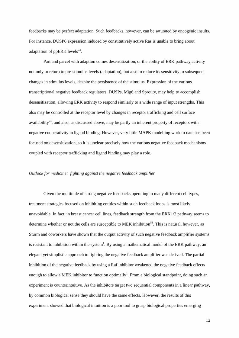
Figure 1. The basic ERK/MAPK pathway backbone. Various external stimuli can cause changes in
the levels of RasGTP (RasT), which induces activation of the three-tiered ERK1/2 cascade. Both
MEK and ERK need two phosphorylations to become fully activated kinases. Doubly phosphorylated
ERK (ppERK) enters the nucleus to affect gene transcription, but also has cytosolic substrates.
RasD RasT
GEFs
GAPs
Raf Raf*
MEK pMEK ppMEK
ERK pERK ppERK ppERK-nuc
RTK*
Stimulus Fig 1, Birtwistle and Kolch

15
Figure 2. Various negative feedback loops superimposed onto the ERK/MAPK backbone. The
ERK cascade backbone from Fig. 1 with short-term (light blue) and long-term (dark blue) negative
feedback depicted.
RasD RasT
SOS
GAPs
Raf Raf*
MEK pMEK ppMEK
ERK pERK ppERK
RTK*
Stimulus
Fig 2., Birtwistle and Kolch

16
Figure 3. Log-linear relationship between ligand dose and active receptors are explained by a
simple negative cooperativity model. Eq. 1 is plotted here with n=0.31 as measured by Alvarado et
al. for EGF binding to EGFR (solid black line), and with n=1 to give a reference for the case of no
cooperativity (black dashed line). The log-linear range extends approximately 4 to 5 decades for the
negative cooperativity system, and only 1 to 2 for the no cooperativity system.
Negative cooperativity (n=0.31) No
cooperativity (n=1)
Fig 3., Birtwistle and Kolch

17

18
References
1. Sturm OE, Orton R, Grindlay J, Birtwistle M, Vyshemirsky V, Gilbert D, Calder M, Pitt A,
Kholodenko B, Kolch W. The mammalian MAPK/ERK pathway exhibits properties of a negative
feedback amplifier. Sci Signal 2010; 3:ra90.
2. Black HS. Inventing Negative Feedback-Amplifier. Ieee Spectrum 1977; 14:54-60.
3. Katz M, Amit I, Yarden Y. Regulation of MAPKs by growth factors and receptor tyrosine
kinases. Biochim Biophys Acta 2007; 1773:1161-76.
4. Keshet Y, Seger R. The MAP kinase signaling cascades: a system of hundreds of components
regulates a diverse array of physiological functions. Methods Mol Biol 2010; 661:3-38.
5. Kolch W. Coordinating ERK/MAPK signalling through scaffolds and inhibitors. Nat Rev Mol
Cell Biol 2005; 6:827-37.
6. Raman M, Chen W, Cobb MH. Differential regulation and properties of MAPKs. Oncogene
2007; 26:3100-12.
7. Cuenda A, Cohen P, Buee-Scherrer V, Goedert M. Activation of stress-activated protein
kinase-3 (SAPK3) by cytokines and cellular stresses is mediated via SAPKK3 (MKK6); comparison
of the specificities of SAPK3 and SAPK2 (RK/p38). EMBO J 1997; 16:295-305.
8. Tournier C, Dong C, Turner TK, Jones SN, Flavell RA, Davis RJ. MKK7 is an essential
component of the JNK signal transduction pathway activated by proinflammatory cytokines. Genes
Dev 2001; 15:1419-26.
9. Yoon S, Seger R. The extracellular signal-regulated kinase: Multiple substrates regulate
diverse cellular functions. Growth Factors 2006; 24:21-44.
10. Nakakuki T, Birtwistle MR, Saeki Y, Yumoto N, Ide K, Nagashima T, Brusch L, Ogunnaike
BA, Okada-Hatakeyama M, Kholodenko BN. Ligand-specific c-Fos expression emerges from the
spatiotemporal control of ErbB network dynamics. Cell 2010; 141:884-96.
11. Marshall CJ. Specificity of receptor tyrosine kinase signaling: transient versus sustained
extracellular signal-regulated kinase activation. Cell 1995; 80:179-85.

19
12. Murphy LO, Smith S, Chen RH, Fingar DC, Blenis J. Molecular interpretation of ERK signal
duration by immediate early gene products. Nat Cell Biol 2002; 4:556-64.
13. Heinrich R, Neel BG, Rapoport TA. Mathematical models of protein kinase signal
transduction. Mol Cell 2002; 9:957-70.
14. Yao Z, Seger R. The ERK signaling cascade--views from different subcellular compartments.
Biofactors 2009; 35:407-16.
15. Duffy A, Kummar S. Targeting mitogen-activated protein kinase kinase (MEK) in solid
tumors. Target Oncol 2009; 4:267-73.
16. McCubrey JA, Steelman LS, Abrams SL, Chappell WH, Russo S, Ove R, Milella M, Tafuri
A, Lunghi P, Bonati A, Stivala F, Nicoletti F, Libra M, Martelli AM, Montalto G, Cervello M.
Emerging MEK inhibitors. Expert Opin Emerg Drugs 2010; 15:203-23.
17. Scheele JS, Rhee JM, Boss GR. Determination of absolute amounts of GDP and GTP bound
to Ras in mammalian cells: comparison of parental and Ras-overproducing NIH 3T3 fibroblasts. Proc
Natl Acad Sci U S A 1995; 92:1097-100.
18. van den Berghe N, Ouwens DM, Maassen JA, van Mackelenbergh MG, Sips HC, Krans HM.
Activation of the Ras/mitogen-activated protein kinase signaling pathway alone is not sufficient to
induce glucose uptake in 3T3-L1 adipocytes. Mol Cell Biol 1994; 14:2372-7.
19. Yonezawa K, Ando A, Kaburagi Y, Yamamoto-Honda R, Kitamura T, Hara K, Nakafuku M,
Okabayashi Y, Kadowaki T, Kaziro Y, et al. Signal transduction pathways from insulin receptors to
Ras. Analysis by mutant insulin receptors. J Biol Chem 1994; 269:4634-40.
20. Fujioka A, Terai K, Itoh RE, Aoki K, Nakamura T, Kuroda S, Nishida E, Matsuda M.
Dynamics of the Ras/ERK MAPK cascade as monitored by fluorescent probes. J Biol Chem 2006;
281:8917-26.
21. Schoeberl B, Eichler-Jonsson C, Gilles ED, Muller G. Computational modeling of the
dynamics of the MAP kinase cascade activated by surface and internalized EGF receptors. Nat
Biotechnol 2002; 20:370-5.
22. Keel BA, Davis JS. Epidermal growth factor activates extracellular signal-regulated protein
kinases (ERK) in freshly isolated porcine granulosa cells. Steroids 1999; 64:654-8.

20
23. Sasagawa S, Ozaki Y, Fujita K, Kuroda S. Prediction and validation of the distinct dynamics
of transient and sustained ERK activation. Nat Cell Biol 2005; 7:365-73.
24. Ferrell JE, Jr. How responses get more switch-like as you move down a protein kinase
cascade. Trends Biochem Sci 1997; 22:288-9.
25. Brown GC, Hoek JB, Kholodenko BN. Why do protein kinase cascades have more than one
level? Trends Biochem Sci 1997; 22:288.
26. Huang CY, Ferrell JE, Jr. Ultrasensitivity in the mitogen-activated protein kinase cascade.
Proc Natl Acad Sci U S A 1996; 93:10078-83.
27. Qiao L, Nachbar RB, Kevrekidis IG, Shvartsman SY. Bistability and oscillations in the
Huang-Ferrell model of MAPK signaling. PLoS Comput Biol 2007; 3:1819-26.
28. Markevich NI, Hoek JB, Kholodenko BN. Signaling switches and bistability arising from
multisite phosphorylation in protein kinase cascades. J Cell Biol 2004; 164:353-9.
29. Ferrell JE, Jr., Bhatt RR. Mechanistic studies of the dual phosphorylation of mitogen-
activated protein kinase. J Biol Chem 1997; 272:19008-16.
30. Zhao Y, Zhang ZY. The mechanism of dephosphorylation of extracellular signal-regulated
kinase 2 by mitogen-activated protein kinase phosphatase 3. J Biol Chem 2001; 276:32382-91.
31. Schilling M, Maiwald T, Hengl S, Winter D, Kreutz C, Kolch W, Lehmann WD, Timmer J,
Klingmuller U. Theoretical and experimental analysis links isoform-specific ERK signalling to cell
fate decisions. Mol Syst Biol 2009; 5:334.
32. Hagan S, Al-Mulla F, Mallon E, Oien K, Ferrier R, Gusterson B, Garcia JJ, Kolch W.
Reduction of Raf-1 kinase inhibitor protein expression correlates with breast cancer metastasis. Clin
Cancer Res 2005; 11:7392-7.
33. Claperon A, Therrien M. KSR and CNK: two scaffolds regulating RAS-mediated RAF
activation. Oncogene 2007; 26:3143-58.
34. Kholodenko BN, Birtwistle MR. Four-dimensional dynamics of MAPK information
processing systems. Wiley Interdiscip Rev Syst Biol Med 2009; 1:28-44.

21
35. von Kriegsheim A, Baiocchi D, Birtwistle M, Sumpton D, Bienvenut W, Morrice N, Yamada
K, Lamond A, Kalna G, Orton R, Gilbert D, Kolch W. Cell fate decisions are specified by the
dynamic ERK interactome. Nat Cell Biol 2009; 11:1458-64.
36. Kholodenko BN, Hancock JF, Kolch W. Signalling ballet in space and time. Nat Rev Mol
Cell Biol 2010; 11:414-26.
37. Buday L, Warne PH, Downward J. Downregulation of the Ras activation pathway by MAP
kinase phosphorylation of Sos. Oncogene 1995; 11:1327-31.
38. Dong C, Waters SB, Holt KH, Pessin JE. SOS phosphorylation and disassociation of the
Grb2-SOS complex by the ERK and JNK signaling pathways. J Biol Chem 1996; 271:6328-32.
39. Kamioka Y, Yasuda S, Fujita Y, Aoki K, Matsuda M. Multiple decisive phosphorylation sites
for the negative feedback regulation of SOS1 via ERK. J Biol Chem.
40. Lehr S, Kotzka J, Avci H, Sickmann A, Meyer HE, Herkner A, Muller-Wieland D.
Identification of major ERK-related phosphorylation sites in Gab1. Biochemistry 2004; 43:12133-40.
41. Dougherty MK, Muller J, Ritt DA, Zhou M, Zhou XZ, Copeland TD, Conrads TP, Veenstra
TD, Lu KP, Morrison DK. Regulation of Raf-1 by direct feedback phosphorylation. Mol Cell 2005;
17:215-24.
42. Cirit M, Wang CC, Haugh JM. Systematic quantification of negative feedback mechanisms in
the extracellular signal-regulated kinase (ERK) signaling network. J Biol Chem; 285:36736-44.
43. Takishima K, Friedman B, Fujiki H, Rosner MR. Thapsigargin, a novel promoter,
phosphorylates the epidermal growth factor receptor at threonine 669. Biochem Biophys Res
Commun 1988; 157:740-6.
44. Heisermann GJ, Wiley HS, Walsh BJ, Ingraham HA, Fiol CJ, Gill GN. Mutational removal of
the Thr669 and Ser671 phosphorylation sites alters substrate specificity and ligand-induced
internalization of the epidermal growth factor receptor. J Biol Chem 1990; 265:12820-7.
45. Li X, Huang Y, Jiang J, Frank SJ. ERK-dependent threonine phosphorylation of EGF receptor
modulates receptor downregulation and signaling. Cell Signal 2008; 20:2145-55.

22
46. Gan Y, Shi C, Inge L, Hibner M, Balducci J, Huang Y. Differential roles of ERK and Akt
pathways in regulation of EGFR-mediated signaling and motility in prostate cancer cells. Oncogene
2010; 29:4947-58.
47. Birtwistle MR, Hatakeyama M, Yumoto N, Ogunnaike BA, Hoek JB, Kholodenko BN.
Ligand-dependent responses of the ErbB signaling network: experimental and modeling analyses.
Mol Syst Biol 2007; 3:144.
48. Brondello JM, Brunet A, Pouyssegur J, McKenzie FR. The dual specificity mitogen-activated
protein kinase phosphatase-1 and -2 are induced by the p42/p44(MAPK) cascade. Journal of
Biological Chemistry 1997; 272:1368-76.
49. Owens DM, Keyse SM. Differential regulation of MAP kinase signalling by dual-specificity
protein phosphatases. Oncogene 2007; 26:3203-13.
50. Brondello JM, Pouyssegur J, McKenzie FR. Reduced MAP kinase phosphatase-1 degradation
after p42/p44MAPK-dependent phosphorylation. Science 1999; 286:2514-7.
51. Nichols A, Camps M, Gillieron C, Chabert C, Brunet A, Wilsbacher J, Cobb M, Pouyssegur
J, Shaw JP, Arkinstall S. Substrate recognition domains within extracellular signal-regulated kinase
mediate binding and catalytic activation of mitogen-activated protein kinase phosphatase-3. J Biol
Chem 2000; 275:24613-21.
52. Guy GR, Jackson RA, Yusoff P, Chow SY. Sprouty proteins: modified modulators,
matchmakers or missing links? Journal of Endocrinology 2009; 203:191-202.
53. Bundschu K, Walter U, Schuh K. Getting a first clue about SPRED functions. Bioessays
2007; 29:897-907.
54. Anastasi S, Fiorentino L, Fiorini M, Fraioli R, Sala G, Castellani L, Alema S, Alimandi M,
Segatto O. Feedback inhibition by RALT controls signal output by the ErbB network. Oncogene
2003; 22:4221-34.
55. Zhang X, Pickin KA, Bose R, Jura N, Cole PA, Kuriyan J. Inhibition of the EGF receptor by
binding of MIG6 to an activating kinase domain interface. Nature 2007; 450:741-4.

23
56. Frosi Y, Anastasi S, Ballaro C, Varsano G, Castellani L, Maspero E, Polo S, Alema S,
Segatto O. A two-tiered mechanism of EGFR inhibition by RALT/MIG6 via kinase suppression and
receptor degradation. J Cell Biol 2010; 189:557-71.
57. Nakayama K, Satoh T, Igari A, Kageyama R, Nishida E. FGF induces oscillations of Hes1
expression and Ras/ERK activation. Curr Biol 2008; 18:R332-4.
58. Mirzoeva OK, Das D, Heiser LM, Bhattacharya S, Siwak D, Gendelman R, Bayani N, Wang
NJ, Neve RM, Guan Y, Hu Z, Knight Z, Feiler HS, Gascard P, Parvin B, Spellman PT, Shokat KM,
Wyrobek AJ, Bissell MJ, McCormick F, Kuo WL, Mills GB, Gray JW, Korn WM. Basal subtype and
MAPK/ERK kinase (MEK)-phosphoinositide 3-kinase feedback signaling determine susceptibility of
breast cancer cells to MEK inhibition. Cancer Res 2009; 69:565-72.
59. Sauro HM, Kholodenko BN. Quantitative analysis of signaling networks. Prog Biophys Mol
Biol 2004; 86:5-43.
60. Joslin EJ, Shankaran H, Opresko LK, Bollinger N, Lauffenburger DA, Wiley HS. Structure of
the EGF receptor transactivation circuit integrates multiple signals with cell context. Mol Biosyst
2010; 6:1293-306.
61. Borisov N, Aksamitiene E, Kiyatkin A, Legewie S, Berkhout J, Maiwald T, Kaimachnikov
NP, Timmer J, Hoek JB, Kholodenko BN. Systems-level interactions between insulin-EGF networks
amplify mitogenic signaling. Mol Syst Biol 2009; 5:256.
62. Chen WW, Schoeberl B, Jasper PJ, Niepel M, Nielsen UB, Lauffenburger DA, Sorger PK.
Input-output behavior of ErbB signaling pathways as revealed by a mass action model trained against
dynamic data. Mol Syst Biol 2009; 5:239.
63. Alvarado D, Klein DE, Lemmon MA. Structural basis for negative cooperativity in growth
factor binding to an EGF receptor. Cell 2010; 142:568-79.
64. Saji M, Taga M, Matsui H, Suyama K, Kurogi K, Minaguchi H. Gene expression and specific
binding of platelet-derived growth factor and its effect on DNA synthesis in human decidual cells.
Mol Cell Endocrinol 1997; 132:73-80.
65. Whittaker J, Garcia P, Yu GQ, Mynarcik DC. Transmembrane domain interactions are
necessary for negative cooperativity of the insulin receptor. Mol Endocrinol 1994; 8:1521-7.
24
66. De Meyts P. The structural basis of insulin and insulin-like growth factor-I receptor binding
and negative co-operativity, and its relevance to mitogenic versus metabolic signalling. Diabetologia
1994; 37 Suppl 2:S135-48.
67. Kholodenko BN. Negative feedback and ultrasensitivity can bring about oscillations in the
mitogen-activated protein kinase cascades. Eur J Biochem 2000; 267:1583-8.
68. Anjum R, Blenis J. The RSK family of kinases: emerging roles in cellular signalling. Nat Rev
Mol Cell Biol 2008; 9:747-58.
69. Kholodenko BN. Cell-signalling dynamics in time and space. Nat Rev Mol Cell Biol 2006;
7:165-76.
70. Shankaran H, Ippolito DL, Chrisler WB, Resat H, Bollinger N, Opresko LK, Wiley HS.
Rapid and sustained nuclear-cytoplasmic ERK oscillations induced by epidermal growth factor. Mol
Syst Biol 2009; 5:332.
71. Shankaran H, Wiley HS. Oscillatory dynamics of the extracellular signal-regulated kinase
pathway. Curr Opin Genet Dev 2010; 20:650-5.
72. Behar M, Hao N, Dohlman HG, Elston TC. Mathematical and computational analysis of
adaptation via feedback inhibition in signal transduction pathways. Biophys J 2007; 93:806-21.
73. Bluthgen N, Legewie S, Kielbasa SM, Schramme A, Tchernitsa O, Keil J, Solf A, Vingron M,
Schafer R, Herzel H, Sers C. A systems biological approach suggests that transcriptional feedback
regulation by dual-specificity phosphatase 6 shapes extracellular signal-related kinase activity in
RAS-transformed fibroblasts. FEBS J 2009; 276:1024-35.
74. Sorkin A, Goh LK. Endocytosis and intracellular trafficking of ErbBs. Exp Cell Res 2009;
315:683-96.


![S---We - DTIC · 2011-05-14 · II. BLACK'S FORMUJLA GENERALIZED H.S. Black's invention of the negative feedback amplifier was based on the following analysis [3]:. consider the feedback](https://static.fdocuments.in/doc/165x107/5f08c90f7e708231d423b5fc/s-we-dtic-2011-05-14-ii-blacks-formujla-generalized-hs-blacks-invention.jpg)


![POSITIVE AND NEGATIVE FEEDBACK IN POLITICS[ Positive and Negative Feedback in Politics ] 5 5 equilibrium. Positive and negative feedback processes lead alternately to the creation,](https://static.fdocuments.in/doc/165x107/5e6fc60d27274a5c975cef86/positive-and-negative-feedback-in-politics-positive-and-negative-feedback-in-politics.jpg)