Biology Sylvia S. Mader Michael Windelspecht Chapter 9 The Cell Cycle and Cellular Reproduction...
51
Biology Sylvia S. Mader Michael Windelspecht Chapter 9 The Cell Cycle and Cellular Reproduction Lecture Outline Copyright © The McGraw-Hill Companies, Inc. Permission required for reproduction or display. See separate FlexArt PowerPoint slides for all figures and tables pre-inserted into PowerPoint without notes. 1
-
Upload
aubrie-richard -
Category
Documents
-
view
256 -
download
5
Transcript of Biology Sylvia S. Mader Michael Windelspecht Chapter 9 The Cell Cycle and Cellular Reproduction...
BiologySylvia S. Mader
Michael Windelspecht
Chapter 9The Cell Cycle and
Cellular Reproduction
Lecture Outline
Copyright © The McGraw-Hill Companies, Inc. Permission required for reproduction or display.
See separate FlexArt PowerPoint slides for all figures and tables pre-inserted into
PowerPoint without notes.
1
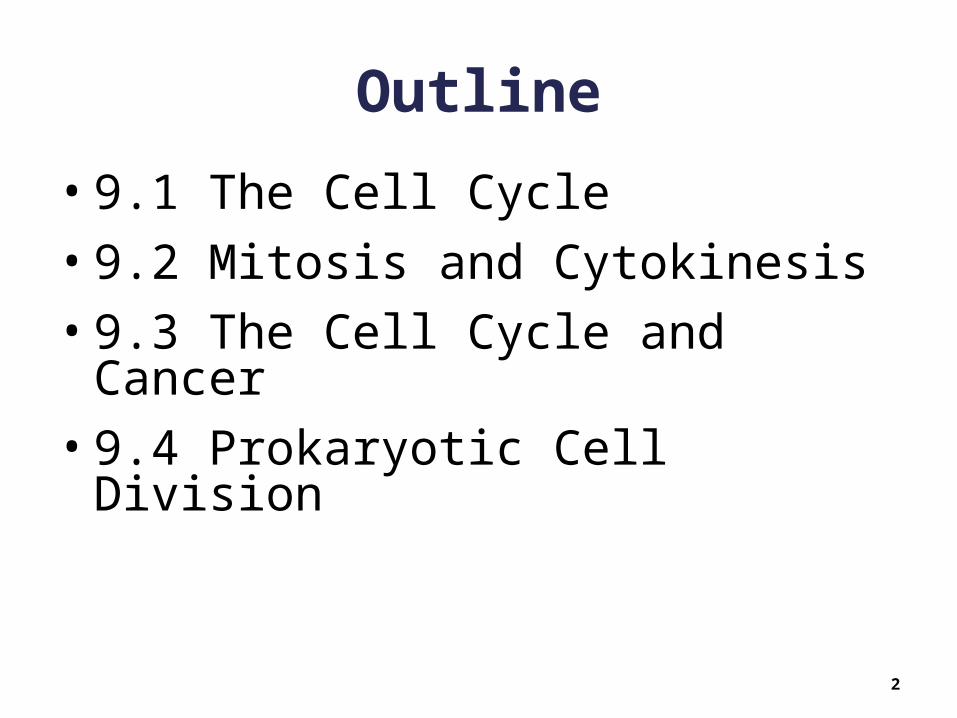
Outline
• 9.1 The Cell Cycle
• 9.2 Mitosis and Cytokinesis
• 9.3 The Cell Cycle and Cancer
• 9.4 Prokaryotic Cell Division
2

9.1 The Cell Cycle
• The cell cycle is an orderly set of stages from the first division to the time the resulting daughter cells divide
• Just prior to the next division: The cell grows larger The number of organelles doubles The DNA is replicated
• The two major stages of the cell cycle: Interphase (includes several stages) Mitotic Stage (includes mitosis and cytokinesis)
3

The Cell Cycle
4
Copyright © The McGraw-Hill Companies, Inc. Permission required for reproduction or display.
G1
(growth)G0
G2 checkpointMitosis checkpoint.Mitosis will occurif DNA hasreplicated properly.Apoptosis willoccur if the DNA isdamaged andcannot be repaired.
S(growth and DNA
replication)
M
Cytokinesis
Telo
phas
eA
nap
has
e
Met
aph
ase
Late
pro
phas
e
Prophase
Interphase
G1 checkpointCell cycle main checkpoint.If DNA is damaged, apoptosiswill occur. Otherwise, the cellis committed to divide whengrowth signals are presentand nutrients are available.
M checkpointSpindle assemblycheckpoint. Mitosiswill not continue ifchromosomes arenot properly aligned.
M
G2
G1
G2
(growth and finalpreparations for
division)

The Cell Cycle
• Interphase Most of the cell cycle is spent in
interphase Cell performs its usual functions Time spent in interphase varies by cell
type Nerve and muscle cells do not complete
the cell cycle (remain in the G0 stage)
5

The Cell Cycle
• Interphase consists of: G1, S, and G2 phases G1 Phase:
• Recovery from previous division• Cell doubles its organelles• Cell grows in size• Cell accumulates raw materials for DNA synthesis
S Phase:• DNA replication • Proteins associated with DNA are synthesized • Chromosomes enter with 1 chromatid each• Chromosomes leave with 2 identical chromatids (sister
chromatids) each G2 Phase:
• Between DNA replication and onset of mitosis• Cell synthesizes proteins necessary for division
6
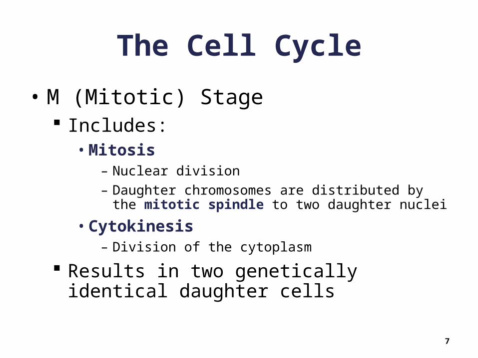
The Cell Cycle
• M (Mitotic) Stage Includes:
• Mitosis – Nuclear division
– Daughter chromosomes are distributed by the mitotic spindle to two daughter nuclei
• Cytokinesis– Division of the cytoplasm
Results in two genetically identical daughter cells
7
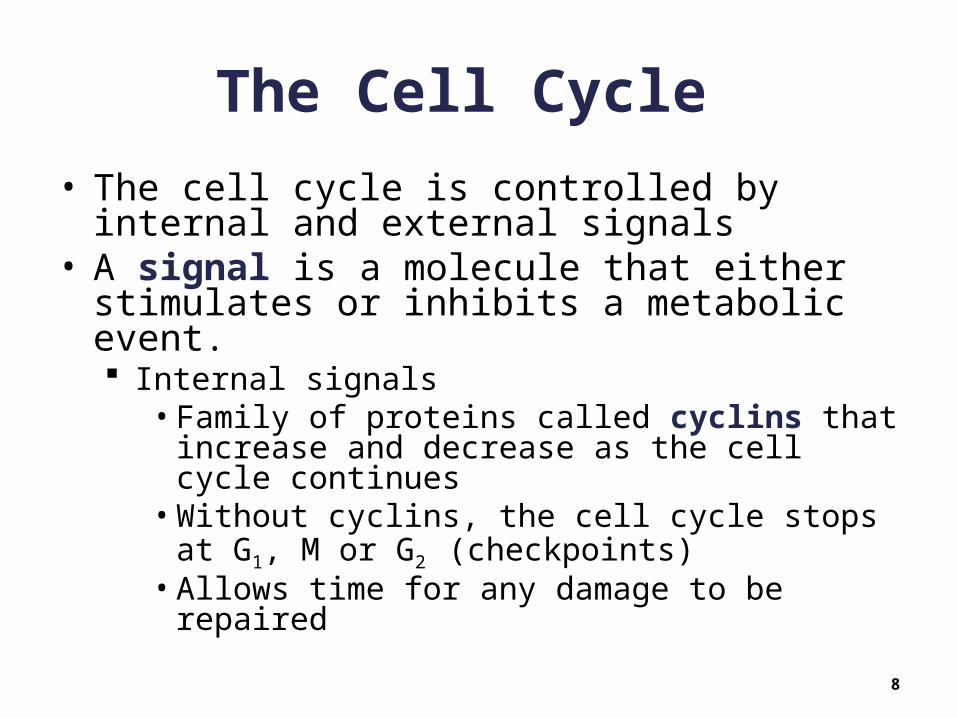
The Cell Cycle
• The cell cycle is controlled by internal and external signals
• A signal is a molecule that either stimulates or inhibits a metabolic event. Internal signals
• Family of proteins called cyclins that increase and decrease as the cell cycle continues
• Without cyclins, the cell cycle stops at G1, M or G2 (checkpoints)
• Allows time for any damage to be repaired
8
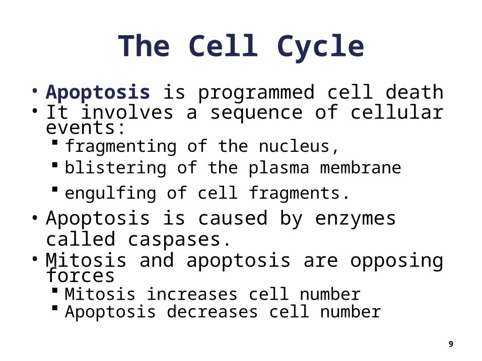
The Cell Cycle
• Apoptosis is programmed cell death• It involves a sequence of cellular events:
fragmenting of the nucleus, blistering of the plasma membrane engulfing of cell fragments.
• Apoptosis is caused by enzymes called caspases.
• Mitosis and apoptosis are opposing forces Mitosis increases cell number Apoptosis decreases cell number
9

Apoptosis
10
Copyright © The McGraw-Hill Companies, Inc. Permission required for reproduction or display.
apoptotic cell
cellfragment
DNAfragment
Cell roundsup, and nucleuscollapses.
Chromatincondenses, andnucleus fragments.
Plasma membraneblisters, and blebsform.
Cell fragmentscontain DNAfragments.
blebs
Courtesy Douglas R. Green/LaJolla Institute for Allergy and Immunology

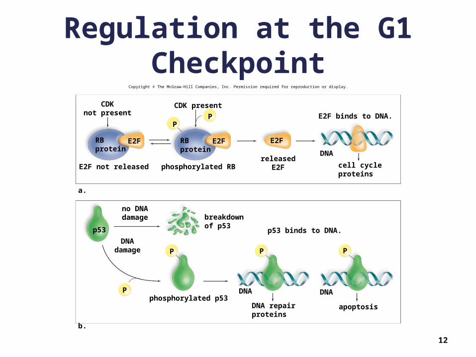
The Cell Cycle
• Apoptosis Cells harbor caspases that are kept in
check by inhibitors• Can be unleashed by internal or external
signals Signal protein p53
• Stops the cell cycle at G1 when DNA is damaged
• Initiates an attempt at DNA repair–If successful, the cycle continues to
mitosis–If not, apoptosis is initiated
11
Regulation at the G1 Checkpoint
12
Copyright © The McGraw-Hill Companies, Inc. Permission required for reproduction or display.
a.
PP
RBprotein
RBprotein
E2FE2F E2F
CDKnot present
E2F not releasedreleased
E2F
E2F binds to DNA.
DNA
cell cycleproteins
phosphorylated RB
CDK present
b.
P
P
P P
breakdownof p53
no DNAdamage
DNAdamage
phosphorylated p53DNA repairproteins
apoptosis
p53 binds to DNA.
DNA DNA
p53
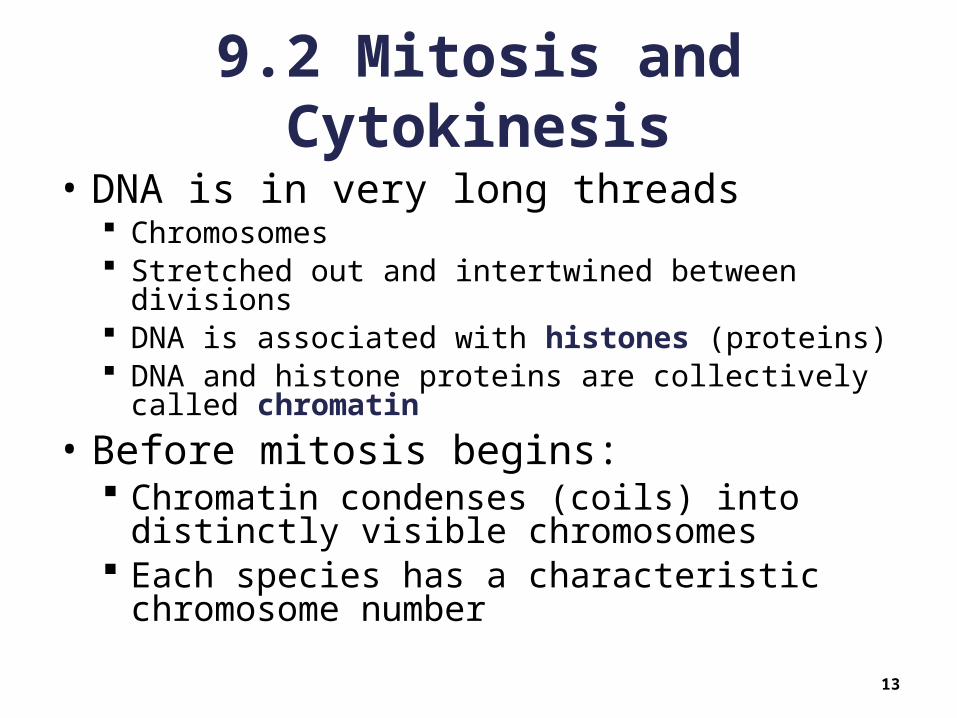
9.2 Mitosis and Cytokinesis
• DNA is in very long threads Chromosomes Stretched out and intertwined between divisions DNA is associated with histones (proteins) DNA and histone proteins are collectively called
chromatin
• Before mitosis begins: Chromatin condenses (coils) into distinctly
visible chromosomes Each species has a characteristic
chromosome number
13
14
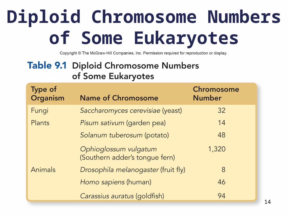
Diploid Chromosome Numbers of Some Eukaryotes

Mitosis and Cytokinesis
• The diploid (2n) number includes two sets of chromosomes of each type Humans have 23 different types of
chromosomes• Each type is represented twice in each body cell
(diploid)• Only sperm and eggs have one of each type
·termed haploid (n) The haploid (n) number for humans is 23
• Two representatives of each chromosome type• Makes a total of 2n = 46 in each nucleus
– One set of 23 from individual’s father (paternal)– Other set of 23 from individual’s mother (maternal)
15

Mitosis and Cytokinesis
• At the end of S phase: Each chromosome internally duplicated Consists of two identical DNA chains
• Sister chromatids (two strands of genetically identical chromosomes)
• Attached together at a single point (called centromere)
• During mitosis: Centromeres holding sister chromatids together
separate Sister chromatids separate Each becomes a daughter chromosome Sisters of each type are distributed to opposite
daughter nuclei
16

Duplicated Chromosomes
17
Copyright © The McGraw-Hill Companies, Inc. Permission required for reproduction or display.
centromere
sister chromatids
one chromatida. b.
kinetochore
9,850© Andrew Syred/Photo Researchers, Inc.
Mitosis and Cytokinesis
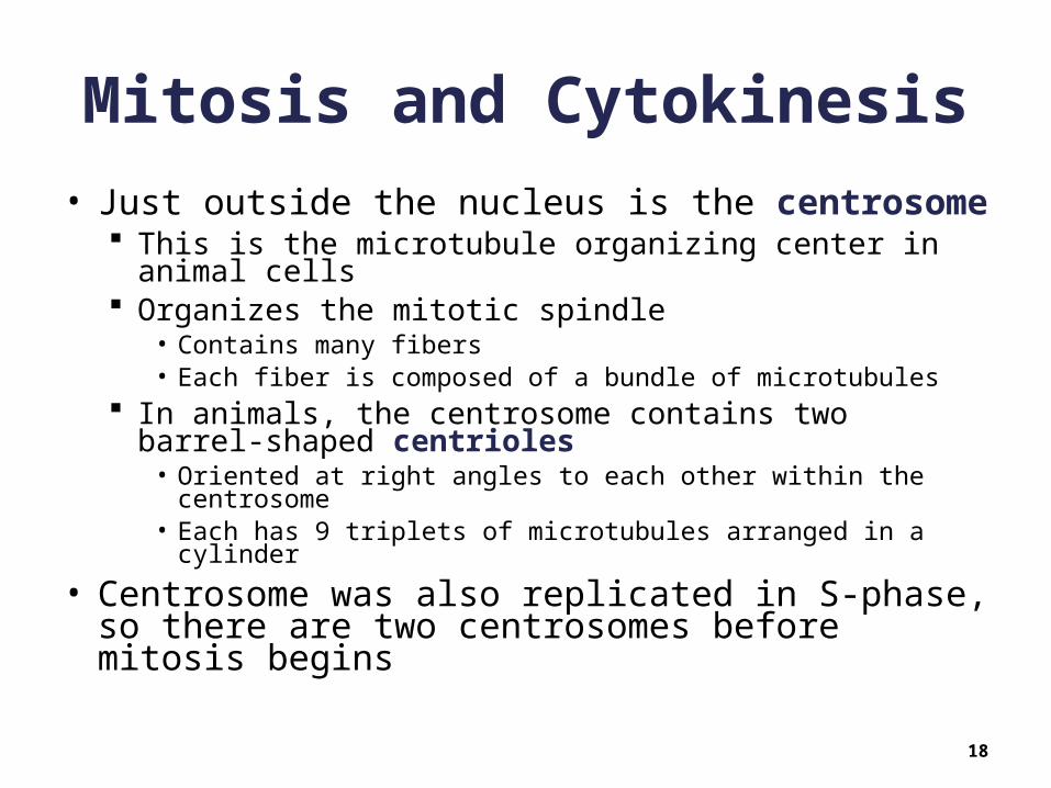
• Just outside the nucleus is the centrosome This is the microtubule organizing center in animal
cells Organizes the mitotic spindle
• Contains many fibers• Each fiber is composed of a bundle of microtubules
In animals, the centrosome contains two barrel-shaped centrioles
• Oriented at right angles to each other within the centrosome• Each has 9 triplets of microtubules arranged in a cylinder
• Centrosome was also replicated in S-phase, so there are two centrosomes before mitosis begins
18
Mitosis and Cytokinesis
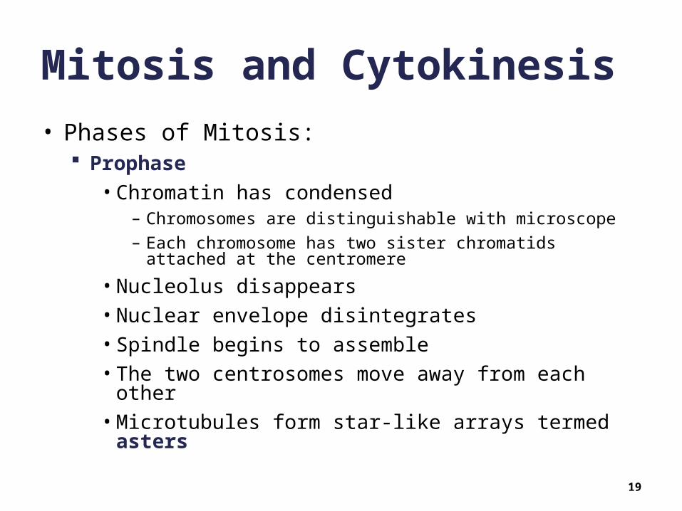
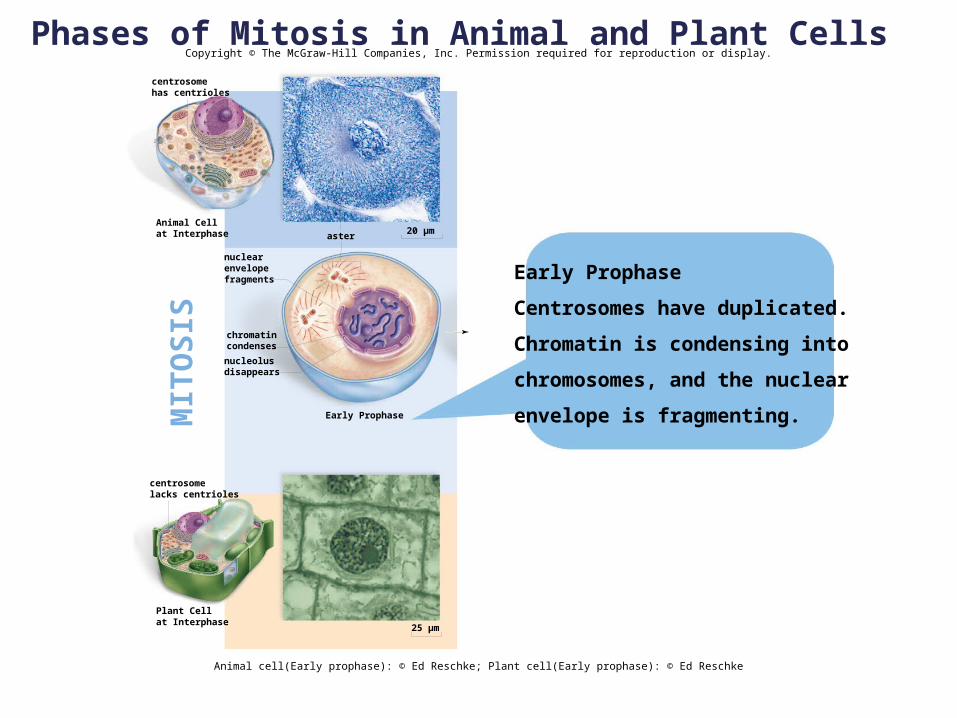
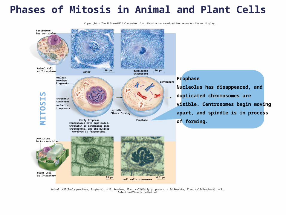
• Phases of Mitosis: Prophase
• Chromatin has condensed– Chromosomes are distinguishable with microscope
– Each chromosome has two sister chromatids attached at the centromere
• Nucleolus disappears
• Nuclear envelope disintegrates
• Spindle begins to assemble
• The two centrosomes move away from each other
• Microtubules form star-like arrays termed asters
19
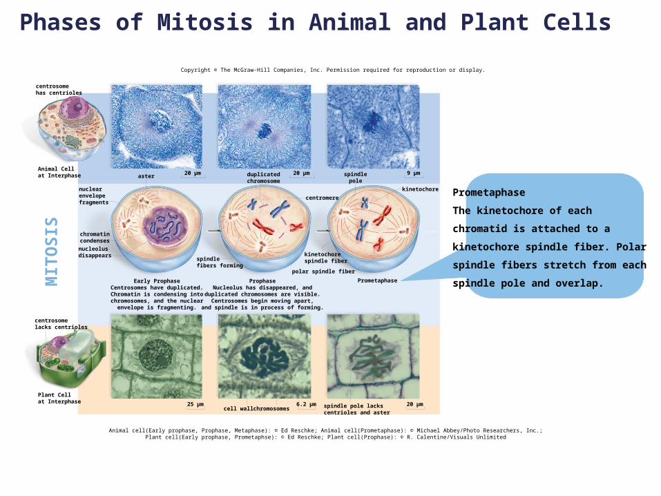
Mitosis and Cytokinesis
• Phases of Mitosis
Prometaphase
• The centromere of each chromosome develops two kinetochores
– Specialized protein complex
– One attached to each sister chromatid
» Physically connect sister chromatids with specialized microtubules (kinetochores)
» These connect sister chromatids to opposite poles of the mother cell
20
Mitosis and Cytokinesis
• Stages of Mitosis Metaphase
• Chromosomes are pulled around by kinetochore fibers
• Forced to align across the equatorial plane of the cell
– Metaphase plate - Represents plane through which mother cell will be divided
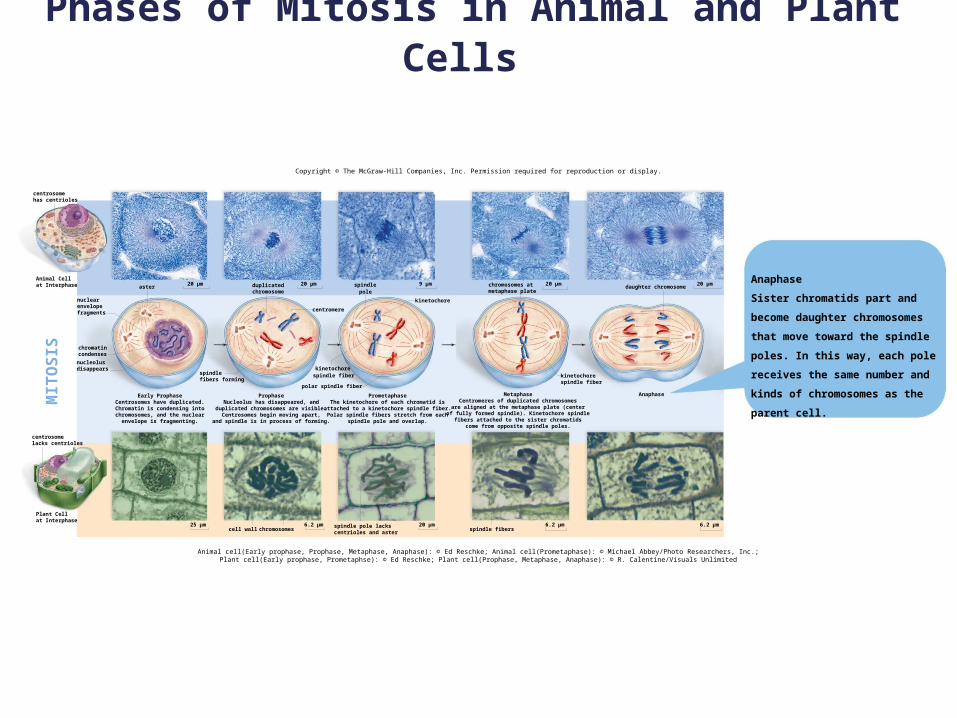
Anaphase• Centromere dissolves, releasing sister chromatids
• Sister chromatids separate
– Now called daughter chromosomes
– Pulled to opposite poles along kinetochore fibers
21

Mitosis and Cytokinesis
• Stages of Mitosis Telophase
• Spindle disappears
• Now two clusters of daughter chromosomes
– Still two of each type with all types represented
– Clusters are incipient daughter nuclei
• Nuclear envelopes form around the two incipient daughter nuclei
– Each daughter nucleus receives one chromosome of each type
22
Mitosis and Cytokinesis
• Cytokinesis = division of cytoplasm• Allocates the mother cell’s cytoplasm equally to
daughter nucleus• Encloses each daughter cell in its own plasma
membrane• Often begins in anaphase• Animal cytokinesis:
A cleavage furrow appears between daughter nuclei Formed by a contractile ring of actin filaments Like pulling on a drawstring Eventually pinches the mother cell in two
23

Mitosis and Cytokinesis
• Cytokinesis in plant cells begins with the formation of a cell plate Rigid cell walls outside plasma membrane do not
permit furrowing Many small membrane-bounded vesicles Eventually fuse into one thin vesicle extending across
the mother cell The membranes of the cell plate become the plasma
membrane between the daughter cells The space between the daughter cells becomes filled
with the middle lamella Daughter cells later secrete primary cell walls on
opposite sides of the middle lamella
24
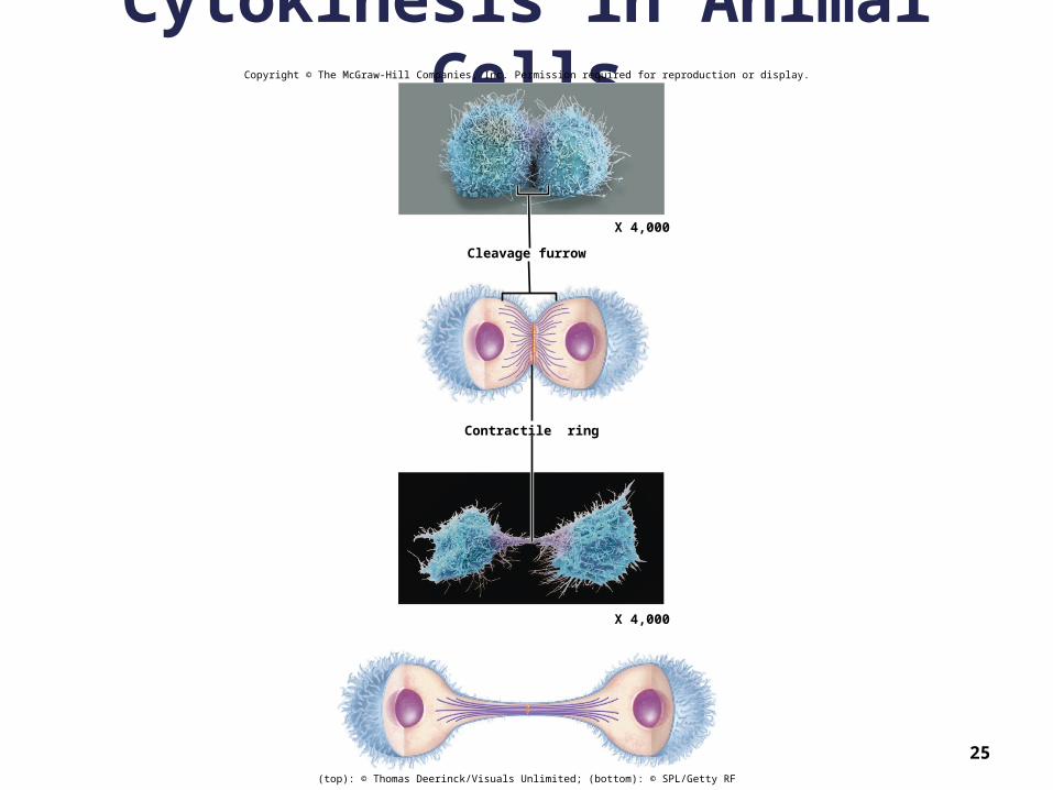
Cytokinesis in Animal Cells
25
Copyright © The McGraw-Hill Companies, Inc. Permission required for reproduction or display.
Cleavage furrow
Contractile ring
X 4,000
X 4,000
(top): © Thomas Deerinck/Visuals Unlimited; (bottom): © SPL/Getty RF

Cytokinesis in Plant Cells
26
Copyright © The McGraw-Hill Companies, Inc. Permission required for reproduction or display.
daughter cells
nucleoli
daughternucleus
cell plate formation
daughter nucleus
vesicles containingmembrane componentsfusing to form cell plate
(left): © B.A. Palevitz and E.H. Newcomb/BPS/Tom Stack & Associates

27
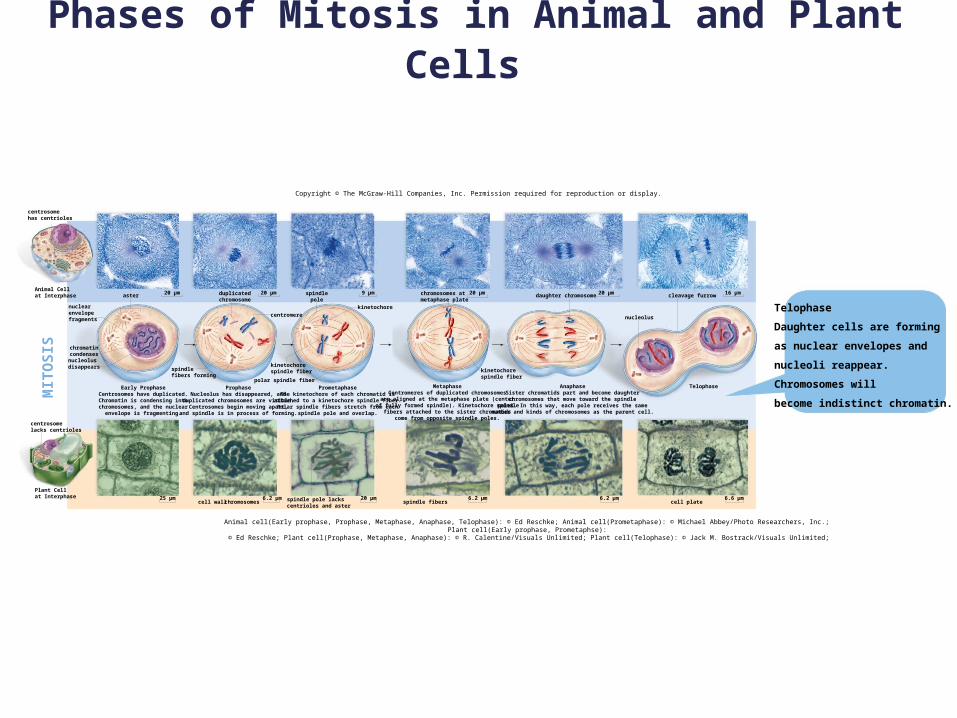
Phases of Mitosis in Animal and Plant Cells
Copyright © The McGraw-Hill Companies, Inc. Permission required for reproduction or display.
Animal cell(Early prophase, Prophase, Metaphase, Anaphase, Telophase): © Ed Reschke; Animal cell(Prometaphase): © Michael Abbey/Photo Researchers, Inc.;Plant cell(Early prophase, Prometaphse): © Ed Reschke; Plant cell(Prophase, Metaphase, Anaphase): © R. Calentine/Visuals Unlimited; Plant cell(Telophase): © Jack M.
Bostrack/Visuals Unlimited;
Plant Cellat Interphase
centromere
aster
kinetochore
polar spindle fiber
chromosomescell wall25µm
centrosome
lacks centrioles
MIT
OS
IS
centrosomehas centrioles
Animal Cellat Interphase
nuclearenvelopefragments
chromatincondenses
nucleolusdisappears
Early ProphaseCentrosomes have duplicated.Chromatin is condensing into
chromosomes, and the nuclearenvelope is fragmenting.
ProphaseNucleolus has disappeared, and
duplicated chromosomes are visible.Centrosomes begin moving apart,
and spindle is in process of forming.
ProphaseNucleolus has disappeared, and
duplicated chromosomes are visible.Centrosomes begin moving apart,
and spindle is in process of forming.
20 µm duplicatedchromosome
20 µm
spindlefibers forming
spindlepole
9 µm
kinetochorespindle fiber
cleavage furrow
spindle fibers
20µm 16µm
kinetochorespindle fiber
AnaphaseSister chromatids part and become daughterchromosomes that move toward the spindle
poles. In this way, each pole receives the samenumber and kinds of chromosomes as the parent cell.
MetaphaseCentromeres of duplicated chromosomesare aligned at the metaphase plate (center
of fully formed spindle). Kinetochore spindlefibers attached to the sister chromatids
come from opposite spindle poles.
chromosomes atmetaphase plate
6.2µm6.2µm20µm6.2µmspindle pole lackscentrioles and aster
TelophaseDaughter cells are formingas nuclear envelopes and
nucleoli reappear. Chromosomes willbecome indistinct chromatin.
daughter chromosome 20µm
nucleolus
cell plate 6.6µm
Phases of Mitosis in Animal and Plant Cells
Animal cell(Early prophase): © Ed Reschke; Plant cell(Early prophase): © Ed Reschke
Copyright © The McGraw-Hill Companies, Inc. Permission required for reproduction or display.
Early Prophase
aster20 µm
25 µm
MIT
OS
IS
Early Prophase
Centrosomes have duplicated.
Chromatin is condensing into
chromosomes, and the nuclear
envelope is fragmenting.
chromatincondenses
nucleolusdisappears
nuclearenvelopefragments
Animal Cellat Interphase
centrosomehas centrioles
centrosomelacks centrioles
Plant Cellat Interphase
Phases of Mitosis in Animal and Plant Cells Copyright © The McGraw-Hill Companies, Inc. Permission required for reproduction or display.
Animal cell(Early prophase, Prophase): © Ed Reschke; Plant cell(Early prophase): © Ed Reschke; Plant cell(Prophase): © R. Calentine/Visuals Unlimited
Prophase
spindlefibers forming
centromere
aster
chromosomescell wall
20 µm 20 µm
6.2 µm25 µm
Prophase
Nucleolus has disappeared, and
duplicated chromosomes are
visible. Centrosomes begin moving
apart, and spindle is in process
of forming.MIT
OS
IS
centrosomelacks centrioles
Animal Cellat Interphase
centrosomehas centrioles
nuclearenvelopefragments
chromatincondenses
nucleolusdisappears
Early ProphaseCentrosomes have duplicated.Chromatin is condensing into
chromosomes, and the nuclearenvelope is fragmenting.
duplicatedchromosome
Plant Cellat Interphase
Phases of Mitosis in Animal and Plant Cells
Copyright © The McGraw-Hill Companies, Inc. Permission required for reproduction or display.
Animal cell(Early prophase, Prophase, Metaphase): © Ed Reschke; Animal cell(Prometaphase): © Michael Abbey/Photo Researchers, Inc.;Plant cell(Early prophase, Prometaphse): © Ed Reschke; Plant cell(Prophase): © R. Calentine/Visuals Unlimited
Prometaphase
centromere
spindlepole
aster
kinetochore
polar spindle fiber
chromosomescell wall
20 µm 20 µm
6.2 µm25 µm 20 µm
9 µm
Prometaphase
The kinetochore of each
chromatid is attached to a
kinetochore spindle fiber. Polar
spindle fibers stretch from each
spindle pole and overlap.
centrosomehas centrioles
Animal Cellat Interphase
nuclearenvelopefragments
chromatincondenses
nucleolusdisappears
spindlefibers forming
Early ProphaseCentrosomes have duplicated.Chromatin is condensing into
chromosomes, and the nuclearenvelope is fragmenting.
ProphaseNucleolus has disappeared, and
duplicated chromosomes are visible.Centrosomes begin moving apart,
and spindle is in process of forming.
kinetochorespindle fiber
duplicatedchromosome
spindle pole lackscentrioles and aster
Plant Cellat Interphase
centrosomelacks centrioles
MIT
OS
IS

Phases of Mitosis in Animal and Plant Cells
Copyright © The McGraw-Hill Companies, Inc. Permission required for reproduction or display.
Animal cell(Early prophase, Prophase, Metaphase): © Ed Reschke; Animal cell(Prometaphase): © Michael Abbey/Photo Researchers, Inc.;Plant cell(Early prophase, Prometaphse): © Ed Reschke; Plant cell(Prophase, Metaphase): © R. Calentine/Visuals Unlimited
duplicatedchromosome
centromere
aster
kinetochore
polar spindle fiber
chromosomescell wall
20 µm
6.2 µm25 µm 20 µm
9 µm
spindle fibers
20 µm
6.2 µm
Metaphase
Centromeres of duplicated
chromosomes are aligned at the
metaphase plate (center of fully
formed spindle). Kinetochore
spindle fibers attached to the
sister chromatids come from
opposite spindle poles.
centrosomehas centrioles
Animal Cellat Interphase
nuclearenvelopefragments
nucleolusdisappears
chromatincondenses
centrosomelacks centrioles
Early ProphaseCentrosomes have duplicated.Chromatin is condensing into
chromosomes, and the nuclearenvelope is fragmenting.
ProphaseNucleolus has disappeared, and
duplicated chromosomes are visible.Centrosomes begin moving apart,
and spindle is in process of forming.
PrometaphaseThe kinetochore of each chromatid is
attached to a kinetochore spindle fiber.Polar spindle fibers stretch from each
spindle pole and overlap.
Metaphase
kinetochorespindle fiber
chromosomes atmetaphase plate
spindlepole
kinetochorespindle fiberspindle
fibers forming
Plant Cellat Interphase
spindle pole lackscentrioles and aster
MIT
OS
IS
20 µm
Phases of Mitosis in Animal and Plant Cells
Copyright © The McGraw-Hill Companies, Inc. Permission required for reproduction or display.
Animal cell(Early prophase, Prophase, Metaphase, Anaphase): © Ed Reschke; Animal cell(Prometaphase): © Michael Abbey/Photo Researchers, Inc.;Plant cell(Early prophase, Prometaphse): © Ed Reschke; Plant cell(Prophase, Metaphase, Anaphase): © R. Calentine/Visuals Unlimited
centromere
spindlepole
aster
kinetochore
kinetochorespindle fiber
polar spindle fiber
chromosomescell wall
20 µm 20 µm
6.2 µm25 µm 20 µm
9 µmdaughter chromosome
spindle fibers
20 µm 20 µm
6.2 µm6.2 µm
centrosomehas centrioles
Animal Cellat Interphase
nuclearenvelopefragments
nucleolusdisappears
chromatincondenses
centrosomelacks centrioles
Early ProphaseCentrosomes have duplicated.Chromatin is condensing into
chromosomes, and the nuclearenvelope is fragmenting.
ProphaseNucleolus has disappeared, and
duplicated chromosomes are visible.Centrosomes begin moving apart,
and spindle is in process of forming.
PrometaphaseThe kinetochore of each chromatid is
attached to a kinetochore spindle fiber.Polar spindle fibers stretch from each
spindle pole and overlap.
MetaphaseCentromeres of duplicated chromosomesare aligned at the metaphase plate (center
of fully formed spindle). Kinetochore spindlefibers attached to the sister chromatids
come from opposite spindle poles.
kinetochorespindle fiber
Anaphase
chromosomes atmetaphase plate
spindle pole lackscentrioles and aster
Plant Cellat Interphase
duplicatedchromosome
spindlefibers forming
MIT
OS
IS
Anaphase
Sister chromatids part and become
daughter chromosomes that move
toward the spindle poles. In this
way, each pole receives the same
number and kinds of chromosomes
as the parent cell.
Phases of Mitosis in Animal and Plant Cells
Copyright © The McGraw-Hill Companies, Inc. Permission required for reproduction or display.
centromere
aster
kinetochore
polar spindle fiber
chromosomescell wall
20 µm 20 µm
6.2 µm25 µm 20 µm
9 µm daughter chromosome
spindle fibers cell plate
20 µm 20 µm 16 µm
6.2 µm 6.6 µm6.2 µm
nucleolus
cleavage furrow
Telophase
Daughter cells are forming
as nuclear envelopes and
nucleoli reappear.
Chromosomes will
become indistinct chromatin.
centrosomehas centrioles
Animal Cellat Interphase
nuclearenvelopefragments
chromatincondensesnucleolusdisappears
centrosomelacks centrioles
Plant Cellat Interphase
Early ProphaseCentrosomes have duplicated.Chromatin is condensing into
chromosomes, and the nuclearenvelope is fragmenting.
spindlefibers forming
duplicatedchromosome
kinetochorespindle fiber
ProphaseNucleolus has disappeared, and
duplicated chromosomes are visible.Centrosomes begin moving apart,
and spindle is in process of forming.
PrometaphaseThe kinetochore of each chromatid is
attached to a kinetochore spindle fiber.Polar spindle fibers stretch from each
spindle pole and overlap.
MetaphaseCentromeres of duplicated chromosomesare aligned at the metaphase plate (center
of fully formed spindle). Kinetochore spindlefibers attached to the sister chromatids
come from opposite spindle poles.
AnaphaseSister chromatids part and become daughterchromosomes that move toward the spindle
poles. In this way, each pole receives the samenumber and kinds of chromosomes as the parent cell.
Telophase
spindle pole lackscentrioles and aster
spindlepole
chromosomes atmetaphase plate
kinetochorespindle fiber
Animal cell(Early prophase, Prophase, Metaphase, Anaphase, Telophase): © Ed Reschke; Animal cell(Prometaphase): © Michael Abbey/Photo Researchers, Inc.; Plant cell(Early prophase, Prometaphse): © Ed Reschke; Plant cell(Prophase, Metaphase, Anaphase): © R. Calentine/Visuals Unlimited; Plant cell(Telophase): © Jack M. Bostrack/Visuals Unlimited;
MIT
OS
IS
Phases of Mitosis in Animal and Plant Cells
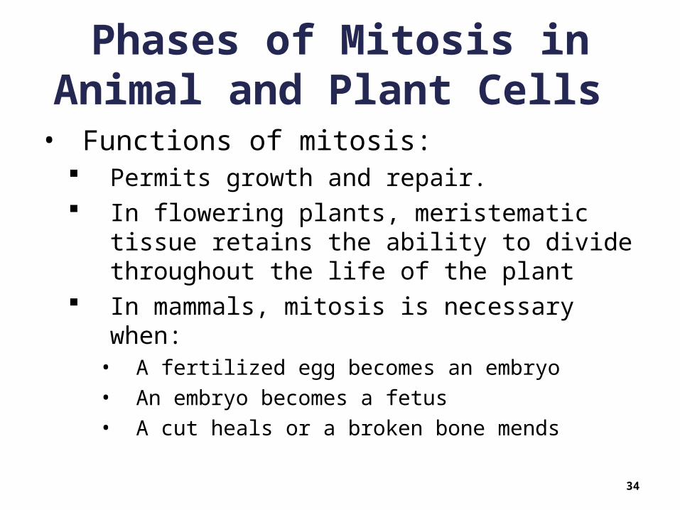
• Functions of mitosis: Permits growth and repair. In flowering plants, meristematic tissue
retains the ability to divide throughout the life of the plant
In mammals, mitosis is necessary when:• A fertilized egg becomes an embryo • An embryo becomes a fetus• A cut heals or a broken bone mends
34

Phases of Mitosis in Animal and Plant Cells
• Stem Cells Many mammalian organs contain stem cells
• Retain the ability to divide • Red bone marrow stem cells divide to produce various
types of blood cells
Therapeutic cloning to produce human tissues can begin with either adult stem cells or embryonic stem cells
Embryonic stem cells can be used for reproductive cloning, the production of a new individual
35
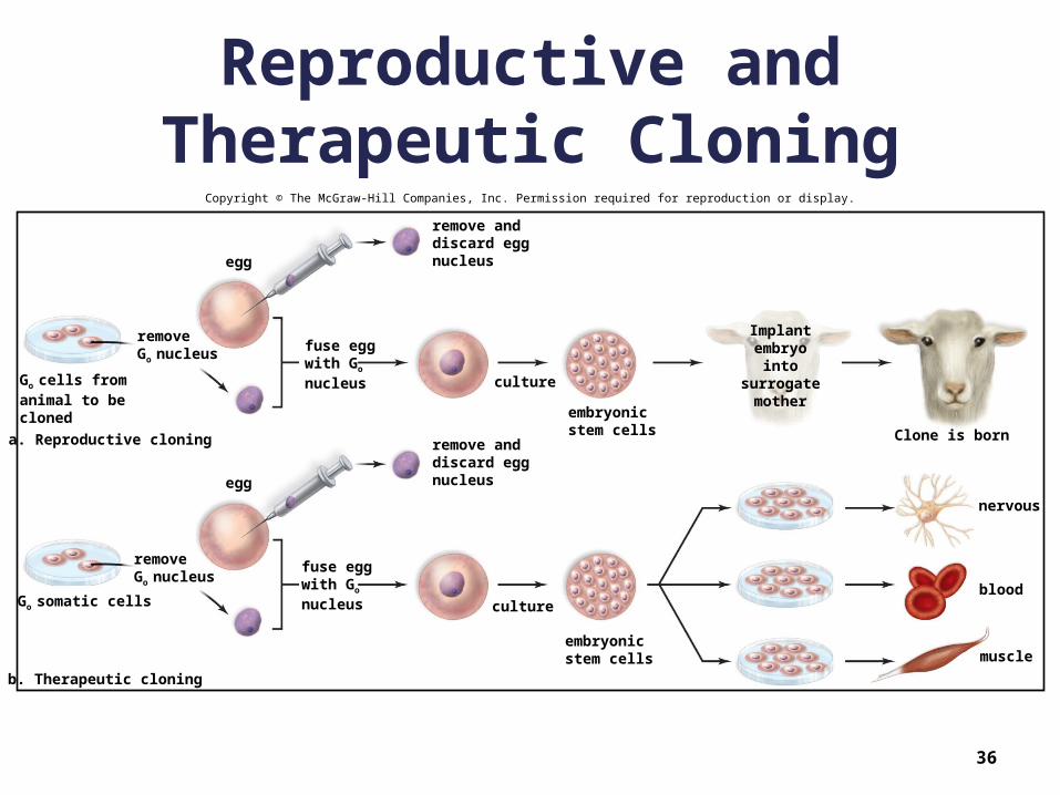
Reproductive and Therapeutic Cloning
36
Copyright © The McGraw-Hill Companies, Inc. Permission required for reproduction or display.
remove anddiscard eggnucleusegg
removeGo nucleus
Go cells fromanimal to becloned
egg
fuse eggwith Go
nucleus
Implantembryo
intosurrogate
mother
Clone is born
culture
embryonicstem cells
nervous
blood
muscleembryonicstem cells
culture
fuse eggwith Go
nucleus
remove anddiscard eggnucleus
Go somatic cells
removeGo nucleus
b. Therapeutic cloning
a. Reproductive cloning

9.3 The Cell Cycle and Cancer
• Abnormal growth of cells is called a tumor Benign tumors are not cancerous
• Encapsulated• Do not invade neighboring tissue or spread
Malignant tumors are cancerous• Not encapsulated• Readily invade neighboring tissues• May also detach and lodge in distant places (metastasis)• Results from mutation of genes regulating the cell cycle
• Development of cancer Tends to be gradual May take years before a cell is obviously cancerous
37

The Cell Cycle and Cancer
• Characteristics of Cancer Cells Lack differentiation
• Are non-specialized• Are immortal (can enter cell cycle repeatedly)
Have abnormal nuclei• May be enlarged• May have abnormal number of chromosomes• Often have extra copies of genes
Do not undergo apoptosis• Normally, cells with damaged DNA undergo apoptosis• The immune system can also recognize abnormal cells and
trigger apoptosis• Cancer cells are abnormal but fail to undergo apoptosis
38

The Cell Cycle and Cancer
• Characteristics of Cancer Cells Form tumors
• Mitosis is normally controlled by contact with neighboring cells – contact inhibition
• Cancer cells have lost contact inhibition Undergo metastasis
• Original tumor easily fragments• New tumors appear in other organs
Undergo angiogenesis• Formation of new blood vessels
– Brings nutrients and oxygen to the tumor
39
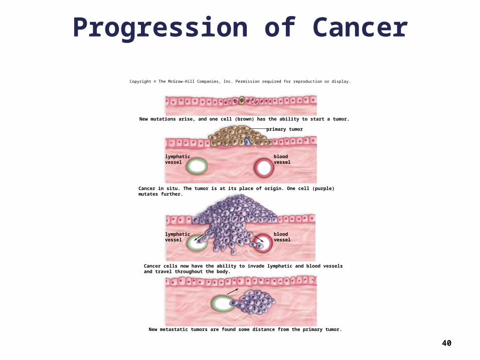
Progression of Cancer
40
Copyright © The McGraw-Hill Companies, Inc. Permission required for reproduction or display.
primary tumor
New mutations arise, and one cell (brown) has the ability to start a tumor.
Cancer in situ. The tumor is at its place of origin. One cell (purple)mutates further.
Cancer cells now have the ability to invade lymphatic and blood vesselsand travel throughout the body.
New metastatic tumors are found some distance from the primary tumor.
lymphaticvessel
bloodvessel
lymphaticvessel
bloodvessel
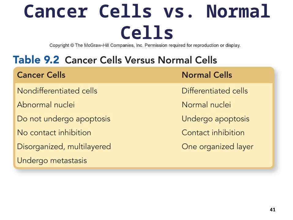
Cancer Cells vs. Normal Cells
41

The Cell Cycle and Cancer
• Origin of Cancer Oncogenes
• Proto-oncogenes promote the cell cycle in various ways
• If a proto-oncogene is mutated, it may become an oncogene
Tumor suppressor genes inhibit the cell cycle in various ways
• If a tumor suppressor gene becomes inactive, it may promote cancer development
Both proto-oncogenes and tumor suppressor genes are normally regulated in coordination with organism’s growth plan
42

The Cell Cycle and Cancer
• Origin of Cancer Chromosomes normally have special material
at each end called telomeres These get shorter each cell division When they get very short, the cell will no
longer divide Telomerase is an enzyme that maintains the
length of telomeres Mutations in telomerase gene:
• Cause telomeres to continue to lengthen, which• Allows cancer cells to continually divide
43

Causes of Cancer
44
Copyright © The McGraw-Hill Companies, Inc. Permission required for reproduction or display.
d: © Biophoto Associates/Photo Researchers, Inc.
activatedsignalingprotein
growthfactor
receptorprotein
signalingprotein
phosphate
b. Effect of growth factor
P
P
P
proto-oncogeneCodes for a growth factor,a receptor protein, or asignaling protein in astimulatory pathway.If a proto-oncogenebecomes an oncogene,the end result can beactive cell division.
tumor suppressor geneCodes for a signalingprotein in an inhibitorypathway. If a tumorsuppressor gene mutates,the end result can beactive cell division.
c. Stimulatory pathway andinhibitory pathway
1,100Xd. Cancerous skin cell
gene productpromotescell cycle
Stimulatorypathway
Inhibitorypathway
gene productinhibitscell cycle
growth factorActivates signalingproteins in a stimulatorypathway that extendsto the nucleus.
a. Influences that cause mutated proto-oncogenes(called oncogenes) and mutated tumorsuppressor genes
Heredity Radiationsources
Pesticides and
herbicides
Viruses
oncogene

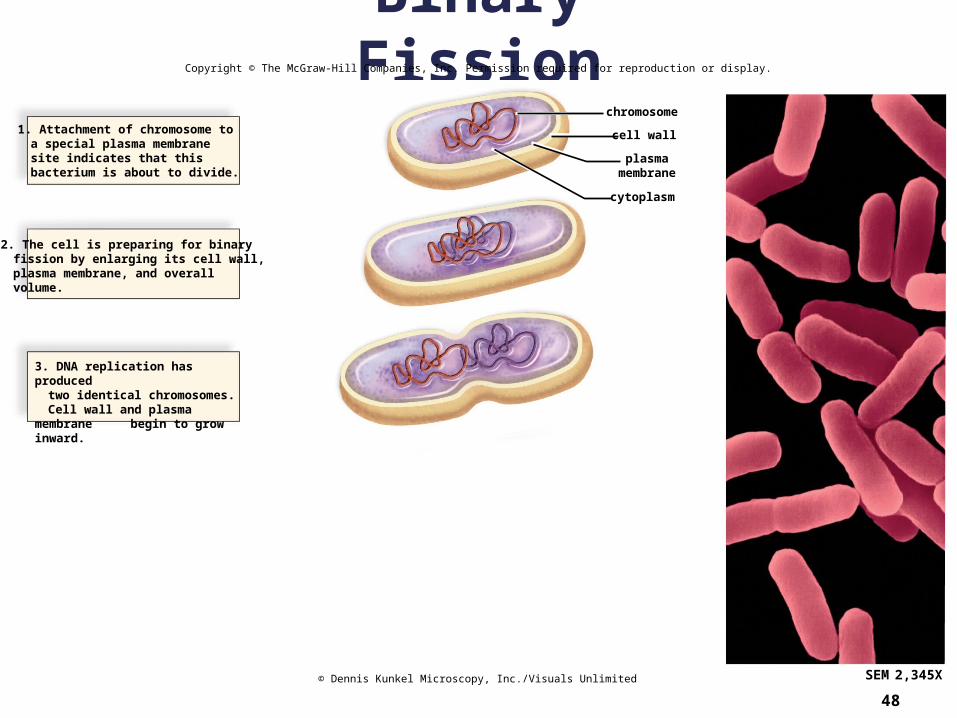
9.4 Prokaryotic Cell Division
• The prokaryotic chromosome is a ring of DNA Folded up in an area called the nucleoid 1,000 X the length of cell Replicated into two rings prior to cell division Replicated rings attach to the plasma membrane
• Binary fission Splitting in two Two replicate chromosomes are distributed to two
daughter cells Produces two daughter cells identical to original cell –
asexual reproduction
45
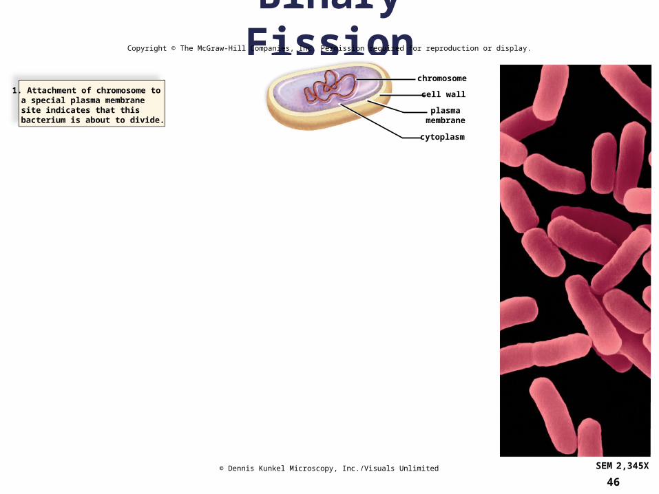
Binary Fission
46
Copyright © The McGraw-Hill Companies, Inc. Permission required for reproduction or display.
chromosome
cell wall
plasmamembrane
cytoplasm
SEM 2,345X
1. Attachment of chromosome toa special plasma membranesite indicates that thisbacterium is about to divide.
© Dennis Kunkel Microscopy, Inc./Visuals Unlimited
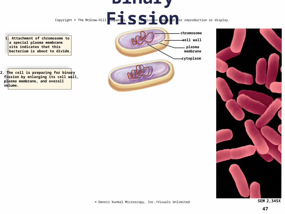
Binary Fission
47
Copyright © The McGraw-Hill Companies, Inc. Permission required for reproduction or display.
chromosome
cell wall
plasmamembrane
cytoplasm
SEM 2,345X
1. Attachment of chromosome toa special plasma membranesite indicates that thisbacterium is about to divide.
2. The cell is preparing for binaryfission by enlarging its cell wall,plasma membrane, and overallvolume.
© Dennis Kunkel Microscopy, Inc./Visuals Unlimited
Binary Fission
48
Copyright © The McGraw-Hill Companies, Inc. Permission required for reproduction or display.
chromosome
cell wall
plasmamembrane
cytoplasm
SEM 2,345X
1. Attachment of chromosome toa special plasma membranesite indicates that thisbacterium is about to divide.
2. The cell is preparing for binaryfission by enlarging its cell wall,plasma membrane, and overallvolume.
3. DNA replication has producedtwo identical chromosomes.Cell wall and plasma membrane begin to grow inward.
© Dennis Kunkel Microscopy, Inc./Visuals Unlimited

Binary Fission
49
Copyright © The McGraw-Hill Companies, Inc. Permission required for reproduction or display.
chromosome
cell wall
plasmamembrane
cytoplasm
SEM 2,345X© Dennis Kunkel Microscopy, Inc./Visuals Unlimited
1. Attachment of chromosome toa special plasma membranesite indicates that thisbacterium is about to divide.
2. The cell is preparing for binaryfission by enlarging its cell wall,plasma membrane, and overallvolume.
3. DNA replication has producedtwo identical chromosomes.Cell wall and plasma membrane begin to grow inward.
4. As the cell elongates, thechromosomes are pulled apart.Cytoplasm is being distributedevenly.
Binary Fission
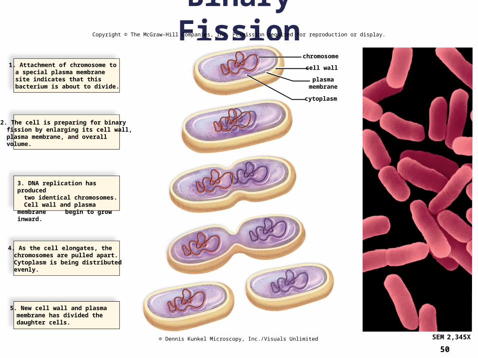
50
Copyright © The McGraw-Hill Companies, Inc. Permission required for reproduction or display.
1. Attachment of chromosome toa special plasma membranesite indicates that thisbacterium is about to divide.
2. The cell is preparing for binaryfission by enlarging its cell wall,plasma membrane, and overallvolume.
3. DNA replication has producedtwo identical chromosomes.Cell wall and plasma membrane begin to grow inward.
4. As the cell elongates, thechromosomes are pulled apart.Cytoplasm is being distributedevenly.
5. New cell wall and plasmamembrane has divided thedaughter cells.
chromosome
cell wall
plasmamembrane
cytoplasm
SEM 2,345X© Dennis Kunkel Microscopy, Inc./Visuals Unlimited

Functions of Cell Division
51