BiologY - Nelson BLAD - final.pdf · In addition to teaching introductory biology classes and an...
17
1 M. B. (Brock) Fenton (Ph.D., University of Toronto) currently teaches at the University of Western Ontario and has received awards for his teaching (Carleton University Faculty of Science Teaching Award; Ontario Confederation of University Faculty Associations Teaching Award; and a 3M Teaching Fellowship, Society for Teaching and Learning in Higher Education) in addition to recognition for his work on public awareness of science (Gordin Kaplan Award from the Canadian Federation of Biological Societies; Honourary Life Membership, Science North, Sudbury, Ontario; Canadian Council of University Biology Chairs Distinguished Canadian Biologist Award; the McNeil Medal for the Public Awareness of Science of the Royal Society of Canada; and the Sir Sanford Fleming Medal for public awareness of Science, the Royal Canadian Institute). He also received the C. Hart Merriam Award from the American Society of Mammalogists for excellence in scientific research. Bats, their biology, behaviour, evolution, and echolocation are the topic of his research, which has been funded by the Natural Sciences and Engineering Research Council of Canada. BIOLOGY EXPLORING THE DIVERSITY OF LIFE First Canadian Edition A great Canadian biology textbook starts with some of Canada’s most respected researchers and instructors.
Transcript of BiologY - Nelson BLAD - final.pdf · In addition to teaching introductory biology classes and an...

1
M. B. (Brock) Fenton (Ph.D., University of Toronto) currently teaches at the University of Western Ontario and has received awards for his teaching (Carleton University Faculty of Science Teaching Award; Ontario Confederation of University Faculty Associations Teaching Award; and a 3M Teaching Fellowship, Society for Teaching and Learning in Higher Education) in addition to recognition for his work on public awareness of science (Gordin Kaplan Award from the Canadian Federation of Biological Societies; Honourary Life Membership, Science North, Sudbury, Ontario; Canadian Council of University Biology Chairs Distinguished Canadian Biologist Award; the McNeil Medal for the Public Awareness of Science of the Royal Society of Canada; and the Sir Sanford Fleming Medal for public awareness of Science, the Royal Canadian Institute). He also received the C. Hart Merriam Award from the American Society of Mammalogists for excellence in scientific research. Bats, their biology, behaviour, evolution, and echolocation are the topic of his research, which has been funded by the Natural Sciences and Engineering Research Council of Canada.
BiologYExploring thE DivErsity of lifE
First Canadian Edition
A great Canadian biology textbook starts with some of Canada’s most respected researchers and instructors.
2 3
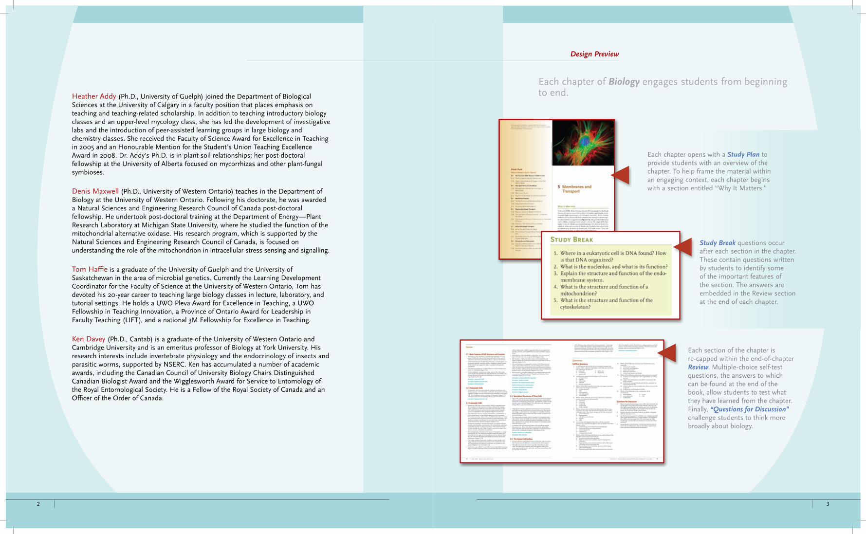
Heather Addy (Ph.D., University of Guelph) joined the Department of Biological Sciences at the University of Calgary in a faculty position that places emphasis on teaching and teaching-related scholarship. In addition to teaching introductory biology classes and an upper-level mycology class, she has led the development of investigative labs and the introduction of peer-assisted learning groups in large biology and chemistry classes. She received the Faculty of Science Award for Excellence in Teaching in 2005 and an Honourable Mention for the Student’s Union Teaching Excellence Award in 2008. Dr. Addy’s Ph.D. is in plant-soil relationships; her post-doctoral fellowship at the University of Alberta focused on mycorrhizas and other plant-fungal symbioses.
Denis Maxwell (Ph.D., University of Western Ontario) teaches in the Department of Biology at the University of Western Ontario. Following his doctorate, he was awarded a Natural Sciences and Engineering Research Council of Canada post-doctoral fellowship. He undertook post-doctoral training at the Department of Energy—Plant Research Laboratory at Michigan State University, where he studied the function of the mitochondrial alternative oxidase. His research program, which is supported by the Natural Sciences and Engineering Research Council of Canada, is focused on understanding the role of the mitochondrion in intracellular stress sensing and signalling.
Tom Haffie is a graduate of the University of Guelph and the University of Saskatchewan in the area of microbial genetics. Currently the Learning Development Coordinator for the Faculty of Science at the University of Western Ontario, Tom has devoted his 20-year career to teaching large biology classes in lecture, laboratory, and tutorial settings. He holds a UWO Pleva Award for Excellence in Teaching, a UWO Fellowship in Teaching Innovation, a Province of Ontario Award for Leadership in Faculty Teaching (LIFT), and a national 3M Fellowship for Excellence in Teaching.
Ken Davey (Ph.D., Cantab) is a graduate of the University of Western Ontario and Cambridge University and is an emeritus professor of Biology at York University. His research interests include invertebrate physiology and the endocrinology of insects and parasitic worms, supported by NSERC. Ken has accumulated a number of academic awards, including the Canadian Council of University Biology Chairs Distinguished Canadian Biologist Award and the Wigglesworth Award for Service to Entomology of the Royal Entomological Society. He is a Fellow of the Royal Society of Canada and an Officer of the Order of Canada.
Each chapter of Biology engages students from beginning to end.
Each chapter opens with a Study Plan to provide students with an overview of the chapter. To help frame the material within an engaging context, each chapter begins with a section entitled “Why It Matters.”
Study Break questions occur after each section in the chapter. These contain questions written by students to identify some of the important features of the section. The answers are embedded in the Review section at the end of each chapter.
Each section of the chapter is re-capped within the end-of-chapter Review. Multiple-choice self-test questions, the answers to which can be found at the end of the book, allow students to test what they have learned from the chapter. Finally, “Questions for Discussion” challenge students to think more broadly about biology.
Design Preview

4 5
While many textbooks use the first few chapters to introduce and/or review, we believe that the first chapters should convey the excitement and interest of biology itself. We, therefore, placed important background information about biology and chemistry in the reference section entitled “The Chemical and Physical Foundations of Biology,” in the middle of the book. These pages are distinct and easy to find with their purple bleed and have become known affectionately as the Purple Pages. The Purple Pages enable information to be readily identifiable and accessible to students as they move through the textbook, rather than tied to one particular chapter. They make background information easy to find when students need to check a topic. The Purple Pages also keep background information out of the main stream of the text, allowing students to focus on the bigger picture.
Science that appears in textbooks is the product of people who have made careful and systematic observations, which led them to formulate hypotheses about these observations, and, where appropriate, design and execute experiments to test these hypotheses.
Boxed features in Biology highlight notable people, important molecules, interesting contexts, and examples of life in extreme conditions.
Unanswered Questions at the end of each chapter emphasize that biologists still have a lot to learn and provide topics for students to tackle should they decide to pursue a career in research.
Life on the Edge boxes provide accounts of organisms thriving “on the edge” at unusual temperatures, pressures, radiation dosages, salt concentrations, etc. These boxes explain how our understanding of “normal” can be increased through the study of the “extreme.”
Molecule Behind Biology boxes give students a sense of the exciting impact of molecular research. From water to progesterone, amanitin and DDT, each chapter features the activity of a relevant chemical.
People Behind Biology boxes contain stories about how particular people have used their ingenuity and creativity to expand our knowledge of biology.
Design Preview Design Preview

6 7
CengageNOW™ and JoinIn™ on TurningPoint® make learning interactive.
Personalized Study Plans (with automatic grading)CengageNOW™ for Biology: Exploring the Diversity of Life saves you time and provides your students with an efficient way to study. After reading a chapter, students can take an assignable pre-test to assess their understanding. Based on their answers, a Personalized Study Plan directs them to animations and sections of the textbook that they need to review to better understand the material. A post-test measures their progress. Students log in with the access card available with each new text.
A Built-In Instructor GradebookWhen you assign CengageNOW™ to your students, the instructor gradebook makes it easy for you to track grades and monitor student progress. You can assign pre-built homework or create your own exams using questions from the text’s test bank. You can also assign How Would You Vote? exercises, which promote critical thinking by asking students to read pro/con articles on controversial issues related to biology and then vote on them. All of these exercises are graded automatically, and results flow directly to your online gradebook. CengageNOW™ can also be easily integrated with a WebCT® or Blackboard® gradebook. Or, if you prefer a hands-off approach, students can benefit from the study system without an instructor setup or involvement.
The Instructor’s Resource DVD saves time on lecture preparation—robust supplements are all a click away.
The Instructor’s Resource DVD for Biology: Exploring the Diversity of Life makes it easy to create customized lectures with Microsoft® PowerPoint®. A single DVD integrates all relevant resources in each chapter’s Microsoft® PowerPoint® lecture, so there’s no hassling with multiple media files or discs.
Each chapter’s lecture slides are organized by section and include the following:
Animations and interactions from • CengageNOW™ Video clips•Slides with book-specific questions that also appear in• JoinIn™ on TurningPoint® Bulleted points listing key concepts•
Editing features and other resources:Ability to edit the copy on lecture slides to fit your needs•Ability to add text art and photos •An• ExamView® computerized test bank—part of our Nelson Education Testing Advantage program (NETA)—ensures quality multiple-choice questions that provide the means to measure higher-level thinking skills as well as recallMicrosoft• ® Word files for the Instructor’s Resource Manual and Test Bank for each chapter
Support for Instructors Teaching and Learning Resources

8
A Tool for Instant In-Class Quizzes and PollsHere’s an easy way to increase students’ participation in class! Nelson Education Ltd. is pleased to offer you book-specific JoinIn™ on TurningPoint® content for classroom response systems tailored to Biology: Exploring the Diversity of Life. Our agreement to offer TurningPoint® software lets you pose book-specific questions and display students’ answers seamlessly within the Microsoft®
PowerPoint® slides of your own lecture, in conjunction with the “clicker” hardware of your choice. It’s a great tool for motivating students to come to class and pay attention. We provide the software and questions for each chapter of the text on ready-to-use Microsoft® PowerPoint® slides.
Teaching and Learning Resources

10 BRIEF CONTENTS BRIEF CONTENTS 11
Unit One Setting the Stage
1 Light and Life 2 Life, Cells, and Origins 3 Selection, Biodiversity, and Biosphere
Unit twO energy-PrOceSS and FacilitatiOn
4 Energy and Enzymes 5 Membranes and Transport 6 Respiration 7 Photosynthesis 8 Cell Communication
Unit three geneS
9 Cell Cycles 10 Recombination 11 Mendel, Genes, Etc. 12 Genes and Chromosomes
Unit FOUr dna and gene exPreSSiOn
13 DNA Structure 14 Gene Expression 15 Control of Gene Expression 16 DNA Technology
Unit Five evOlUtiOn and claSSiFicatiOn
17 Microevolution 18 Species 19 Evolution and Classification 20 Darwin, Fossils, and Developmental Biology
Unit Six diverSity OF liFe
21 Prokaryotes 22 Viruses 23 Protists 24 Fungi 25 Plants 26 Protostomes 27 Deuterostomes
Unit Seven SyStemS and PrOceSS—PlantS
28 Plant Body 29 Transport in Plants 30 Reproduction and Development in Plants 31 Control of Plant Growth and Development
Unit eight SyStemS and PrOceSS—animalS
32 Animal Organization 33 Nerves 34 Sensory 35 Endocrine 36 Muscles, Bones 37 Circulation 38 Animal Reproduction 39 Animal Development 40 Animal Behaviour
Unit nine liFe PrOceSSeS—an integrated view
41 Nutrition 42 Gaseous Exchange 43 Internal Regulation 44 Defense
Unit ten ecOlOgy
45 Population Ecology 46 Population Interactions and Community Ecology 47 Ecosystems
Unit eleven BiOlOgy in actiOn
48 Conservation of Biodiversity49 Domestication
Brief Contents

1
Study Plan
1.1 The Physical Nature of Light
1.1a What Is Light?
1.1b Light Interacts with Matter
1.1c Why Chlorophyll Is Green
1.2 Light as a Source of Energy
1.3 Light as a Source of Information
1.3a Rhodopsin, a Highly Conserved Photoreceptor
1.3b Sensing Light Without Eyes
1.3c The Eye
1.3d Darwin and the Evolution of the Eye
1.4 Light Can Damage Biological Molecules
1.4a Damage by Light: Direct Effects
1.4b Damage by Light: Indirect Effects
1.5 Role of Light in Ecology and Behaviour
1.5a Using Light to Tell Time: Circadian Rhythms
1.5b Avoiding Detection: Camoufl age
1.5c Using Colour as Signals
1.5d Light in Aquatic Habitats
1.5e Ecological Light Pollution
1.6 Life in the Dark
1.7 Organisms Making Their Own Light: Bioluminescence
1 Light and Life
Why It Matters
Claude Monet (1840–1926), a French painter, is considered by many to be the master of the impressionist form that rose to prominence in the late nineteenth century. Other well-known impressionists include Edgar Degas and Paul Cézanne. Impressionism as an art movement was characterized by the use of small visible brush strokes that emphasized light and colour, rather than lines, to defi ne an object. The artists used pure, unmixed colour, not smoothly blended, as was the custom at the time. For example, instead of physically mixing yellow and blue paint, they placed unmixed yellow paint on the canvas next to unmixed blue paint so that the colours would mingle in the eye of the viewer to create the “impression” of green. The Impressionists found that they could capture the momentary and transient eff ects of sunlight and changing colour of a scene by painting en plein air, in the open air, outside of the studio, where they could more accurately paint the refl ected light of an immediate scene.
Interestingly, compared with his early works, Monet’s later paintings verge on the abstract, with colours bleeding into each other and a lack of rational shape and perspective. For example, “The Japanese Footbridge” is an explosion of orange, yellow, and red hues,
Pain
tings
by
Clau
de M
onet
. (a)
“W
ater
-Lily
Pon
d, S
ymph
ony
in G
reen
” (p
aint
ed 1
899)
; (b) “
The
Japa
nese
Foo
tbrid
ge”
(pai
nted
192
0–19
22)
a. b.
NEL
40947_01_Ch01_p001-022 pp.indd 1 12/16/08 11:17:21 AM
Preview • First Pass Pages • Preview • First Pass Pages
Preview • First Pass Pages • Preview • First Pass Pages
UNIT ONE MOLECULES AND CELLS2 NEL
with heavy, broad brush strokes, leaving the viewer barely able to discern the vague shape of the arched bridge. In many of Monet’s later works, the colours in his paintings became more muted, far less vibrant and bright, with a pronounced colour shift from blue-green to red-yellow and an almost total absence of light blues. The sense of atmosphere and light that he was famous for in his earlier works disappeared.
Although the change in Monet’s paintings could easily be explained by an intentional change in style or perhaps an age-related change in manual dexterity, Monet himself realized that it was not his style or dex-terity that had changed but, rather, his ability to see. Monet suff ered from cataracts, the vision- deteriorating disease that was diagnosed in both eyes by a Parisian ophthalmologist in 1912 when Monet was 72. A cata-ract is a change in the lens of the eye, making it more opaque. The under-lying cause is a progressive denatur-ation of one of the proteins that make up the lens. The increased opaque-ness of the lens absorbs certain wavelengths of light, decreasing the transmittance of blue light. Thus, to a cataract suff erer such as Monet, the world appears more yellow.
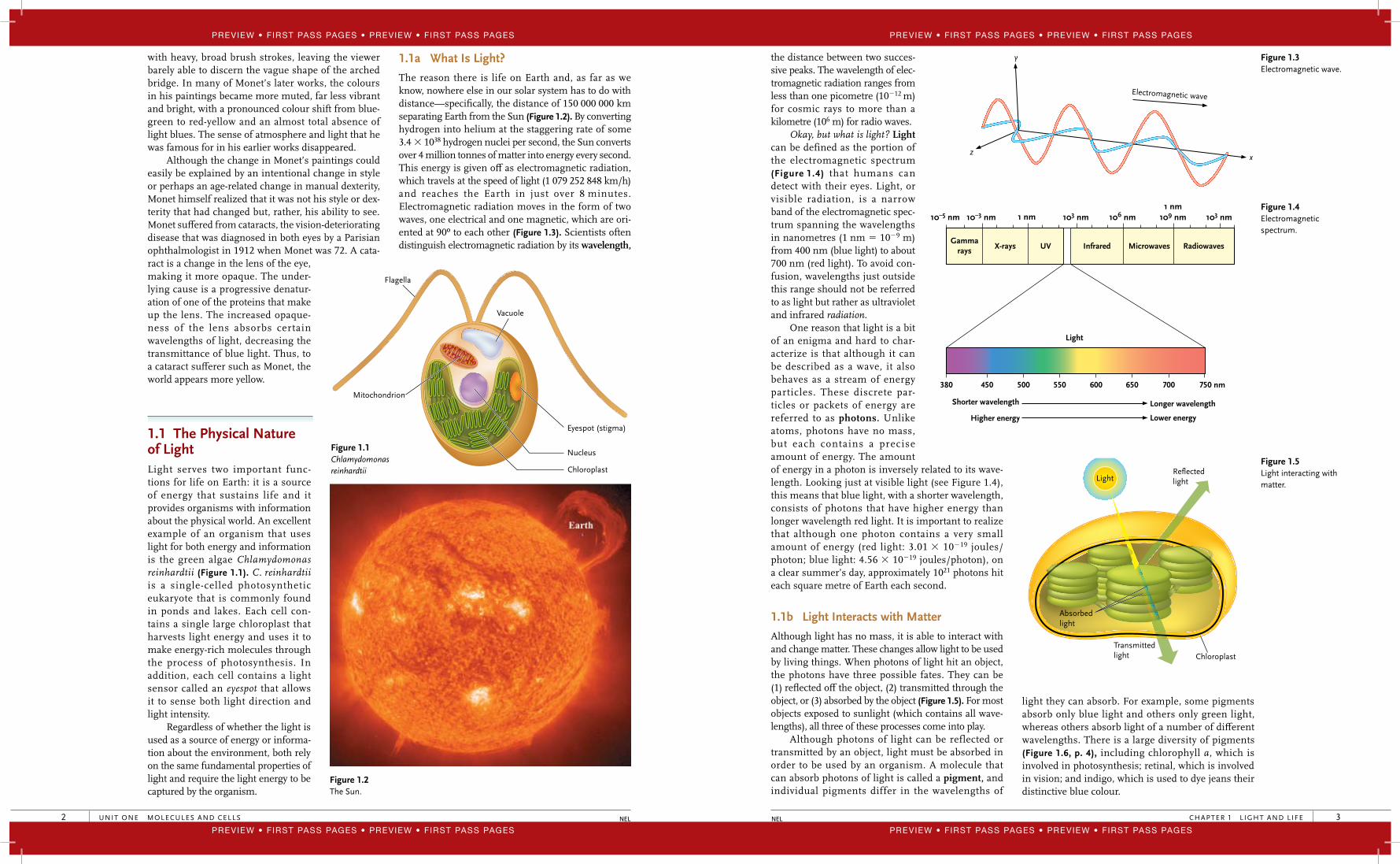
1.1 The Physical Nature of LightLight serves two important func-tions for life on Earth: it is a source of energy that sustains life and it provides organisms with information about the physical world. An excellent example of an organism that uses light for both energy and information is the green algae Chlamydomonas reinhardtii (Figure 1.1). C. reinhardtii is a single-celled photosynthetic eukaryote that is commonly found in ponds and lakes. Each cell con-tains a single large chloroplast that harvests light energy and uses it to make energy-rich molecules through the process of photosynthesis. In addition, each cell contains a light sensor called an eyespot that allows it to sense both light direction and light intensity.
Regardless of whether the light is used as a source of energy or informa-tion about the environment, both rely on the same fundamental properties of light and require the light energy to be captured by the organism.
Flagella
Vacuole
Eyespot (stigma)
Nucleus
Chloroplast
Mitochondrion
Figure 1.1 Chlamydomonas reinhardtii
Figure 1.2 The Sun.
1.1a What Is Light?
The reason there is life on Earth and, as far as we know, nowhere else in our solar system has to do with distance—specifi cally, the distance of 150 000 000 km separating Earth from the Sun (Figure 1.2). By converting hydrogen into helium at the staggering rate of some 3.4 � 1038 hydrogen nuclei per second, the Sun converts over 4 million tonnes of matter into energy every second. This energy is given off as electromagnetic radiation, which travels at the speed of light (1 079 252 848 km/h) and reaches the Earth in just over 8 minutes. Electromagnetic radiation moves in the form of two waves, one electrical and one magnetic, which are ori-ented at 90º to each other (Figure 1.3). Scientists often distinguish electromagnetic radiation by its wavelength,
40947_01_Ch01_p001-022 pp.indd 2 12/16/08 11:17:23 AM
CHAPTER 1 L IGHT AND LIFE 3NEL
the distance between two succes-sive peaks. The wavelength of elec-tromagnetic radiation ranges from less than one picometre (10�12 m) for cosmic rays to more than a kilometre (106 m) for radio waves.
Okay, but what is light? Light can be defined as the portion of the electromagnetic spectrum (Figure 1.4) that humans can detect with their eyes. Light, or visible radiation, is a narrow band of the electromagnetic spec-trum spanning the wavelengths in nanometres (1 nm � 10�9 m) from 400 nm (blue light) to about 700 nm (red light). To avoid con-fusion, wavelengths just outside this range should not be referred to as light but rather as ultraviolet and infrared radiation.
One reason that light is a bit of an enigma and hard to char-acterize is that although it can be described as a wave, it also behaves as a stream of energy particles. These discrete par-ticles or packets of energy are referred to as photons. Unlike atoms, photons have no mass, but each contains a precise amount of energy. The amount of energy in a photon is inversely related to its wave-length. Looking just at visible light (see Figure 1.4), this means that blue light, with a shorter wavelength, consists of photons that have higher energy than longer wavelength red light. It is important to realize that although one photon contains a very small amount of energy (red light: 3.01 � 10�19 joules/photon; blue light: 4.56 � 10�19 joules/photon), on a clear summer’s day, approximately 1021 photons hit each square metre of Earth each second.
1.1b Light Interacts with Matter
Although light has no mass, it is able to interact with and change matter. These changes allow light to be used by living things. When photons of light hit an object, the photons have three possible fates. They can be (1) refl ected off the object, (2) transmitted through the object, or (3) absorbed by the object (Figure 1.5). For most objects exposed to sunlight (which contains all wave-lengths), all three of these processes come into play.
Although photons of light can be reflected or transmitted by an object, light must be absorbed in order to be used by an organism. A molecule that can absorb photons of light is called a pigment, and individual pigments differ in the wavelengths of
Electromagnetic wave
z
y
x
Figure 1.3 Electromagnetic wave.
10–5 nm 10–3 nm 103 nm 106 nm 109 nm 103 nm 1 nm
1 nm
380
Shorter wavelength Longer wavelength
Higher energy Lower energy
450 500 550 600 650 700 750 nm
Light
Gammarays X-rays UV Infrared Microwaves Radiowaves
Figure 1.4 Electromagnetic spectrum.
ReflectedlightLight
Absorbedlight
Transmittedlight Chloroplast
Figure 1.5 Light interacting with matter.
light they can absorb. For example, some pigments absorb only blue light and others only green light, whereas others absorb light of a number of diff erent wavelengths. There is a large diversity of pigments (Figure 1.6, p. 4), including chlorophyll a, which is involved in photosynthesis; retinal, which is involved in vision; and indigo, which is used to dye jeans their distinctive blue colour.
40947_01_Ch01_p001-022 pp.indd 3 12/16/08 11:17:23 AM
Preview • First Pass Pages • Preview • First Pass Pages
Preview • First Pass Pages • Preview • First Pass Pages
Preview • First Pass Pages • Preview • First Pass Pages
Preview • First Pass Pages • Preview • First Pass Pages

UNIT ONE MOLECULES AND CELLS4 NEL
What is it about pigments that enable them to capture light? At first glance, the pigments shown in Figure 1.6 seem to be very different from each other structurally; however, they all share a common feature critical to light absorption: a region where carbon atoms are covalently bonded with alternating single and double bonds. This bonding arrangement is called a conjugated system and results in the delo-calization of electrons. None of these electrons are closely associated with a particular atom, and because of this, they are more available to interact with a photon of light.
1.1c Why Chlorophyll Is Green
Absorption of light occurs when the energy of a photon is transferred to an electron of the pigment molecule. For example, Figure 1.7 shows this in a single molecule
Figure 1.6 Assortment of pigments.
O
Mg
ONH
NH
OH
O
O
CH3
H3C
H3C
H3C
CH3CH3 CH3
CH3 CH3
OH
O
HOCOO–
Protein
O
HO
H3C
HO
HO HO OH
O
HO
N
N N
N
H
HH
CH3CH3
H3C
H2C=CH
CH2CH3
CO2CH3
O
CH3
CH2CH2CO2CH2CH=C(CH2CH2CH2CH)3CH3
CH3
H
CH3
O
CH3
N N
H
HSH3C
H3C
H
NN
COO–
H H
H
Chlorophyll a
11-cis-Retinal Indigo
Phycoerythrobilin Carmine
Beta-carotene
Blue photons excite electrons to aneven higher energy state
Red photons excite electrons to ahigher energy state
Energy state of electrons in chlorophyll
e–
0 1 2
e–
Figure 1.7 Absorption of light by chlorophyll.
40947_01_Ch01_p001-022 pp.indd 4 12/16/08 11:17:23 AM
CHAPTER 1 L IGHT AND LIFE 5NEL
of chlorophyll. Recall from chemistry that electrons occupy discrete energy levels, or excited states, in their orbits around the nucleus of an atom. Before absorbing a photon of light, an electron exists in the ground state, which we can designate as 0. Upon absorption of a photon of light, the energy is transferred to the electron, moving it from the ground state to a higher energy, excited state. For a chlorophyll molecule, the electron involved in photon capture can exist in two, and only two, excited states (see Figure 1.7). The lower excited state, designated as 1, is reached by chlorophyll absorbing a photon of red light. The higher excited state, designated as 2, is reached by the absorption of a photon of blue light. Absorption of blue light excites an electron to a higher energy state than absorption of red light because blue photons contain more energy.
Two important principles must be kept in mind when thinking about light absorption by pigments: fi rst, a single photon results in the excitation of one, and only one, electron in a pigment molecule. Second, the energy of the photon must match the energy diff erence between the ground state and one of the excited states in order for the photon to be absorbed. If the energies do not match, the photon is not absorbed. In the chlo-rophyll molecule, the energy of a blue photon or a red photon matches perfectly with the energy required for an electron to reach either the fi rst or the second excited state.
So why is chlorophyll green in colour? The colour of a pigment is determined by the wavelengths of light it cannot absorb. Chlorophyll is green because although it can trap photons of blue light and red light, it cannot absorb photons of green light. As shown in Figure 1.7, a chlorophyll molecule cannot absorb a photon of green light because it does not have an energy level matching that of a green photon. Whereas red and blue photons are captured, green photons are refl ected or transmitted, giving chlorophyll (and plants) its dis-tinctive green colour.
Because pigments do not absorb all wavelengths of light equally, the eff ectiveness of light in driving processes that use the absorbed light, such as pho-tosynthesis or vision, varies depending on the wave-length of the light. A plot of the effectiveness of diff erent wavelengths of light on a biological process is called an action spectrum. Figure 1.8 illustrates the action spectrum for photosynthesis in the leaf of a plant.
Figure 1.8 shows that red and blue wavelengths of light are more eff ective at driving photosynthesis than green wavelengths are. This fi ts well with what we know about the wavelengths of light that are absorbed by chlorophyll. You may notice in Figure 1.8 that some photosynthesis still occurs under green light. This is because photosynthesis involves many different pigments besides just chlorophyll, and some of these absorb wavelengths of light between red and blue.
Study Break
1. What form does light take?2. What do the structures of all pigment molecules
have in common?
1.2 Light as a Source of EnergyWe have already seen that after a photon of light is absorbed, an electron within a pigment molecule is raised to a higher excited state. This excited state electron is a source of potential energy that can be used to do work. As we will see in Chapter 7, this potential energy is used in photosynthetic electron transport to synthesize energy-rich compounds NADPH (the reduced form of nicotinamide adenine dinucleotide phosphate) and ade-nosine triphosphate (ATP), which are used to convert carbon dioxide into carbohydrates (Figure 1.9). In addition,
400 500 600 700
Wavelength (nm)
Rate
of O
2 re
leas
ein
pho
tosy
nthe
sis
Figure 1.8 Action spectrum.
Sugar
Released chemical energy is made available for other metabolic processes.
and
Photosynthesis captures electromagnetic energy
from sunlight.
Electromagnetic energy in sunlight
Oxygen Oxygen
Energy is stored as chemical energy.
Cellular respiration releases chemical energy
from sugar molecules.
Carbon dioxide
Water
Figure 1.9 Photosynthesis.
40947_01_Ch01_p001-022 pp.indd 5 12/16/08 11:17:24 AM
Preview • First Pass Pages • Preview • First Pass Pages
Preview • First Pass Pages • Preview • First Pass Pages
Preview • First Pass Pages • Preview • First Pass Pages
Preview • First Pass Pages • Preview • First Pass Pages
UNIT ONE MOLECULES AND CELLS6 NEL
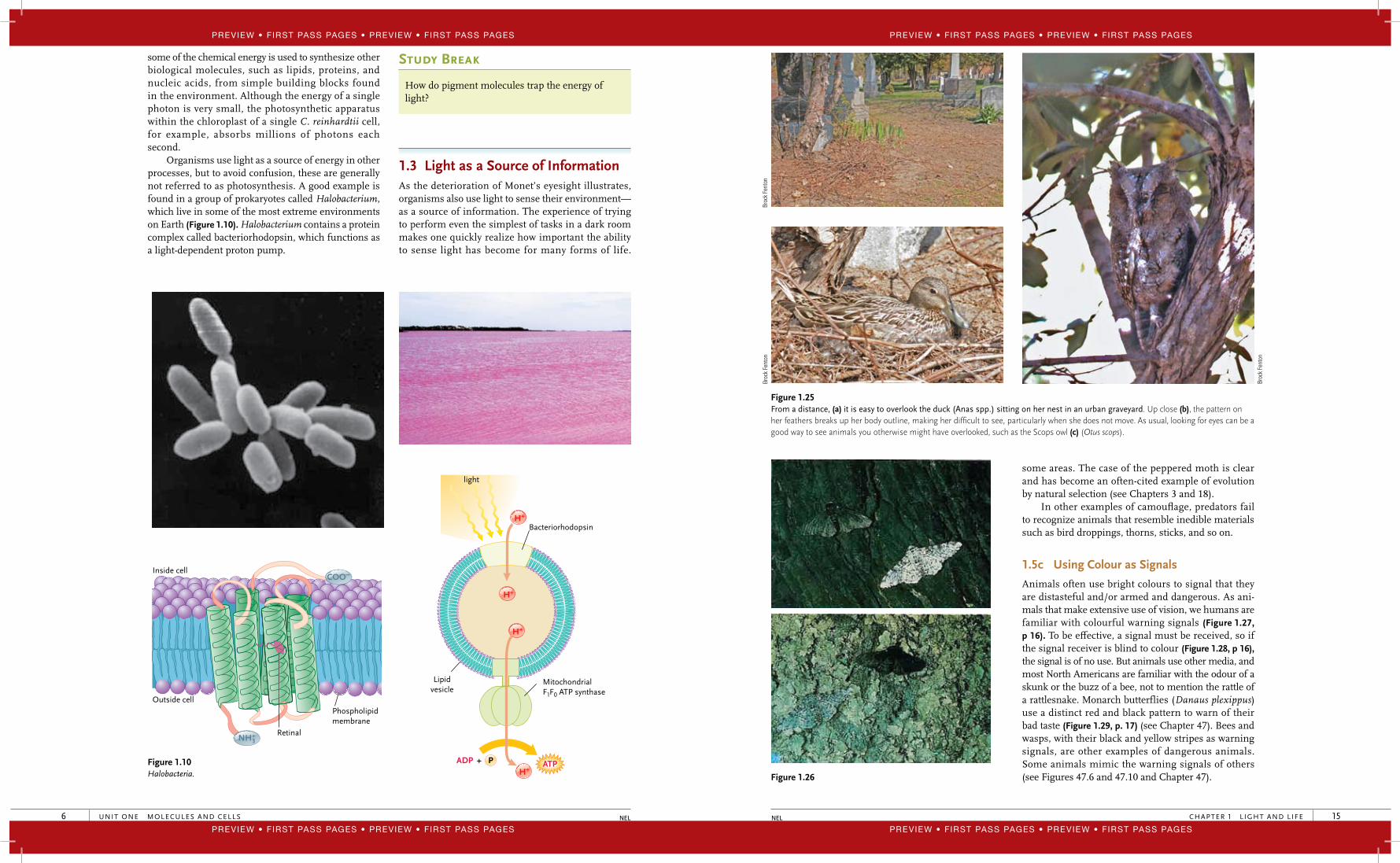
some of the chemical energy is used to synthesize other biological molecules, such as lipids, proteins, and nucleic acids, from simple building blocks found in the environment. Although the energy of a single photon is very small, the photosynthetic apparatus within the chloroplast of a single C. reinhardtii cell, for example, absorbs millions of photons each second.
Organisms use light as a source of energy in other processes, but to avoid confusion, these are generally not referred to as photosynthesis. A good example is found in a group of prokaryotes called Halobacterium, which live in some of the most extreme environments on Earth (Figure 1.10). Halobacterium contains a protein complex called bacteriorhodopsin, which functions as a light-dependent proton pump.
Study Break
How do pigment molecules trap the energy of light?
1.3 Light as a Source of InformationAs the deterioration of Monet’s eyesight illustrates, organisms also use light to sense their environment—as a source of information. The experience of trying to perform even the simplest of tasks in a dark room makes one quickly realize how important the ability to sense light has become for many forms of life.
Figure 1.10 Halobacteria.
COO–
NH+3
Inside cell
Outside cell
Retinal
Phospholipidmembrane
Lipidvesicle
Bacteriorhodopsin
MitochondrialF1F0 ATP synthase
H+
H+
H+
ATPP ADP +
light
H+
40947_01_Ch01_p001-022 pp.indd 6 12/16/08 11:17:24 AM
CHAPTER 1 L IGHT AND LIFE 15NEL
some areas. The case of the peppered moth is clear and has become an often-cited example of evolution by natural selection (see Chapters 3 and 18).
In other examples of camoufl age, predators fail to recognize animals that resemble inedible materials such as bird droppings, thorns, sticks, and so on.
1.5c Using Colour as Signals
Animals often use bright colours to signal that they are distasteful and/or armed and dangerous. As ani-mals that make extensive use of vision, we humans are familiar with colourful warning signals (Figure 1.27, p 16). To be eff ective, a signal must be received, so if the signal receiver is blind to colour (Figure 1.28, p 16), the signal is of no use. But animals use other media, and most North Americans are familiar with the odour of a skunk or the buzz of a bee, not to mention the rattle of a rattlesnake. Monarch butterflies (Danaus plexippus) use a distinct red and black pattern to warn of their bad taste (Figure 1.29, p. 17) (see Chapter 47). Bees and wasps, with their black and yellow stripes as warning signals, are other examples of dangerous animals. Some animals mimic the warning signals of others (see Figures 47.6 and 47.10 and Chapter 47).
Figure 1.25 From a distance, (a) it is easy to overlook the duck (Anas spp.) sitting on her nest in an urban graveyard. Up close (b), the pattern on her feathers breaks up her body outline, making her diffi cult to see, particularly when she does not move. As usual, looking for eyes can be a good way to see animals you otherwise might have overlooked, such as the Scops owl (c) (Otus scops).
Figure 1.26
Broc
k Fe
nton
Broc
k Fe
nton
Broc
k Fe
nton
40947_01_Ch01_p001-022 pp.indd 15 12/16/08 11:17:28 AM
Preview • First Pass Pages • Preview • First Pass Pages
Preview • First Pass Pages • Preview • First Pass Pages
Preview • First Pass Pages • Preview • First Pass Pages
Preview • First Pass Pages • Preview • First Pass Pages
UNIT ONE MOLECULES AND CELLS16 NEL
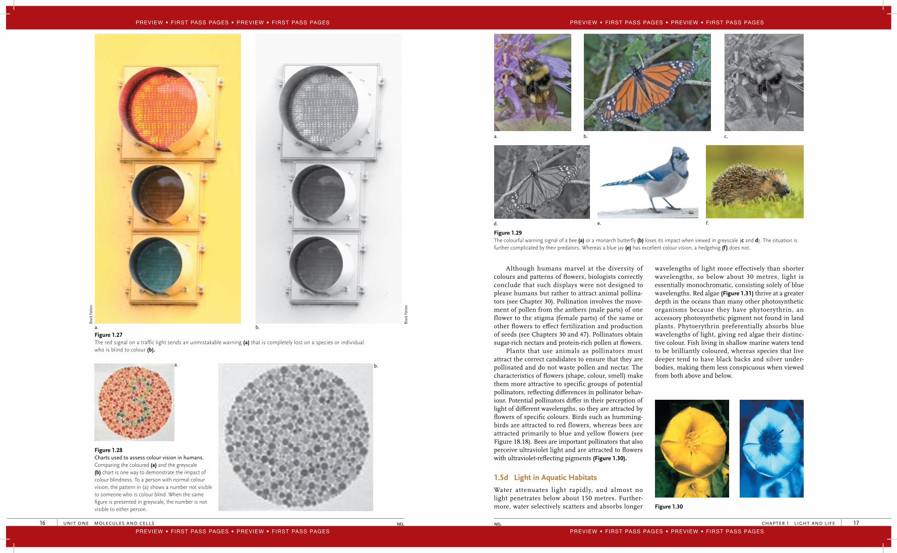
Figure 1.27 The red signal on a traffi c light sends an unmistakable warning (a) that is completely lost on a species or individual who is blind to colour (b).
Figure 1.28 Charts used to assess colour vision in humans. Comparing the coloured (a) and the greyscale (b) chart is one way to demonstrate the impact of colour blindness. To a person with normal colour vision, the pattern in (a) shows a number not visible to someone who is colour blind. When the same fi gure is presented in greyscale, the number is not visible to either person.
Broc
k Fe
nton
Broc
k Fe
nton
a. b.
a. b.
40947_01_Ch01_p001-022 pp.indd 16 12/16/08 11:17:30 AM
CHAPTER 1 L IGHT AND LIFE 17NEL
Figure 1.29 The colourful warning signal of a bee (a) or a monarch butterfl y (b) loses its impact when viewed in greyscale (c and d). The situation is further complicated by their predators. Whereas a blue jay (e) has excellent colour vision, a hedgehog (f ) does not.
Although humans marvel at the diversity of colours and patterns of fl owers, biologists correctly conclude that such displays were not designed to please humans but rather to attract animal pollina-tors (see Chapter 30). Pollination involves the move-ment of pollen from the anthers (male parts) of one flower to the stigma (female parts) of the same or other fl owers to eff ect fertilization and production of seeds (see Chapters 30 and 47). Pollinators obtain sugar-rich nectars and protein-rich pollen at fl owers.
Plants that use animals as pollinators must attract the correct candidates to ensure that they are pollinated and do not waste pollen and nectar. The characteristics of fl owers (shape, colour, smell) make them more attractive to specific groups of potential pollinators, refl ecting diff erences in pollinator behav-iour. Potential pollinators diff er in their perception of light of diff erent wavelengths, so they are attracted by fl owers of specifi c colours. Birds such as humming-birds are attracted to red flowers, whereas bees are attracted primarily to blue and yellow flowers (see Figure 18.18). Bees are important pollinators that also perceive ultraviolet light and are attracted to fl owers with ultraviolet-refl ecting pigments (Figure 1.30).
1.5d Light in Aquatic Habitats
Water attenuates light rapidly, and almost no light penetrates below about 150 metres. Further-more, water selectively scatters and absorbs longer
wavelengths of light more effectively than shorter wavelengths, so below about 30 metres, light is essentially monochromatic, consisting solely of blue wavelengths. Red algae (Figure 1.31) thrive at a greater depth in the oceans than many other photosynthetic organisms because they have phytoerythrin, an accessory photosynthetic pigment not found in land plants. Phytoerythrin preferentially absorbs blue wavelengths of light, giving red algae their distinc-tive colour. Fish living in shallow marine waters tend to be brilliantly coloured, whereas species that live deeper tend to have black backs and silver under-bodies, making them less conspicuous when viewed from both above and below.
Figure 1.30
a. b. c.
d. e. f.
40947_01_Ch01_p001-022 pp.indd 17 12/16/08 11:17:31 AM
Preview • First Pass Pages • Preview • First Pass Pages
Preview • First Pass Pages • Preview • First Pass Pages
Preview • First Pass Pages • Preview • First Pass Pages
Preview • First Pass Pages • Preview • First Pass Pages
UNIT ONE MOLECULES AND CELLS20 NEL
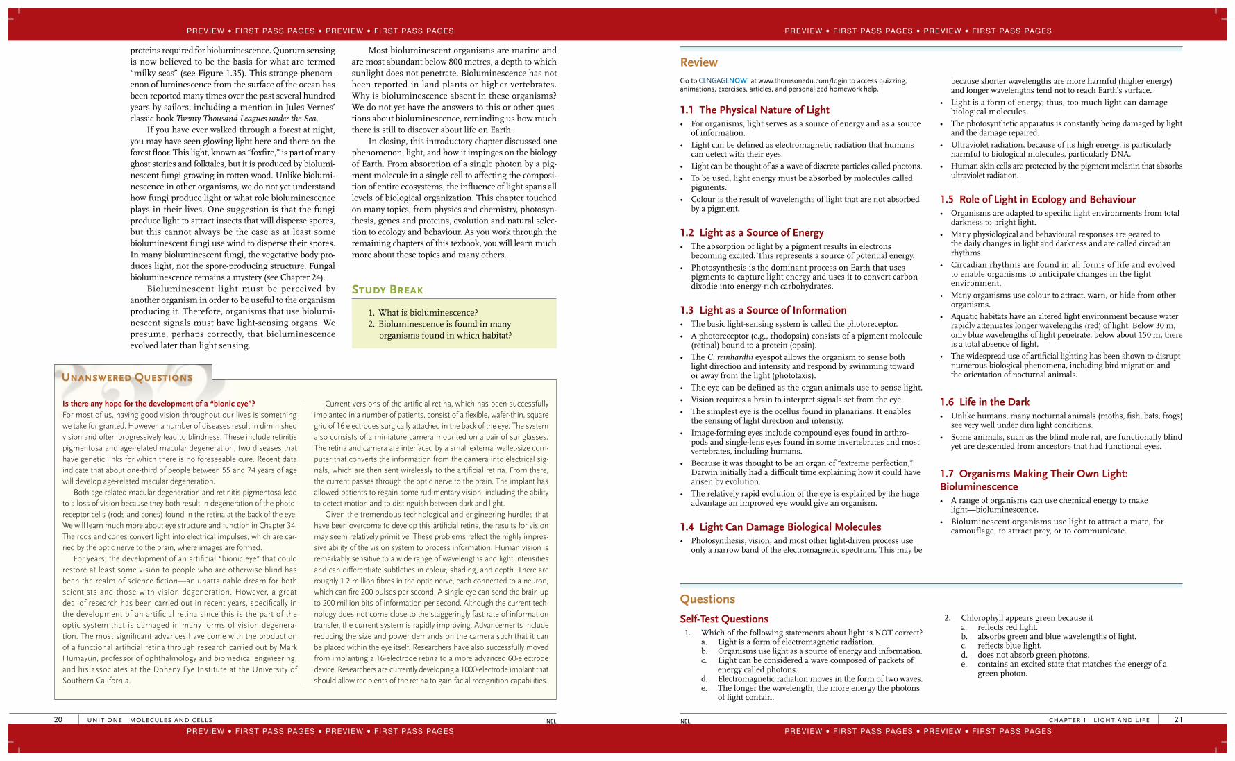
proteins required for bioluminescence. Quorum sensing is now believed to be the basis for what are termed “milky seas” (see Figure 1.35). This strange phenom-enon of luminescence from the surface of the ocean has been reported many times over the past several hundred years by sailors, including a mention in Jules Vernes’ classic book Twenty Thousand Leagues under the Sea.
If you have ever walked through a forest at night, you may have seen glowing light here and there on the forest fl oor. This light, known as “foxfi re,” is part of many ghost stories and folktales, but it is produced by biolumi-nescent fungi growing in rotten wood. Unlike biolumi-nescence in other organisms, we do not yet understand how fungi produce light or what role bioluminescence plays in their lives. One suggestion is that the fungi produce light to attract insects that will disperse spores, but this cannot always be the case as at least some bioluminescent fungi use wind to disperse their spores. In many bioluminescent fungi, the vegetative body pro-duces light, not the spore-producing structure. Fungal bioluminescence remains a mystery (see Chapter 24).
Bioluminescent light must be perceived by another organism in order to be useful to the organism producing it. Therefore, organisms that use biolumi-nescent signals must have light-sensing organs. We presume, perhaps correctly, that bioluminescence evolved later than light sensing.
Most bioluminescent organisms are marine and are most abundant below 800 metres, a depth to which sunlight does not penetrate. Bioluminescence has not been reported in land plants or higher vertebrates. Why is bioluminescence absent in these organisms? We do not yet have the answers to this or other ques-tions about bioluminescence, reminding us how much there is still to discover about life on Earth.
In closing, this introductory chapter discussed one phenomenon, light, and how it impinges on the biology of Earth. From absorption of a single photon by a pig-ment molecule in a single cell to aff ecting the composi-tion of entire ecosystems, the infl uence of light spans all levels of biological organization. This chapter touched on many topics, from physics and chemistry, photosyn-thesis, genes and proteins, evolution and natural selec-tion to ecology and behaviour. As you work through the remaining chapters of this texbook, you will learn much more about these topics and many others.
Study Break
1. What is bioluminescence?2. Bioluminescence is found in many
organisms found in which habitat?
Unanswered Questions
Is there any hope for the development of a “bionic eye”?For most of us, having good vision throughout our lives is something we take for granted. However, a number of diseases result in diminished vision and often progressively lead to blindness. These include retinitis pigmentosa and age-related macular degeneration, two diseases that have genetic links for which there is no foreseeable cure. Recent data indicate that about one-third of people between 55 and 74 years of age will develop age-related macular degeneration.
Both age-related macular degeneration and retinitis pigmentosa lead to a loss of vision because they both result in degeneration of the photo-receptor cells (rods and cones) found in the retina at the back of the eye. We will learn much more about eye structure and function in Chapter 34. The rods and cones convert light into electrical impulses, which are car-ried by the optic nerve to the brain, where images are formed.
For years, the development of an artifi cial “bionic eye” that could restore at least some vision to people who are otherwise blind has been the realm of science fi ction—an unattainable dream for both scientists and those with vision degeneration. However, a great deal of research has been carried out in recent years, specifi cally in the development of an artifi cial retina since this is the part of the optic system that is damaged in many forms of vision degenera-tion. The most signifi cant advances have come with the production of a functional artifi cial retina through research carried out by Mark Humayun, professor of ophthalmology and biomedical engineering, and his associates at the Doheny Eye Institute at the University of Southern California.
Current versions of the artifi cial retina, which has been successfully implanted in a number of patients, consist of a fl exible, wafer-thin, square grid of 16 electrodes surgically attached in the back of the eye. The system also consists of a miniature camera mounted on a pair of sunglasses. The retina and camera are interfaced by a small external wallet-size com-puter that converts the information from the camera into electrical sig-nals, which are then sent wirelessly to the artifi cial retina. From there, the current passes through the optic nerve to the brain. The implant has allowed patients to regain some rudimentary vision, including the ability to detect motion and to distinguish between dark and light.
Given the tremendous technological and engineering hurdles that have been overcome to develop this artifi cial retina, the results for vision may seem relatively primitive. These problems refl ect the highly impres-sive ability of the vision system to process information. Human vision is remarkably sensitive to a wide range of wavelengths and light intensities and can differentiate subtleties in colour, shading, and depth. There are roughly 1.2 million fi bres in the optic nerve, each connected to a neuron, which can fi re 200 pulses per second. A single eye can send the brain up to 200 million bits of information per second. Although the current tech-nology does not come close to the staggeringly fast rate of information transfer, the current system is rapidly improving. Advancements include reducing the size and power demands on the camera such that it can be placed within the eye itself. Researchers have also successfully moved from implanting a 16- electrode retina to a more advanced 60-electrode device. Researchers are currently developing a 1000-electrode implant that should allow recipients of the retina to gain facial recognition capabilities.
40947_01_Ch01_p001-022 pp.indd 20 12/16/08 11:17:35 AM
CHAPTER 1 L IGHT AND LIFE 21NEL
Review Go to at www.thomsonedu.com/login to access quizzing, animations, exercises, articles, and personalized homework help.
1.1 The Physical Nature of Light• For organisms, light serves as a source of energy and as a source
of information.• Light can be defi ned as electromagnetic radiation that humans
can detect with their eyes.• Light can be thought of as a wave of discrete particles called photons.• To be used, light energy must be absorbed by molecules called
pigments.• Colour is the result of wavelengths of light that are not absorbed
by a pigment.
1.2 Light as a Source of Energy• The absorption of light by a pigment results in electrons
becoming excited. This represents a source of potential energy.• Photosynthesis is the dominant process on Earth that uses
pigments to capture light energy and uses it to convert carbon dixodie into energy-rich carbohydrates.
1.3 Light as a Source of Information• The basic light-sensing system is called the photoreceptor.• A photoreceptor (e.g., rhodopsin) consists of a pigment molecule
(retinal) bound to a protein (opsin). • The C. reinhardtii eyespot allows the organism to sense both
light direction and intensity and respond by swimming toward or away from the light (phototaxis).
• The eye can be defi ned as the organ animals use to sense light. • Vision requires a brain to interpret signals set from the eye. • The simplest eye is the ocellus found in planarians. It enables
the sensing of light direction and intensity.• Image-forming eyes include compound eyes found in arthro-
pods and single-lens eyes found in some invertebrates and most vertebrates, including humans.
• Because it was thought to be an organ of “extreme perfection,” Darwin initially had a diffi cult time explaining how it could have arisen by evolution.
• The relatively rapid evolution of the eye is explained by the huge advantage an improved eye would give an organism.
1.4 Light Can Damage Biological Molecules• Photosynthesis, vision, and most other light-driven process use
only a narrow band of the electromagnetic spectrum. This may be
because shorter wavelengths are more harmful (higher energy) and longer wavelengths tend not to reach Earth’s surface.
• Light is a form of energy; thus, too much light can damage biological molecules.
• The photosynthetic apparatus is constantly being damaged by light and the damage repaired.
• Ultraviolet radiation, because of its high energy, is particularly harmful to biological molecules, particularly DNA.
• Human skin cells are protected by the pigment melanin that absorbs ultraviolet radiation.
1.5 Role of Light in Ecology and Behaviour• Organisms are adapted to specifi c light environments from total
darkness to bright light. • Many physiological and behavioural responses are geared to
the daily changes in light and darkness and are called circadian rhythms.
• Circadian rhythms are found in all forms of life and evolved to enable organisms to anticipate changes in the light environment.
• Many organisms use colour to attract, warn, or hide from other organisms.
• Aquatic habitats have an altered light environment because water rapidly attenuates longer wavelengths (red) of light. Below 30 m, only blue wavelengths of light penetrate; below about 150 m, there is a total absence of light.
• The widespread use of artifi cial lighting has been shown to disrupt numerous biological phenomena, including bird migration and the orientation of nocturnal animals.
1.6 Life in the Dark• Unlike humans, many nocturnal animals (moths, fi sh, bats, frogs)
see very well under dim light conditions. • Some animals, such as the blind mole rat, are functionally blind
yet are descended from ancestors that had functional eyes.
1.7 Organisms Making Their Own Light: Bioluminescence• A range of organisms can use chemical energy to make
light—bioluminescence. • Bioluminescent organisms use light to attract a mate, for
camouflage, to attract prey, or to communicate.
Questions
Self-Test Questions 1. Which of the following statements about light is NOT correct?
a. Light is a form of electromagnetic radiation.b. Organisms use light as a source of energy and information.c. Light can be considered a wave composed of packets of
energy called photons.d. Electromagnetic radiation moves in the form of two waves.e. The longer the wavelength, the more energy the photons
of light contain.
2. Chlorophyll appears green because it a. refl ects red light.b. absorbs green and blue wavelengths of light.c. refl ects blue light.d. does not absorb green photons.e. contains an excited state that matches the energy of a
green photon.
40947_01_Ch01_p001-022 pp.indd 21 12/16/08 11:17:36 AM
Preview • First Pass Pages • Preview • First Pass Pages
Preview • First Pass Pages • Preview • First Pass Pages
Preview • First Pass Pages • Preview • First Pass Pages
Preview • First Pass Pages • Preview • First Pass Pages
UNIT ONE MOLECULES AND CELLS22 NEL
3. To be used as a source of information or energy, a. photons of light must fi rst be absorbed by a pigment.b. light must fi rst be trapped by a molecule of chlorophyll.c. a protein must bind to a photon of light.d. light energy must be refl ected off a substance.e. None of the above statements are correct.
4. A photoreceptor consists of a. a pigment molecule bound to a protein.b. a protein that is involved in photosynthesis.c. a group of many pigment molecules.d. a molecule of chlorophyll. e. None of the above is correct.
5. Compared to the eyespot of C. reinhardtii, the human eye a. is composed of photoreceptors.b. can detect changes in light intensity.c. can activate a signal transduction pathway when it
absorbs light. d. is not damaged by ultraviolet radiation.e. is image forming.
6. Which of the following statements is NOT correct?a. Rapid eye evolution is explained by the huge advantage
an improved eye would give an organism.b. The ocellus is common in a number of insects, arthropods,
and molluscs. c. “Vision” requires not only eyes but also a brain.d. All eyes consist of a single large photoreceptor cell.e. The ommatidium of insects is very adept at detecting
movement. 7. Light represents only a very narrow region of the electromagnetic
spectrum, yet it is used for a diversity of processes, including vision, photosynthesis, phototaxis, and navigation. This is becausea. light contains the most energy.b. light can excite molecules without destroying them.c. all other wavelengths of light are too destructive to
biological molecules.d. light is the dominant form of radiation that reaches
Earth’s surface. e. Both b and d are correct.
8. Which of the following statements about circadian rhythms is correct?a. They have a period of approximately 12 hours.b. They stop if an organism is placed in complete
darkness.c. They are found only in animals and plants.d. They enable organisms to anticipate changes to their
light environment. e. They are not aff ected by airplane travel.
9. The Mexican cavefi sh illustrate that a. animals can still see in complete darkness.b. you don’t need eyes for vision.c. eyes can still function without photoreceptors.d. organs that are no longer of use can degenerate over
time.e. None of the above is correct.
10. Bioluminescence isa. the process whereby organisms capture light and then
release it.b. the production of light energy from chemical energy.c. found only in bacteria.d. commonly found in organisms that are found in the
deep ocean.e. Both b and d are correct.
Questions for Discussion 1. In writing this chapter, the authors found it diffi cult to defi ne
the “eye.” Why do you think this was diffi cult? 2. Are eyes perfect? 3. What is the biochemical basis of circadian rhythms and
biological clocks? What are the components of this clock, and how does it work?
40947_01_Ch01_p001-022 pp.indd 22 12/16/08 11:17:36 AM
23
Study Plan
2.1 What Is Life?
2.1a Seven Characteristics that All Forms of Life Share
2.1b The Fundamental Unit of Life Is the Cell
2.2 The Chemical Origins of Life
2.2a 4.6 Billion Years Condensed into 1 Year
2.2b Conditions on Primordial Earth
2.2c The Miller–Urey Experiment
2.2d The Synthesis of Polymers from Monomers
2.2e Protobionts: The First Cells
2.3 The Origins of Information and Metabolism
2.3a The Origin of the Information System
2.3b Ribozymes Are Biological Catalysts that Are Not Proteins
2.3c The Evolution of Proteins and DNA
2.3d The Development of Energy-Harnessing Reaction Pathways
2.4 Early Life
2.4a Earliest Evidence of Life
2.4b Could Life Have Come to Earth from Space?
2.4c Prokaryotes Have Properties Common to All Cells
2.4d Prokaryotes Display Remarkable Diversity
2.4e Oxygenic Photosynthesis and the Rise of Atmospheric Oxygen
2.5 Eukaryotic Cells
2.5a The Endomembrane System Is Derived from the Plasma Membrane
2.5b Endosymbiosis: The Origin of Mitochondria and Chloroplasts
2.5c Several Lines of Evidence Support the Theory of Endosymbiosis
2.5d The Cytoskeleton Supports and Moves Cell Structures
2.5e The Flagella of Eukaryotes and Prokaryotes Are Not Evolutionarily Related
2.5f Why Are Eukaryotic Cells Larger than Prokaryotes?
2.5g The Evolution of Multicellular Eukaryotes
2.5h Life May Have Been the Inevitable Consequence of the Physical Conditions of Primitive Earth
Micrograph of a meteorite.
2 Origins of Life
Why It Matters
In 1984, a group of scientists in the Antarctic discovered a 1.9 kg meteorite that they catalogued as ALH84001. Initial studies of the meteorite showed that it was about 4.5 billion years old, which is about the same age as the solar system. As well, its chemical compo-sition indicated that it had originated from Mars and had impacted Earth approximately 13,000 years ago. The meteorite garnered head-lines around the world in 1996 when an article was published in the prestigious journal Science with evidence that ALH84001 contained distinct evidence that life had at one time existed on Mars.
Chemical analysis showed that, when on Mars, ALH84001 had at one time been fractured and subsequently infi ltrated by liquid water. Using scanning electron microscopy, the coauthors of the article observed very small, elliptical, ropelike, and tubular structures in the fractured surfaces of ALH84001 that look very similar to fossilized prokaryotes. Furthermore, the scientists found microscopic mineral “globules,” which bear strong resemblance to mineral alterations caused by primitive prokaryotes on Earth. One last piece of evidence is that the meteorite contains an abundance of polycyclic aromatic hydrocarbons (PAHs). These compounds are commonly formed when microorganisms die and break down.
NEL
40947_02_Ch02_p023-046 pp.indd 23 12/16/08 11:20:09 AM
Preview • First Pass Pages • Preview • First Pass Pages
Preview • First Pass Pages • Preview • First Pass Pages
Preview • First Pass Pages • Preview • First Pass Pages
Preview • First Pass Pages • Preview • First Pass Pages

UNIT ONE MOLECULES AND CELLS32 NEL
Study Break
1. What are ribozymes, and what is their signifi -cance in our understanding of the origins of life?
2. In what ways was DNA better than RNA as a means of storing genetic information?
2.4 Early Life2.4a Earliest Evidence of Life
The earliest conclusive evidence of life is found in the fossilized remains of structures called stromato-lites, which have been dated to about 3.5 billion years ago. Stromatolites are a type of layered rock that is formed when microorganisms bind particles of sedi-ment together, forming thin sheets (Figure 2.15). Con-fi dence that stromatolites were formed by microbial activity comes from the fact that modern-day stromat-olites containing living microbes, although rare, do exist in habitats characterized by warm shallow water (see Figure 2.15).
Modern-day stromatolites are formed by the action of a group of photosynthetic prokaryotes called cyano-bacteria. As we discuss in a later section, cyanobacteria possess a sophisticated metabolism that suggests that earlier life forms must have preceeded their evolution. Indirect (nonfossil) evidence of life existing as early as 3.9 billion years ago comes from research looking at the carbon composition of ancient rocks. Early organisms would have required the ability to take CO2 from the atmosphere and “fi x it” by incorporating it into various organic forms (sugars, amino acids, etc.). Interestingly, organisms preferentially incorporate the carbon-12 isotope over other isotopes, such as carbon-13. Researchers have discovered sedimentary rocks, originating from the ocean fl oor, that contain deposits that are depleted in 13C. This fi nding suggests that the deposits are remnants of ancient microbes.
2.4b Could Life Have Come to Earth from Space?
It is a well-regarded hypothesis that life on Earth could have had an extraterrestrial origin. Panspermia is the name given to the hypothesis that very simple
Molecule behind Biology
L1 Ligase Ribozyme
RNA molecules may have been critical in the development of life on Earth since it is thought that they not only could store information but also could act as biological catalysts prior to the evolution of DNA and proteins. However, to replicate RNA, individual nucleotide triphosphate monomers need to be joined, or ligated, together to form an RNA polymer. Today, this
ligation reaction, carried out by a group of protein enzymes called polymerases, can result in RNA stands being many thousands of nucleotides in length. How this polymerization reaction would have been catalyzed in an RNA-only world stumped scientists for years.
Using what is called in vitro evolution and selection, scientists recently produced a range of synthetic
ribozymes that do not currently exist in nature. One of these synthetic ribozymes is called the L1 ligase ribozyme, and it has been shown to catalzye the joining of two RNA mono-mers together. This fi nding clearly suggests that, although not currently found in nature, a ribozyme capable of ligating nucleotides together may have existed on primitive Earth.
a.
Figure 2.15 (a) Stromatolites exposed at low tide in Western Australia’s Shark Bay. These mounds, which consist of mineral deposits made by photosynthetic cyanobacteria, are about 2000 years old; they are highly similar in structure to fossil stromatolites that formed more than 3 billion years ago. As a result of photosynthesis by cyanobac-teria, oxygen began to accumulate in the atmosphere. (b) Structures that are believed to be a strand of fossil prokaryote cells in a rock sample 3.5 billion years old.
5 μm
b.
40947_02_Ch02_p023-046 pp.indd 32 12/16/08 11:20:13 AM
CHAPTER 2 ORIGINS OF LIFE 33NEL
forms of life are present in outer space and may have seeded early Earth. Two points of discussion support the extraterrestrial origin of life on Earth:
• Although life seems very complex, it arose rela-tively quickly after the formation of Earth. The Earth formed 4.6 billion years ago, and we have clear fossil evidence of life dated to about 3.5 billion years ago and chemical evidence to about 3.9 billion years ago. Given that primordial Earth had to cool after being formed, many scientists argue that this window for the development of life is very narrow.
• Research in the past decade has shown that life is far more resilient than previously thought and could possibly survive for years in space. Extremo-philes, which are mostly prokaryotes, can thrive under very harsh conditions of temperature, pres-sure, and nutrients and might be able to survive in a dormant state in interstellar space. Prolonged dormancy is a property of the spores of a range of organisms, including a number of prokaryotes and simple eukaryotes. Spores are highly resis-tant to changes in the external environment and can be restored to active growth after exposure to high levels of radiation, water defi ciency, and/or exposure to extreme temperatures. Given this, one cannot discount the possibility that simple life forms came to Earth about 4 billion years ago and initiated the evolution of life as we know it.
2.4c Prokaryotes Have Properties Common to All Cells
All forms of life are based on two fundamentally distinct types of cells: prokaryotic and eukaryotic. The earliest forms of life, including those found in
stromatolites, are the simplest organisms known, pro-karyotes (Figure 2.16). As we discuss in Chapter 21, prokaryotic organisms are found in two domains of life: the bacteria and the Archaea. Although the lack of a nucleus makes prokaryotes distinctly different from eukaryotic cells, it is important to realize that all cells share many fundamental features. All cells pos-sess a selectively permeable plasma membrane, which separates the external environment from the cytoplasm of the cell. The cytoplasm consists of the cytosol, which is mostly water, salts, and various organic molecules, along with the various structural features within the cell, including organelles. The plasma membrane con-tains protein complexes that allow the controlled trans-port of materials into and out of the cells. In addition, the prokaryotic plasma membrane also contains protein complexes that form electron transport chains, used to link the oxidation of various molecules to the synthesis of ATP. In photosynthetic prokaryotes, the plasma membrane, or internal membranes derived from the plasma membrane, are the sites of photosynthetic electron transport chains, which harvest light energy for the synthesis of energy-rich molecules, including ATP. We will see that in eukaryotes, energy transduc-tion machinery is found in organelles called mitochon-dria and chloroplasts. The DNA of both prokaryotic and eukaryotic cells is organized into chromosomes. How-ever, as you will learn in subsequent chapters, the struc-ture of the chromosome is distinctly diff erent between prokaryotes and eukaryotes. Lacking a nucleus, the DNA of a prokaryote is found localized in a central region of the cell called a nucleoid. The processes of transcription and translation, which are discussed in detail in Chapter 14, are also fundamentally similar in prokaryotes and eukaryotes relying on ribosomes for the synthesis of proteins from an RNA template.
Figure 2.16 Prokaryotic cell structure. An electron micrograph (left) and a diagram (right) of the bacterium Escherichia coli. The pili extending from the cell wall attach bacterial cells to other cells of the same species or to eukaryotic cells as a part of infection. A typical E. coli has four fl agella.
0.5 μm
Plasmamembrane
Nucleoid Cytoplasm
Bacterialflagellum
Cell wall
Pili Plasmamembrane
Cellwall
NucleoidCytoplasm
Ribosomes
Capsule
40947_02_Ch02_p023-046 pp.indd 33 12/16/08 11:20:14 AM
Preview • First Pass Pages • Preview • First Pass Pages
Preview • First Pass Pages • Preview • First Pass Pages
Preview • First Pass Pages • Preview • First Pass Pages
Preview • First Pass Pages • Preview • First Pass Pages
UNIT ONE MOLECULES AND CELLS34 NEL
2.4d Prokaryotes Display Remarkable Diversity
Prokaryotic cells are usually not much more than a few micrometres in length and a micrometre or less in diameter, which makes them about 10 times smaller than a typical eukaryotic cell. Also, compared with eukaryotic cells, prokaryotes have much less internal membrane organization. Although prokary-otic cells appear to be relatively simple, their simplicity is deceptive. As we discuss further in Chapter 21, prokaryotes display remarkable metabolic fl exibility, being able to use a variety of substances as energy and carbon sources and to synthesize almost all of their required organic molecules from simple inorganic raw materials. In many respects, prokaryotes are biochem-ically more versatile than eukaryotes. Their small size and metabolic versatility are refl ected in their abun-dance; prokaryotes vastly outnumber all other types of organisms and live successfully in almost all regions of Earth’s surface, from the Antarctic to hot springs. Chapter 21 outlines the diversity of prokaryotes and extends the discussion of the prokaryotic structure.
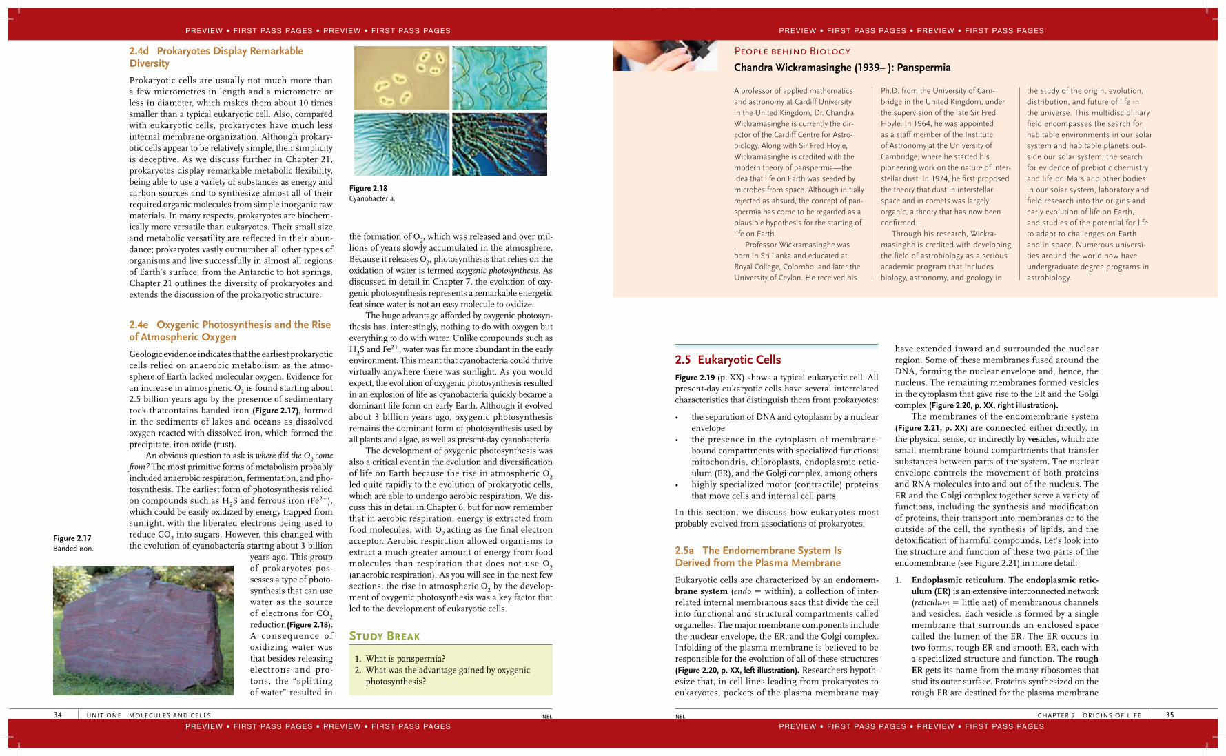
2.4e Oxygenic Photosynthesis and the Rise of Atmospheric Oxygen
Geologic evidence indicates that the earliest prokaryotic cells relied on anaerobic metabolism as the atmo-sphere of Earth lacked molecular oxygen. Evidence for an increase in atmospheric O2 is found starting about 2.5 billion years ago by the presence of sedimentary rock thatcontains banded iron (Figure 2.17), formed in the sediments of lakes and oceans as dissolved oxygen reacted with dissolved iron, which formed the precipitate, iron oxide (rust).
An obvious question to ask is where did the O2 come from? The most primitive forms of metabolism probably included anaerobic respiration, fermentation, and pho-tosynthesis. The earliest form of photosynthesis relied on compounds such as H2S and ferrous iron (Fe2�), which could be easily oxidized by energy trapped from sunlight, with the liberated electrons being used to reduce CO2 into sugars. However, this changed with the evolution of cyanobacteria startng about 3 billion
years ago. This group of prokaryotes pos-sesses a type of photo-synthesis that can use water as the source of electrons for CO2 reduction (Figure 2.18). A consequence of oxidizing water was that besides releasing electrons and pro-tons, the “splitting of water” resulted in
the formation of O2, which was released and over mil-lions of years slowly accumulated in the atmosphere. Because it releases O2, photosynthesis that relies on the oxidation of water is termed oxygenic photosynthesis. As discussed in detail in Chapter 7, the evolution of oxy-genic photosynthesis represents a remarkable energetic feat since water is not an easy molecule to oxidize.
The huge advantage aff orded by oxygenic photosyn-thesis has, interestingly, nothing to do with oxygen but everything to do with water. Unlike compounds such as H2S and Fe2�, water was far more abundant in the early environment. This meant that cyanobacteria could thrive virtually anywhere there was sunlight. As you would expect, the evolution of oxygenic photosynthesis resulted in an explosion of life as cyanobacteria quickly became a dominant life form on early Earth. Although it evolved about 3 billion years ago, oxygenic photosynthesis remains the dominant form of photosynthesis used by all plants and algae, as well as present-day cyanobacteria.
The development of oxygenic photosynthesis was also a critical event in the evolution and diversifi cation of life on Earth because the rise in atmospheric O2 led quite rapidly to the evolution of prokaryotic cells, which are able to undergo aerobic respiration. We dis-cuss this in detail in Chapter 6, but for now remember that in aerobic respiration, energy is extracted from food molecules, with O2 acting as the final electron acceptor. Aerobic respiration allowed organisms to extract a much greater amount of energy from food molecules than respiration that does not use O2 (anaerobic respiration). As you will see in the next few sections, the rise in atmospheric O2 by the develop-ment of oxygenic photosynthesis was a key factor that led to the development of eukaryotic cells.
Study Break
1. What is panspermia?2. What was the advantage gained by oxygenic
photosynthesis?
Figure 2.17 Banded iron.
Figure 2.18 Cyanobacteria.
40947_02_Ch02_p023-046 pp.indd 34 12/16/08 11:20:15 AM
CHAPTER 2 ORIGINS OF LIFE 35NEL
2.5 Eukaryotic CellsFigure 2.19 (p. XX) shows a typical eukaryotic cell. All present-day eukaryotic cells have several interrelated characteristics that distinguish them from prokaryotes:
• the separation of DNA and cytoplasm by a nuclear envelope
• the presence in the cytoplasm of membrane-bound compartments with specialized functions: mitochondria, chloroplasts, endoplasmic retic-ulum (ER), and the Golgi complex, among others
• highly specialized motor (contractile) proteins that move cells and internal cell parts
In this section, we discuss how eukaryotes most probably evolved from associations of prokaryotes.
2.5a The Endomembrane System Is Derived from the Plasma Membrane
Eukaryotic cells are characterized by an endomem-brane system (endo � within), a collection of inter-related internal membranous sacs that divide the cell into functional and structural compartments called organelles. The major membrane components include the nuclear envelope, the ER, and the Golgi complex. Infolding of the plasma membrane is believed to be responsible for the evolution of all of these structures (Figure 2.20, p. XX, left illustration). Researchers hypoth-esize that, in cell lines leading from prokaryotes to eukaryotes, pockets of the plasma membrane may
have extended inward and surrounded the nuclear region. Some of these membranes fused around the DNA, forming the nuclear envelope and, hence, the nucleus. The remaining membranes formed vesicles in the cytoplasm that gave rise to the ER and the Golgi complex (Figure 2.20, p. XX, right illustration).
The membranes of the endomembrane system (Figure 2.21, p. XX) are connected either directly, in the physical sense, or indirectly by vesicles, which are small membrane-bound compartments that transfer substances between parts of the system. The nuclear envelope controls the movement of both proteins and RNA molecules into and out of the nucleus. The ER and the Golgi complex together serve a variety of functions, including the synthesis and modifi cation of proteins, their transport into membranes or to the outside of the cell, the synthesis of lipids, and the detoxifi cation of harmful compounds. Let’s look into the structure and function of these two parts of the endomembrane (see Figure 2.21) in more detail:
1. Endoplasmic reticulum. The endoplasmic retic-ulum (ER) is an extensive interconnected network (reticulum � little net) of membranous channels and vesicles. Each vesicle is formed by a single membrane that surrounds an enclosed space called the lumen of the ER. The ER occurs in two forms, rough ER and smooth ER, each with a specialized structure and function. The rough ER gets its name from the many ribosomes that stud its outer surface. Proteins synthesized on the rough ER are destined for the plasma membrane
People behind Biology
Chandra Wickramasinghe (1939– ): Panspermia
A professor of applied mathematics and astronomy at Cardiff University in the United Kingdom, Dr. Chandra Wickramasinghe is currently the dir-ector of the Cardiff Centre for Astro-biology. Along with Sir Fred Hoyle, Wickramasinghe is credited with the modern theory of panspermia—the idea that life on Earth was seeded by microbes from space. Although initially rejected as absurd, the concept of pan-spermia has come to be regarded as a plausible hypothesis for the starting of life on Earth.
Professor Wickramasinghe was born in Sri Lanka and educated at Royal College, Colombo, and later the University of Ceylon. He received his
Ph.D. from the University of Cam-bridge in the United Kingdom, under the supervision of the late Sir Fred Hoyle. In 1964, he was appointed as a staff member of the Institute of Astronomy at the University of Cambridge, where he started his pioneering work on the nature of inter-stellar dust. In 1974, he fi rst proposed the theory that dust in interstellar space and in comets was largely organic, a theory that has now been confi rmed.
Through his research, Wickra-masinghe is credited with developing the field of astrobiology as a serious academic program that includes biology, astronomy, and geology in
the study of the origin, evolution, distribution, and future of life in the universe. This multidisciplinary field encompasses the search for habitable environments in our solar system and habitable planets out-side our solar system, the search for evidence of prebiotic chemistry and life on Mars and other bodies in our solar system, laboratory and field research into the origins and early evolution of life on Earth, and studies of the potential for life to adapt to challenges on Earth and in space. Numerous universi-ties around the world now have undergraduate degree programs in astrobiology.
40947_02_Ch02_p023-046 pp.indd 35 12/16/08 11:20:16 AM
Preview • First Pass Pages • Preview • First Pass Pages
Preview • First Pass Pages • Preview • First Pass Pages
Preview • First Pass Pages • Preview • First Pass Pages
Preview • First Pass Pages • Preview • First Pass Pages

UNIT ONE MOLECULES AND CELLS36 NEL
or for release outside the cell. After being synthe-sized, these proteins enter the lumen where they fold into their fi nal form. The proteins are then delivered to cell surface within vesicles that pinch off from the ER and move to join with the Golgi
complex. In comparison, the smooth ER does not have ribosomes attached to its surface. Instead of protein synthesis, smooth ER serves various functions, including the synthesis of lipids that become part of cell membranes.
Proteins made by the ribosomes that are freely suspended in the cytosol remain in the cytosol, pass through the nuclear pores to enter the nucleus, or become parts of mitochondria, chloroplasts, the cytoskeleton, or other cytoplasmic structures.
2. Golgi complex. The Golgi complex consists of a stack of flattened membranous sacs and is usu-ally located between the rough ER and the plasma membrane. The Golgi complex receives proteins made in the ER and transported to the complex in vesicles. Within the Golgi complex, further chem-ical modifi cations of the proteins occur. The modi-fi ed proteins are then sorted into other vesicles that pinch off from the margins of Golgi sacs on the side of the complex that faces the plasma mem-brane. The Golgi complex regulates the movement of several types of proteins. Some are secreted from the cell, others become embedded in the plasma membrane, and yet others are placed in lysosomes.
Vesicle
Ribosome (freein cytosol)
Nuclearpore complex
Microfilaments
Microbody
Nuclearenvelope
Chromatin
Nucleolus
Rough ER
Ribosome (attachedto rough ER)
Smooth ER
Cytosol
MitochondrionEnergymetabolism
PlasmamembraneTransport
Microtubulesradiating fromcell centre
LysosomeDegradation;recycling
Golgi complexModification,distribution ofproteins
NucleusMembrane-enclosedregion of DNA;hereditary control
Endoplasmic reticulumSynthesis, modification,transport of proteins;membrane synthesis
Pair ofcentriolesin cell centre
Figure 2.19 Eukaryotic cell.
Figure 2.20 A hypothetical route for formation of the nuclear envelope and endoplasmic reticulum, through segments of the plasma membrane that were brought into the cytoplasm by endocytosis.
Cytoplasm
Endoplasmicreticulum
Nuclearenvelope
Nuclear region
40947_02_Ch02_p023-046 pp.indd 36 12/16/08 11:20:16 AM
CHAPTER 2 ORIGINS OF LIFE 37NEL
For example, proteins secreted from the cell are transported to the plasma membrane by secretory vesicles, which release their contents to the exte-rior by exocytosis. Vesicles may also form by the reverse process, called endocytosis, which brings molecules into the cell from the exterior.
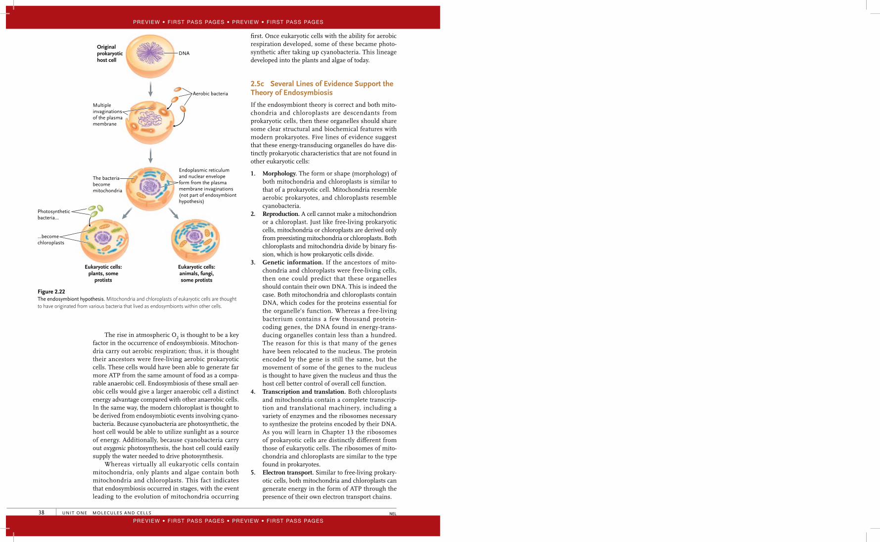
2.5b The Theory of Endosymbiosis Suggests that Mitochondria and Chloroplasts Evolved from Ingested Prokaryotes
Besides the extensive endomembrane system, another clear characteristic of eukaryotic cells is the presence of energy-transducing organelles: the chloroplast and
the mitochondrion (plural � mitochondria). From the last section, recall that all the membrane structures of the endomembrane systems are thought to have been derived from an infolding of the plasma membrane. By comparison, a large body of evidence supports a model of eukaryotic evolution that involves endosymbiosis—the mitochondria and chloroplasts are descendants of free-living prokaryotes (Figure 2.22, p. XX). The estab-lished theory of endosymbiosis states that the prokary-otic ancestors of modern mitochondria and chloroplasts were engulfed by larger prokaryotic cells, forming a mutually advantageous relationship called a symbiosis, and that slowly, over time, the host cell and the endosym-bionts became inseparable parts of the same organism.
Proteins made by ER ribosomes enter ER membranes or the space inside ER cisternae. Chemical modification of some proteins begins. Membrane lipids are also made in the ER.
1
Vesicles bud fromthe ER membrane and then transport unfinishedproteins and lipids to the Golgi complex.
2
Protein and lipid modification is completedin the Golgi complex, and products are sorted into vesicles that bud fromthe complex.
3
Secretory vesicles buddingfrom the Golgi membranestransport finished products to theplasma membrane. The productsare released by exocytosis. Othervesicles remain in storage inthe cytoplasm.
4
Lysosomes budding from the Golgi membranes contain hydrolytic enzymes that digest damaged organelles or the contents of endocytic vesicles that fuse with them. Endocytic vesicles form at the plasma membrane and move into the cytoplasm.
5
Instructions for buildingproteins leave the nucleusand enter the cytoplasm.
Proteins (green)are assembled onribosomes attachedto the ER or free inthe cytoplasm.
Nucleus
RoughER
Ribosomes
Golgicomplex
Endocyticvesicle
Lysosomes
Damagedorganelle
Secretoryvesicles
Vesicles
Figure 2.21 Vesicle traffi c in the cytoplasm. The ER and Golgi complex are part of the endomembrane system., which releases proteins and other substances to the cell exterior and gathers materials from outside the cell.
40947_02_Ch02_p023-046 pp.indd 37 12/16/08 11:20:17 AM
Preview • First Pass Pages • Preview • First Pass Pages
Preview • First Pass Pages • Preview • First Pass Pages
Preview • First Pass Pages • Preview • First Pass Pages
Preview • First Pass Pages • Preview • First Pass Pages
UNIT ONE MOLECULES AND CELLS38 NEL
fi rst. Once eukaryotic cells with the ability for aerobic respiration developed, some of these became photo-synthetic after taking up cyanobacteria. This lineage developed into the plants and algae of today.
2.5c Several Lines of Evidence Support the Theory of Endosymbiosis
If the endosymbiont theory is correct and both mito-chondria and chloroplasts are descendants from prokaryotic cells, then these organelles should share some clear structural and biochemical features with modern prokaryotes. Five lines of evidence suggest that these energy-transducing organelles do have dis-tinctly prokaryotic characteristics that are not found in other eukaryotic cells:
1. Morphology. The form or shape (morphology) of both mitochondria and chloroplasts is similar to that of a prokaryotic cell. Mitochondria resemble aerobic prokaryotes, and chloroplasts resemble cyanobacteria.
2. Reproduction. A cell cannot make a mitochondrion or a chloroplast. Just like free-living prokaryotic cells, mitochondria or chloroplasts are derived only from preexisting mitochondria or chloroplasts. Both chloroplasts and mitochondria divide by binary fi s-sion, which is how prokaryotic cells divide.
3. Genetic information. If the ancestors of mito-chondria and chloroplasts were free-living cells, then one could predict that these organelles should contain their own DNA. This is indeed the case. Both mitochondria and chloroplasts contain DNA, which codes for the proteins essential for the organelle’s function. Whereas a free-living bacterium contains a few thousand protein-coding genes, the DNA found in energy-trans-ducing organelles contain less than a hundred. The reason for this is that many of the genes have been relocated to the nucleus. The protein encoded by the gene is still the same, but the movement of some of the genes to the nucleus is thought to have given the nucleus and thus the host cell better control of overall cell function.
4. Transcription and translation. Both chloroplasts and mitochondria contain a complete transcrip-tion and translational machinery, including a variety of enzymes and the ribosomes necessary to synthesize the proteins encoded by their DNA. As you will learn in Chapter 13 the ribosomes of prokaryotic cells are distinctly diff erent from those of eukaryotic cells. The ribosomes of mito-chondria and chloroplasts are similar to the type found in prokaryotes.
5. Electron transport. Similar to free-living prokary-otic cells, both mitochondria and chloroplasts can generate energy in the form of ATP through the presence of their own electron transport chains.
The rise in atmospheric O2 is thought to be a key factor in the occurrence of endosymbiosis. Mitochon-dria carry out aerobic respiration; thus, it is thought their ancestors were free-living aerobic prokaryotic cells. These cells would have been able to generate far more ATP from the same amount of food as a compa-rable anaerobic cell. Endosymbiosis of these small aer-obic cells would give a larger anaerobic cell a distinct energy advantage compared with other anaerobic cells. In the same way, the modern chloroplast is thought to be derived from endosymbiotic events involving cyano-bacteria. Because cyanobacteria are photosynthetic, the host cell would be able to utilize sunlight as a source of energy. Additionally, because cyanobacteria carry out oxygenic photosynthesis, the host cell could easily supply the water needed to drive photosynthesis.
Whereas virtually all eukaryotic cells contain mitochondria, only plants and algae contain both mitochondria and chloroplasts. This fact indicates that endosymbiosis occurred in stages, with the event leading to the evolution of mitochondria occurring
DNA
Aerobic bacteria
Multipleinvaginationsof the plasmamembrane
The bacteriabecomemitochondria
Endoplasmic reticulum and nuclear envelope form from the plasma membrane invaginations (not part of endosymbionthypothesis)
Originalprokaryotichost cell
Eukaryotic cells:plants, some
protists
Eukaryotic cells:animals, fungi,some protists
Photosyntheticbacteria…
…becomechloroplasts
Figure 2.22 The endosymbiont hypothesis. Mitochondria and chloroplasts of eukaryotic cells are thought to have originated from various bacteria that lived as endosymbionts within other cells.
40947_02_Ch02_p023-046 pp.indd 38 12/16/08 11:20:17 AM
Preview • First Pass Pages • Preview • First Pass Pages
Preview • First Pass Pages • Preview • First Pass Pages