BIOLOGY 524 SKULL – I BRAINCASE - CHONDROCRANIUM S. S. Sumida
Upload
verity-bradleyCategory
view
219download
0
Biology 323Human Anatomy for Biology MajorsLecture 15Dr. Stuart Sumida
Development and Structure, of the Reproductive System
X
X
X
X
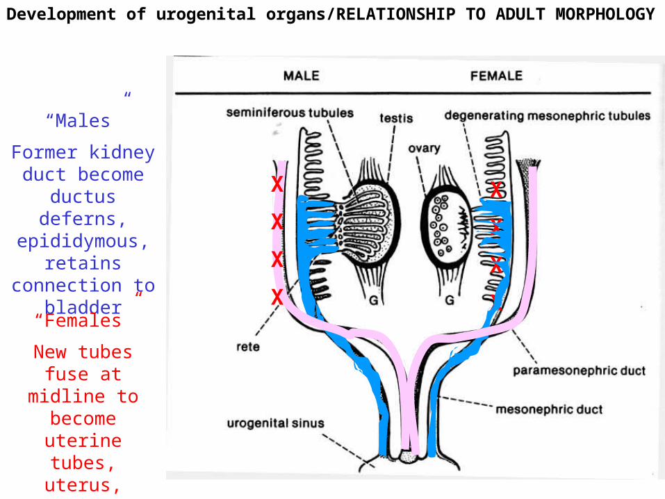
Development of urogenital organs/RELATIONSHIP TO ADULT MORPHOLOGY
“Males”
Former kidney duct become
ductus deferns, epididymous,
retains connection to bladder
“Females”
New tubes fuse at midline to become
uterine tubes, uterus, superior
2/3 vagina
X
X
X
X
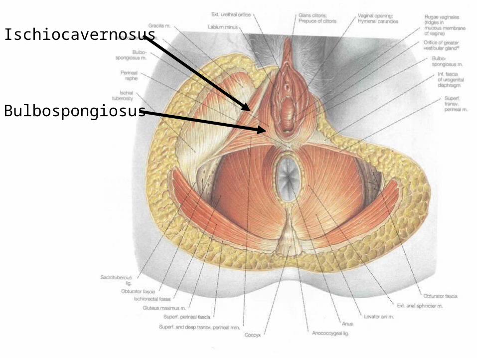
Ischiocavernosus
Bulbospongiosus
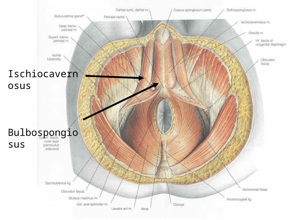
Ischiocavernosus
Bulbospongiosus
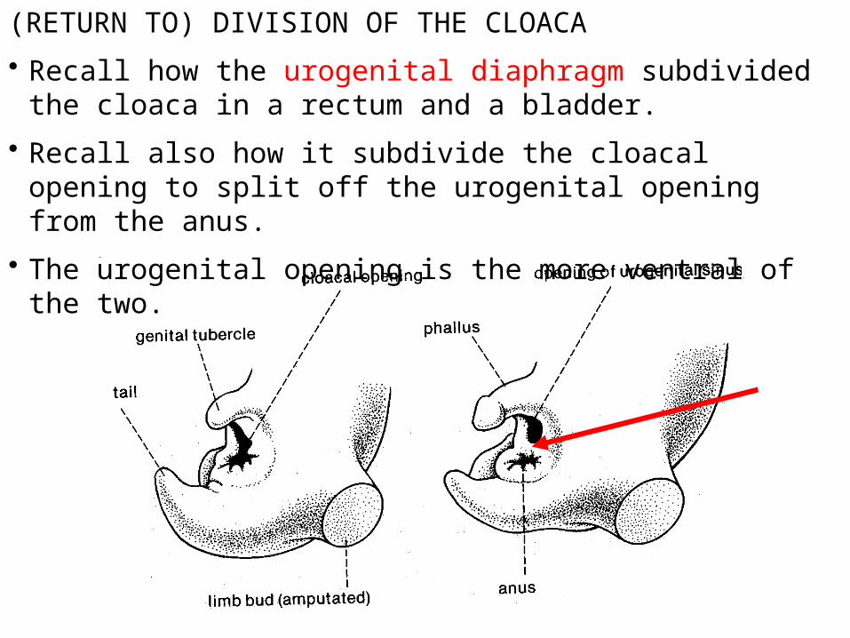
(RETURN TO) DIVISION OF THE CLOACA
• Recall how the urogenital diaphragm subdivided the cloaca in a rectum and a bladder.
• Recall also how it subdivide the cloacal opening to split off the urogenital opening from the anus.
• The urogenital opening is the more ventral of the two.
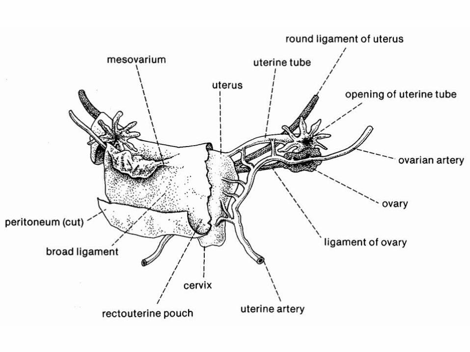
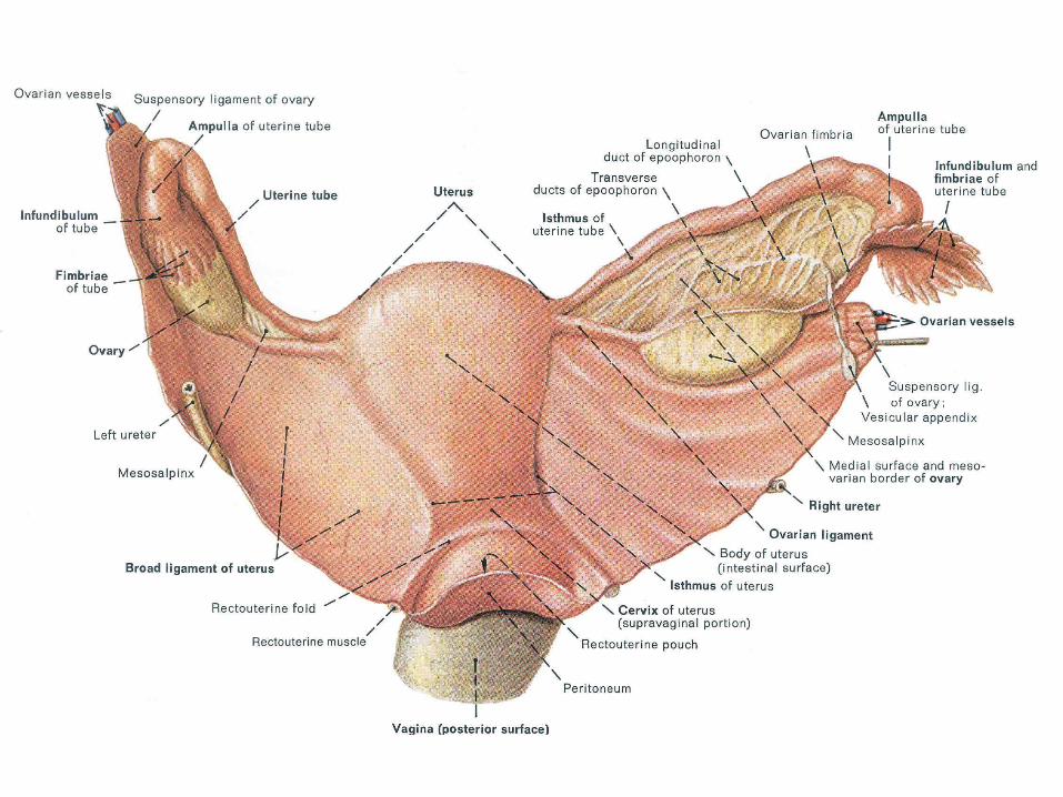
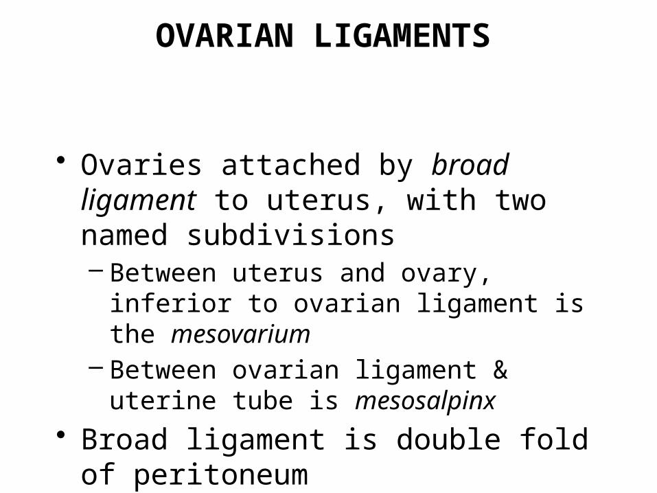
• Ovaries attached by broad ligament to uterus, with two named subdivisions– Between uterus and ovary, inferior to ovarian
ligament is the mesovarium– Between ovarian ligament & uterine tube is
mesosalpinx
• Broad ligament is double fold of peritoneum
OVARIAN LIGAMENTS
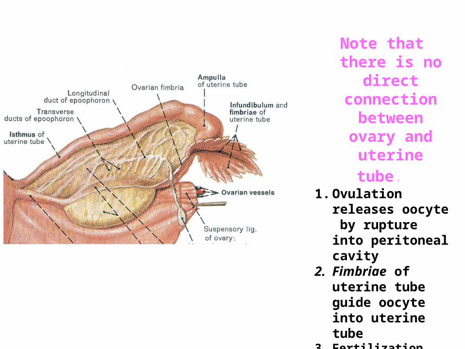
Note that there is no direct
connection between ovary
and uterine tube.
1. Ovulation releases oocyte by rupture into peritoneal cavity
2. Fimbriae of uterine tube guide oocyte into uterine tube
3. Fertilization occurs immediately after ovulation, high in uterine tube at or near fimbriae.

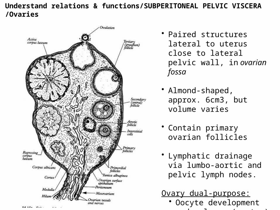
• Paired structures lateral to uterus close to lateral pelvic wall, in ovarian fossa
• Almond-shaped, approx. 6cm3, but volume varies
• Contain primary ovarian follicles
• Lymphatic drainage via lumbo-aortic and pelvic lymph nodes.
Ovary dual-purpose: • Oocyte development and
release (cortex)• Endocrine gland (cortex
and medulla)
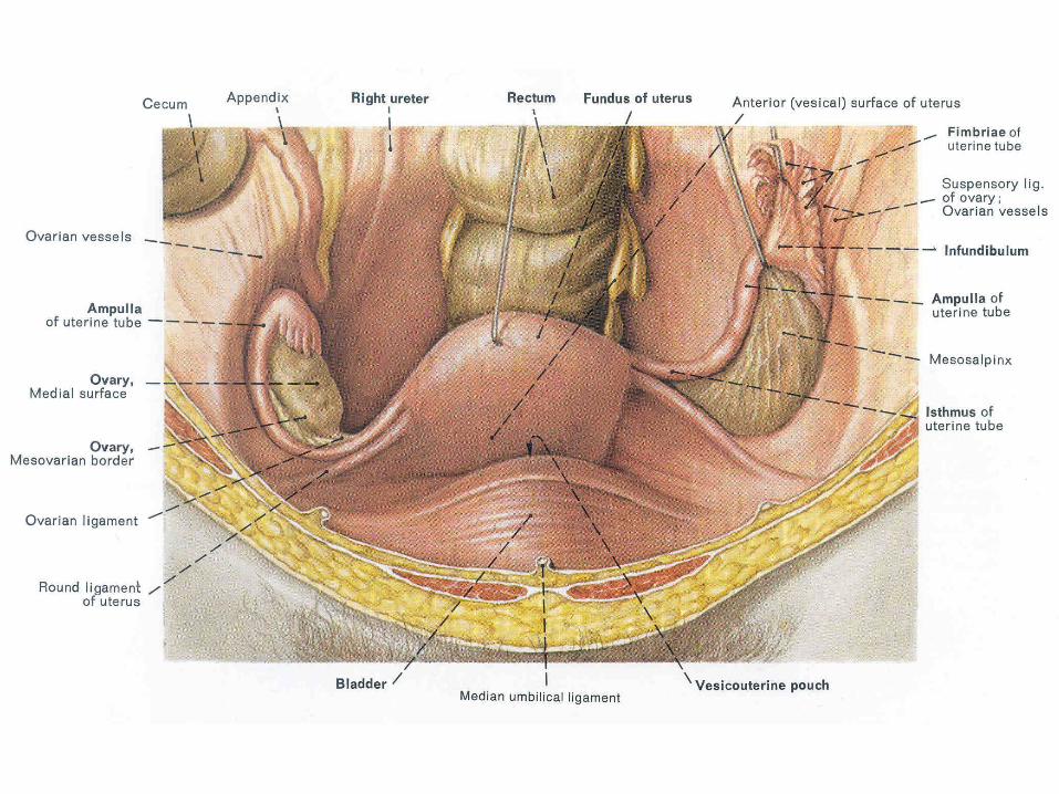
Understand relations & functions/SUBPERITONEAL PELVIC VISCERA /Ovaries
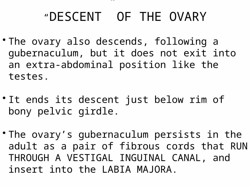
“DESCENT” OF THE OVARY
• The ovary also descends, following a gubernaculum, but it does not exit into an extra-abdominal position like the testes.
• It ends its descent just below rim of bony pelvic girdle.
• The ovary’s gubernaculum persists in the adult as a pair of fibrous cords that RUN THROUGH A VESTIGAL INGUINAL CANAL, and insert into the LABIA MAJORA.
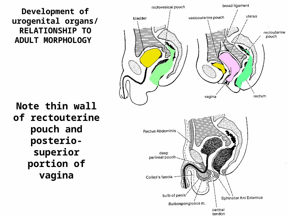
Development of urogenital organs/ RELATIONSHIP TO
ADULT MORPHOLOGY
Note thin wall of rectouterine pouch
and posterio-superior portion of
vagina
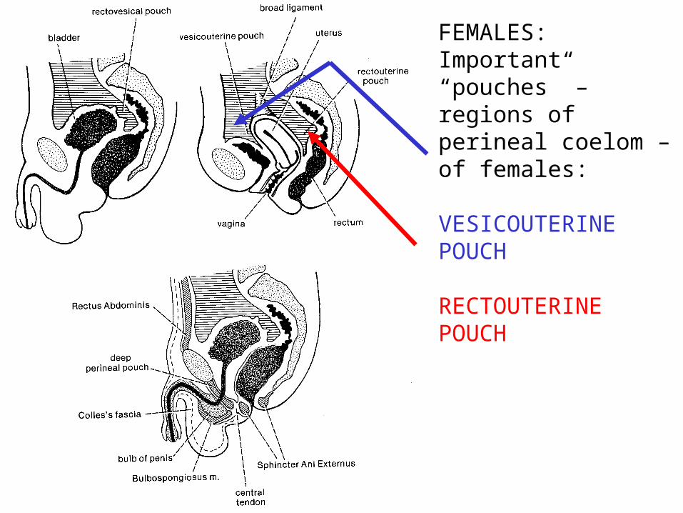
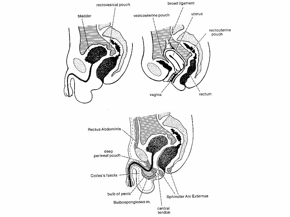
FEMALES:Important “pouches” – regions of perineal coelom – of females:
VESICOUTERINE POUCH
RECTOUTERINE POUCH
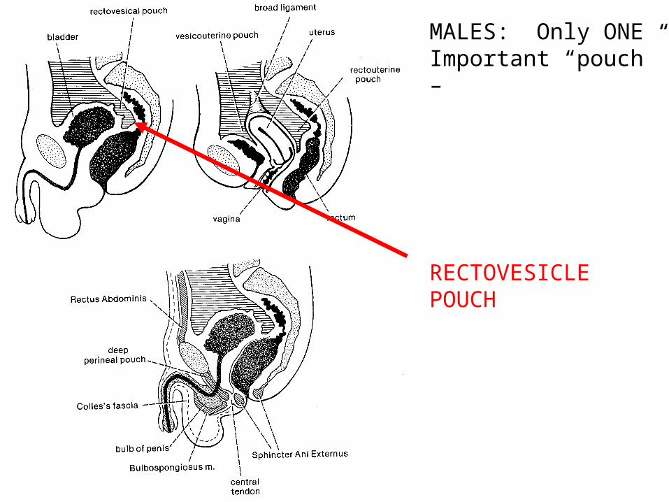
MALES: Only ONE Important “pouch” –
RECTOVESICLE POUCH
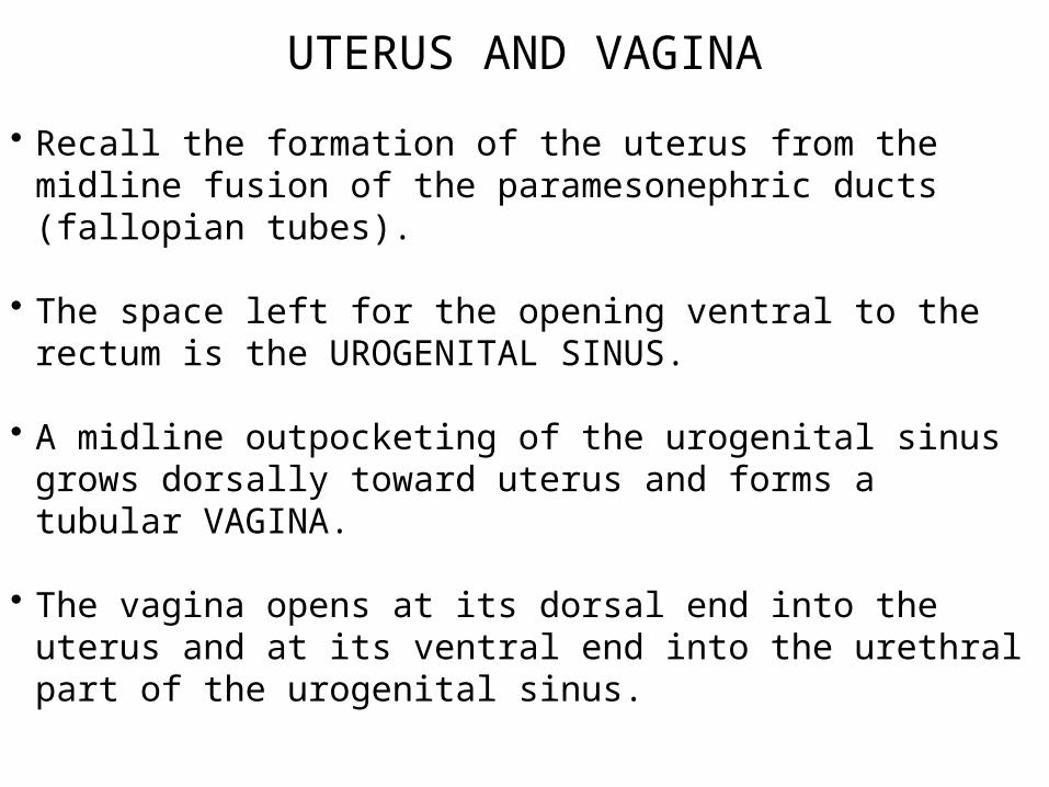
UTERUS AND VAGINA
• Recall the formation of the uterus from the midline fusion of the paramesonephric ducts (fallopian tubes).
• The space left for the opening ventral to the rectum is the UROGENITAL SINUS.
• A midline outpocketing of the urogenital sinus grows dorsally toward uterus and forms a tubular VAGINA.
• The vagina opens at its dorsal end into the uterus and at its ventral end into the urethral part of the urogenital sinus.
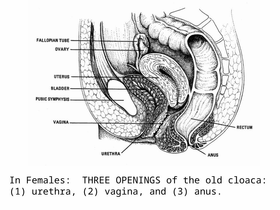
In Females: THREE OPENINGS of the old cloaca: (1) urethra, (2) vagina, and (3) anus.
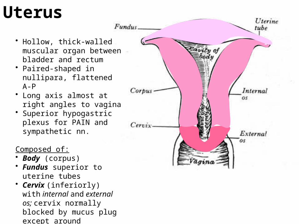
• Hollow, thick-walled muscular organ between bladder and rectum
• Paired-shaped in nullipara, flattened A-P
• Long axis almost at right angles to vagina
• Superior hypogastric plexus for PAIN and sympathetic nn.
Composed of:• Body (corpus)• Fundus superior to uterine
tubes• Cervix (inferiorly) with
internal and external os; cervix normally blocked by mucus plug except around ovulation
Uterus
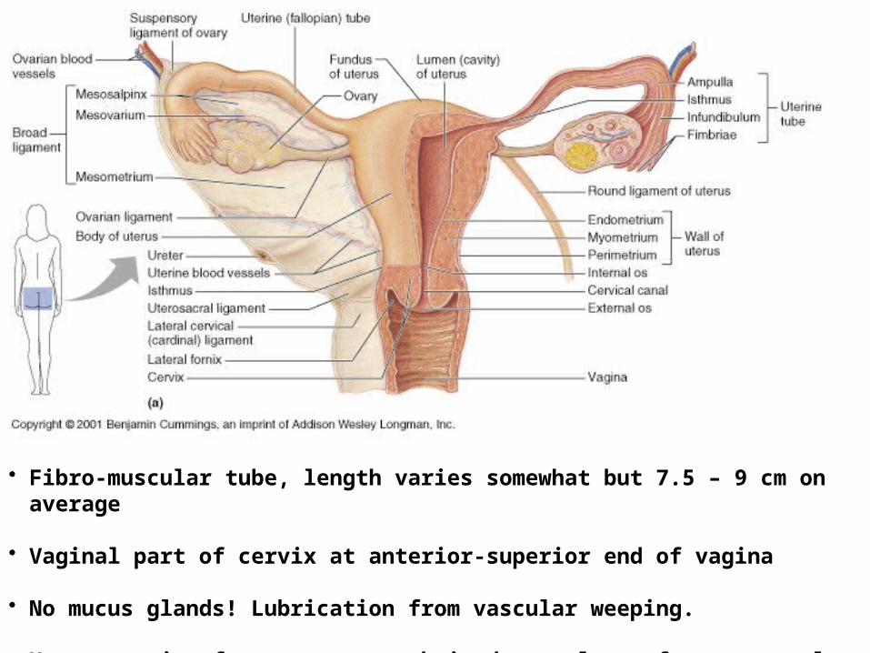
• Fibro-muscular tube, length varies somewhat but 7.5 – 9 cm on average
• Vaginal part of cervix at anterior-superior end of vagina
• No mucus glands! Lubrication from vascular weeping.
• Upper portion from paramesonephric ducts, lower from external invagination
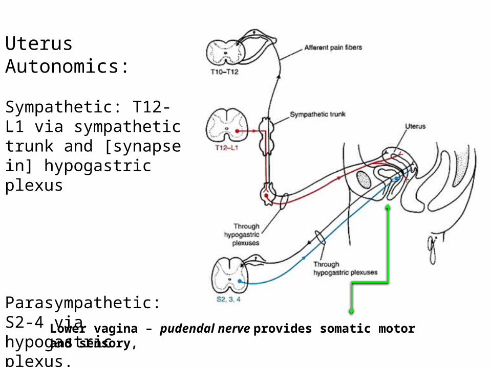
Uterus Autonomics:
Sympathetic: T12-L1 via sympathetic trunk and [synapse in] hypogastric plexus
Parasympathetic: S2-4 via hypogastric plexus.
Lower vagina – pudendal nerve provides somatic motor and sensory,
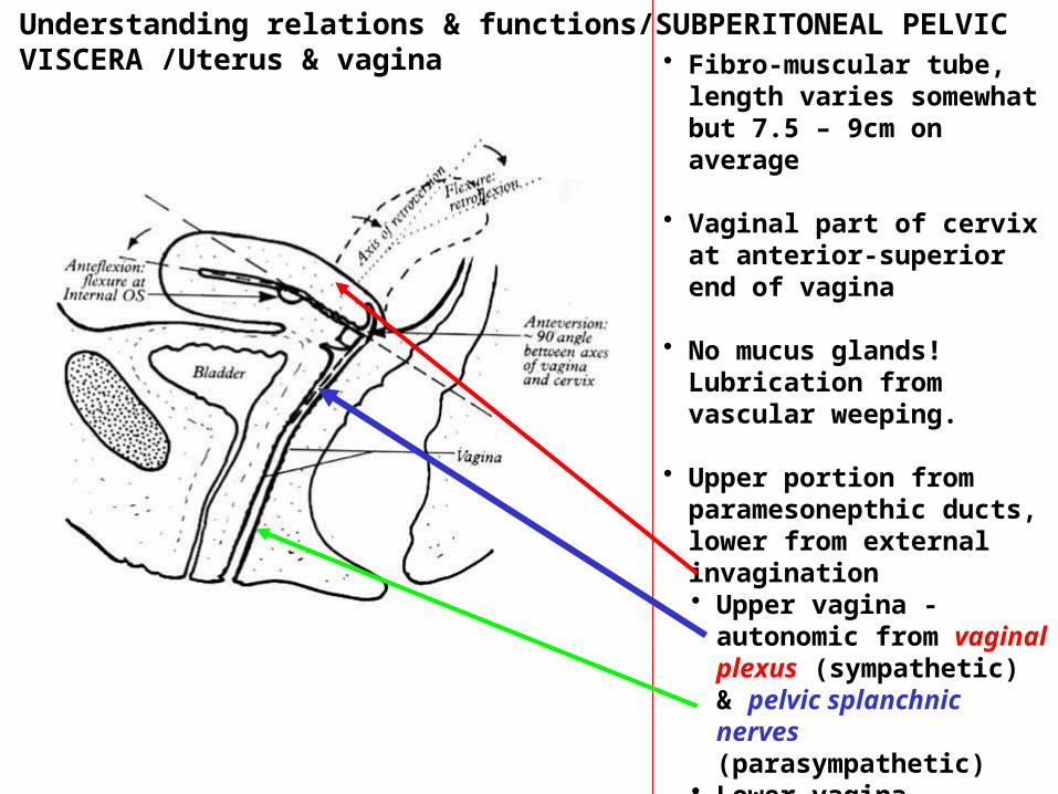
• Fibro-muscular tube, length varies somewhat but 7.5 – 9cm on average
• Vaginal part of cervix at anterior-superior end of vagina
• No mucus glands! Lubrication from vascular weeping.
• Upper portion from paramesonepthic ducts, lower from external invagination• Upper vagina - autonomic
from vaginal plexus (sympathetic) & pelvic splanchnic nerves (parasympathetic)
• Lower vagina – pudendal nerve (somatic motor and sensory)
Understanding relations & functions/SUBPERITONEAL PELVIC VISCERA /Uterus & vagina
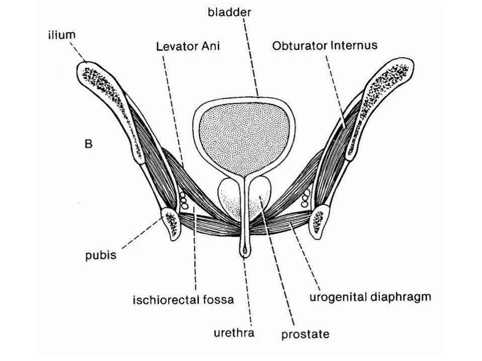
DESCENT OF THE TESTES:
• Recall from the previous lecture that the male testes descend from their initially intraperitoneal position, through the body wall, into a pouch protruding from the body wall called the SCROTUM.
• Everything gets drug along in this descent: ductus deferens, nerves, blood vessels.
• All of these together form a connection (“leash”) of testicular connections called the SPERMATIC CORD.
RETROPERITONEAL POSITION OF THE TESTES
• The serial homolog of the coelom and its peritoneal boundaries together are called the TUNICA VAGINALIS.
• (Another way of saying this is that each testis is surrounded by its own little coelomic sac.
• Remember, each testis started out retroperitoneal on the dorsal side of the body wall with the coelom ventral to it.
• Appropriately, tunica vaginalis is wrapped around only part of each testis – the ventral side, leaving it retroperitoneal even in the scrotal sac.
WHY DESCEND???
Preserve male fertility – sperm must be kept a bit cooler than standard mammalian body temperature. Otherwise they degenerate and lose motility.
Recall from the previous lecture:• As a transitory stage of kidney degenerates, a ligament
called the GUBERNACULUM descends on each side of abdomen from inferior pole of gonad.
• Gubernaculum passes obliquely through developing anterior abdominal wall at site of future inguinal canal and attaches at internal surface of labioscrotal swelling (future position of scrotum in males or labium majorum in females).
• Gubernaculum is thought to guide descent of testes into scrotum, and ultimately anchors testis to scrotal wall.
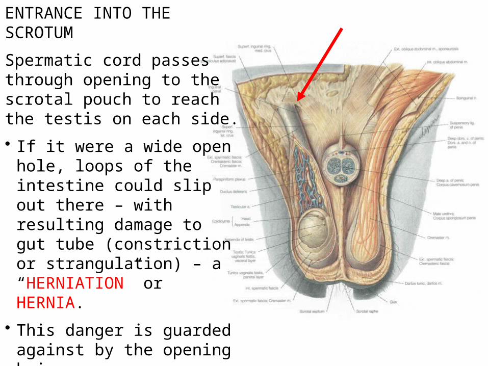
ENTRANCE INTO THE SCROTUM
• Spermatic cord passes through opening to the scrotal pouch to reach the testis on each side.
• If it were a wide open hole, loops of the intestine could slip out there – with resulting damage to gut tube (constriction or strangulation) – a “HERNIATION” or HERNIA.
• This danger is guarded against by the opening being a very narrow slit – the INGUINAL CANAL.
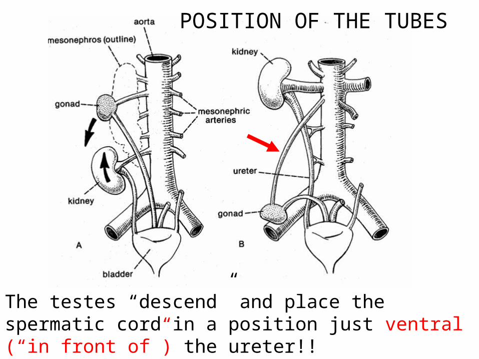
The testes “descend” and place the spermatic cord in a position just ventral (“in front of”) the ureter!!
POSITION OF THE TUBES
ENTRANCE INTO THE SCROTUM
Spermatic cord passes through opening to the scrotal pouch to reach the testis on each side.
• If it were a wide open hole, loops of the intestine could slip out there – with resulting damage to gut tube (constriction or strangulation) – a “HERNIATION” or HERNIA.
• This danger is guarded against by the opening being a very narrow slit – the INGUINAL CANAL.
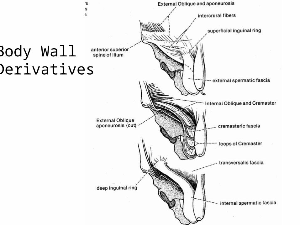
Body WallDerivatives
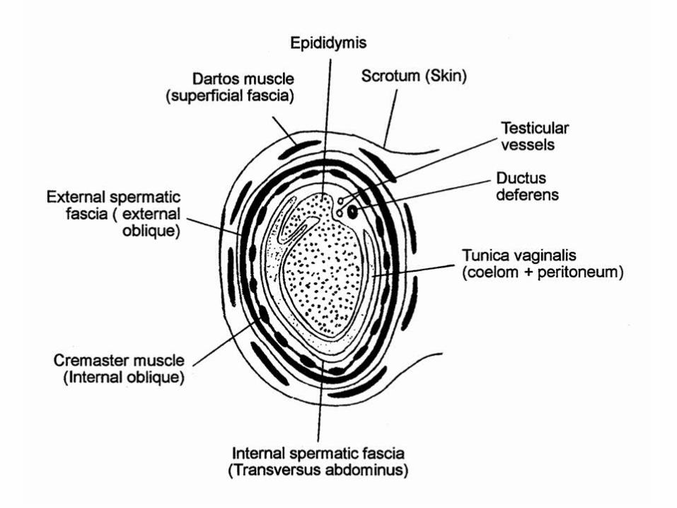
SERIAL HOMOLOGS OF SCROTAL STRUCTURES
As testes push through body wall, they carry with them all layers and a bit of coelomic space. The equivalents are:
• Skin: SCROTAL SAC
• Superficial fascia: DARTOS MUSCLE
• External oblique: EXTERNAL SPERMATIC FASCIA
• Internal oblique: CREMASTER MUSCLE
• Transversus abdominus: INTERNAL SPERMATIC FASCIA
• Coelom + peritoneum: TUNICA VAGINALIS
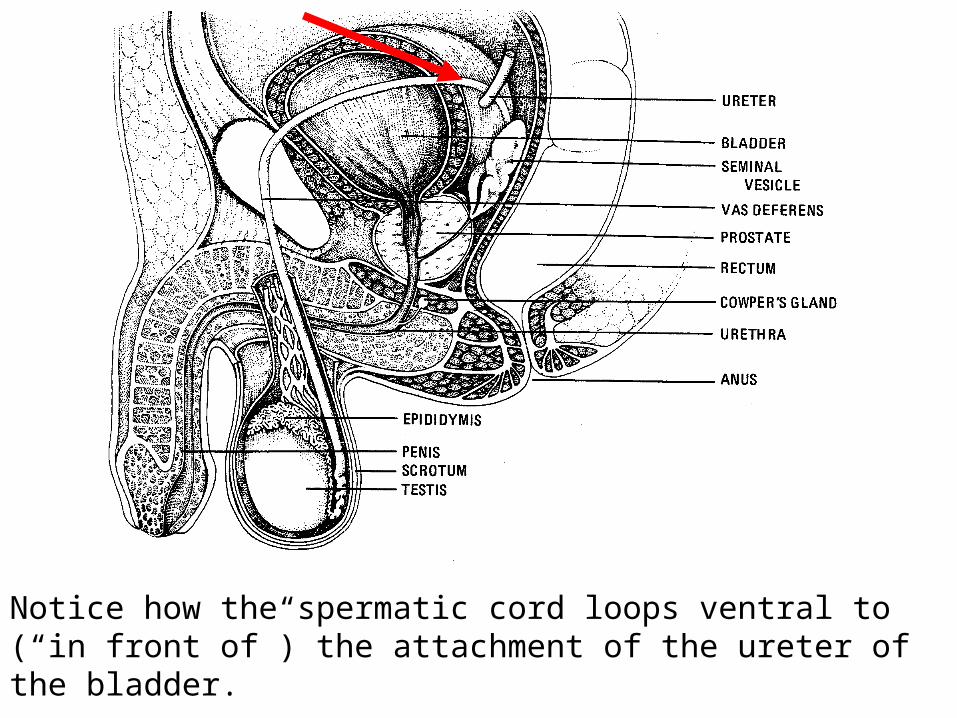
Notice how the spermatic cord loops ventral to (“in front of”) the attachment of the ureter of the bladder.
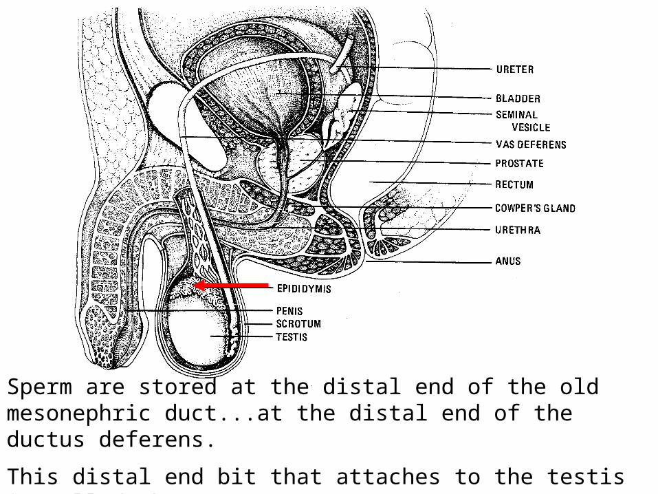
Sperm are stored at the distal end of the old mesonephric duct...at the distal end of the ductus deferens.
This distal end bit that attaches to the testis is called the EPIDIDYMIS.
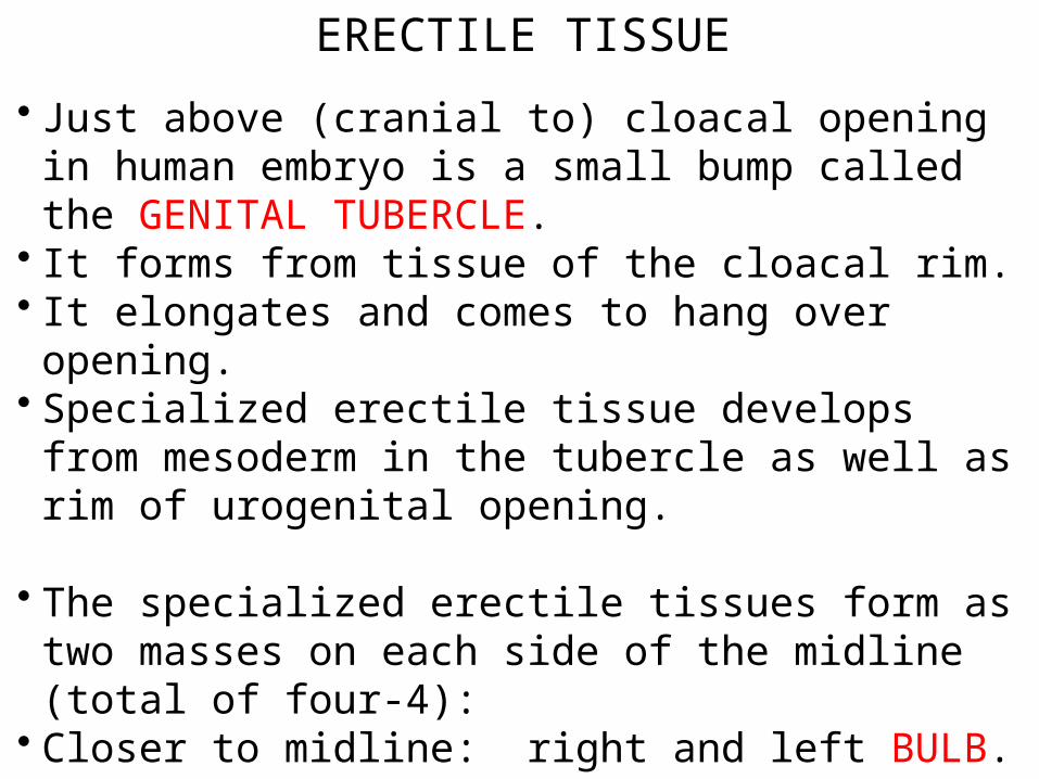
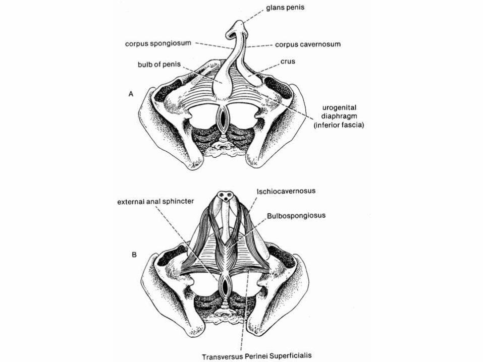
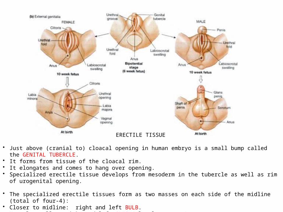
ERECTILE TISSUE
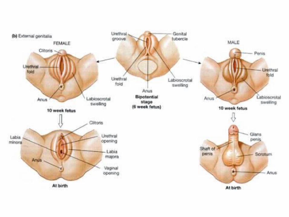
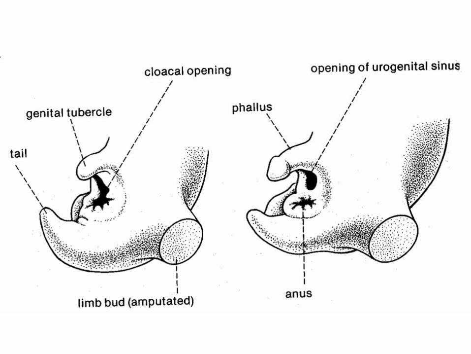
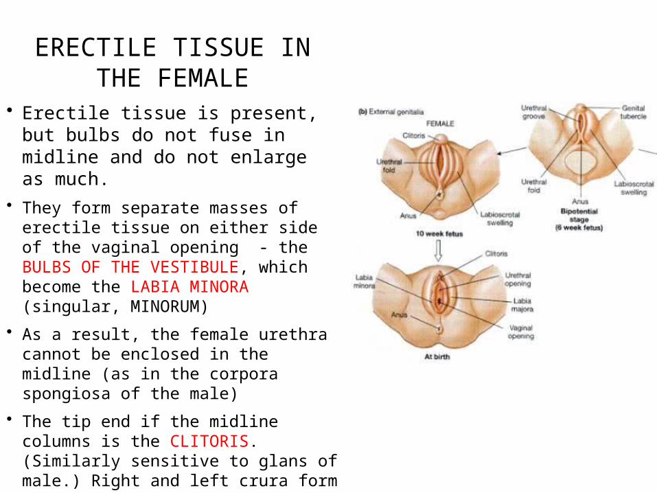
• Just above (cranial to) cloacal opening in human embryo is a small bump called the GENITAL TUBERCLE.
• It forms from tissue of the cloacal rim.• It elongates and comes to hang over opening.• Specialized erectile tissue develops from mesoderm in the tubercle as well as rim of urogenital opening.
• The specialized erectile tissues form as two masses on each side of the midline (total of four-4):
• Closer to midline: right and left BULB.• More laterally: right and left CRUS (plural – curura).
ERECTILE TISSUE
• Just above (cranial to) cloacal opening in human embryo is a small bump called the GENITAL TUBERCLE.
• It forms from tissue of the cloacal rim.• It elongates and comes to hang over opening.• Specialized erectile tissue develops from mesoderm in the tubercle as well as rim of urogenital
opening.
• The specialized erectile tissues form as two masses on each side of the midline (total of four-4): • Closer to midline: right and left BULB.• More laterally: right and left CRUS (plural – curura).

(RETURN TO) DIVISION OF THE CLOACA
• Recall how the urogenital diaphragm subdivided the cloaca in a rectum and a bladder.
• Recall also how it subdivide the cloacal opening to split off the urogenital opening from the anus.
• The urogenital opening is the more ventral of the two.
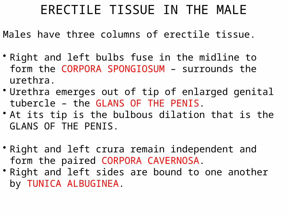
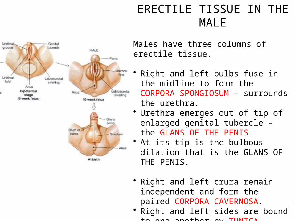
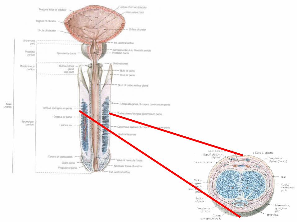
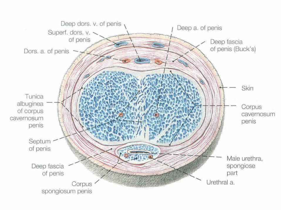
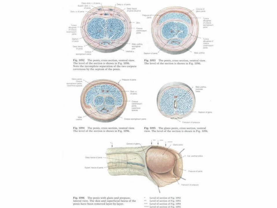
ERECTILE TISSUE IN THE MALE
Males have three columns of erectile tissue.
• Right and left bulbs fuse in the midline to form the CORPORA SPONGIOSUM – surrounds the urethra.
• Urethra emerges out of tip of enlarged genital tubercle – the GLANS OF THE PENIS.
• At its tip is the bulbous dilation that is the GLANS OF THE PENIS.
• Right and left crura remain independent and form the paired CORPORA CAVERNOSA.
• Right and left sides are bound to one another by TUNICA ALBUGINEA.
ERECTILE TISSUE IN THE FEMALE• Erectile tissue is present, but bulbs do not fuse in midline and do not enlarge as much.
• They form separate masses of erectile tissue on either side of the vginal opening - the BULBS OF THE VESTIBULE, which become the LABIA MINORA (singular, MINORUM)
• As a result, the female urethra cannot be enclosed in the midline (as in the corpora spongiosa of the male)
• The tip end if the midline columns is the CLITORIS. (Similarly sensitive to glans of male.) Right and left crura form (much smaller) corpora cavernosa of clitoris.
ERECTILE TISSUE IN THE FEMALE
• Erectile tissue is present, but bulbs do not fuse in midline and do not enlarge as much.
• They form separate masses of erectile tissue on either side of the vaginal opening - the BULBS OF THE VESTIBULE, which become the LABIA MINORA (singular, MINORUM)
• As a result, the female urethra cannot be enclosed in the midline (as in the corpora spongiosa of the male)
• The tip end if the midline columns is the CLITORIS. (Similarly sensitive to glans of male.) Right and left crura form (much smaller) corpora cavernosa of clitoris.
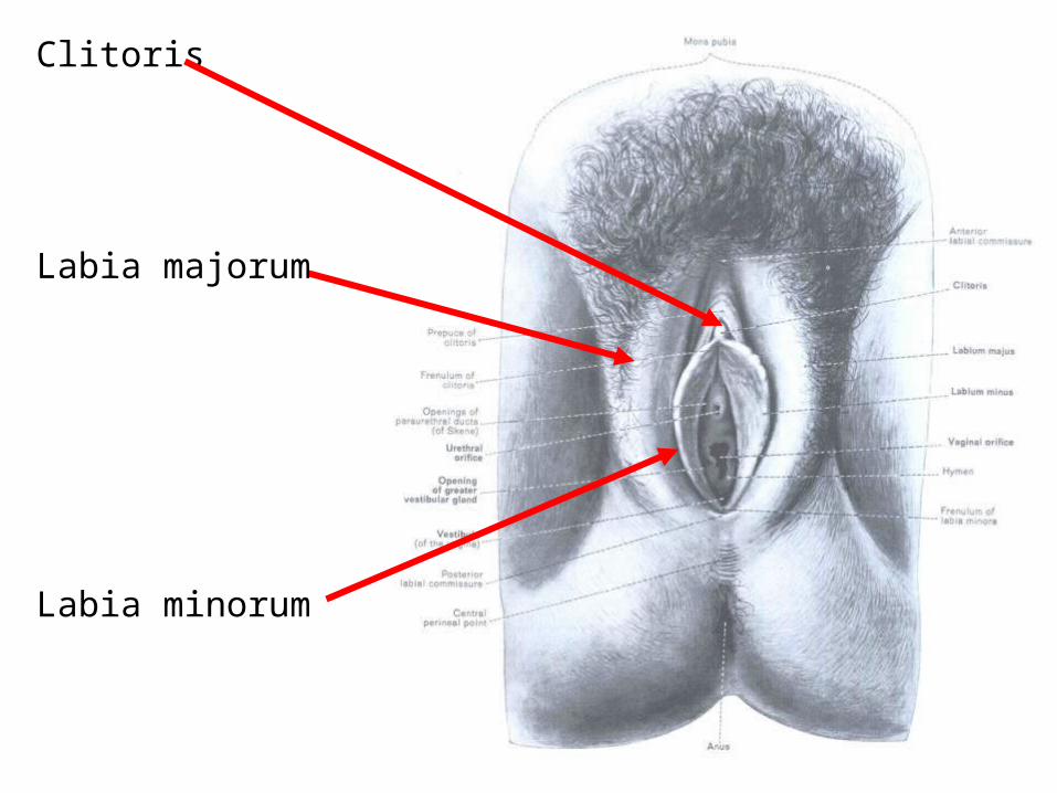
Clitoris
Labia majorum
Labia minorum
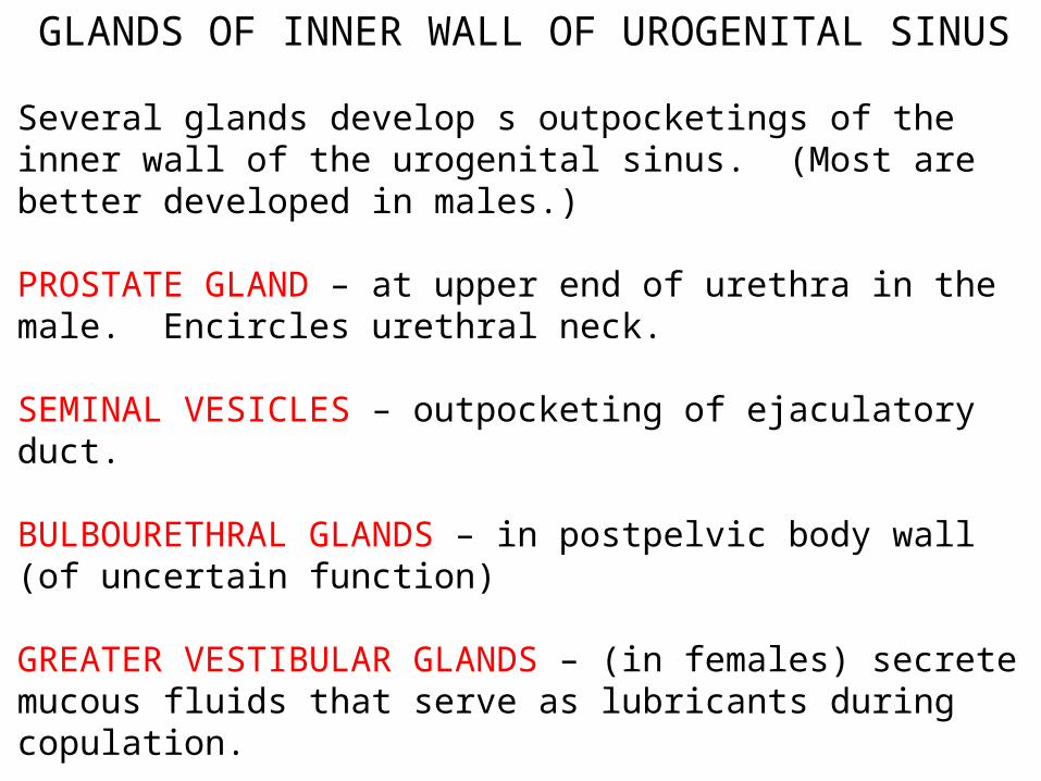
GLANDS OF INNER WALL OF UROGENITAL SINUS
Several glands develop s outpocketings of the inner wall of the urogenital sinus. (Most are better developed in males.)
PROSTATE GLAND – at upper end of urethra in the male. Encircles urethral neck.
SEMINAL VESICLES – outpocketing of ejaculatory duct.
BULBOURETHRAL GLANDS – in postpelvic body wall (of uncertain function)
GREATER VESTIBULAR GLANDS – (in females) secrete mucous fluids that serve as lubricants during copulation.
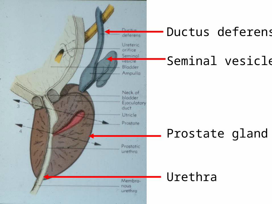
Ductus deferens
Seminal vesicle
Prostate gland
Urethra
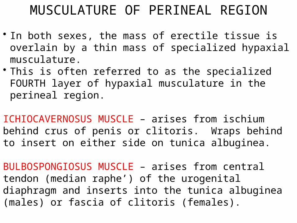
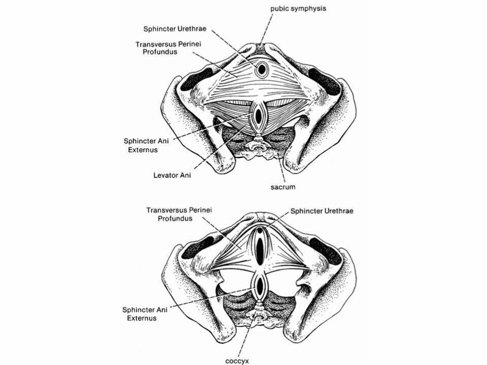
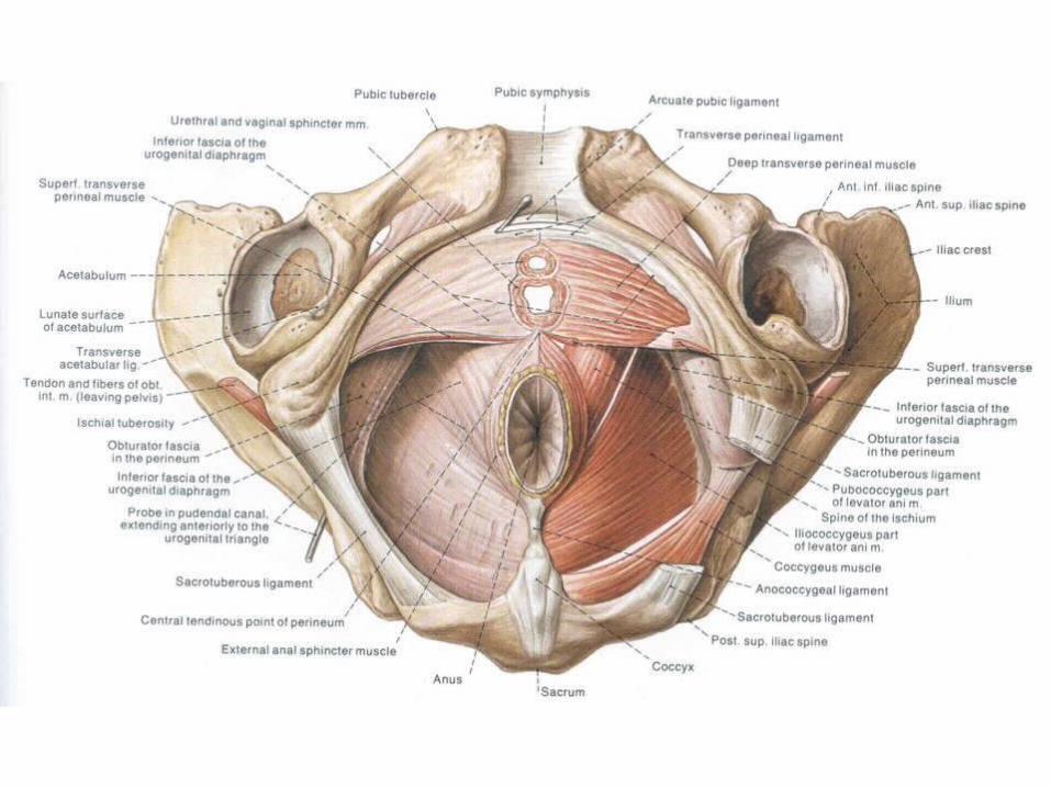
MUSCULATURE OF PERINEAL REGION
• In both sexes, the mass of erectile tissue is overlain by a thin mass of specialized hypaxial musculature.
• This is often referred to as the specialized FOURTH layer of hypaxial musculature in the perineal region.
ICHIOCAVERNOSUS MUSCLE – arises from ischium behind crus of penis or clitoris. Wraps behind to insert on either side on tunica albuginea.
BULBOSPONGIOSUS MUSCLE – arises from central tendon (median raphe’) of the urogenital diaphragm and inserts into the tunica albuginea (males) or fascia of clitoris (females).
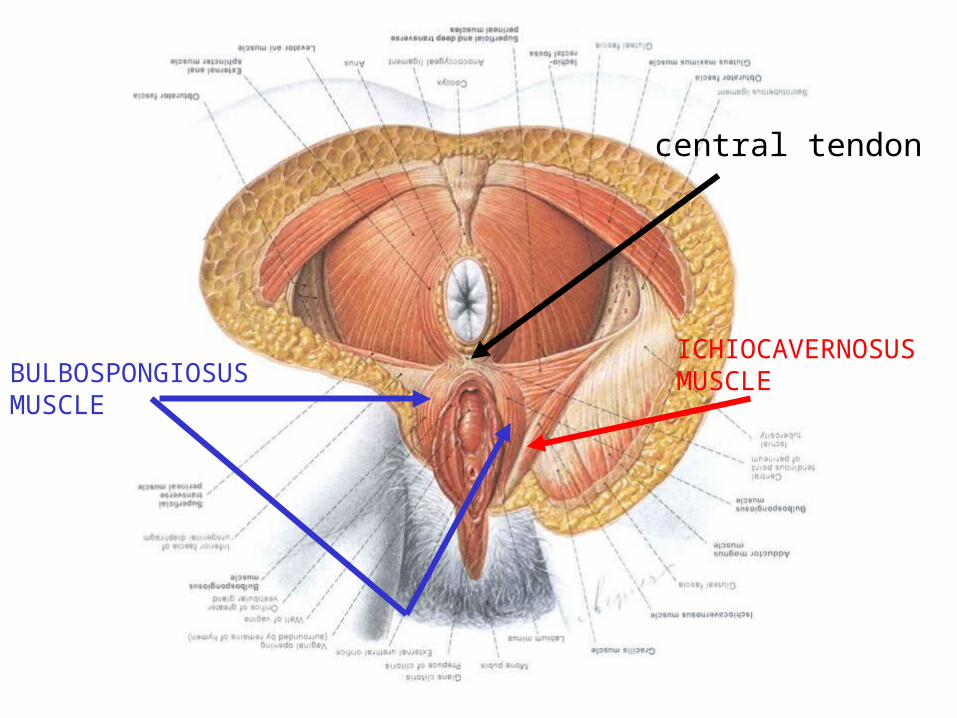
ICHIOCAVERNOSUS MUSCLEBULBOSPONGIOSUS
MUSCLE
central tendon
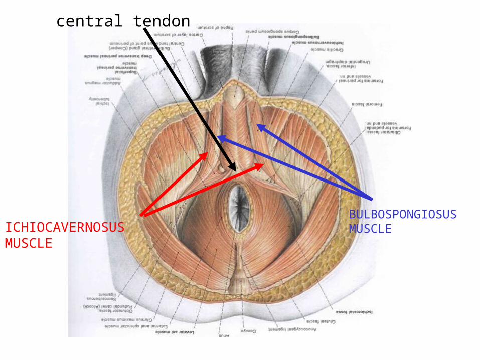
ICHIOCAVERNOSUS MUSCLE
BULBOSPONGIOSUS MUSCLE
central tendon
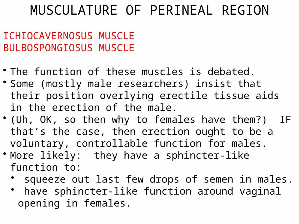
MUSCULATURE OF PERINEAL REGION
ICHIOCAVERNOSUS MUSCLEBULBOSPONGIOSUS MUSCLE
• The function of these muscles is debated.• Some (mostly male researchers) insist that their position
overlying erectile tissue aids in the erection of the male.• (Uh, OK, so then why to females have them?) IF that’s
the case, then erection ought to be a voluntary, controllable function for males.
• More likely: they have a sphincter-like function to:• squeeze out last few drops of semen in males.• have sphincter-like function around vaginal opening in
females.