BIOLOGICAL OXIDATION/ ETC/ OXIDATIVE PHOSPHORYLATION
131
-
Upload
yesanna -
Category
Health & Medicine
-
view
437 -
download
23
Transcript of BIOLOGICAL OXIDATION/ ETC/ OXIDATIVE PHOSPHORYLATION


Bioenergetics or biochemical thermodynamics,
is the study of the energy changes
accompanying biochemical reactions.
Three fundamental thermodynamic variables:
Enthalpy (H):
The heat content of physical object or body
(system)
Derived from first law of thermodynamics.
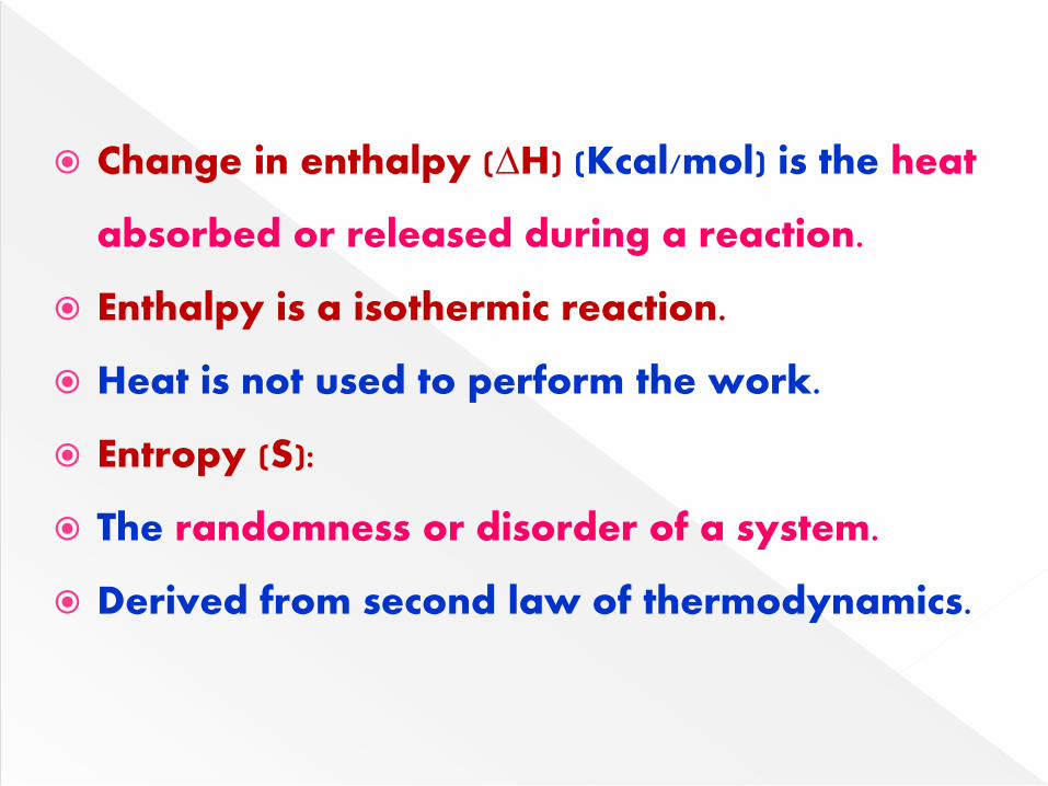
Change in enthalpy (∆H) (Kcal/mol) is the heat
absorbed or released during a reaction.
Enthalpy is a isothermic reaction.
Heat is not used to perform the work.
Entropy (S):
The randomness or disorder of a system.
Derived from second law of thermodynamics.

Change in entropy (∆S) is the degree of
randomness or disorders created during the
reaction.
Free energy (G):
The maximum usable work that can be
obtained from a system at constant pressure,
temperature and volume.

Free energy change (∆G) is the change in free
energy occurring during biological reactions.
It is related to enthalpy & entropy
Change in free energy can be expressed as:
∆G= ∆H - T ∆S
∆H is the change in enthalpy
∆S is the change in entropy
T is the absolute temperature

Standard free energy change (∆G):
It is defined as free energy change under
standard conditions.
Standard condition is defined as pH 7.0,
temperature 25◦C, all reactant concentration at
1m conc, all gases at pressure 1 atmosphere.

Exergonic reactions:
If the free energy change ∆G is negative in
sign, the reaction proceeds spontaneously
with loss of free energy & it is exergonic
Exergonic is usually by breaking the bonds.

Endergonic reactions:
If the free energy change ∆G is positive, the
reaction proceeds only if free energy can be
gained & it is endergonic.
Endergonic is usually by formation of the
bonds.
Reactions at equilibrium: If the free energy
change is zero, the reaction is at equilibrium.


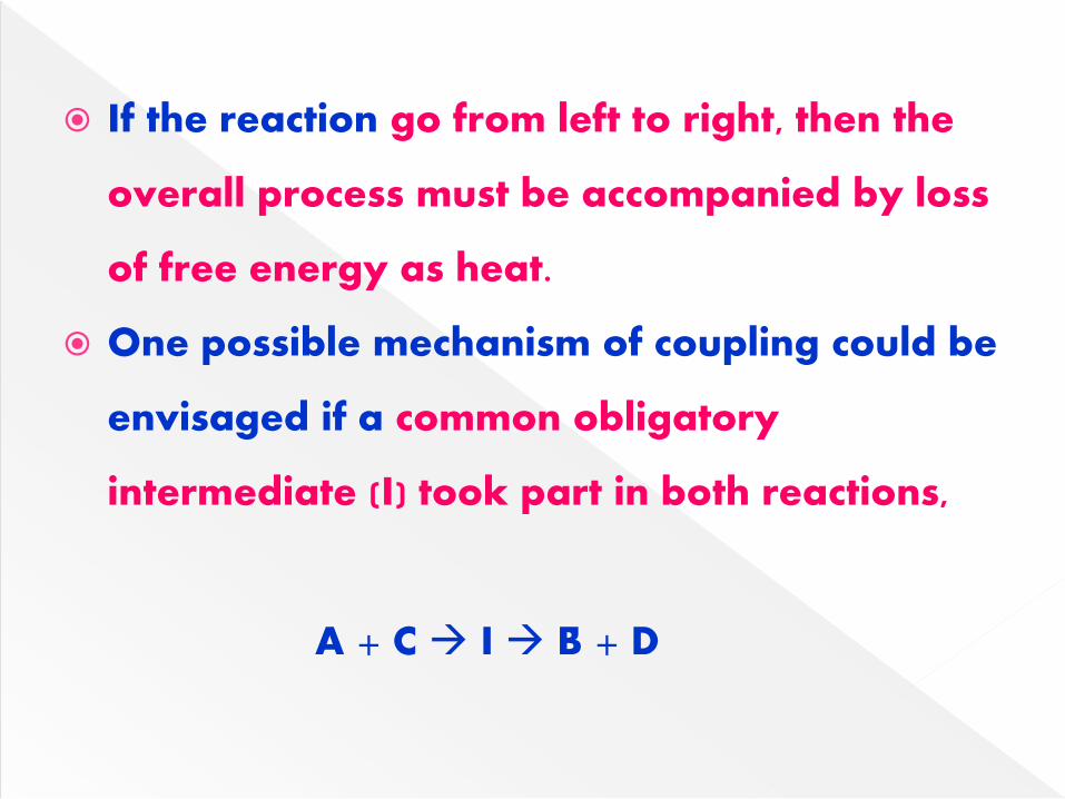
If the reaction go from left to right, then the
overall process must be accompanied by loss
of free energy as heat.
One possible mechanism of coupling could be
envisaged if a common obligatory
intermediate (I) took part in both reactions,
A + C I B + D

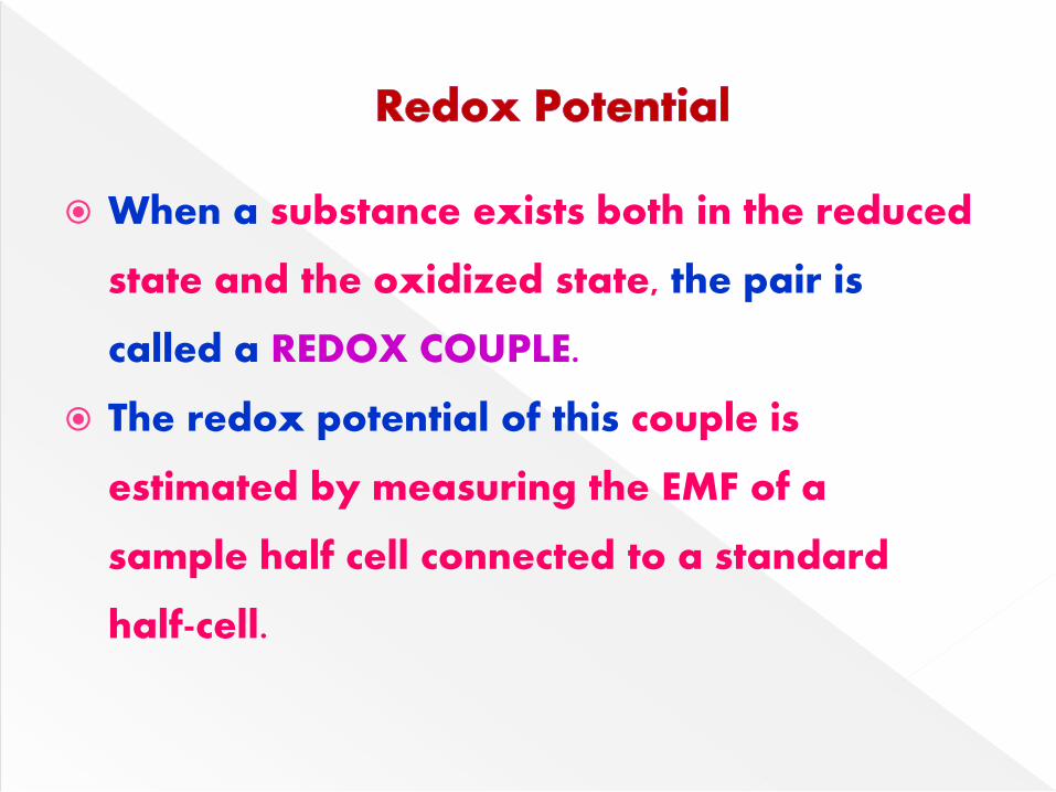
When a substance exists both in the reduced
state and the oxidized state, the pair is
called a REDOX COUPLE.
The redox potential of this couple is
estimated by measuring the EMF of a
sample half cell connected to a standard
half-cell.

When a substance has lower affinity for
electrons than hydrogen it has a negative
redox potential.
Lower affinity for electrons = Neg. Redox
potential.
Electrons move always from more
electronegative to electropositive.

Oxidation is defined as loss of electrons
Loss of electrons occurs in three ways
(1) Direct loss of electrons
(2) Removal of hydrogen
(3) Addition of oxygen
Electrons are transferred as
(1) Hydride ions(H:-)
(2) Hydrogen atoms (H)
(3) Electrons (e-)

Direct loss of electrons:
Electrons are lost directly & passed on to
second acceptor molecule.
Eg: Conversion of ferrous iron to ferric iron
Removal of hydrogen:
Electrons are lost during dehydrogenation.
Loss of hydrogen may occur as loss of
hydrogen atoms or as hydride ion which has
two electrons.

Reduction:
Reduction is defined as the gain of electrons.
Eg: ferric iron(Fe3+) to ferrous iron (Fe2+)
Oxidation-Reduction Reactions:
Oxidation-reduction reactions involve
transfer of electrons from one compound to
another.
When one substrate is oxidized, another
substrate is simultaneously reduced.

Oxidoreductases:
Catalyzes oxidation & reduction reactions.
They catalyze the addition of oxygen, transfer of
hydrogen & transfer of electrons.
Subclass: Subclasses are oxidases, dehydrogenases,
oxygenases & hydroperoxidases.
Oxidases:
Catalyze the transfer of hydrogen or electrons from
the donor, using oxygen as hydrogen acceptor.
The reaction product may be H2O or H2O2


They contain flavoprotein (FAD or FMN) as
coenzymes.
They transfer hydrogen atoms from the
substrate to oxygen via flavin carriers.
They are called as aerobic dehydrogenases.
They are also capable of transferring
hydrogen to acceptors other than oxygen.

Perform 2 main functions:
Transfer hydrogen from one substrate to another in a
coupled oxidation-reduction reactions.
As components of ETC Dehydrogenases use
coenzymes – nicotinamides & riboflavin - as
hydrogen carriers
Dehydrogenae
Specific for ADehydrogenae
Specific for B

Dehydrogenases;
Catalyze the transfer of hydrogen (or
electrons), but the hydrogen acceptor is
molecule other than oxygen.
The hydrogen acceptors are coenzymes,
E.g. NAD, NADP, FAD & FMN.
NAD linked dehydrogenases :
H2 H + H+ + e-
AH2 + NAD + A + NADH + H+

NADP+ linked dehydrogenases:
Reductive biosynthesis of various substances
e.g Fatty acid biosynthesis
FAD linked Dehydrogenases:
FAD is the coenzymes instead of NAD. e.g
Succinate dehydrogenase
Cytochomes:
All cytochromes (Except Cytochrome Oxidase)
are anaerobic dehydrogenases

Oxygenases catalyze the direct incorporation
of oxygen into the substrate.
Oxygen is bound to active site of the enzyme.
There are two types of oxygenases:
Monooxygenases & Dioxygenases.
Monooxygenases:
These will catalyze the incorporation of only
one oxygen to the substrate.

They are also called as hydroxylases or
mixed function oxygenases.
Dioxygenases: These will catalyze the
incorporation of both atoms of oxygen into
the substrate.
Phenylalanine +O2+Biopterin Tyrosine + H2O+Dihydrobiopterin
Homogenstisic acid + O2 Maleylacetoacetate

Hydroperoxidases:
These enzymes will utilize hydrogen
peroxide as (H2O2) as the substrate.
These are two types:
Peroxidases:
Catalase:

Peroxidases:
Utilize H2O2 as oxygen donor but O2 acceptor
is a molecule other than H2O2.
Eg. Glutathione peroxidase.
Catalase:
It is a unique enzyme & utilizes H2O2 as both
donor & acceptor of oxygen (electrons).
E.g: H2O2 + H2O2 2H2O + O2

Catalase functions in the cell to detoxify
H2O2.
Peroxisomes are rich in oxidases and
catalases.
Coenzymes involved in Biological Oxidations
are:
NAD+,NADP+,FAD+,FMN+
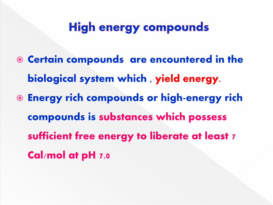
Certain compounds are encountered in the
biological system which , yield energy.
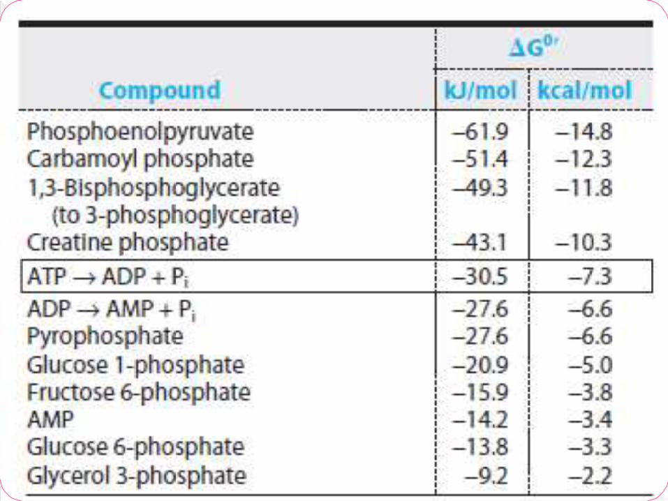
Energy rich compounds or high-energy rich
compounds is substances which possess
sufficient free energy to liberate at least 7
Cal/mol at pH 7.0

Certain other compounds which liberate less
than 7.0 cal/mol.
Are referred to as low energy compounds
Indicated by SQUIGGLE bond (~)
Free energy varies from -7 to -15 kcal/mol
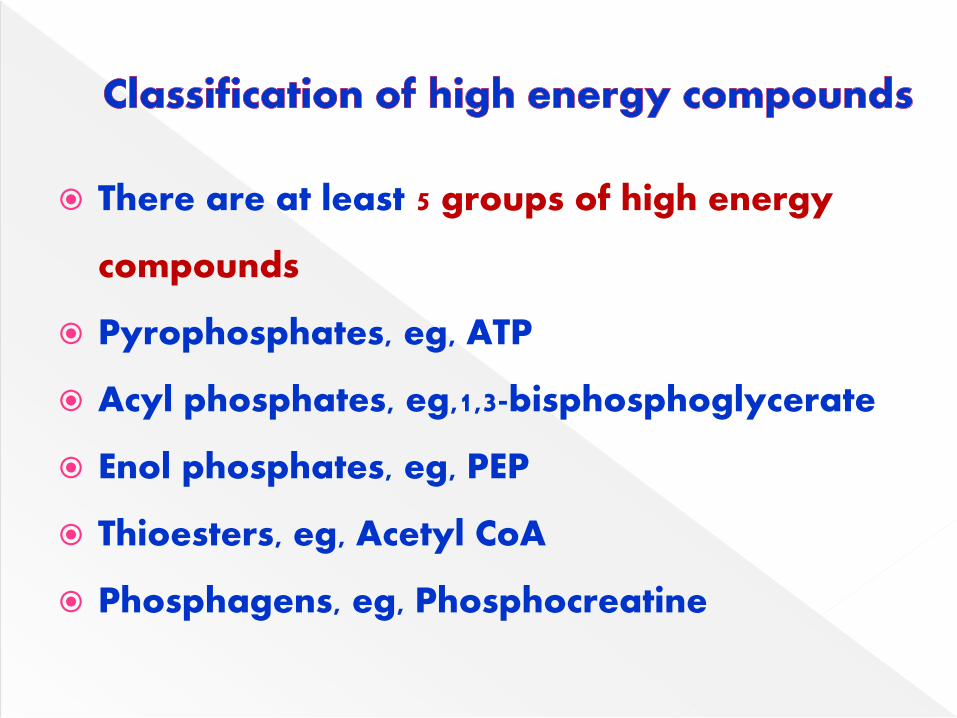
There are at least 5 groups of high energy
compounds
Pyrophosphates, eg, ATP
Acyl phosphates, eg,1,3-bisphosphoglycerate
Enol phosphates, eg, PEP
Thioesters, eg, Acetyl CoA
Phosphagens, eg, Phosphocreatine

The high-energy compounds possess acid
anhydride bonds (mostly phosphoanhydride
bonds) which are formed by the condensation
of two acidic groups or related compounds.
These bonds are referred as high-energy
bonds.
Free energy is liberated when these bonds
are hydrolysed.
ATP is most important high-energy compound

The hydrolysis of ATP is associated with the release of
large amount of energy.
The energy liberated is utilized for various process
like muscle contraction, active transport etc.
ATP can also acts as a donor of high-energy
phosphate to low-energy compounds, to make them
energy rich.
ADP can accepts phosphate to form ATP.
ATP + H2O ADP + Pi + 7.3 Cal

Oxidative Phosphorylation
Substrate level Phosphorylation
~P
ATP
ADP
~P
Muscle Contraction
Active transport
Biosynthesis
Phosphorylation
P
Creatine Creatine ~P
~P

ATP serves as an immediately available
energy currency of the cell which is
constantly being utilized & regenerated.
ATP acts as an energy link between the
catabolism & anabolism in the biological
systems.
Hydrolysis of ATP releases 7.3kcal/mol.
At rest, Na+ - K+ - ATPase uses up one-third of
all ATP formed.
An average person at rest consumes &
regenerates ATP at a rate of approximately
3 molecules per second, i.e. about 1.5 kg/day.

ATP can be synthesized in two ways
Oxidative phosphorylation:
Major source of ATP in aerobic organisms.
It is linked with mitochondrial ETC.
Substrate level phosphorylation:
When the energy of high energy compound is directly
transferred to nucleoside diphosphate to form a
triphosphate without the help from ETC.

The high-energy compounds such as
PEP
1,3-bisphosphoglycerate
Succinyl CoA can transfer high-energy
phosphate to ultimately produce ATP.

Storage forms:
Phosphocreatine ( creatine phosphate)
provides high energy reservoir of ATP to
regenerate ATP rapidly, catalyzed by
creatine kinase.
Stored mainly in muscle & brain.
In invertebrates, phosphoarginine ( arginine
phosphate ) is storage form.


The transfer of electrons from the reduced
coenzymes through the respiratory chain to
oxygen is known as biological oxidation.
Energy released during this process is
trapped as ATP.
This coupling of oxidation with
phosphorylation is called oxidative
phosphorylation.

Oxidation:
Oxidation is defined as the loss of electrons
and reduction as the gain in electrons.
When a substance exists both in the reduced
state & in the oxidized state, the pair is
called a redox couple.

Redox potential(E0):
The oxidation-reduction potential or redox
potential, is a quantitative measure of the
tendency of a redox pair to lose or gain
electrons.
The redox pairs are assigned specific
standard redox potential at pH 7.0 & 250C

Redox pair E0 Volts
Succinate/α -ketoglutarate -0.67
2H+/H2 -0.42
NAD+/NADH -0.32
FMN/FMNH2 -0.30
Lipoate (ox/red) -0.29
FAD/FADH2 -0.22
Puruvate/lactate -0.19
Fumarate/succinate +0.03
Cytochrome b (Fe3+/Fe2+) +0.07
CoenzymeQ (ox/red) +0.10
Cytochrome c1 (Fe3+/Fe2+) +0.23
Cytochrome c (Fe3+/Fe2+) +0.25
Cytochrome a (Fe3+/Fe2+) +0.29
½ O2/H2O +0.82

The more negative redox potential represents a
greater tendency to lose electrons.
A more positive redox potential indicates a
greater tendency to accept electrons
The electrons flow from a redox pair with more
negative E0 to another redox pair with more
positive E0
The redox potential (E0) is directly related to the
change in the free energy (∆G0)

The inner mitochondrial is impermeable to
NADH.
Therefore, the NADH produced in the cytosol
cannot directly enter the mitochondria.
Two pathways
Glycerol-phosphate shuttle
Malate-aspartate shuttle

Cytosolic glycerol 3-phosphate dehydrogenase
oxidizes NADH to NAD+
The reducing equivalents are transported
through glycerol 3-phosphate into the
mitochondria.
Glycerol 3-phosphate dehydrogenase-present
on outer surface of inner mitochondrial
membrane – reduces FAD to FADH2.

Dihydroxyacetone phosphate (DHAP)
escapes into the cytosol & the shuttling
continues.
FADH2 gets oxidized via ETC to generate
2ATP

CH2OH
I
C=O
I
CH2O-P
CH2OH
I
HO- C=H
I
CH2O-P
CH2OH
I
HO- C=H
I
CH2O-P
CH2OH
I
C=O
I
CH2O-P
Cytosolic Gly-3P-DH
NADH+H NAD+
DHAP
Gly-3-P
CYTOSOL
Mitochondrial -matrix
Gly-3-PDHAP
Mitochondrial Gly-3P-DH
FAD+FADH2
H2O
ETC2ATP

In the cytosol, oxaloacetate accepts the
reducing equivalents (NADH) & becomes
malate.
Malate enters the mitochondria where it is
oxidized by mitochondrial MDH
In this reaction, NADH & oxaloacetate are
regenerated.
NADH gets oxidized via ETC & 3 ATP are
produced.

Oxaloacetate
Malate
NADH + H+
NAD+
Malate
Oxaloacetate
NADH + H+
NAD+
H2O
ETC3ATP
Aspartate
Aspartate
Glutamate
α-ketoglutarate
α-ketoglutarate
glutamate
Cytosolic MDH
Mitochondrial
MDH Aminotransferase
Aminotransferase
CYTOSOL
Mitochondrial Matrix

In the mitochondria, oxaloacetate
participates in transamination reaction with
glutamate to produce aspartate & α-
ketoglutarate.
The aspartate enters the cytosol &
transaminates with α-ketoglutarate to give
oxaloacetate & glutamate.

The flow of electrons occurs through successive
dehydrogenase enzymes in mitochondria ,
together known as the ETC.
(the electrons are transferred from higher to
lower potential.)
o Significance:
o The free energy released during the transport
of electrons is utilized for the formation of ATP.

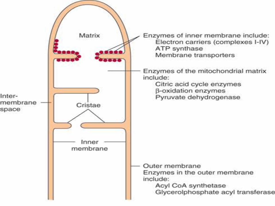
Mitochondria consists of five distinct parts
Outer membrane, inner membrane,
intermembrane space, cristae & matrix
Inner mitochondrial membrane:
The ETC & ATP synthesizing system are located
on inner mitochondrial membrane, which is
specialized structure, rich in proteins.

Inner membrane is highly folded to form
cristae.
Surface area of inner mitochondrial
membrane is increased due to cristae.
The inner surface of inner mitochondrial
membrane possesses specialized particles,
the phosphorylating subunits which are
centres for ATP production.
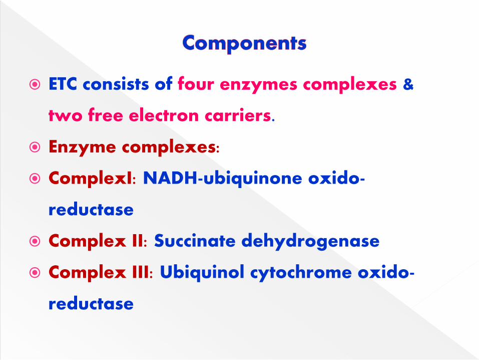
ETC consists of four enzymes complexes &
two free electron carriers.
Enzyme complexes:
ComplexI: NADH-ubiquinone oxido-
reductase
Complex II: Succinate dehydrogenase
Complex III: Ubiquinol cytochrome oxido-
reductase

Complex IV: Cytochrome oxidase
Two free electron carriers are coenzyme Q
& Cytochrome C.
Complex V: It is ATP synthase.
The complexes I-IV are carriers of electrons
while complex V is responsible for ATP
synthesis.

The enzyme complexes & mobile carriers are
collectively involved in the transport of
electrons which, ultimately, combine with
oxygen to produce water.
Largest proportion of O2 supplied to body is
utilized by mitochondria for the operation of
ETC.

Of the two coenzymes NAD+& NADP+, NAD+ is more
actively involved in ETC.
Tightly bound to the inner membrane
NAD+ is reduced to NADH+ H+ by dehydrogenases
with the removal of two hydrogen atoms from the
substrates, the substrates includes pyruvate, gly-3-P.
etc.
NADPH is more effectively utilized for anabolic
reactions - fatty acid synthesis, cholesterol synthesis.

N
CONH2
N
CONH2
H H
XH2
X
oxidised coenzyme
NAD+ or NADP
+
reduced coenzymeNADH or NADPH
+ H+


The enzyme NADH dehydrogenase (NADH-
coenzyme Q reductase) is a flavoprotein
with FMN as the prosthetic group.
The coenzyme FMN accepts two electrons &
a proton to form FMNH2.
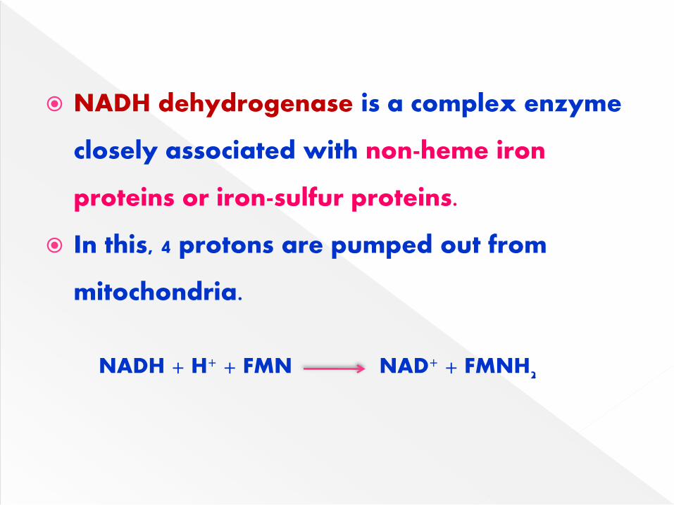
NADH dehydrogenase is a complex enzyme
closely associated with non-heme iron
proteins or iron-sulfur proteins.
In this, 4 protons are pumped out from
mitochondria.
NADH + H+ + FMN NAD+ + FMNH2

The electrons from FADH2 enter ETC at the level of Co Q.
Succinate DH is an enzyme found in inner mitochondrial
membrane.
It is also a flavoprotein with FAD as coenzyme.
The 3 major enzyme systems that transfer their electrons
directly to ubiquinone are:
a. Succinate dehydrogenase
b. Fatty acyl CoA dehydrogenase
c. Mitochondrial glycerol phosphate dehydrogenase.

C
CCH
C
C
HC
NC
CN
NC
NHC
H3C
H3C
O
O
CH2
HC
HC
HC
H2C
OH
O P O-
O
O-
OH
OH
C
CCH
C
C
HC
NC
C
HN
NC
NHC
H3C
H3C
O
O
CH2
HC
HC
HC
H2C
OH
O P O-
O
O-
OH
OH
C
CCH
C
C
HC
NC
C
HN
NH
C
NHC
H3C
H3C
O
O
CH2
HC
HC
HC
H2C
OH
O P O-
O
O-
OH
OH
e + H
+ e
+ H
+
FMN FMNH2 FMNH·

Iron-sulfur centers (Fe-S) are prosthetic groups
containing 1-4 iron atoms
Iron-sulfur (Fe-S) proteins exist in the oxidized
(Fe3+) or reduced (Fe2+) state.
Iron-sulfur centers transfer only one electron,
even if they contain two or more iron atoms

Fe-S participates in the transfer of electrons
from FMN to coenzyme Q.
Other Fe-S proteins associated with
cytochrome b & cytochrome c1 participate in
the transport of electrons.


It is also known as ubiquinone.
It is a quinone derivative with isoprenoid side
chain
Mammalian tissues possess a quinone with 10
isoprenoid units which is known as coenzyme
Q10
The ubiquinone is reduced successively to
semiquinone (QH) & finally to quinol (QH2)

It accepts a pair of electrons from NADH or
FADH2 through complex I or complex II
respectively.
2 molecules of cytochrome c are reduced.
The Q cycle facilitates the switching from the
2 electron carrier ubiquinol to the single
electron carrier cytochrome c.
This is a mobile carrier.

Coenzyme Q
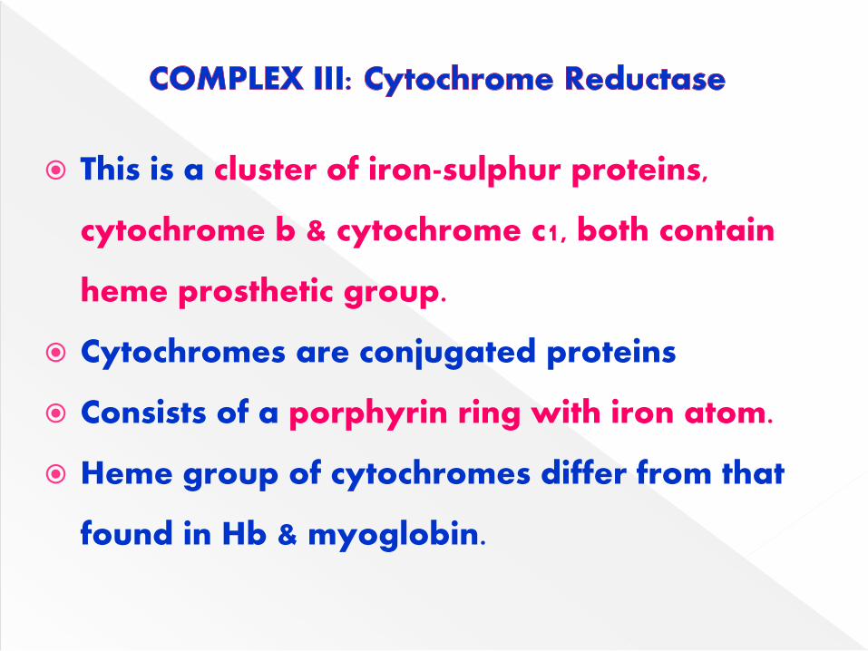
This is a cluster of iron-sulphur proteins,
cytochrome b & cytochrome c1, both contain
heme prosthetic group.
Cytochromes are conjugated proteins
Consists of a porphyrin ring with iron atom.
Heme group of cytochromes differ from that
found in Hb & myoglobin.

The iron of heme in cytochromes is
alternately oxidized (Fe3+) & reduced (Fe2+)
Which is essential for transport of electrons
in the ETC.
In this, 4 protons are pumped out.
The electrons transported from coenzyme Q
to cytochromes b, c1, c, a & a3.

The property of reversible oxidation-
reduction of heme iron present in
cytochromes allows them to function as
effective carriers of electrons in ETC.
Cytochrome C:
It is a small protein containing 104 amino
acids & a heme group.
It is a loosely bound to inner mitochondrial
membrane & can be easily extracted.

Contains cytochrome a and cytochrome a3
Which is the terminal component of ETC
Tightly bound to inner mitochondrial
membrane.
Cytochrome oxidase is the only electron
carrier, heme iron of which can directly
react with molecular oxygen.
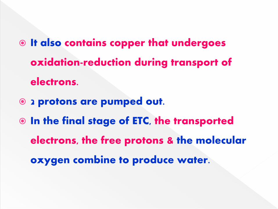
It also contains copper that undergoes
oxidation-reduction during transport of
electrons.
2 protons are pumped out.
In the final stage of ETC, the transported
electrons, the free protons & the molecular
oxygen combine to produce water.

Electrons donors:
NADH & FADH2
NADH: It is produced in the following
reactions
PDH complex: It transfers electrons from
pyruvate to NAD+
α-ketoglutarate DH: It transfers electrons
from alpha-ketoglutarate to NAD+

Isocitrate DH: It transfers electrons from
isocitrate to NAD+
Malate DH: It transfers electrons from malate
to NAD+
Hydroxyacyl CoA DH: It transfers electrons
from hydroxy acyl CoA to NAD+
FADH2:
FAD is tightly bound to enzymes called
flavoproteins.
FADH2 is produced in the following reactions.
Succinate DH (complex II):
It transfers electrons from succinate to FAD.
Glycerol 3-P DH: It transfers electrons from
glycerol 3-P to FAD.
Fatty acyl CoA DH: It transfers electrons from
fatty acids to FAD.
FADH2 donates electrons to coenzyme Q

Complex V
ATP synthase
(F0,F1)
Complex II
Succinate CoQ
Reductase
FADH2
FeS
Coenzyme Q
Substrate
NADH+ H+
FeS
FMNH2
Complex I
NADH-CoQ
Reductase
Cyt b FeS Cyt c1 Cyt c Cyt a Cyt a3 H2OO2
Complex III
CoQ-Cytochrome C
Reductase
Complex IV
Cytochrome
Oxidase
Succinate
ADP+Pi ATP

The inhibitors bind to one of the components
of ETC & block the transport of electrons
This causes the accumulation of reduced
components before the inhibitor blockade
step & oxidized components after that step.
The synthesis of ATP is dependent on ETC.

All the site-specific inhibitors of ETC also
inhibit ATP formation.
NADH & coenzyme Q (Complex I):
Fish poison rotenone, barbiturate drug
amytol & antibiotic piercidin A inhibit this
site.
Complex II: Carboxin inhibit this site.

Between cytochrome b & c1 ( Complex III):
Antimycin A –an antibiotic, British antilewisite
(BAL) –an antidote used against war-gas-
Naphthoquinone are important inhibitors of
the site between cytochrome b & c1.
Cytochrome oxidase (Complex IV):
Carbon monoxide, cyanide, hydrogen sulphide & azide
Effectively inhibit cytochrome while cyanide &
azide react with oxidized form of cytochrome.
Cyanide is most potent inhibitor of ETC
It binds to Fe3+ of cytochrome oxidase
blocking mitochondrial respiration leading to
cell death.
Cyanide poisoning causes death due to tissue
asphyxia (mostly of CNS)

Substrate NAD+ FMN CoQ Cyt b Cyt c1 Cyt c
Cyt a
Cyt a3
O2
AmytolRotenone
Piericidin A_
Antimycin ABAL
_
CyanideSodium Azide
Carbon monoxide
ATP
(Site 1)
ATP
(Site 2)
ATP
(Site 3)

Biological Oxidation:
The transfer of electrons from the reduced co-
enzymes though the respiratory chain to oxygen is
known as biological oxidation.
Energy released during this process is trapped as
ATP.
This coupling of oxidation with phosphorylation is
called as OXIDATIVE PHOSPHORYLATION.

Complex V of the inner mitochondrial
membrane is the site of oxidative
phosphorylation.
Phosphagens act as storage forms of high-
energy phosphate and include creatine
phosphate, which occurs in vertebrate
skeletal muscle, heart, spermatozoa & brain
Arginine phosphate, in invertebrate muscle.

When ATP is rapidly being utilized as a
source of energy for muscular contraction,
phosphagens permit its concentrations to be
maintained, but when the ATP/ADP ratio is
high, their concentration can increase to act
as a store of high-energy phosphate.

The P:O ratio refers to the number of
inorganic phosphate molecules utilized for
ATP generation for every atom of oxygen
consumed.
Approximately P:O ratio represents the
number of molecules of ATP synthesized per
pair of electrons carried through ETC.

P:O Ratio of 3:
P/O ratio is 3 for oxidation of substrates
producing NADH.
For each molecule of NADH that is oxidized
through ETC 3 ATP are produced.
Ex: Malate, Pyruvate, Isocitrate, α-Ketoglutarate
P/O ratio of 2:
P/O ratio is 2 for oxidation of substrates
producing FADH2.

FADH2 transfers electrons to coenzyme Q thus
missing the first site of oxidative phosphorylation.
For each molecule of FADH2 produces 2 ATP.
Ex: Succinate, fatty acyl CoA, glycerol 3-P.
P/O Ratio of 1:
P/O ratio is 1 for compounds that transfer electrons
to cytochrome oxidase complex.
Ex: Ascorbic acid.
NOTE:
Studies on isolated mitochondria indicate P/O ratio
of 2.5 for NADH & 1.5 for FADH2

There are 3 reactions in the ETC that are exergonic,
Where the energy change is sufficient to drive the
synthesis of ATP from ADP and Pi.
Site1:
Oxidation of FMNH2 by coenzyme Q.
Site2:
Oxidation of cytochrome b by cytochrome c1
Site3:
Cytochrome oxidase.

½ O2 + NADH + H+ H2O + NAD+
The redox potential difference between these two redox paires
is 1.14V, which is equivalent to an energy 52 Cal/mol
3 ATP are synthesized in ETC when NADH is oxidized which
equals to 21.9 Cal.
(each ATP=7.3 Cal)
The efficiency of energy conservation is calculated as
21.9 × 100
52 =
42%
When NADH is oxidized, about 42% of energy
is trapped in the form of 3ATP & remaining is
lost as heat.
The heat liberation is not a wasteful process,
since it allows ETC to go on continuously to
generate ATP.
This heat is necessary to maintain body
temperature.


Two important hypothesis to explain the
process of oxidative phosporylation.
Namely chemical coupling & chemiosmotic
Chemical coupling hypothesis:
This hypothesis was put forth by Edward
Slater (1953)
According to this, during the course of electron
transfer in respiratory chain, a series of
phosphorylated high-energy intermediates are
first produced which are utilized for the
synthesis of ATP.
These reactions are believed to be analogous
to the substrate level phosphorylation that
occurs in glycolysis or citric acid cycle.
This hypothesis lacks experimental evidence.

The transport of electrons through the
respiratory chain is effectively utilized to
produce ATP from ADP + Pi.
Proton gradient:
The inner mitochondrial membrane, is
impermeable to protons (H+) & hydroxyl ions
(OH-).

The transport of electrons through ETC is
coupled with the translocation of protons
(H+)across the inner mitochondrial
membrane from the matrix to the inter
membrane space.
The pumping of protons results in an
electrochemical or proton gradient .

This is due to the accumulation of more H+
ions (low pH) on the outer side of the inner
mitochondrial membrane than the inner side.
The proton gradient developed due to the
electron flow in the respiratory chain is
sufficient to result in the synthesis of ATP
from ADP +Pi.


Enzyme systems for ATP synthesis:
ATP synthase, present in the complex V,
utilizes the proton gradient for the synthesis
of ATP.
This enzyme is also known as ATPase, since it
can hydrolyze ATP to ADP + Pi.
ATP synthase is a complex enzyme & consists
of two functional subunits, namely F1 & Fo.

Fo unit: O stands for oligomycin,
Fo inhibited by oligomycin.
Fo spans inner mitochondrial membrane acting
as a proton channel through which protons
enter the mitochondria
Fo unit has 4 polypeptide chains & is connected
to F1.
Fo is water insolube whereas F1 is a water
soluble peripheral membrane protein.

o F1 unit: It projects into the matrix.
o F1 has 9 polypeptide chains, (3 alpha, 3 beta, 1 gamma, 1
delta, 1 epsilon)
o The α chains have binding sites for ATP & ADP & beta
chains have catalytic activity.
o ATP synthesis requires Mg +2 Ions.
Its structure is comparable with lollipops.
The protons that accumulate on the intermembrane
space re-enter the mitochondrial matrix leading to the
synthesis of ATP.

ATP Synthase


Paul Boyer in 1964 proposed that a
conformational change in the mitochondrial
membrane proteins leads to the synthesis of
ATP
This is now considered as rotary motor/engine
driving model or binding change model, is
widely accepted for the generation of ATP.

The enzyme ATP synthase is Fo & F1 complex
The Fo sub complex is composed of channel
protein ‘C’ subunits to which F1-ATP synthase
is attached.
F1-ATP synthase consists of a central gamma-
subunit surrounded by alternating alpha &
beta subunits ( α3 & β3).
In response to the proton flux, the gamma
subunit physically rotates.

This induces conformational changes in the β3
subunits that finally lead to the release of ATP.
According to the binding change mechanism,
the three β subunits of F1 - ATP synthase adopt
different conformations.
One subunit has Open (O) conformation, the
second has loose (L) conformation while the
third one has tight (T) conformation.

By an known mechanism, protons induce the
rotation of gamma subunit, which in turn
induces conformation changes in β subunits,.
The substrates ADP & Pi bind to β subunit in L
conformation.
The L site changes to T conformation, & this
leads to the synthesis of ATP.

The O site changes to L conformation which
binds to ADP + Pi.
The T site changes to O conformation &
releases ATP.
This cycle of conformation changes of β
subunits is repeated.
Three ATP are generated for each revolution.

Protons entering the system, cause conformational changes in F1 particle. The 3 beta subunits are in three functional states, O (open ), L (loose) & T(tight).Conformational change induces catalytic activity.Open form is regained after release of ATP.
The mitochondrial transport of electrons is
tightly coupled with oxidative
phosphorylation.
Oxidation & phosphorylation proceed
simultaneously.
There are certain compounds that can
uncouple (or delink) the electron transport
from oxidative phosphorylation.

Such compounds are known as uncouplers,
increase in the permeability of inner
mitochondrial membrane to protons (H+).
The result is that ATP synthesis does not
occur
The energy linked with the transport of
electrons is dissipated as HEAT.
The uncouplers allow (often at accelerated
rate) oxidation of substrates (via NADH or
FADH2) without ATP formation.

Examples:
2,4-dinitrophenol (DNP):
It is small lipophilic molecule.
DNP is a proton – carrier & easily diffuse
through the inner mitochondrial membrane.
Others –dinitrocressol, pentachlorophenol,
trifluorocarbonylcyanide, phenylhydrazone

Certain physiological substances which act as
uncouplers at higher concentration.
These are thermogenin, thyroxine and long
chain fatty acids & unconjugated bilirubin

Significance of uncoupling:
The maintenance of body temperature is
particularly important in hairless animals,
hibernating animals & the animals adopted
to cold
These animals possess a specialized tissue
called brown adipose tissue in the upper
back & neck portions.

The mitochondria of brown adipose tissue
are rich in electron carriers & are specialized
to carry out an oxidation uncoupled from
phosphorylation.
This causes liberation of heat when fat is
oxidized in the brown adipose tissue.
The presence of brown adipose tissue in
certain individuals is believed to protect
them from becoming obese.
Thermogenin is a natural uncoupler located in
the inner mitochondrial membrane of brown
adipose tissue
It acts like an uncoupler, blocks the formation
of ATP, & liberates heat.
Ionophores: These are lipophilic substances
that promote the transport of ions across
biological membranes.
Valinomycin & nigercin also act as uncouplers.

Oligomycin: This antibiotic prevents the
mitochondrial oxidation as well as
phosphorylation.
It binds with enzyme ATP synthase & blocks
the proton(H+) channels.
Thus it prevents the translocation (re-entry)
of protons into the mitochondrial matrix.

Due to this, protons get accumulated at
higher concentration in the inter membrane
space
Electron transport is stoped.
Atractyloside: It is a plant toxin & inhibits
oxidative phosphorylation.
It blocks the adequate supply of ADP.

100 polypeptides are required for oxidative
phosphorylation.
Of these, 13 are coded by mitochondrial DNA &
synthesized in the mitochondria, while the rest
are produced in the cytosol (coded by nuclear
DNA) & transported.
mtDNA is maternally inherited since
mitochondria from the sperm do not enter the
fertilized ovum.

Mitochondrial DNA is 10 times more
susceptible to mutations than nuclear DNA.
mtDNA mutations are commonly seen in
tissues with high rate of oxidative
phosphorylation (e.g. CNS, skeletal & heart
muscle, liver).

Diseases:
Lethal infantile mitochondrial opthalmoplegia
Leber’s hereditary optic neuropathy (LHON)
Myoclonic epilepsy
Mitochondrial encephalopathy lactic acidosis
stroke like episodes (MELAS)
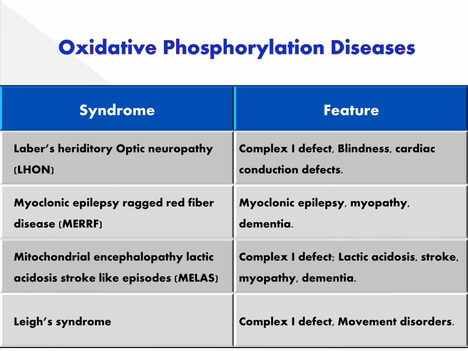
Syndrome Feature
Laber’s heriditory Optic neuropathy
(LHON)
Complex I defect, Blindness, cardiac
conduction defects.
Myoclonic epilepsy ragged red fiber
disease (MERRF)
Myoclonic epilepsy, myopathy,
dementia.
Mitochondrial encephalopathy lactic
acidosis stroke like episodes (MELAS)
Complex I defect; Lactic acidosis, stroke,
myopathy, dementia.
Leigh’s syndrome Complex I defect, Movement disorders.

Text book of Biochemistry – AR Aroor
Text book of Biochemistry-Harper 25th edition
Text book of Biochemistry – DM Vasudevan
Text book of Biochemistry – U Satyanarayana