Biological implications of somatic DDX41 p.R525H mutation ...
14
Biological implications of somatic DDX41 p.R525H mutation in acute myeloid leukemia Moe Kadono a,b , Akinori Kanai a , Akiko Nagamachi a , Satoru Shinriki c,d , Jin Kawata d , Koji Iwato e , Taiichi Kyo e , Kumi Oshima b , Akihiko Yokoyama f , Takeshi Kawamura g , Reina Nagase h , Daichi Inoue h , Toshio Kitamura h , Toshiya Inaba a , Tatsuo Ichinohe b , and Hirotaka Matsui a,c,d a Department of Molecular Oncology and Leukemia Program Project, ResearchInstitute for Radiation Biology and Medicine, Hiroshima University, Hiroshima, Japan; b Department of Hematology and Oncology, Research Institute for Radiation Biology and Medicine, Hiroshima University, Hiroshima, Japan; c Department of Molecular Laboratory Medicine, Graduate School of Medical Sciences, Kumamoto University, Kumamoto, Japan; d Central Clinical Laboratory, Kumamoto University Hospital, Kumamoto, Japan; e Department of Hematology, Hiroshima Red Cross Hospital & Atomic-bomb Survivors Hospital, Hiroshima, Japan; f Laboratory for Malignancy Control Research, Medical Innovation Center, Kyoto University Graduate School of Medicine, Kyoto, Japan; g Department of Molecular Biology and Medicine, Laboratory for System Biology and Medicine (LSBM), Research Center for Advanced Science and Technology (RCAST), University of Tokyo, Tokyo, Japan; h Division of Cellular Therapy, Institute of Medical Science, The University of Tokyo, Tokyo, Japan (Received 13 April 2016; revised 28 April 2016; accepted 29 April 2016) The DDX41 gene, encoding a DEAD-box type ATP-dependent RNA helicase, is rarely but reproducibly mutated in myeloid diseases. The acquired mutation in DDX41 is highly concen- trated at c.G1574A (p.R525H) in the conserved motif VI located at the C-terminus of the heli- case core domain where ATP interacts and is hydrolyzed. Therefore, it is likely that the p.R525H mutation perturbs ATPase activity in a dominant-negative manner. In this study, we screened for the DDX41 mutation of CD34-positive tumor cells based on mRNA sequencing and identified the p.R525H mutation in three cases among 23 patients. Intrigu- ingly, these patients commonly exhibited acute myeloid leukemia (AML) with peripheral blood cytopenias and low blast counts, suggesting that the mutation inhibits the growth and differentiation of hematopoietic cells. Data from cord blood cells and leukemia cell lines sug- gest a role for DDX41 in preribosomal RNA processing, in which the expression of the p.R525H mutant causes a certain ribosomopathy phenotype in hematopoietic cells by sup- pressing MDM2-mediated RB degradation, thus triggering the inhibition of E2F activity. This study uncovered a pathogenic role of p.R525H DDX41 in the slow growth rate of tumor cells. Age-dependent epigenetic alterations or other somatic changes might collaborate with the mutation to cause AML. Copyright Ó 2016 ISEH - International Society for Experi- mental Hematology. Published by Elsevier Inc. Current comprehensive sequencing approaches led to the identification of rare but reproducible somatic gene mutations in myeloid malignancies. Among them, there is a somatic mutation in the DDX41 gene encoding a DEAD-box type ATP-dependent RNA helicase. The so- matic mutation in DDX41 is highly concentrated at c.G1574A (p.R525H) in the conserved motif VI, located at the C-terminus of the RecA-like helicase core domain where ATP interacts and is hydrolyzed. Therefore, it is likely that the p.R525H mutation in the DDX41 protein per- turbs ATPase activity in a dominant-negative manner. In addition, germline mutations in DDX41 were recently iso- lated in a subset of familial acute myeloid leukemia (AML)/myelodysplastic syndrome (MDS) pedigrees [1,2]. Because the roles of these somatic and germline mutations in the pathogenesis of myeloid diseases are not completely understood, the researchers who first described these muta- tions advocate a role for DDX41 in mRNA splicing via Offprint requests to: Hirotaka Matsui, Department of Molecular Labora- tory Medicine, Graduate School of Medical Sciences, Kumamoto Univer- sity, 1-1-1 Honjo, Chuo-ku, Kumamoto, Japan 860-8556; E-mail: [email protected] or Tatsuo Ichinohe, MD, PhD, Department of Hematology and Oncology, Research Institute for Radiation Biology and Medicine, Hiroshima University, 1-2-3 Kasumi, Minami-ku, Hiroshi- ma, Japan 784-8553; E-mail: [email protected] Supplementary data related to this article can be found at http://dx.doi. org/10.1016/j.exphem.2016.04.017. 0301-472X/Copyright Ó 2016 ISEH - International Society for Experimental Hematology. Published by Elsevier Inc. http://dx.doi.org/10.1016/j.exphem.2016.04.017 Experimental Hematology 2016;44:745–754
Transcript of Biological implications of somatic DDX41 p.R525H mutation ...

Experimental Hematology 2016;44:745–754
Biological implications of somatic DDX41 p.R525H mutation in acutemyeloid leukemia
Moe Kadonoa,b, Akinori Kanaia, Akiko Nagamachia, Satoru Shinrikic,d, Jin Kawatad, Koji Iwatoe,Taiichi Kyoe, Kumi Oshimab, Akihiko Yokoyamaf, Takeshi Kawamurag, Reina Nagaseh, Daichi Inoueh,
Toshio Kitamurah, Toshiya Inabaa, Tatsuo Ichinoheb, and Hirotaka Matsuia,c,d
aDepartment of Molecular Oncology and Leukemia Program Project, Research Institute for Radiation Biology and Medicine, Hiroshima University,
Hiroshima, Japan; bDepartment of Hematology and Oncology, Research Institute for Radiation Biology and Medicine, Hiroshima University,
Hiroshima, Japan; cDepartment of Molecular Laboratory Medicine, Graduate School of Medical Sciences, Kumamoto University, Kumamoto, Japan;dCentral Clinical Laboratory, Kumamoto University Hospital, Kumamoto, Japan; eDepartment of Hematology, Hiroshima Red Cross Hospital &
Atomic-bomb Survivors Hospital, Hiroshima, Japan; fLaboratory for Malignancy Control Research, Medical Innovation Center, Kyoto University
Graduate School of Medicine, Kyoto, Japan; gDepartment of Molecular Biology and Medicine, Laboratory for System Biology and Medicine (LSBM),
Research Center for Advanced Science and Technology (RCAST), University of Tokyo, Tokyo, Japan; hDivision of Cellular Therapy, Institute of
Medical Science, The University of Tokyo, Tokyo, Japan
(Received 13 April 2016; revised 28 April 2016; accepted 29 April 2016)
Offprint requests to
tory Medicine, Gradu
sity, 1-1-1 Honjo,
hmatsui@kumamoto-
of Hematology and O
and Medicine, Hirosh
ma, Japan 784-8553;
Supplementary data
org/10.1016/j.exphem
0301-472X/Copyright
http://dx.doi.org/10
The DDX41 gene, encoding a DEAD-box type ATP-dependent RNA helicase, is rarely butreproducibly mutated in myeloid diseases. The acquired mutation in DDX41 is highly concen-trated at c.G1574A (p.R525H) in the conserved motif VI located at the C-terminus of the heli-case core domain where ATP interacts and is hydrolyzed. Therefore, it is likely that thep.R525H mutation perturbs ATPase activity in a dominant-negative manner. In this study,we screened for the DDX41 mutation of CD34-positive tumor cells based on mRNAsequencing and identified the p.R525H mutation in three cases among 23 patients. Intrigu-ingly, these patients commonly exhibited acute myeloid leukemia (AML) with peripheralblood cytopenias and low blast counts, suggesting that the mutation inhibits the growth anddifferentiation of hematopoietic cells. Data from cord blood cells and leukemia cell lines sug-gest a role for DDX41 in preribosomal RNA processing, in which the expression of thep.R525H mutant causes a certain ribosomopathy phenotype in hematopoietic cells by sup-pressing MDM2-mediated RB degradation, thus triggering the inhibition of E2F activity.This study uncovered a pathogenic role of p.R525H DDX41 in the slow growth rate of tumorcells. Age-dependent epigenetic alterations or other somatic changes might collaborate withthe mutation to cause AML. Copyright � 2016 ISEH - International Society for Experi-mental Hematology. Published by Elsevier Inc.
Current comprehensive sequencing approaches led tothe identification of rare but reproducible somatic genemutations in myeloid malignancies. Among them, there isa somatic mutation in the DDX41 gene encoding a
: Hirotaka Matsui, Department of Molecular Labora-
ate School of Medical Sciences, Kumamoto Univer-
Chuo-ku, Kumamoto, Japan 860-8556; E-mail:
u.ac.jp or Tatsuo Ichinohe, MD, PhD, Department
ncology, Research Institute for Radiation Biology
ima University, 1-2-3 Kasumi, Minami-ku, Hiroshi-
E-mail: [email protected]
related to this article can be found at http://dx.doi.
.2016.04.017.
� 2016 ISEH - International Society for Experimental He
.1016/j.exphem.2016.04.017
DEAD-box type ATP-dependent RNA helicase. The so-matic mutation in DDX41 is highly concentrated atc.G1574A (p.R525H) in the conserved motif VI, locatedat the C-terminus of the RecA-like helicase core domainwhere ATP interacts and is hydrolyzed. Therefore, it islikely that the p.R525H mutation in the DDX41 protein per-turbs ATPase activity in a dominant-negative manner. Inaddition, germline mutations in DDX41 were recently iso-lated in a subset of familial acute myeloid leukemia(AML)/myelodysplastic syndrome (MDS) pedigrees [1,2].Because the roles of these somatic and germline mutationsin the pathogenesis of myeloid diseases are not completelyunderstood, the researchers who first described these muta-tions advocate a role for DDX41 in mRNA splicing via
matology. Published by Elsevier Inc.

746 M. Kadono et al./ Experimental Hematology 2016;44:745–754
interaction with the U2 and U5 complexes [1]. However,unlike canonical spliceosomal mutations, the DDX41 muta-tion causes, not only MDS, but also primary AML.
In this study, we propose a role for DDX41 as a precur-sor ribosome RNA (pre-rRNA) processing factor in whichthe p.R525H mutation affects ribosome biogenesis. Wefound that ribosome biogenesis was affected widely whencord blood–derived CD34-positive cells were transfectedwith DDX41 p.R525H, thus compromising cell cycle pro-gression through impaired E2F function. Because molecu-lar mechanisms for the development of AML withcytopenias have not been elucidated, we propose here thatthe DDX41 p.R525H mutation in hematopoietic stem/pro-genitor cells is involved in the pathogenesis of a certainsubset of such AML cases.
Methods
Patient samples and cell fractionationBone marrow aspirates or peripheral blood specimens werecollected from 23 patients with AML who participated in the studyaccording to the protocol approved by the ethics committee on hu-man genome research at Hiroshima University and KumamotoUniversity. The protocol was based on an opt-out or on writteninformed consent obtained by the patients. The protocol includedthe use of pooled samples at initial diagnosis. Total RNA was ex-tracted from CD34-positive cell fractions isolated by magneticactivated cell sorting. Where possible, CD3-positive and CD34-negative/CD3-negative fractions were isolated.
mRNA sequencingIn this study, gene mutations were screened upon mRNAsequencing. The libraries for mRNA sequencing were preparedaccording to the SureSelect library preparation kit (Agilent Tech-nologies, Santa Clara, CA) and subjected to massively parallelsequencing with a GAIIX or Hiseq2500 sequencer (Illumina,San Diego, CA) using a single-end 36-bp or 50-bp sequencinglength protocol. Sequenced tags were aligned to the human refer-ence genome (build hg19) using ELAND (Illumina), and geneexpression was normalized to the amount of reads per kilobaseof exon per million mapped (rpkm).
AntibodiesThe following antibodies were used in this study: anti-DDX41(ab182007; Abcam, Cambridge, UK), anti-Actin (MAB1501; MerckMillipore, Darmstadt, Germany), anti-FLAG M2 (F3165; Sigma-Aldrich, St. Louis, MO), anti-Myc-tag (2272; Cell Signaling Tech-nology, Danvers, MA), anti-Nucleolin (M019-3S; MBL, Nagoya,Japan), anti-RPL5 (ab86863; Abcam), anti-RPL11 (ab79352;Abcam), anti-RB (9313; Cell Signaling Technology), anti-phospho-RB (9301; Cell Signaling Technology), anti-MDM2 (ab3110; Ab-cam), and anti–multi-ubiquitin (D058-3; MBL).
Vector constructionDDX41 and other cDNAs used in this study were obtained by po-lymerase chain reaction (PCR) amplification from human cordblood–derived total RNA and cloned into pMYs-IG retrovirus vec-tor [3]. Point mutations and truncations were generated using asite-directed mutagenesis approach.
Cell culture and plasmid transfectionCord blood–derived CD34-positive cells were purchased from theRIKEN Cell Bank (Ibaraki, Japan). The cells were cultured onTst4/min feeder cells (kindly provided by Dr. H. Kawamoto atKyoto University) in Dulbecco’s modified Eagle medium supple-mented with 20% fetal bovine serum (FBS) in the presence of hu-man stem cell factor (SCF), FMS-related tyrosine kinase 3 ligand(FLT3-L), and thrombopoietin (TPO; 100 ng/mL each). THP-1and K562 cells were cultured in RPMI1640 medium containing10% FBS. For enforced gene expression, pMYs-IG retroviral vec-tor and PLAT-E or PLAT-F packaging cells were used.
Immunofluorescence analysisMurine lung fibroblasts grown on coverslips were infected with aretrovirus and cultured for 2 days. THP-1 cells were first trans-duced with retrovirus and then attached to a glass slide using a cy-tospin apparatus. The cells were fixed with 4% paraformaldehyde,permeabilized with 0.1% Triton X-100, and blocked with 1%bovine serum albumin (BSA), followed by staining with primaryantibodies. After staining with Cy3-or Alexa Fluor 488-labeledsecondary antibodies and Hoechst 33342, fluorescent signalswere observed, and the images were taken using a confocal laserscanning microscope (LSM 5; Carl Zeiss Microscopy, Jena,Germany).
Immunoblot and immunoprecipitation analysisCells (1 � 106) were extracted with NP40 lysis buffer (50 mMTris-HCl pH 8.0, 150 mM NaCl, and 1.0% NP40) containing pro-teinase and phosphatase inhibitor cocktails (Complete and Phos-STOP; Roche Life Sciences, Indianapolis, IN). Forimmunoprecipitation analysis, protein G-coated magnet beads(20 mL in each experiment) were conjugated with an antibody(4 mg) and mixed with cell extracts overnight at 4�C. The beadswere washed five times with NP40 lysis buffer and then resus-pended with 50 mL of sample buffer (NP40 lysis buffer containing1% sodium dodecyl sulfate, 0.5% b-mercaptoethanol, and 12.5%glycerol), followed by denaturation and elution by heating at95�C for 2 minutes. The samples were loaded and electrophoresedon 8.5%, 12.5%, or 15% polyacrylamide gels according to the mo-lecular weight of the protein of interest.
Northern blotting analysisNorthern blotting analysis was performed based on a non-RI pro-tocol according to the manufacturer’s instructions (DIG NorthernStarter Kit; Roche Life Sciences). Briefly, 1.5 mg of total RNAwasdenatured and fractionated using a denaturing gel (2% formalde-hyde/1.2% agarose), followed by transfer to a nylon membrane.The membranes were hybridized with digoxigenin-labeled RNAprobes, and the signals were detected using alkaline-phospha-tase–labeled antidigoxigenin antibody and a chemiluminescentsubstrate. RNA probes in this assay were prepared by PCR ampli-fication of internal transcribed spacer 1 (ITS1) and ITS2 regions(for ITS1, forward primer: 50-acggagcccggagggcgaggcccgc-30,reverse primer: 50-cgtctccctcccgagttctcggctc-30; and for ITS2, for-ward primer: 50- ctaagcgcagacccggcggcgtccg-30, reverse primer:50- acgggaactcggcccgagccggctc-30), followed by in vitro transcrip-tion using DIG-11-UTP for probe labeling. The ITS1 probe wasused for the identification of 47S, 45S, 30S, and 21S pre-rRNA,whereas the ITS2 probe was used for 47S, 45S, 32S, and 12Sdetection (Supplementary Figures E1A and E1B, online only,
747M. Kadono et al./ Experimental Hematology 2016;44:745–754
available at www.exphem.org). Schematic pre-rRNA–processingpathways are shown in Supplementary Figure E1B.
ATPase assayThe assay reaction (10 mL) contained the following components:50 mM Tris-HCl, pH 7.4, 20 mM MgCl2, 50 mM KCl, 3.33 pM[a-32P]ATP, and 0.2 mg of helicase domain of DDX41 purifiedby an in vitro translation system (Trans-direct insect cell; Shi-madzu, Kyoto, Japan). BSA was used as a negative control. Sam-ples were incubated for 15 or 30 minutes at 37�C, and the reactionwas stopped by adding 1 mL of 0.5M EDTA, pH 8.0. The reactionmixture (1 mL) was then spotted on a cellulose PEI-F plate (Avan-tor Performance Materials, Center Valley, PA) and developed byascending chromatography in 0.75 M LiCl/1 M formic acid solu-tion for 1 hour. The plate was air dried and the ATP/ADP signalswere detected by autoradiography.
B
UPN Age/Sex Karyotype WB(103/
6 72/M 45,X,-Y (8/20cells), 46,XY (12/20cells) 1.
10 81/M 47,XY,+der(1;19)(q10;p10) (3/20cells), 46,XY (17/20cells) 0.
17 64/M 46,XY 3.
2995 (TCGA) 67/M 46,XY 0.
*muta�ons previously reported as recurrent. N.I.: not indicated.
UPN6 UPNA
R/H S G N T GGTRGIRHV
R S G N T GGTRGIRHV
CD34-CD3- cells
CD3+ cells
Amino acid
amino acid
R/H S G N T GGTRGIRHVAmino acid
p.R525(c.G1574)
CD34+ cells
CGC TCG GGA AACACAGGCGGGACCCGCGGCATTCGGCACGTAnucleo�de
Pa�ent: UPN17
Figure 1. Identification of the somatic DDX41 p.R525H mutation in AML patie
harboring the p.R525H DDX41 mutation. The images were taken at 400� magnifi
bottom. (B) Confirmation of somatic DDX41 p.R525H mutation by Sanger re
negative, and CD3-positive cells from patient UPN17 are shown. Arrow indicate
Mouse bone marrow transplantation (BMT)C57BL/6 (Ly5.1) mice (Sankyo Labo Service Corporation, Tokyo,Japan) and C57BL/6 (Ly5.2) mice (Charles River LaboratoriesJapan, Kanagawa, Japan) were used for BMT experiments. A totalof 100,000 cells (Ly5.1) transduced with a DDX41/GFP expressionvector was transplanted into sublethally irradiated recipient mice(Ly5.2), and peripheral white blood cell (WBC) numbers andgreen fluorescent protein signals were measured 2–3 months later.
Results
Identification of the DDX41 p.R525H mutation in threeAML patientsWe screened for the DDX41 mutation of CD34-positive tu-mor cells based on mRNA sequencing and identified a
C µL)
Blast(%) (PB)
Blast(%) (BM)
Hb(g/dL)
Plt(104/µL)
Other gene muta�ons*
6 1 25 11.3 6.7 SF1
6 1 32 6.4 1.6 ー
1 3.3 28 4.8 16.6 ー
6 0 35 N.I. N.I. MYLK2
10 UPN17
I A
I A
I A
ATC GCC
nts exhibiting cytopenias. (A) Bone marrow blasts of three AML patients
cation. Clinical manifestations of the patients are shown in the table at the
sequencing. Representative data of CD34-positive, CD34-negative/CD3-
s the p.R525 position.
748 M. Kadono et al./ Experimental Hematology 2016;44:745–754
heterozygous DDX41 c.G1574A (p.R525H) mutation(chr5: 176,939,370 on build GRCh37/hg19) in three casesamong 23 patients (Fig. 1A). Although we did not performcomprehensive sequencing of paired germline DNA, weconfirmed the mutation as somatic by the direct genomesequencing of CD3-positive cells (Fig. 1B). Intriguingly,these patients, in addition to a patient with the same muta-tion enlisted in the Cancer Genome Atlas [4], exhibitedAML with peripheral and bone marrow cytopenias andlow blast counts (Fig. 1A), although the precise cellularityof the bone marrow was undetermined because a bonemarrow biopsy was not performed. In addition, a recentstudy described a germline p.R525H mutation associatedwith bone marrow hypocellularity and low WBC count ina primary AML patient [2]. According to our transcriptomedata on patients’ CD34-positive cells, almost one half of thesequenced tags at c.G1574 were considered mutated in eachpatient (18 of 37, 19 of 32, and 23 of 47 reads were called‘‘A’’). Therefore, almost all CD34-positive cells in these
DDX41 (p.R525H)-MycFLAG-STING
DDX41 (WT)-MycFLAG-STING
mergedAn�-FLAG (Alexa488)
An�-My(Cy3)
FLAG-STING
A
PERKRARTD MPredicted NLS
2nd methionine
127↓
6↓
N
52-
DDX41
70-
200↓
C
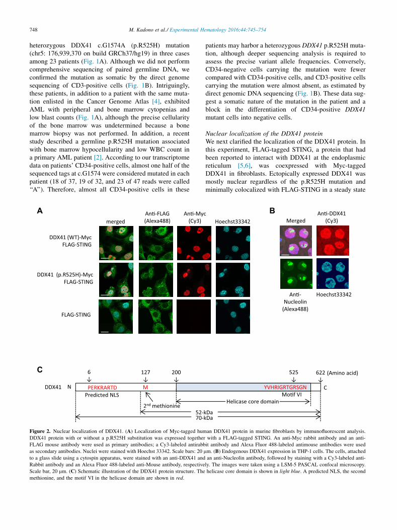
Figure 2. Nuclear localization of DDX41. (A) Localization of Myc-tagged hum
DDX41 protein with or without a p.R525H substitution was expressed together
FLAG mouse antibody were used as primary antibodies; a Cy3-labeled antirabb
as secondary antibodies. Nuclei were stained with Hoechst 33342. Scale bars: 20
to a glass slide using a cytospin apparatus, were stained with an anti-DDX41 and
Rabbit antibody and an Alexa Fluor 488-labeled anti-Mouse antibody, respective
Scale bar, 20 mm. (C) Schematic illustration of the DDX41 protein structure. The
methionine, and the motif VI in the helicase domain are shown in red.
patients may harbor a heterozygous DDX41 p.R525H muta-tion, although deeper sequencing analysis is required toassess the precise variant allele frequencies. Conversely,CD34-negative cells carrying the mutation were fewercompared with CD34-positive cells, and CD3-positive cellscarrying the mutation were almost absent, as estimated bydirect genomic DNA sequencing (Fig. 1B). These data sug-gest a somatic nature of the mutation in the patient and ablock in the differentiation of CD34-positive DDX41mutant cells into negative cells.
Nuclear localization of the DDX41 proteinWe next clarified the localization of the DDX41 protein. Inthis experiment, FLAG-tagged STING, a protein that hadbeen reported to interact with DDX41 at the endoplasmicreticulum [5,6], was coexpressed with Myc-taggedDDX41 in fibroblasts. Ectopically expressed DDX41 wasmostly nuclear regardless of the p.R525H mutation andminimally colocalized with FLAG-STING in a steady state
BcHoechst33342
An�-DDX41 (Cy3)
An�-Nucleolin
(Alexa488)
Hoechst33342
Merged
YVHRIGRTGRSGN
525↓
Helicase core domainMo�f VI
C
622↓
kDa
(Amino acid)
kDa
an DDX41 protein in murine fibroblasts by immunofluorescent analysis.
with a FLAG-tagged STING. An anti-Myc rabbit antibody and an anti-
it antibody and Alexa Fluor 488-labeled antimouse antibodies were used
mm. (B) Endogenous DDX41 expression in THP-1 cells. The cells, attached
an anti-Nucleolin antibody, followed by staining with a Cy3-labeled anti-
ly. The images were taken using a LSM-5 PASCAL confocal microscopy.
helicase core domain is shown in light blue. A predicted NLS, the second
749M. Kadono et al./ Experimental Hematology 2016;44:745–754
(Fig. 2A). In the THP-1 leukemia cell line, the endogenousDDX41 protein was also mostly nuclear (Fig. 2B). In thenucleus, DDX41 protein localized both in the nucleoplasmand in the nucleolus (stained with nucleolin), suggestingthat DDX41 plays a role in the nucleoplasm as well as inthe nucleolus. Although previous studies describedDDX41 as a cytosolic DNA sensor that recognizes nucleicacids of pathogens [6], our study did not suggest this func-tion based on the protein localization. However, we identi-fied a 52-kDa DDX41 short isoform translated from thesecond methionine in addition to the full-length 70-kDaDDX41 (Fig. 2C; Supplementary Figures E2A and E2B,online only, available at www.exphem.org). This shortform, which lacks a putative nuclear localizing signal(NLS) and is detected both in the cytoplasm and in the nu-cleus (Fig. 2C; Supplementary Figure E2C, online only,available at www.exphem.org), might function as a sensorof pathogenic nucleic acids.
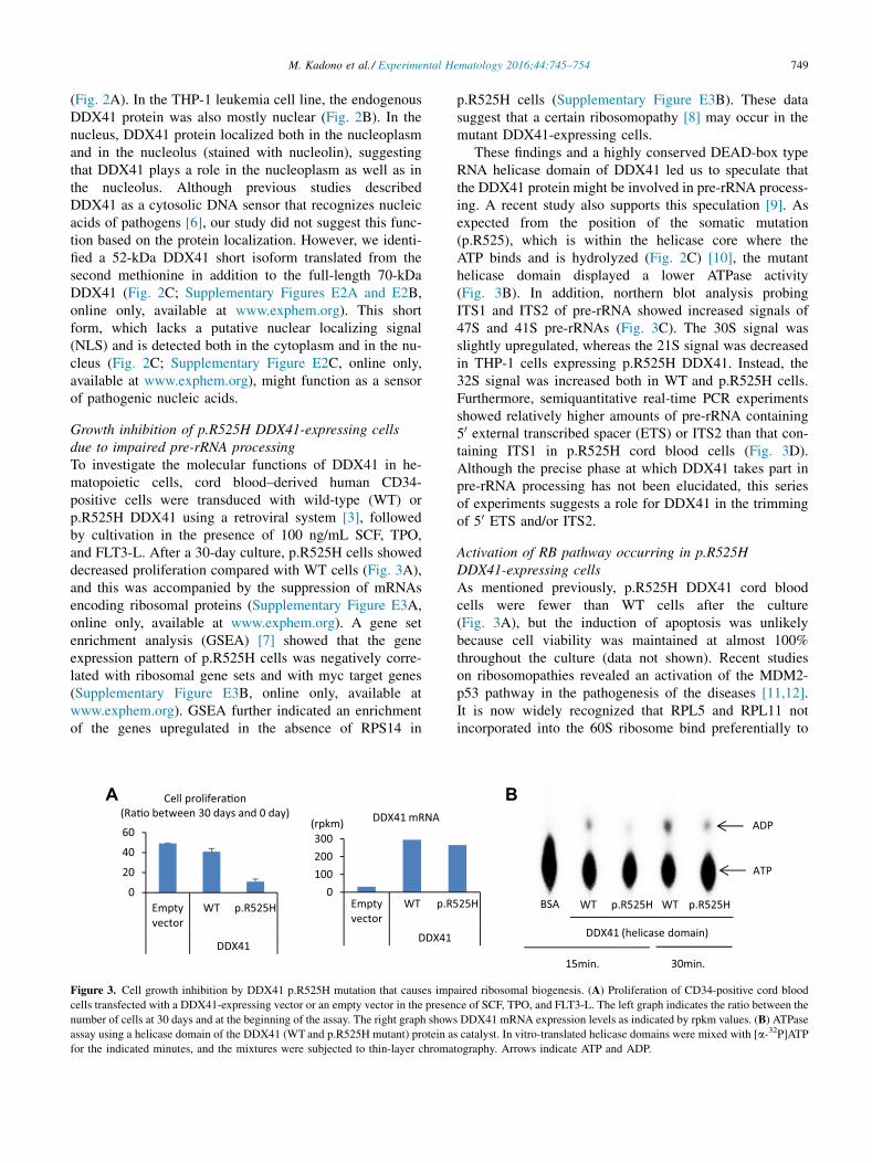
Growth inhibition of p.R525H DDX41-expressing cellsdue to impaired pre-rRNA processingTo investigate the molecular functions of DDX41 in he-matopoietic cells, cord blood–derived human CD34-positive cells were transduced with wild-type (WT) orp.R525H DDX41 using a retroviral system [3], followedby cultivation in the presence of 100 ng/mL SCF, TPO,and FLT3-L. After a 30-day culture, p.R525H cells showeddecreased proliferation compared with WT cells (Fig. 3A),and this was accompanied by the suppression of mRNAsencoding ribosomal proteins (Supplementary Figure E3A,online only, available at www.exphem.org). A gene setenrichment analysis (GSEA) [7] showed that the geneexpression pattern of p.R525H cells was negatively corre-lated with ribosomal gene sets and with myc target genes(Supplementary Figure E3B, online only, available atwww.exphem.org). GSEA further indicated an enrichmentof the genes upregulated in the absence of RPS14 in
A
0100200300
Emptyvector
WT p.R5
DDX41
DDX41 mRNA
0
20
40
60
Emptyvector
WT p.R525H
DDX41
Cell prolifera�on(Ra�o between 30 days and 0 day)
(rpkm)
Figure 3. Cell growth inhibition by DDX41 p.R525H mutation that causes imp
cells transfected with a DDX41-expressing vector or an empty vector in the presen
number of cells at 30 days and at the beginning of the assay. The right graph show
assay using a helicase domain of the DDX41 (WT and p.R525H mutant) protein a
for the indicated minutes, and the mixtures were subjected to thin-layer chroma
p.R525H cells (Supplementary Figure E3B). These datasuggest that a certain ribosomopathy [8] may occur in themutant DDX41-expressing cells.
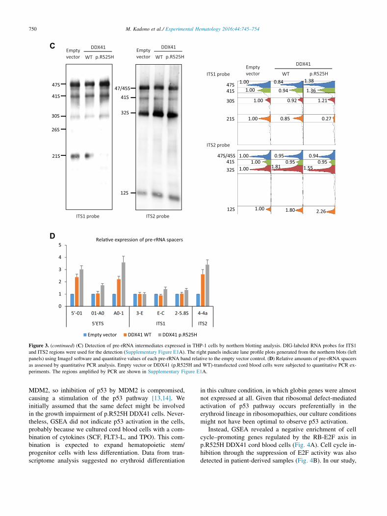
These findings and a highly conserved DEAD-box typeRNA helicase domain of DDX41 led us to speculate thatthe DDX41 protein might be involved in pre-rRNA process-ing. A recent study also supports this speculation [9]. Asexpected from the position of the somatic mutation(p.R525), which is within the helicase core where theATP binds and is hydrolyzed (Fig. 2C) [10], the mutanthelicase domain displayed a lower ATPase activity(Fig. 3B). In addition, northern blot analysis probingITS1 and ITS2 of pre-rRNA showed increased signals of47S and 41S pre-rRNAs (Fig. 3C). The 30S signal wasslightly upregulated, whereas the 21S signal was decreasedin THP-1 cells expressing p.R525H DDX41. Instead, the32S signal was increased both in WT and p.R525H cells.Furthermore, semiquantitative real-time PCR experimentsshowed relatively higher amounts of pre-rRNA containing50 external transcribed spacer (ETS) or ITS2 than that con-taining ITS1 in p.R525H cord blood cells (Fig. 3D).Although the precise phase at which DDX41 takes part inpre-rRNA processing has not been elucidated, this seriesof experiments suggests a role for DDX41 in the trimmingof 50 ETS and/or ITS2.
Activation of RB pathway occurring in p.R525HDDX41-expressing cellsAs mentioned previously, p.R525H DDX41 cord bloodcells were fewer than WT cells after the culture(Fig. 3A), but the induction of apoptosis was unlikelybecause cell viability was maintained at almost 100%throughout the culture (data not shown). Recent studieson ribosomopathies revealed an activation of the MDM2-p53 pathway in the pathogenesis of the diseases [11,12].It is now widely recognized that RPL5 and RPL11 notincorporated into the 60S ribosome bind preferentially to
15min. 30min.
ATP
WT
DDX41 (helicase domain)
p.R525HBSA WT p.R525H
ADP
B
25H
aired ribosomal biogenesis. (A) Proliferation of CD34-positive cord blood
ce of SCF, TPO, and FLT3-L. The left graph indicates the ratio between the
s DDX41 mRNA expression levels as indicated by rpkm values. (B) ATPase
s catalyst. In vitro-translated helicase domains were mixed with [a-32P]ATP
tography. Arrows indicate ATP and ADP.
0
1
2
3
4
5
5'-01 01-A0 A0-1 3-E E-C 2-5.8S 4-4a
5'ETS ITS1 ITS2
Empty vector DDX41 WT DDX41 p.R525H
Rela�ve expression of pre-rRNA spacers
C
D
47S
41S
21S
30S
26S
Emptyvector WT p.R525H
DDX41
ITS1 probe
47/45S
41S
32S
12S
ITS2 probe
Emptyvector WT p.R525H
DDX41
Emptyvector WT p.R525H
DDX41
47S41S
21S
30S
0.84ITS1 probe
1.381.00
0.94 1.361.00
0.92 1.211.00
0.85 0.271.00
47S/45S41S32S
ITS2 probe
0.95 0.941.000.95 0.951.00
1.81 1.551.00
1.80 2.261.0012S
Figure 3. (continued) (C) Detection of pre-rRNA intermediates expressed in THP-1 cells by northern blotting analysis. DIG-labeled RNA probes for ITS1
and ITS2 regions were used for the detection (Supplementary Figure E1A). The right panels indicate lane profile plots generated from the northern blots (left
panels) using ImageJ software and quantitative values of each pre-rRNA band relative to the empty vector control. (D) Relative amounts of pre-rRNA spacers
as assessed by quantitative PCR analysis. Empty vector or DDX41 (p.R525H and WT)-transfected cord blood cells were subjected to quantitative PCR ex-
periments. The regions amplified by PCR are shown in Supplementary Figure E1A.
750 M. Kadono et al./ Experimental Hematology 2016;44:745–754
MDM2, so inhibition of p53 by MDM2 is compromised,causing a stimulation of the p53 pathway [13,14]. Weinitially assumed that the same defect might be involvedin the growth impairment of p.R525H DDX41 cells. Never-theless, GSEA did not indicate p53 activation in the cells,probably because we cultured cord blood cells with a com-bination of cytokines (SCF, FLT3-L, and TPO). This com-bination is expected to expand hematopoietic stem/progenitor cells with less differentiation. Data from tran-scriptome analysis suggested no erythroid differentiation
in this culture condition, in which globin genes were almostnot expressed at all. Given that ribosomal defect-mediatedactivation of p53 pathway occurs preferentially in theerythroid lineage in ribosomopathies, our culture conditionsmight not have been optimal to observe p53 activation.
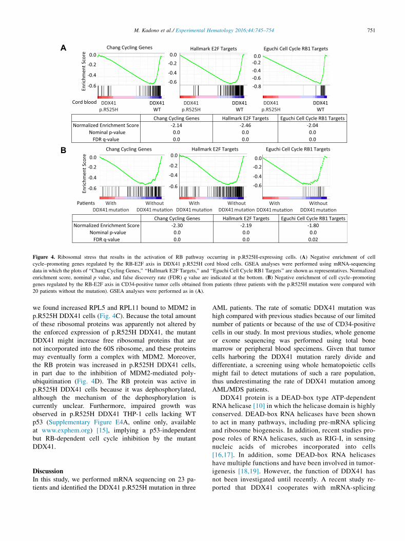
Instead, GSEA revealed a negative enrichment of cellcycle–promoting genes regulated by the RB-E2F axis inp.R525H DDX41 cord blood cells (Fig. 4A). Cell cycle in-hibition through the suppression of E2F activity was alsodetected in patient-derived samples (Fig. 4B). In our study,
Enric
hmen
t Sco
re
0.0-0.2
-0.4
-0.6
Hallmark E2F Targets
DDX41WT
DDX41p.R525H
0.0-0.2-0.4-0.6-0.8
Eguchi Cell Cycle RB1 Targets
DDX41WT
DDX41p.R525H
DDX41WT
0.0
-0.2
-0.4
-0.6En
richm
ent S
core
Chang Cycling Genes
DDX41p.R525H
Cord blood
WithDDX41 muta�on
WithoutDDX41 muta�on
0.0
-0.2
-0.4
-0.6
Hallmark E2F Targets Eguchi Cell Cycle RB1 Targets
0.0-0.2
-0.4
-0.6
WithDDX41 muta�on
WithoutDDX41 muta�on
0.0
-0.2
-0.4
-0.6
Chang Cycling Genes
WithDDX41 muta�on
WithoutDDX41 muta�on
Pa�ents
Chang Cycling Genes Hallmark E2F Targets Eguchi Cell Cycle RB1 TargetsNormalized Enrichment Score -2.14 -2.46 -2.04
Nominal p-value 0.0 0.0 0.0 FDR q-value 0.0 0.0 0.0
Chang Cycling Genes Hallmark E2F Targets Eguchi Cell Cycle RB1 TargetsNormalized Enrichment Score -2.30 -2.19 -1.80
Nominal p-value 0.0 0.0 0.0 FDR q-value 0.0 0.0 0.02
A
B
Figure 4. Ribosomal stress that results in the activation of RB pathway occurring in p.R525H-expressing cells. (A) Negative enrichment of cell
cycle–promoting genes regulated by the RB-E2F axis in DDX41 p.R525H cord blood cells. GSEA analyses were performed using mRNA-sequencing
data in which the plots of ‘‘Chang Cycling Genes,’’ ‘‘Hallmark E2F Targets,’’ and ‘‘Eguchi Cell Cycle RB1 Targets’’ are shown as representatives. Normalized
enrichment score, nominal p value, and false discovery rate (FDR) q value are indicated at the bottom. (B) Negative enrichment of cell cycle–promoting
genes regulated by the RB-E2F axis in CD34-positive tumor cells obtained from patients (three patients with the p.R525H mutation were compared with
20 patients without the mutation). GSEA analyses were performed as in (A).
751M. Kadono et al./ Experimental Hematology 2016;44:745–754
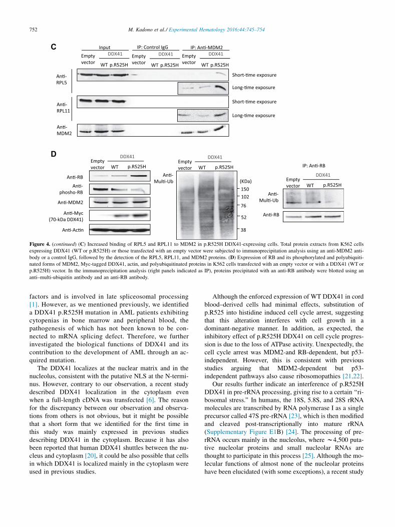
we found increased RPL5 and RPL11 bound to MDM2 inp.R525H DDX41 cells (Fig. 4C). Because the total amountof these ribosomal proteins was apparently not altered bythe enforced expression of p.R525H DDX41, the mutantDDX41 might increase free ribosomal proteins that arenot incorporated into the 60S ribosome, and these proteinsmay eventually form a complex with MDM2. Moreover,the RB protein was increased in p.R525H DDX41 cells,in part due to the inhibition of MDM2-mediated poly-ubiquitination (Fig. 4D). The RB protein was active inp.R525H DDX41 cells because it was dephosphorylated,although the mechanism of the dephosphorylation iscurrently unclear. Furthermore, impaired growth wasobserved in p.R525H DDX41 THP-1 cells lacking WTp53 (Supplementary Figure E4A, online only, availableat www.exphem.org) [15], implying a p53-independentbut RB-dependent cell cycle inhibition by the mutantDDX41.
DiscussionIn this study, we performed mRNA sequencing on 23 pa-tients and identified the DDX41 p.R525H mutation in three
AML patients. The rate of somatic DDX41 mutation washigh compared with previous studies because of our limitednumber of patients or because of the use of CD34-positivecells in our study. In most previous studies, whole genomeor exome sequencing was performed using total bonemarrow or peripheral blood specimens. Given that tumorcells harboring the DDX41 mutation rarely divide anddifferentiate, a screening using whole hematopoietic cellsmight fail to detect mutations of such a rare population,thus underestimating the rate of DDX41 mutation amongAML/MDS patients.
DDX41 protein is a DEAD-box type ATP-dependentRNA helicase [10] in which the helicase domain is highlyconserved. DEAD-box RNA helicases have been shownto act in many pathways, including pre-mRNA splicingand ribosome biogenesis. In addition, recent studies pro-pose roles of RNA helicases, such as RIG-I, in sensingnucleic acids of microbes incorporated into cells[16,17]. In addition, some DEAD-box RNA helicaseshave multiple functions and have been involved in tumor-igenesis [18,19]. However, the function of DDX41 hasnot been investigated until recently. A recent study re-ported that DDX41 cooperates with mRNA-splicing
An�-Mul�-Ub
Emptyvector WT p.R525H
DDX41
IP: An�-RB
An�-RB
Emptyvector WT p.R525H
DDX41
An�-Mul�-Ub
102
76
52
38
150(KDa)
Emptyvector WT p.R525H
DDX41
An�-RB
An�-phosho-RB
An�-MDM2
An�-Myc(70-kDa DDX41)
An�-Ac�n
An�-RPL5
An�-RPL11
Short-�me exposure
Long-�me exposure
Short-�me exposure
Long-�me exposure
Emptyvector WT p.R525H
Input IP: Control IgG IP: An�-MDM2DDX41 Empty
vector WT p.R525H
DDX41 Emptyvector WT p.R525H
DDX41
An�-MDM2
C
D
Figure 4. (continued) (C) Increased binding of RPL5 and RPL11 to MDM2 in p.R525H DDX41-expressing cells. Total protein extracts from K562 cells
expressing DDX41 (WT or p.R525H) or those transfected with an empty vector were subjected to immunoprecipitation analysis using an anti-MDM2 anti-
body or a control IgG, followed by the detection of the RPL5, RPL11, and MDM2 proteins. (D) Expression of RB and its phosphorylated and polyubiquiti-
nated forms of MDM2, Myc-tagged DDX41, actin, and polyubiquitinated proteins in K562 cells transfected with an empty vector or with a DDX41 (WT or
p.R525H) vector. In the immunoprecipitation analysis (right panels indicated as IP), proteins precipitated with an anti-RB antibody were blotted using an
anti–multi-ubiquitin antibody and an anti-RB antibody.
752 M. Kadono et al./ Experimental Hematology 2016;44:745–754
factors and is involved in late spliceosomal processing[1]. However, as we mentioned previously, we identifieda DDX41 p.R525H mutation in AML patients exhibitingcytopenias in bone marrow and peripheral blood, thepathogenesis of which has not been known to be con-nected to mRNA splicing defect. Therefore, we furtherinvestigated the biological functions of DDX41 and itscontribution to the development of AML through an ac-quired mutation.
The DDX41 localizes at the nuclear matrix and in thenucleolus, consistent with the putative NLS at the N-termi-nus. However, contrary to our observation, a recent studydescribed DDX41 localization in the cytoplasm evenwhen a full-length cDNA was transfected [6]. The reasonfor the discrepancy between our observation and observa-tions from others is not obvious, but it might be possiblethat a short form that we identified for the first time inthis study was mainly expressed in previous studiesdescribing DDX41 in the cytoplasm. Because it has alsobeen reported that human DDX41 shuttles between the nu-cleus and cytoplasm [20], it could be also possible that cellsin which DDX41 is localized mainly in the cytoplasm wereused in previous studies.
Although the enforced expression of WT DDX41 in cordblood–derived cells had minimal effects, substitution ofp.R525 into histidine induced cell cycle arrest, suggestingthat this alteration interferes with cell growth in adominant-negative manner. In addition, as expected, theinhibitory effect of p.R525H DDX41 on cell cycle progres-sion is due to the loss of ATPase activity. Unexpectedly, thecell cycle arrest was MDM2-and RB-dependent, but p53-independent. However, this is consistent with previousstudies arguing that MDM2-dependent but p53-independent pathways also cause ribosomopathies [21,22].
Our results further indicate an interference of p.R525HDDX41 in pre-rRNA processing, giving rise to a certain ‘‘ri-bosomal stress.’’ In humans, the 18S, 5.8S, and 28S rRNAmolecules are transcribed by RNA polymerase I as a singleprecursor called 47S pre-rRNA [23], which is then modifiedand cleaved post-transcriptionally into mature rRNA(Supplementary Figure E1B) [24]. The processing of pre-rRNA occurs mainly in the nucleolus, where w4,500 puta-tive nucleolar proteins and small nucleolar RNAs arethought to participate in this process [25]. Although the mo-lecular functions of almost none of the nucleolar proteinshave been elucidated (with some exceptions), a recent study

753M. Kadono et al./ Experimental Hematology 2016;44:745–754
that performed siRNA-mediated depletion of 625 candidatenucleolar proteins helped to explain the pre-rRNA process-ing machinery [9]. Because it has been shown that somespliceosomal factors, such as hPrp43/DHX15 RNA heli-case, also participate in pre-rRNA processing [26,27],DDX41 could play multiple roles. We think that thispathway can at least partly account for the developmentof AML with cytopenias harboring the DDX41 p.R525Hmutation. Hematopoietic stem cells have a low level of pro-tein synthesis compared with differentiating or growing pro-genitor cells [28]. Assuming that AML stem cells with theDDX41 p.R525H mutation are constitutively in a low-protein synthesis status due to ribosomal stress, the cellscould be able to be maintained under this stress, but theycannot proliferate or differentiate, which would explain inpart the pathophysiology of a slowly growing AML.
In summary, we propose a mechanism of growth defectin hematopoietic cells triggered by p.R525H DDX41 occur-ring in the following order: (1) the p.R525H mutant inhibitspre-rRNA processing; (2) compromised ribosomal biogen-esis as a result of impaired rRNA synthesis causes a releaseof ribosomal proteins that bind to MDM2; and (3) MDM2-mediated RB degradation is suppressed, eventually acti-vating the RB pathway and resulting in the inhibition ofE2F activity. Although this study uncovered a pathogenicrole of p.R525H DDX41 in the slow growth rate of tumorcells, how the mutation induces AML development and in-hibits cell differentiation is still not understood. Lethallyirradiated mice transplanted with hematopoietic stem/pro-genitor cells overexpressing p.R525H DDX41 did notdevelop myeloid malignancy, even in the p53-deficientbackground (Supplementary Figure E4B, online only, avail-able at www.exphem.org). Considering the late occurrenceof AML in patients harboring the mutation, age-dependentepigenetic alterations or other somatic changes may berequired for this mutation to transform hematopoietic cellsfully.
AcknowledgmentsThe authors thank Dr. H. Kawamoto for providing the cells andR. Tai, E. Kanai, and M. Nakamura for excellent technical assis-tance. This work was partly supported by the Daiichi SankyoFoundation of Life Science, the NOVARTIS Foundation (Japan)for the Promotion of Science, the SGH Foundation, the PrincessTakamatsu Cancer Research Fund, and the Relay for Life programfounded by the Japan Cancer Society.
Conflict of interest disclosureThe authors declare no competing financial interests.
References1. Polprasert C, Schulze I, Sekeres MA, et al. Inherited and somatic
defects in DDX41 in myeloid neoplasms. Cancer Cell. 2015;27:
658–670.
2. Lewinsohn M, Brown AL, Weinel LM, et al. Novel germ line DDX41
mutations define families with a lower age of MDS/AML onset and
lymphoid malignancies. Blood. 2016;127:1017–1023.
3. Sekine R, Kitamura T, Tsuji T, Tojo A. Efficient retroviral transduc-
tion of human B-lymphoid and myeloid progenitors: Marked inhibi-
tion of their growth by the Pax5 transgene. Int J Hematol. 2008;87:
351–362.
4. Ding L, Ley TJ, Larson DE, et al. Clonal evolution in relapsed acute
myeloid leukemia revealed by whole-genome sequencing. Nature.
2012;481:506–510.
5. Parvatiyar K, Zhang Z, Teles RM, et al. The helicase DDX41 recog-
nizes the bacterial secondary messengers cyclic di-GMP and cyclic
di-AMP to activate a type I interferon immune response. Nat Immu-
nol. 2012;13:1155–1161.
6. Zhang Z, Yuan B, Bao M, Lu N, Kim T, Liu YJ. The helicase DDX41
senses intracellular DNA mediated by the adaptor STING in dendritic
cells. Nat Immunol. 2011;12:959–965.
7. Subramanian A, Tamayo P, Mootha VK, et al. Gene set enrichment
analysis: A knowledge-based approach for interpreting genome-wide
expression profiles. Proc Natl Acad Sci U S A. 2005;102:15545–15550.
8. Aspesi A, Pavesi E, Robotti E, et al. Dissecting the transcriptional
phenotype of ribosomal protein deficiency: Implications for
Diamond-Blackfan Anemia. Gene. 2014;545:282–289.
9. Tafforeau L, Zorbas C, Langhendries JL, et al. The complexity of hu-
man ribosome biogenesis revealed by systematic nucleolar screening
of pre-rRNA processing factors. Mol Cell. 2013;51:539–551.
10. Linder P, Jankowsky E. From unwinding to clamping: The DEAD box
RNA helicase family. Nat Rev Mol Cell Biol. 2011;12:505–516.
11. Dutt S, Narla A, Lin K, et al. Haploinsufficiency for ribosomal protein
genes causes selective activation of p53 in human erythroid progenitor
cells. Blood. 2011;117:2567–2576.
12. Ebert BL, Pretz J, Bosco J, et al. Identification of RPS14 as a 5q-
syndrome gene by RNA interference screen. Nature. 2008;451:
335–339.
13. Fumagalli S, Ivanenkov VV, Teng T, Thomas G. Suprainduction of
p53 by disruption of 40S and 60S ribosome biogenesis leads to the
activation of a novel G2/M checkpoint. Genes Dev. 2012;26:1028–
1040.
14. Sun XX, Wang YG, Xirodimas DP, Dai MS. Perturbation of 60 S ri-
bosomal biogenesis results in ribosomal protein L5- and L11-
dependent p53 activation. J Biol Chem. 2010;285:25812–25821.
15. Durland-Busbice S, Reisman D. Lack of p53 expression in human
myeloid leukemias is not due to mutations in transcriptional regulatory
regions of the gene. Leukemia. 2002;16:2165–2167.
16. Sato S, Li K, Kameyama T, et al. The RNA sensor RIG-I dually func-
tions as an innate sensor and direct antiviral factor for hepatitis B vi-
rus. Immunity. 2015;42:123–132.
17. Jiang F, Ramanathan A, Miller MT, et al. Structural basis of RNA
recognition and activation by innate immune receptor RIG-I. Nature.
2011;479:423–427.
18. Fuller-Pace FV. DEAD box RNA helicase functions in cancer. RNA
Biol. 2013;10:121–132.
19. Dardenne E, Pierredon S, Driouch K, et al. Splicing switch of an
epigenetic regulator by RNA helicases promotes tumor-cell invasive-
ness. Nat Struct Mol Biol. 2012;19:1139–1146.
20. Abdul-Ghani M, Hartman KL, Ngsee JK. Abstrakt interacts with and
regulates the expression of sorting nexin-2. J Cell Physiol. 2005;204:
210–218.
21. Donati G, Brighenti E, Vici M, et al. Selective inhibition of rRNA
transcription downregulates E2F-1: A new p53-independent mecha-
nism linking cell growth to cell proliferation. J Cell Sci. 2011;
124(Pt 17):3017–3028.
22. Uchida C, Miwa S, Kitagawa K, et al. Enhanced Mdm2 activity in-
hibits pRB function via ubiquitin-dependent degradation. EMBO J.
2005;24:160–169.

754 M. Kadono et al./ Experimental Hematology 2016;44:745–754
23. Baatout S. Staining of the nucleolar organizer regions: relevance in he-
matology. Blood Rev. 1996;10:185–188.
24. Mullineux ST, Lafontaine DL. Mapping the cleavage sites on mamma-
lian pre-rRNAs: Where do we stand? Biochimie. 2012;94:1521–1532.
25. Ahmad Y, Boisvert FM, Gregor P, Cobley A, Lamond AI. NOPdb:
Nucleolar Proteome Databased2008 update. Nucleic Acids Res.
2009;37(Database issue):D181–D184.
26. Bohnsack MT, Martin R, Granneman S, Ruprecht M, Schleiff E,
Tollervey D. Prp43 bound at different sites on the pre-rRNA
performs distinct functions in ribosome synthesis. Mol Cell. 2009;
36:583–592.
27. Yoshimoto R, Okawa K, Yoshida M, Ohno M, Kataoka N. Identifica-
tion of a novel component C2ORF3 in the lariat-intron complex: Lack
of C2ORF3 interferes with pre-mRNA splicing via intron turnover
pathway. Genes Cells. 2014;19:78–87.
28. Buszczak M, Signer RA, Morrison SJ. Cellular differences in
protein synthesis regulate tissue homeostasis. Cell. 2014;159:
242–251.

18S 5.8S 28S
01 A0 1 3 E C 2 4’ 4a
Northern probes ITS1 ITS2
5’ 3’
5’ETS region ITS1 region ITS2 region 3’ETS region
47S pre-rRNA
4aA0 1
3 E
4’
015’01 A0
E C2 5.8SqPCR amplicons
023’
5’ end 3’ end
01 02
47S
45S
1 0241S 230S 01 2 02 32S
Pathway 1 Pathway 2
A
B
2 02 32S221S 1
28S12S 3’21S-C C1
7S 4aE18S-E 1
5.8S 4’318S 1
221S 28S12S 3’1
E18S-E 5.8S 4’1
21S-C C 7S 4a1
318S 1
Supplementary Figure E1. Structure and processing pathways of pre-rRNA. (A) Structure of 47S pre-rRNA. The 50 and 30 ETS and ITS 1 and 2 are spacer
regions that will be removed during pre-rRNA processing, and there are many pre-rRNA intermediates partially retaining these spacers, as shown in (B).
Detailed pre-rRNA cleavage sites and their names are described in a recent review [24]. The regions used for northern probes (ITS1 and ITS2) and amplified
by quantitative PCR analysis (50-01, 01-A0, A0-1, 3-E, E-C, 2-5.8S, and 40-4a) are shown in orange and gray, respectively. (B) Schematic processing path-
ways of pre-rRNA. Three rRNAs (18S, 5.8S, and 28S) are first transcribed as a long polycistronic precursor designated as 47S pre-rRNA. The latter will
subsequently be processed to produce mature rRNAs through pathways, as shown in this schema.
754.e1M. Kadono et al./ Experimental Hematology 2016;44:745–754

Supplementary Figure E2. Expression and localization of DDX41 and its short isoform. (A) Endogenous DDX41 expression in four AML cell lines (upper
panels). DDX41 was detected by an antibody against the C-terminus of DDX41. A full-length image of DDX41 immunoblot is shown at right. (B) Exogenous
DDX41 proteins expressed in HEK293 cells. The cells were transfected with a Myc-tagged DDX41 cDNA (cDNA starting from the first and second methi-
onine, designated as p70 and p52, respectively), and the extracts were subjected to immunoblot analysis. Tagging positions are indicated as C (C-terminus) or
N (N-terminus). The C-terminally tagged p70 cDNA also showed the 52-kDa band weakly (lanes 1 and 2), whereas the p70 cDNA tagged at the N-terminus
did not show the 52-kDa band (lane 5 and 6), suggesting that the 52-kDa band is a truncated protein originated from the c-terminal part of DDX41. Indeed,
the p70 cDNA, where the p.M132 (the third methionine) was substituted with an alanine, expressed a very weak 52-kDa band (lane 11), which disappeared
when the p.M127 (the second methionine) was substituted with alanine (lanes 13 and 14). Actin was used as a loading control. A schematic diagram is shown
in the center, and full-length images of DDX41 immunoblot are shown at bottom right. (C) Localization of 52-kDa DDX41 tagged with Myc at its c-terminus.
The experiment was performed as in (A). Scale bars, 20 mm.
754.e2 M. Kadono et al./ Experimental Hematology 2016;44:745–754

Supplementary Figure E3. Suppression of ribosomal genes in p.R525H DDX41-expressing cord blood cells. (A) Decrease in ribosomal genes (RPLs and
RPSs) in p.R525H DDX41 cord blood cells compared with the cells transfected with WT DDX41-expressing vector. Each bar indicates the ratio of p.R525H
cells to WT cells. (B) GSEA analysis indicating negative enrichment of gene sets related to ribosome biogenesis and positive enrichment of genes upregulated
in RPS14-deficient p.R525H DDX41 cord blood cells. Normalized enrichment score, nominal p value, and FDR q value are shown at the bottom.
754.e3M. Kadono et al./ Experimental Hematology 2016;44:745–754

Supplementary Figure E4. There was no growth advantage of p.R525H DDX41-expressing cells in vitro or in vivo. (A) DDX41 (WT or p.R525H)-
expressing THP-1 cell number. THP cells lacking functional p53 were transduced with a DDX41-expressing vector, and viable cells were counted. (B) There
was no growth advantage of p.R525H DDX41-expressing hematopoietic cells in murine BMT experiments. Donor cells (with or without p53þ/� background)
were transduced with an empty/GFP-expressing vector or a DDX41/GFP-expressing vector, followed by transplantation into lethally irradiated recipients.
GFP positivity (%) and peripheral WBC numbers (�103/mL) were measured 2–3 months after BMT. No significant difference in GFP signals was observed
between the groups, suggesting that enforced expression of DDX41 pR525H alone does not account for the acquisition of clonogenicity.
754.e4 M. Kadono et al./ Experimental Hematology 2016;44:745–754


![MedullaryThyroidCarcinoma:MolecularSignalingPathways ...downloads.hindawi.com/journals/jtr/2011/815826.pdf · most common somatic mutation in sporadic MTC [27]. This mutation is associated](https://static.fdocuments.in/doc/165x107/5eb9b8cca5f68134660fa242/medullarythyroidcarcinomamolecularsignalingpathways-most-common-somatic-mutation.jpg)