BIOLOGICAL CONSEQUENCES OF OXYGEN DESATURATION …digital.csic.es/bitstream/10261/33423/3/Biological...
21
1 BIOLOGICAL CONSEQUENCES OF OXYGEN DESATURATION AND RESPIRATORY EFFORT IN AN ACUTE ANIMAL MODEL OF OBSTRUTIVE SLEEP APNEA (OSA). Maria Nácher BSc 1,2 , Ramon Farré PhD 3,4 , Josep M. Montserrat MD 1,4 , Marta Torres 1,4 , Daniel Navajas PhD 3,4 , Oriol Bulbena PhD 2 , Anna Serrano-Mollar PhD 2 . 1 Sleep Lab, Hospital Clinic Provincial-IDIBAPS, Barcelona, Spain; 2 Department of Experimental Pathology, IIBB-CSIC-IDIBAPS, Barcelona, Spain; 3 Unitat Biofísica i Bioenginyeria, Facultat Medicina, Universitat Barcelona-IDIBAPS, Barcelona, Spain; 4 CibRes. Spain Running title: Hypoxia, normoxia, respiratory effort and obstructive apnea. Subject Code: Sleep-disordered breathing: biological consequences. Mailing address: Dr. J.M Montserrat Laboratori de la Son. Servei de Pneumologia C/ Casanoves, 170 E-08036 Barcelona, Spain Fax: 34-93-2275746; Phone: 34-93-2275746 E-mail: [email protected]
Transcript of BIOLOGICAL CONSEQUENCES OF OXYGEN DESATURATION …digital.csic.es/bitstream/10261/33423/3/Biological...
1
BIOLOGICAL CONSEQUENCES OF OXYGEN DESATURATION AND RESPIRATORY
EFFORT IN AN ACUTE ANIMAL MODEL OF OBSTRUTIVE SLEEP APNEA (OSA).
Maria Nácher BSc1,2, Ramon Farré PhD3,4, Josep M. Montserrat MD1,4, Marta Torres 1,4,
Daniel Navajas PhD3,4, Oriol Bulbena PhD2, Anna Serrano-Mollar PhD2.
1Sleep Lab, Hospital Clinic Provincial-IDIBAPS, Barcelona, Spain; 2Department of
Experimental Pathology, IIBB-CSIC-IDIBAPS, Barcelona, Spain; 3Unitat Biofísica i
Bioenginyeria, Facultat Medicina, Universitat Barcelona-IDIBAPS, Barcelona, Spain;
4CibRes. Spain
Running title: Hypoxia, normoxia, respiratory effort and obstructive apnea.
Subject Code: Sleep-disordered breathing: biological consequences.
Mailing address:
Dr. J.M Montserrat
Laboratori de la Son. Servei de Pneumologia
C/ Casanoves, 170
E-08036 Barcelona, Spain
Fax: 34-93-2275746; Phone: 34-93-2275746
E-mail: [email protected]
2
ABSTRACT
Background. An animal model mimicking all the factors involved in obstructive sleep apnea
(OSA) is useful for investigating mechanisms because the associated comorbididy usually
present in such patients is an important limitation. Aim: To test the hypothesis that
hypoxia/normoxia and the respiratory effort have different effects on the induction of the
inflammatory response and endothelial dysfunction in an acute rat model of OSA.
Methods: Four groups of anesthetized rats were studied (n=8): 1) Sham; 2) Apnea:
obstructions (15 s each, 60/h, for 3 h); 3) Apnea+O2: obstructions and breathing oxygen-
enriched air to avoid hypoxia and 4) Intermittent hypoxia/normoxia. Inflammatory and
endothelial mediators were measured as outcomes along with NF-kB in the lung and
diaphragm..
Results: TNF-α and IL-1β significantly increased in all groups compared with Sham. NF-kB
in the lung was increased in Apnea and Hypoxia/Normoxia groups, but not in Apnea+O2
group. In diaphragm tissue, NF-κB was only significant in Apnea compared to Sham.
Significant differences were found in the ratio thromboxane-B2/6-keto-Prostaglandin-F1α
between Apnea and Hypoxia/Normoxia compared to Sham but not in Apnea+O2.
Conclusions: Oxygen desaturations and respiratory efforts play a role in the induction of
systemic inflammation but only hypoxia/normoxia induces endothelial dysfunction. These
data suggest a potential role for oxygen therapy in patients with OSA.
Word count: 208
Key words: obstructive apnea, inflammation, endothelial dysfunction, respiratory effort,
hypoxia, animal model.
3
INTRODUCTION
Obstructive sleep apnea (OSA) is the most common condition involving sleep-disordered
breathing, affecting between 4-6% of the population (1). OSA is characterized by intermittent
episodes of partial or complete obstruction of the upper airway during sleep and these
disrupt normal ventilation and sleep architecture. OSA is typically associated with excessive
daytime sleepiness, snoring, and witnessed apneas. This syndrome is also a cardiovascular
risk factor (2), its association with hypertension (2-4) being one of the most clearly
determined cardiovascular consequences.
The current evidence available shows that OSA is associated with systemic inflammation
and endothelial dysfunction (2, 5-15), and this has been considered one of the potential
mechanisms linking OSA with cardiovascular disease. In most cases, however, the data are
partial and sometimes even contradictory. Associated comorbidity, such as obesity, may
represent an important confounding factor, possibly leading to a distortion of the results
obtained from patient studies. Animal models will therefore be useful for exploring the
different mechanisms of OSA, as they not only avoid any associated comorbidity but will also
allow us to analyse the roles of the various factors involved in the syndrome.
In our current research, we are testing the hypothesis that hypoxia/normoxia and respiratory
effort have different effects on the induction of the inflammatory response and endothelial
dysfunction present in OSA. To this end, we have used a previously described acute animal
model of OSA (3 h, in anesthetized animals) that mimics the recurrent upper airway
obstructions characteristic of this sleep breathing disorder (16). The model avoids the
associated comorbidiity found in humans and allows us to individualize the various factors
present in OSA. Of course, that this acute model can not be completely related to the
chronic cardiovascular effects.
4
We analysed as outcomes of the different group experiments the circulating levels TNF-α
and IL-1β, which served as markers of inflammatory response. Furthermore, in order to
ascertain the source of these pro- inflammatory cytokines, we measured the transcription
factor nuclear kappa B (NF-κB) in two target tissues involved in recurrent obstructive
apneas: lung and diaphragm. Additionally, thromboxane-A2 (TxA2) and prostacyclin (PGI2)
were measured to evaluate the endothelial dysfunction. TxA2 is a powerful starting agent of
platelet aggregation and a potent vasoconstrictive, whereas, PGI2 has vasodilatadory effects
and acts against platelet aggregation. An imbalance of vasoconstrictor and vasodilator
mediators has been implicated in vascular diseases.
A better knowledge of the mechanisms involved in the biological response of OSA might be
very important, not only for understanding the disease but also for its potential relevance to
treatment strategy. For instance, in the case that hypoxia/normoxia proved to be the major
factor responsible for the systemic inflammatory/endothelial response, oxygen administration
could also play a role in treatment, especially in patients with severe OSA who do not accept
or tolerate CPAP.
2. MATERIALS AND METHODS
2.1 Animals
Pathogen-free male Sprague-Dawley rats, weighing 350-375 g, were obtained from Charles
River Laboratories (Saint Germain sur L'arbresle, France). The rats were housed in a
controlled environment and fed rodent chow (A04; Panlab, Barcelona, Spain) and tap water
ad libitum. This study conformed to European Community (Directive 86/609/EEC) and
Spanish guidelines for the use of experimental animals and was approved by the institutional
committees of animal care and research.

5
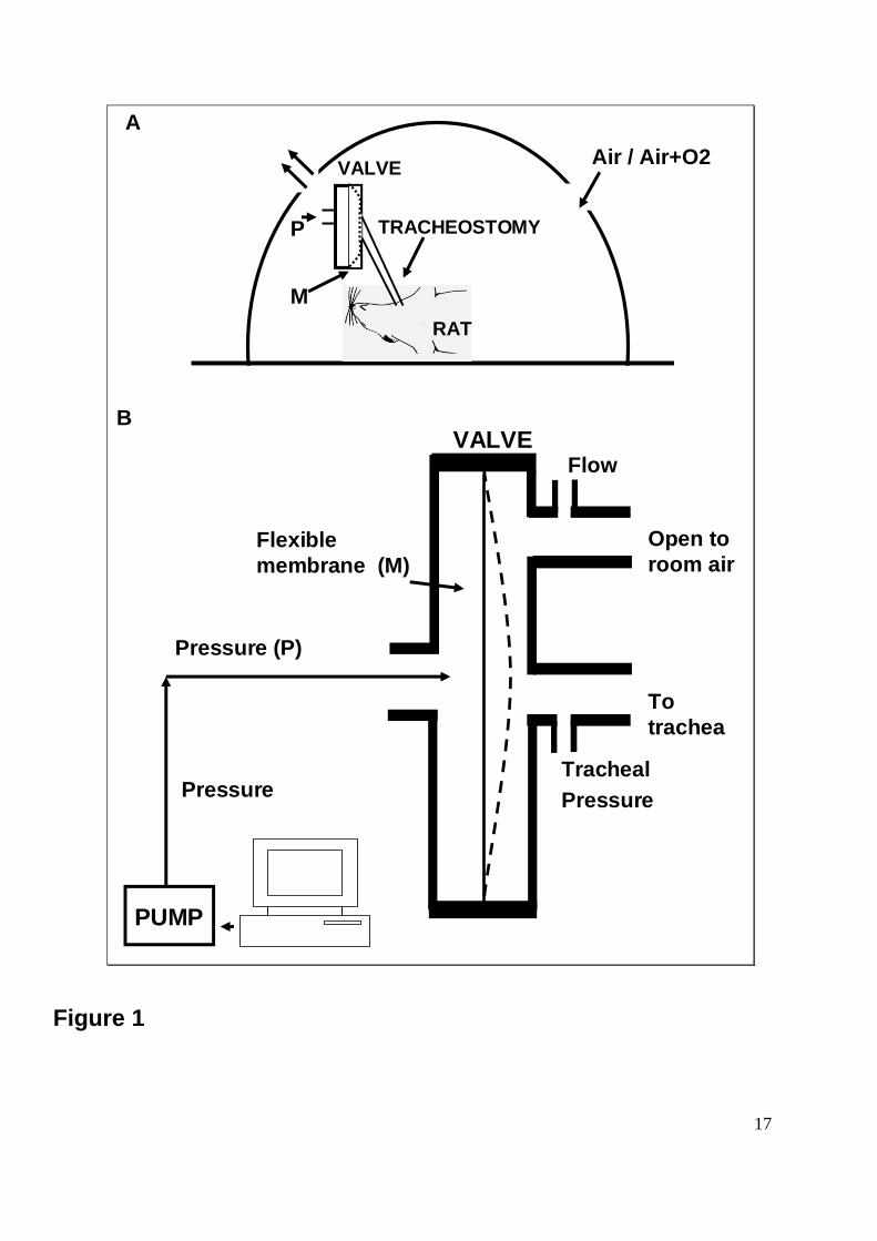
2.2 Model of recurrent obstructive apneas
Recurrent obstructive apneas in rats were applied by means of a previously described
computer-controlled collapsible segment based on a Starling resistor placed in the upper
airway of the animal (16,17) (Figure 1). Briefly, the collapsible segment consisted of two
identical cylindrical chambers separated by a circular flexible membrane. The base of one of
the chambers had a tube connected to a computer-driven source of external pressure that
produced controlled obstruction of the segment. A tube at the centre of the base of the other
chamber connected the collapsible segment to the rat trachea. Another tube in this chamber
wall was open to the atmosphere and acted as a pneumotachograph to measure breathing
flow. A pressure port in the tube connecting the collapsible segment to the trachea enabled
us to measure tracheal pressure. Arterial oxygen saturation in the rat leg was monitored by
pulse oximetry (504 Inc Wauseda, WI, USA).
2.3 Experimental groups
Experimental animals were randomly divided into four groups (n=8 for each group): Sham,
Apnea, Apnea+O2 and Hypoxia/Normoxia groups. The animals were anesthetized
(Urethane 1.2 g/kg, intraperitoneal), a tracheotomy was performed, a cannula (2 mm ID) was
inserted into the trachea, and the inlet of the cannula was connected to an upper airway
collapsible segment (16). In the Sham group, no obstructions were applied to the upper
airway collapsible segment (Figure 2). In the Apnea group, the animals were subjected to 60
obstructive apneas per hour lasting 15 s each for a period of 3 h. As shown in Figure 2, the
respiratory efforts during obstructive apneas were three times that of the basal pressure. The
Apnea+O2 group was treated in the same way as the Apnea group but a rich atmosphere of
oxygen was applied: the oxygen concentration of the air was adjusted to maintain baseline
oxygen saturation and avoid desaturations during apneas. The Hypoxia/Normoxia group was
instrumented by a special device (Figure 1) that allows the induction of hypoxia/normoxia
periods 60 times per hour. The cycles applied comprised a hypoxic phase (ambient O2
6
concentration 5 %) of 15 s followed by a rapid return to ambient O2 at 21 % for 45 s for a
period of 3 h (Figure 2). The animals were sacrificed at the end of the experiment by
exsanguination from the abdominal aorta.
2.4 Biochemical assays in plasma
The blood samples were put into microcentrifuge tubes containing ETDA, placed on ice and
centrifuged at 3000 rpm for 15 min. The plasma was collected and frozen at −80°C until
used. Tumor necrosis factor-α (TNF-α) and Interleukin-1β (IL-1β) concentrations were
measured by a commercial solid-phase sandwich enzymelinked immuno-sorbent assay
(ELISA) from R&D Systems (Minneapolis, MN, U.S.A.). Circulating levels of Thromboxane-
B2 (TxB2) and 6-keto-Prostaglandin-F1α (6kPGF1α) were measured with commercially
available high sensitivity ELISA kits (Cayman Chemical, Tallinn, Estonia).
2.5 Nuclear protein extraction and determination of nuclear factor- B (NF-kB) binding
activity.
Samples of lung and diaphragm tissue were obtained immediately after sacrifice, frozen in
liquid nitrogen and kept at -80 ºC for further studies. For the measurement of NF-κB
activation, nuclear fractions were prepared from lung and diaphragm tissues using a Nuclear
Extract Kit (Active Motif, Rixensart, Belgium). Levels of nuclear p65 concentrations were
determined by TransAMTM NF-κB p65 Chemi kit (Active Motiff, Rixensart, Belgium).
2.6 Statistics
Data are expressed as mean ± SEM. Statistical analysis was carried out by ANOVA. When
differences were significant, appropriate post hoc tests, including the Newman-Keuls test
(GraphPad Software Inc, San Diego, CA, USA), were performed. A value of p< 0.05 was
considered significant.
7
3. RESULTS
3.1 Inflammatory biochemical markers
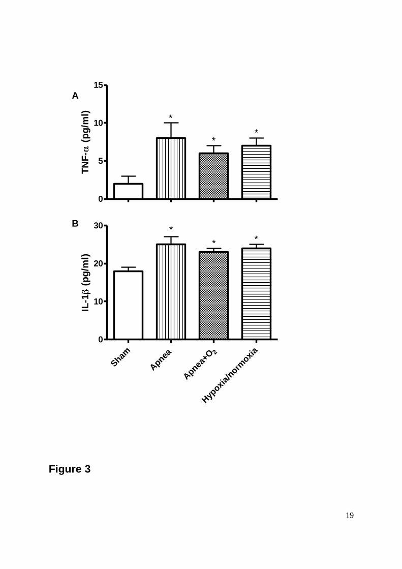
The inflammatory response was reflected by an increase in TNF-α and IL-1 . Both markers
showed a significant increase in all the experimental groups, compared to the Sham group
(Figure 3). There were no differences between groups. These data indicate that respiratory
effort and recurrent hypoxia/normoxia induce systemic inflammation.
3.2 Nuclear factor-kB binding activity
NF-kB binding activity was increased in nuclear fractions from lungs in both the Apnea and
the Hypoxia/Normoxia group compared to the Sham animals. This enhancement in NF-kB
activity was not observed in the Apnea+O2 group (Figure 4-top). Figure 4-bottom shows NF-
κB activation in nuclear fractions from diaphragm tissue, indicating that, compared to Sham,
there was a significant increase only in the Apnea group.
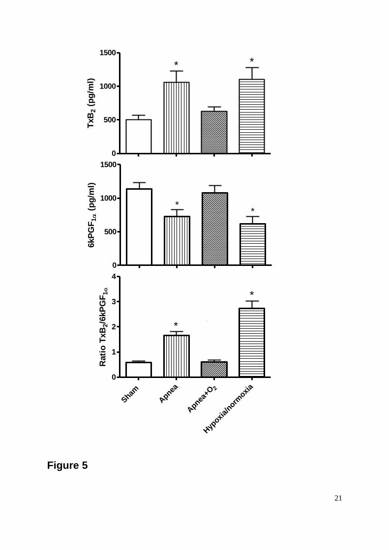
3.3 Vascular endothelial markers
As an indication of endothelial dysfunction, TxB2 and 6kPGF1α were measured in plasma
samples (Figure 5). The levels of TxB2 showed a significant increase between the Apnea
and Hypoxia/Normoxia group compared to the Sham group. However, the TxB2 level in the
Apnea+O2 group did not show a significant increase compared to the Sham group. The
levels of 6kPGF1α showed a significant decrease between the Apnea and Hypoxia/Normoxia
group compared to the Sham group. This decrease of 6kPGF1α level was not found in the
Apnea+O2 compared to the Sham group (Figure 5). An increase in TxB2 accompanied by a
decrease in the 6kPGF1α, which translates into an increase of the TxB2/6kPGF1α ratio, an
index of a vasoconstrictor effect, was observed in the Apnea and Hypoxia/Normoxia
8
experimental groups compared to Sham animals. An oxygen-rich atmosphere reversed
these changes.
4. DISCUSSION
There is a growing body of evidence in the literature to indicate that OSA is associated with
increased levels of proinflammatory cytokines, and subsequent increased endothelial
dysfunction. These could lead to cardiovascular disease (10-15). However, it is not clear to
what extent hypoxia/normoxia and respiratory effort play a role as mechanisms that trigger
the inflammatory and endothelial processes in OSA patients. Both stimuli could play an
important role in the development of cardiovascular diseases. On the one hand, it has been
reported in experimental models that hypoxia/normoxia without respiratory effort can trigger
a vascular disease (18). On the other hand, the respiratory effort caused by the upper airway
obstruction results in large negative swings in intrathoracic pressure that can also trigger
systemic inflammation. (19).
In our study, we have discriminated between the different challenges that contribute to the
pathology of OSA in order to identify the importance of these stimuli in the induction of
systemic inflammation and endothelial dysfunction. The data from our study show that 3
hours of recurrent obstructive apneas were associated with an increase in inflammatory
mediators, in line with various studies on OSA patients that have identified increased levels
of circulating TNF-α and IL-1β. This increase occurs when either the hypoxia/normoxia or
the respiratory effort appear on their own. We have also observed that apneas and
hypoxia/normoxia episodes induce endothelial dysfunction, but this was not the case with
recurrent respiratory efforts (obstructive apneas plus oxygen).
The experimental model allowed us to determine the importance of the different stimuli. It is
important to take into account that in this experimental model the group of rats with only
9
respiratory efforts maintained a normal oxygen, whereas experimental models of resistive
breathing are always linked to tissue hypoxia. On the basis of our data, it seems reasonable
to assume that the activation of inflammatory pathways is caused by the dual presence of
hypoxia/normoxia and respiratory effort, as has also been suggested by Vassilakopoulos et
al. (19).
It has been reported that subjects with OSA have a selective activation of NF-κB in
monocytes compared with control subjects. Thus, the activation of NF-κB may be a
molecular mechanism implicated in the development of OSA pathology,(7; 12; 20-22) as it is
well- known that oxidative stress is considered the initial and the major stimulus for NF-κB
activation, caused by resistive breathing and the hypoxia normoxia episodes (23,24). Our
results therefore demonstrate an activation of NF-κB triggered by recurrent obstructive
apneas, in the lung as well as in the diaphragm. Hypoxia/normoxia alone in lung tissue also
contributes to NF-κB activation; respiratory effort alone does not activate the NF-κB in any of
these tissues, however. These data suggest that oxygen desaturation plays the major role in
the activation of NF-κB in the lung, whereas in the diaphragm the activation of NF-κB
depends on both stimuli being present at the same time. Thus, the increased levels
observed in TNF-α and IL-1β as a result of respiratory effort alone would have no
relationship with NF-κB activation in the lung or diaphragm. In this case, TNF- and IL-1
plasma levels do not correlate with NF-kB activity from either the lung or the diaphragm.
These cytokines might be upregulated through a non-NF-kB mediated pathway in both
tissues, or produced by circulating monocytes, as has been reported in OSA patients (7,20-
22).
Furthermore, OSA patients present an increase in vascular endothelial dysfunction (12-14).
Dysfunctional endothelium is characterized by an imbalance in the production of vasoactive
hormones, increased adherence of inflammatory mediators to endothelial cells and
10
hypercoagulability, and it is a known risk factor for cardiovascular events (25). Another point
that should be considered is the role of the CO2. We have not measured the CO2 levels and
therefore unable to analyse its role. In fact, the results as regards CO2 are conflicting. On
the one hand, Fletcher et al. (26) have suggested that exposure to hypercapnia during
intermittent hypoxia may not be critical because the effect of intermittent hypoxia on diurnal
blood pressure in rats does not vary according to any increase in the level of carbon dioxide.
On the other hand, Tamisier et al. (27) have shown that hypercapnic hypoxia does lead to
greater sympathetic activation than hypocapnic hypoxia.
6kPGF1α has vasodilatadory effects, whereas TxB2 results in vasoconstriction; more
specifically, since they have markedly opposite effects on vascular tone, the ratio of
TxB2/6kPGF1α, is an index of vasoconstriction. In this way, it has been reported that patients
with vascular disease and OSA, show a higher TxB2/6kPGF1α ratio, compared with controls,
reflecting a predominance of vasoconstrictor activity. This could have cardiovascular
consequences and suggests that OSA pathology could be associated with an abnormal
release of prostanoids during sleep (28-29). The results obtained in this study show an
increase in the TxB2/6kPGF1α ratio induced by recurrent obstructive apneas and
hypoxia/normoxia. Therefore, these findings suggest a vasoconstrictor effect in apnea and
hypoxia/normoxia conditions, and this could correlate with our previous studies in the chronic
animal model, where the animals subjected to 5-s obstructions at a rate of 60 per hour, 6
h/day for 4 weeks showed higher TxB2/6kPGF1α ratio compared with controls (30).
In summary, in a rat model of sleep apnea we have shown that after only 3 hours any one of
the tested stimuli – repetitive apneas, oxygen desaturations alone or respiratory efforts alone
– plays a major role in the induction of an inflammatory process. However, hypoxia/normoxia
episodes proved to be the main trigger for endothelial dysfunction. Even though this
11
information is derived from an acute animal model, it suggests a possible role for oxygen
therapy in SAHS treatment, especially in patients where CPAP treatment is not possible.
ACKNOWLEDGEMENTS
This work was supported in part by Ministerio de Ciencia y Tecnología (SAF 2004-00684
and SAF2005-0110), SEPAR, FUCAP.
12
FIGURE LEGENDS
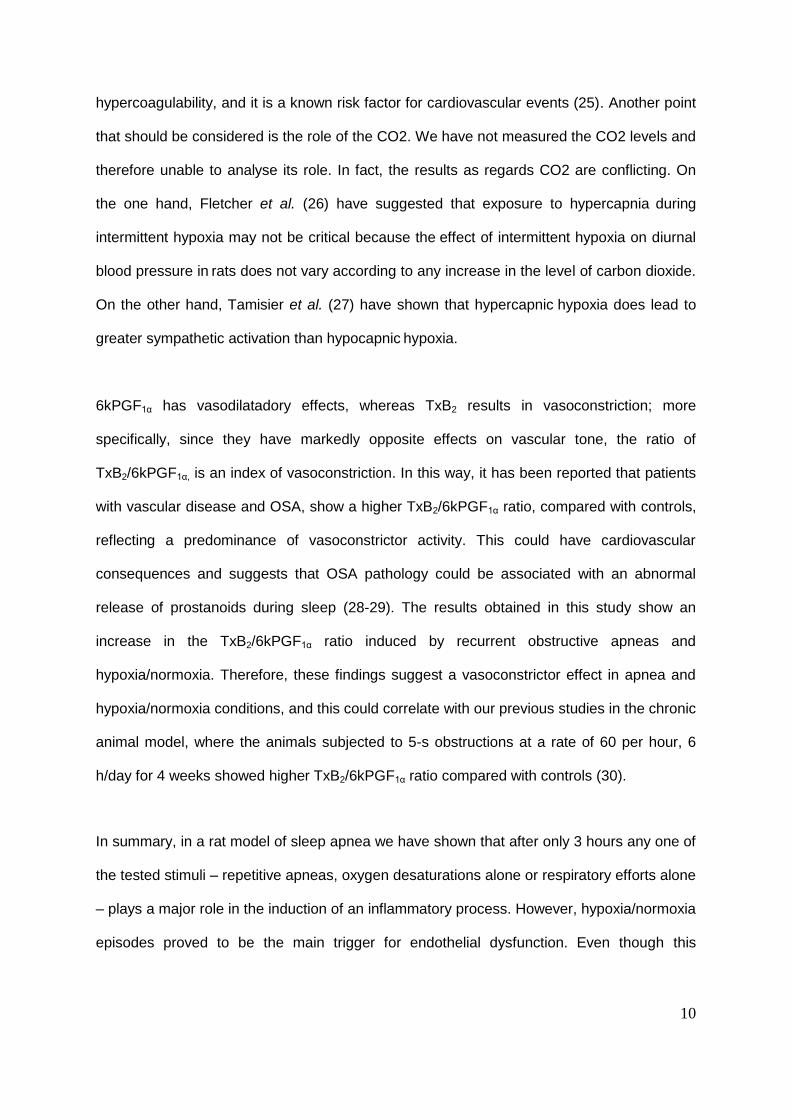
Figure 1: The system to induce realistic obstructive events in rats is depicted, with the
general set-up on the top (A). A tracheostomized rat is surrounded by a transparent box. The
tracheal tube is connected to an upper airway collapsible segment that is able to induce
obstruction (see below). The box has two large holes. Air is flushed from one of these into
the sham or apnea groups. A variable FiO2 was applied in the apnea + O2 group. In the
case of the hypoxia/normoxia group, a number of cycles were applied to form a hypoxic
phase (ambient O2 concentration 5 %) of 15 s followed by a rapid return to ambient O2 at 21
% for 45 s. Gas circulates through the box and reaches the other hole. On the bottom (B),
the collapsible segment is shown. This consists of two chambers and a membrane (M)
between them. One chamber is connected to a pressure (P) pump. If no pressure is applied,
the membrane is in its central position (solid line) and there is no obstruction of the trachea.
When an external pressure is applied, the membrane (dashed line) occludes the tracheal
port. The other chamber is connected to the tracheostomy tube. Another tube in this clamber
was open to the atmosphere and acted as pheumotachograph. The pump is controlled by a
computer-driven source that is able to increase the pressure and move the membrane that
occludes the thracheostomy. The obstruction can be performed at any frequency and for any
duration and periodicity. Tracheal pressure and breathing flow can be measured by
connecting pressure transducers to the corresponding ports in the valve
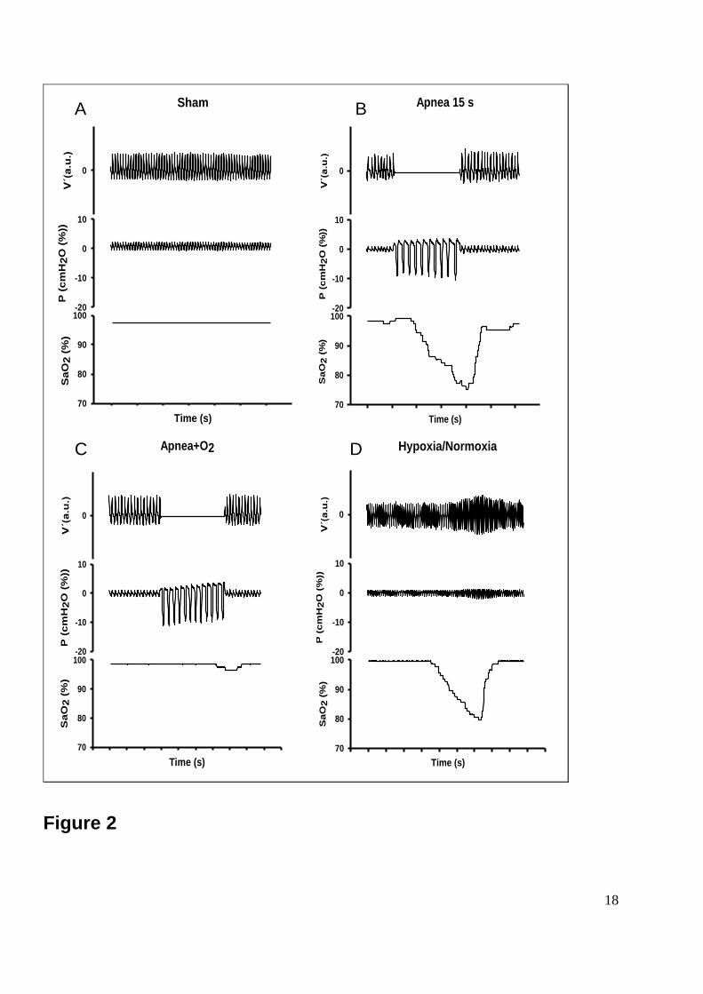
Figure 2: Examples of the signals recorded during the experiment. Figure 2A shows sham
conditions; flow, pressure and the rat’s arterial oxygen saturation (SaO2) exhibited normal
values. Figure 2B shows that during the apnea, flow was nil owing to valve closure, pressure
swings in the trachea were markedly increased as a result of breathing effort and the SaO2
exhibited a transient decrease. Figure 2C shows that during the apnea+O2, flow was nil
owing to valve closure, pressure swings in the trachea were markedly increased as a result
of breathing effort but the SaO2 showed no change. Figure 2D shows that during the
13
hypoxia/normoxia, flow was nil owing to valve closure, there were no pressure swings in the
trachea because of no breathing effort, while the SaO2 exhibited a transient decrease.
Figure 3: Figure 3A shows a significant increase in plasma levels of Interleukin-1β (IL-1β)
among the Apnea, Hypoxia/Normoxia and Apnea+O2 groups compared to the Sham group.
Figure 3B shows significant differences in plasma levels of Tumor necrosis factor-α (TNF-α)
among the Apnea, Hypoxia/Normoxia and Apnea+O2 groups compared to the Sham group.
Data are means SEM of 8 animals per group (*p<0.05 vs Sham group).
Figure 4: Figure -upper shows a significant increase in the activation of NF-κB in the Apnea
and the Hypoxia/Normoxia group, compared to the Sham group in nuclear fractions in lung
tissue. Figure 4-bottom shows a significant in the activation of NF-κB in the Apnea group
compared to the Sham group in nuclear fractions in diaphragm tissue. Data are means
SEM of 8 animals per group (*p<0.05 vs Sham group).
Figure 5: Figure 5-upper shows a significant increase in plasma levels of Thromboxane-B2
(TxB2) between the Apnea and the Hypoxia/Normoxia groups compared to the Sham group.
Figure 5-middle shows a significant decrease in plasma levels of 6-keto-Prostaglandin-F1α
(6kPGF1α) between the Apnea and the Hypoxia/Normoxia groups compared to the Sham
group. Figure 5-bottom shows the ratio of TxB2/6kPGF1α between the Apnea and the
Hypoxia/Normoxia groups compared to the Sham group. Data are means SEM of 8
animals per group (*p<0.05 vs Sham group).
14
REFERENCES
1. Durán J, Esnaola S. Rubio R, Iztueta A. Obstructive sleep apnea-hypopnea and
related clinical features in a population-based sample of subjects aged 30 to 70 year.
Am J Respir Crit Care Med 2001; 163: 685-689.
2. Caples SM, Garcia-Touchard A, Somers VK. Sleep-disordered breathing and
cardiovascular risk. Sleep 2007; 30(3):291-303.
3. Pepperell JCT, Ramdassingh-Dow S, Crosthwaite N, Mullins R, Jenkinson C,
Stradling JR, Davies RJ. Ambulatory blood pressure after therapeutic and sub-
therapeutic continuous positive airway pressure for obstructive sleep apnoea: a
randomised controlled trial. Lancet 2002;359:204–209.
4. Becker HF, Jerrentrup A, Ploch T, Psych D, Grote L, Penzel T, Sullivan CE, Peter
JH. Effect of nasal continuous positive airway pressure treatment on blood pressure
in patients with obstructive sleep apnea. Circulation 2003;107:68–73.
5. Kasasbeh E, Chi DS, Krishnaswamy G. Inflammatory aspects of sleep apnea and
their cardiovascular consequences. Southern Medical Journal 2006; 99(1):58-67.
6. Entzian P, Linnemann K, Schlaak M, Zabel P. Obstructive sleep apnea syndrome
and circadian rhythms of hormones and cytokines. American Journal of Respiratory
and Critical Care Medicine 1996; 153(3):1080-1086.
7. Ryan S, Taylor CT, McNicholas WT. Selective activation of inflammatory pathways
by intermittent hypoxia in obstructive sleep apnea syndrome. Circulation 2005;
112(17):2660-2667.
8. Von Kanel R, Loredo JS, Ancoli-Israel S, Dimsdale JE. Association between sleep
apnea severity and blood coagulability: treatment effects of nasal continuous positive
airway pressure. Sleep and Breathing 2006; 10(3):139-146.
9. Prabhakar NR, Fields RD, Baker T, Fletcher EC. Intermittent hypoxia: cell to system.
American Journal of Physiology-Lung Cellular and Molecular Physiology 2001;
281(3):L524-L528.
10. Minoguchi K, Tazaki T, Yokoe T et al. Elevated production of tumor necrosis factor-
alpha by monocytes in patients with obstructive sleep apnea syndrome. Chest 2004;
126(5):1473-1479.
15
11. Maulik N, Das DK. Redox signaling in vascular angiogenesis. Free Radical Biology
and Medicine 2002; 33(8):1047-1060.
12. Ip MSM, Tse HF, Lam B, Tsang KWT, Lam WK. Endothelial function in obstructive
sleep apnea and response to treatment. American Journal of Respiratory and Critical
Care Medicine 2004; 169(3):348-353.
13. Nieto FJ, Herrington DM, Redline S, Benjamin EJ, Robbins JA. Sleep apnea and
markers of vascular endothelial function in a large community sample of older adults.
American Journal of Respiratory and Critical Care Medicine 2004; 169(3):354-360.
14. Lavie L, Kraiczi H, Hefetz A et al. Plasma vascular endothelial growth factor in sleep
apnea syndrome - Effects of nasal continuous positive air pressure treatment.
American Journal of Respiratory and Critical Care Medicine 2002; 165(12):1624-
1628.
15. Budhiraja R, Tuder RM, Hassoun PM. Endothelial dysfunction in pulmonary
hypertension. Circulation 2004; 109(2):159-165.
16. Nacher M, Serrano-Mollar A, Farre R, Panes J, Segui J, Montserrat JM. Recurrent
obstructive apneas trigger early systemic inflammation in a rat model of sleep apnea.
Respiratory Physiology & Neurobiology 2007; 155(1):93-96.
17. Farre R, Rotger M, Montserrat JM, Calero G, Navajas D. Collapsible upper airway
segment to study the obstructive sleep apnea/hypopnea syndrome in rats.
Respiratory Physiology & Neurobiology 2003; 136(2-3):199-209.
18. Fletcher EC. Physiological and genomic consequences of intermittent hypoxia -
Invited Review: Physiological consequences of intermittent hypoxia: systemic blood
pressure. Journal of Applied Physiology 2001; 90(4):1600-1605.
19. Vassilakopoulos T, Katsaounou P, Karatza MH, Kollintza A, Zakynthinos S, Roussos
C. Strenuous resistive breathing induces plasma cytokines - Role of antioxidants and
monocytes. American Journal of Respiratory and Critical Care Medicine 2002;
166(12):1572-1578.
20. Greenberg H, Ye XB, Wilson D, Htoo AK, Hendersen T, Liu SF. Chronic intermittent
hypoxia activates nuclear factor-kappa B in cardiovascular tissues in vivo.
Biochemical and Biophysical Research Communications 2006; 343(2):591-596.
16
21. Htoo AK, Greenberg H, Tongia S et al. Activation of nuclear factor kappa B in
obstructive sleep apnea: a pathway leading to systemic inflammation. Sleep and
Breathing 2006; 10(1):43-50.
22. Ryan S, Taylor CT, McNicholas WT. Predictors of elevated nuclear factor-kappa B-
dependent genes in obstructive sleep apnea syndrome. American Journal of
Respiratory and Critical Care Medicine 2006; 174(7):824-830.
23. Williams A, Scharf SM. Obstructive sleep apnea, cardiovascular disease, and
inflammation - is NF-kappa B the key? Sleep and Breathing 2007; 11(2):69-76.
24. Lavie L. Obstructive sleep apnoea syndrome--an oxidative stress disorder. Sleep
Med Rev 2003; 7: 35–51.
25. Budhiraja R, Tuder RM, Hassoun PM. Endothelial dysfunction in pulmonary
hypertension. Circulation 2004; 109(2):159-165.
26. Fletcher EC, Bao G & Miller CC 3rd (1995). Effect of recurrent episodic hypocapnic,
eucapnic, and hypercapnic hypoxia on systemic blood pressure. J Appl Physiol 78,
1516–1521.
27. Tamisier R, Nieto L, Anand A, Cunnington D & Weiss JW (2004). Sustained muscle
sympathetic activity after hypercapnic but not hypocapnic hypoxia in normal humans.
Respir Physiol Neurobiol 141, 145–155.
28. Krieger J, Benzoni D, Sforza E, Sassard J. Urinary-Excretion of prostanoids During
Sleep in Obstructive Sleep-Apnea Patients. Clinical and Experimental Pharmacology
and Physiology 1991; 18(8):551-555.
29. Bolla M, You D, Loufrani L, et al. Cyclooxygenase involvement in thromboxane-
dependent contraction in rat mesenteric resistance arteries. Hypertension.
2004;43:1264-9.
30. Farré R, Nácher M, Serrano-Mollar A, Gáldiz JB, Alvarez FJ, Navajas D, Montserrat
JM. Rat model of chronic recurrent airway obstructions to study the sleep apnea
syndrome. Sleep 2007; 1;30 (7):930-3.
17
VALVE
TRACHEOSTOMY
Air / Air+O2
Tracheal
Pressure
Open to
room air
To
trachea
Flexible
membrane (M)
PUMP
A
B
Pressure (P)
VALVE
Pressure
P
M
RAT
Flow
Figure 1
18
P (
cm
H2O
(%
))
-20
-10
0
10
Time (s)
SaO
2 (
%)
70
80
90
100
Sham
V´(a.u
.)
0
P (
cm
H2
O (
%))
-20
-10
0
10
Time (s)
SaO
2 (
%)
70
80
90
100
Apnea 15 s
V´(a.u
.)
0
Hypoxia/Normoxia
V´(a.u
.)
0
P (
cm
H2O
(%
))
-20
-10
0
10
Time (s)
Sa
O2
(%
)
70
80
90
100
Apnea+O2
V´(a.u
.)
0
P (
cm
H2O
(%
))
-20
-10
0
10
Time (s)
SaO
2 (
%)
70
80
90
100
A B
C D
Figure 2
19
Figure 3
Sham
Apnea
2
Apnea
+O
Hyp
oxia/
normoxi
a
0
5
10
15
*
**
TN
F-
(p
g/m
l)
Sham
Apnea
2
Apnea
+O
Hyp
oxia/
normoxi
a
0
10
20
30 ***
IL-1
(p
g/m
l)
A
B

20
Figure 4
Sham
Apnea
2
Apnea
+O
Hyp
oxia/
normoxi
a
0
10000
20000
30000
40000
50000
*
NF
-B
dia
ph
rag
m
(RL
U)
Sham
Apnea
2
Apnea
+O
Hyp
oxia/
normoxi
a
0
10000
20000
30000
40000
50000
*
*
NF
-B
lu
ng
(R
LU
)
Sham
Apnea
2
Apnea
+O
Hyp
oxia/
normoxi
a
0
10000
20000
30000
40000
50000
*
NF
-B
dia
ph
rag
m
(RL
U)
Sham
Apnea
2
Apnea
+O
Hyp
oxia/
normoxi
a
0
10000
20000
30000
40000
50000
*
*
NF
-B
lu
ng
(R
LU
)
21
Sham
Apnea 2
Apnea
+O
Hyp
oxia/
normoxi
a
0
500
1000
1500
**
TxB
2 (
pg
/ml)
Sham
Apnea
2
Apnea
+O
Hyp
oxia/
normoxi
a
0
500
1000
1500
**
6kP
GF
1 (
pg
/ml)
Sham
Apnea 2
Apnea
+O
Hyp
oxia/
normoxi
a
0
1
2
3
4
*
*
Rati
o T
xB
2/6
kP
GF
1
Figure 5