Biological and chemical interest in selenium: a brief ...
35
DOI: https://doi.org/10.24820/ark.5550190.p009.784 Page 457 © ARKAT USA, Inc The Free Internet Journal for Organic Chemistry Review Archive for Organic Chemistry Arkivoc 2017, part ii, 457-491 Biological and chemical interest in selenium: a brief historical account João B. T. Rocha, a,b, * Bruna C. Piccoli, b and Cláudia S. Oliveira b a Departamento de Bioquímica e Biologia Molecular, Centro de Ciências Naturais e Exatas, Universidade Federal de Santa Maria,Santa Maria, RS, Brazil b Programa de Pós-graduação em Ciências Biológicas: Bioquímica Toxicológica, Centro de Ciências Naturais e Exatas, Universidade Federal de Santa Maria,Santa Maria, RS, Brazil E-mail: [email protected] Dedicated to Prof. Dr. Jacek Młochowski on the occasion of his 80 th anniversary Received 07-06-2016 Accepted 09-24-2016 Published on line 12-27-2016 Abstract This review presents a brief account of the discovery, importance, and use of selenium. Based in the importance of selenoproteins, their mechanism of reaction with the participation of selenium, as a selenol (-SeH) group, are indicated. Since the selenol group is the softest nucleophile center found in life, a brief discussion about the synthesis and possible antioxidant and selenoprotein mimetic effects of the organoselenium compounds that can generate the selenol group is presented. Keywords: Selenol, organoselenium compounds, selenoprotein mimetic, selenite toxicity
Transcript of Biological and chemical interest in selenium: a brief ...

DOI: https://doi.org/10.24820/ark.5550190.p009.784 Page 457 ©ARKAT USA, Inc
The Free Internet Journal
for Organic Chemistry Review
Archive for
Organic Chemistry Arkivoc 2017, part ii, 457-491
Biological and chemical interest in selenium: a brief historical account
João B. T. Rocha,a,b,* Bruna C. Piccoli,b and Cláudia S. Oliveirab
a Departamento de Bioquímica e Biologia Molecular, Centro de Ciências Naturais e Exatas,
Universidade Federal de Santa Maria,Santa Maria, RS, Brazil b Programa de Pós-graduação em Ciências Biológicas: Bioquímica Toxicológica,
Centro de Ciências Naturais e Exatas, Universidade Federal de Santa Maria,Santa Maria, RS, Brazil
E-mail: [email protected]
Dedicated to Prof. Dr. Jacek Młochowski on the occasion of his 80th anniversary
Received 07-06-2016 Accepted 09-24-2016 Published on line 12-27-2016
Abstract
This review presents a brief account of the discovery, importance, and use of selenium. Based in the
importance of selenoproteins, their mechanism of reaction with the participation of selenium, as a selenol
(-SeH) group, are indicated. Since the selenol group is the softest nucleophile center found in life, a brief
discussion about the synthesis and possible antioxidant and selenoprotein mimetic effects of the
organoselenium compounds that can generate the selenol group is presented.
Keywords: Selenol, organoselenium compounds, selenoprotein mimetic, selenite toxicity

Arkivoc 2017, ii, 457-491 Rocha, J. et al
Page 458 ©ARKAT USA, Inc
Table of Contents
1. Introduction
1.1 The selenol group: the softest of the nucleophiles in cell biology
1.2 The essentiality of selenium to life
2. Chemical Interest in the Use of Selenium in Organic Synthesis
2.1 A brief history of the synthesis of organoselenium compounds
2.2 Re-discovery of Ebselen: can the chemist imitate chemistry of life?
3. Biological Interest in Selenium: a Brief Historical Account
3.1 Early history of inorganic selenium toxicology
3.2 Oxidation of thiol groups and toxicity of inorganic selenium forms
3.3 Therapeutic use of inorganic selenium
3.4 Toxicology of naturally occurring organoselenium compounds: selenium as a causative factor of
livestock poisoning
4. The Biological and Pharmacological Interest in Synthetic Organoselenium Compounds: an Overview
4.1 The early pharmacological and nutritional use of synthetic organoselenium compounds
5. Concluding Remarks and Perspectives
References
1.Introduction
1.1 The selenol group: the softest of the nucleophiles in cell biology Since the discovery of selenium by Berzelius in 1817, the chemical and the biological interest in this element has been growing along the years.1-3 Nowadays, we have a good understanding about its chemistry but only a partial knowledge about its role in the living cell – physiology or toxicology. The most common inorganic and organic forms of selenium found in the environment and in living cells are depicted in Tables 1 and 2. Table 1. Inorganic and simple organic forms of selenium found in the environment and/or in living cells
Name Molecular structure
Molecular formulae
Distribution
Selenium, elemental [COMPLEX STRUCTURE] Se0 Soil
Hydrogen selenide Selenide
H2Se; Se2-
Mammals
Selenate
SeO42-
Soil, plants and mammals
Selenite
SeO32-
Soil, plants and mammals

Arkivoc 2017, ii, 457-491 Rocha, J. et al
Page 459 ©ARKAT USA, Inc
Table 1. Continued
Name Molecular structure
Molecular formulae
Distribution
Dimethyl diselenide
C2H6Se2 Plants
Trimethylselenonium ion
C3H9Se+ Plants
Selenophosphate
SePO3H3 Mammals
Table 2. Amino acids-, amino acid derivatives-, peptides-, and protein-containing selenium found in plants
and/or mammals
Name Molecular structure
Molecular formulae
Distribution
Selenocysteine
C3H7NO2Se Plants
Seleno-homocysteine
C4H9NO2Se Plants and mammals
Seleno-methionine
C5H11NO2Se Plants and mammals
Methyl selenocysteine
C4H9NO2Se Plants
Arkivoc 2017, ii, 457-491 Rocha, J. et al
Page 460 ©ARKAT USA, Inc
Table 2. Continued
Name Molecular structure
Molecular formulae
Distribution
γ-glutamyl-methyl-
selenocysteine
C9H16N2O5Se Plants
Se-cystathionine
C7H14N2O4Se
Plants
Se-containing protein
Plants and mammals: the
aminoacid selenomethionine
Selenoprotein
Mammals: the selenocysteinyl
residues
In mammals, the physiological chemistry of selenium is apparently played by a single functional group, i.e.,
the selenol (-SeH) group of a selenocysteinyl residues found in a few types of selenoprotein.4-14 The selenol (or
selenohydryl) is an analog of the thiol (or sulfhydryl) group of cysteine and the or hydroxyl group found in
serine (Figure 1). These functional groups have different roles in cell physiology and here we will focus on the
properties of the selenol group. The general chemical structure of these amino acids is depicted in Figure 1.

Arkivoc 2017, ii, 457-491 Rocha, J. et al
Page 461 ©ARKAT USA, Inc
Figure 1. Structure of the naturally occurring amino acids with selenol, thiol and hydroxy groups.
Serine and cysteine can be found in body fluid as free amino acids in the low millimolar range (e.g., 0.05-
0.2 mmoL/L).12 In contrast, selenocysteine is stable only inside the structure of selenoproteins and it cannot be
detected in biological fluids as a free amino acid. The selenol group has lower redox potential than the
analogous thiol group.13,14 Serine has 6 codons, cysteine has 2 codons and selenocysteine has only one codon
(UGA, which normally is the termination codon in mRNA molecules coding a protein). The incorporation of
selenocysteine in selenoproteins is co-translational and involves the participation of structural factors both
from the mRNA structure and from specific proteins that interact in the ribosomes with the UGA codon5 (Table 3).
Table 3. Some biological and chemical properties of the lateral groups of the amino acids selenocysteine,
cysteine and serine
L-Selenocysteine L-Cysteine L-Serine
Lateral group (reduced forms)
-SeH -SH -OH
Lateral group (oxidized forms)
-Se-S- (selanyl-sulfide) -SeOH (selenenic)27,28
–S-S- (dissulfide) –SO2H (sulfinic)
–SO2OH (sulfonic)15-19
pKa (lateral group) 5.213 8.313
Distribution in life Small number of selenoproteins
Widespread Widespread
Physiological role outside protein Component of GSH15,21 Neurotransmitter
(D-Serine)22-24
Redox potential -488 mV13 -233 mV13
Codon(s) UGA UGU and UGC UGA UCU, UCC, UCA, UCG, ACG, and AGC
The human genome encodes 25 selenoproteins, but only about half of them present a definite
biochemical role.5-11,14,25-30 A schematic representation of the chemical reactions catalyzed by well-studied
selenoenzymes is depicted in Table 4.

Arkivoc 2017, ii, 457-491 Rocha, J. et al
Page 462 ©ARKAT USA, Inc
Table 4. Diagrammatic representation of the reactions of the seleno groups in mammalian cells27,28. For commentary and explanation see the paragraph that follows.
No. Reagents Products Enzymes
1 GPx
2
GPx
3
GPx
4
TrxR
5
TrxR
6
MsrB
7
MsrB
8
MsrB
9
Dio
10 Dio
The reaction of peroxides with the selenol group can form either H2O or alcohols. In the living cell,
reaction 1 indicated for R1-SeH is catalyzed by isoforms of glutathione peroxidase (GPx). The GPx isoforms
reduce peroxides, using reduced glutathione (GSH) as the source of reducing equivalents. Since H2O2 and
organic peroxides (ROOH) can react with Fe2+, forming extremely reactive intermediates (for instance, •OH and
ROO•), the enzyme protects cells from the oxidative stress induced by peroxide metabolites. In reaction 2, the
Se atom of the selenenic acid is attacked by an S from the thiol group of a reduced GSH forming the –S-Se-
bond. This type of bond is central in the biological chemistry of selenium and illustrates the importance of
sulfur-selenium interaction in cell biology. In reaction 3, a second molecule of GSH attacks the sulfur in the
intermediate –S-Se-, regenerating the R1-SeH. The reactions catalyzed by the thioredoxin reductase (TrxR)
isoforms are represented in reactions 4 and 5: first the –S-Se- bond is reduced by two vicinal thiol groups, then
the selenol and thiol group of TrxR reacts with the disulfide bond (-S-S-) found in the oxidized Trx, reducing it
to the reduced Trx. In the methionine sulfoxide reductase (MsrB) (reactions 6 to 8), the selenol group of the
enzyme is supposed to interact with the sulfur atom of the sulfoxide, the selenol group is oxidized to a
selenenic acid and the methionine is regenerated (reaction 6). The thiol group of MsrB, which is in close

Arkivoc 2017, ii, 457-491 Rocha, J. et al
Page 463 ©ARKAT USA, Inc
proximity to a selenenic group in the active center of the enzyme, attacks this oxidized group, releasing H2O
and forming a –S-Se- inside the active center of MsrB (reaction 7). The oxidized enzyme (-S-Se-) can be
regenerated by reduced Trx (reaction 8). The sequence of reactions 4-9 demonstrate that the substrate of
TrxR (i.e., the Trx) is important to maintaining the MsrB in its active form and indicate an intricate relationship
among the selenoenzymes. The reactions 1-8 indicate that sulfur-selenium interactions are fundamental for
selenoprotein physiological roles. Reactions 9 and 10 represent the steps of the reaction catalyzed by
iodothyronine deiodinase (Dio) isoforms. In the first step, the selenol group of Dio attacks the C-I bond,
forming the intermediate –Se-I. In the second step, an endogenous thiol (still not identified) regenerates the
active Dio.
In this mini review, we will discuss basic aspects of the role of selenoproteins in cell physiology but will not
give details about the discoveries that led to the establishment of selenocysteine as the twenty first amino
acid. For a comprehensive view of this exciting field of selenium biology, we refer the reader to several
important reviews and references (for instance, refs 31-33).
1.2 The essentiality of selenium to life
From the chemical point of view, life is based on carbon and on other relatively light elements bound
covalently to carbon or existing as free ions in the aqueous environment of living cells (Figure 2). The
exceptions are lithium, fluorine and beryllium that probably because of their size and reactivity could not be
selected to exist in the living cell as ions. Iodine is the heaviest element found in organic molecules in
mammals, but it has a limited role as a component of the thyroid hormones 3,3',5,5'-tetraiodothyronine
(thyroxine or T4) and by 3,3',5-triiodothyronine (T3). Selenium is the second heaviest element found
covalently bound to carbon, but it has a much broader physiological role than iodine. Of note, the activation of
T4 to T3, by 5'-deiodination, is catalyzed by the selenoenzyme iodothyronine deiodinase (Dio). The catalysis
involves a direct interaction of selenium (as selenol group) with iodine27,28,35,36 (Table 4).
Figure 2. Essential elements in mammalian cell physiology.
The role played by selenium in life is much narrower than sulfur, which is found adjacent to selenium in
the periodic table. In contrast to selenium, which is found only in selenocysteinyl residues2,4-10,13,14,25-28,37,38,
sulfur is found in thousands of proteins as the thiol group of cysteine or as thioether group in methionine,

Arkivoc 2017, ii, 457-491 Rocha, J. et al
Page 464 ©ARKAT USA, Inc
either as part of methionine initiator tRNA or as internal residues of innumerable proteins.39-44 Thiol-
containing proteins can play a much more diverse catalytic role than selenol-containing proteins.40-43 The
selenoenzymes are oxidoreductases that catalyze a narrow range of reactions.5,7-9,27-30,38 As cited above, the
human genome encodes only 25 selenoproteins and the maximum number of selenoproteins found in
vertebrates is about 40.45 The limited participation of selenium in the physiology of living cells indicates that
the selenol group was selected by evolutional pressures to perform only a restricted set of chemical reactions.
The restricted role of selenium in living cells and the negligible occurrence of the selenol group out of
selenoproteins is certainly determined by the extreme reactivity of the selenol groups under physiological
conditions.46,47 The unstable nature of the selenol group found in low molecular mass molecules has been
recognized for a long time in pure chemical systems.48 In fact, the stabilisation of the selenol group can only be
accomplished in the complex microenvironment of high molecular mass selenoproteins.28 In mammals, the
precise fine tuned exploitation of selenium in cell biochemistry is further exemplified by the hierarchy of
selenium utilization by different tissues under dietary selenium shortage.49-51 For instance, when compared
with liver and other tissues, the brain is spared from selenium deficiency, indicating that evolutionary
pressures have also modulated the preferential bioavailability of selenium to specific organs.51-53 Accordingly,
the importance of selenoproteins for proper brain functioning has become much more apparent in the last
decades.52-61
Two intriguing facts about the selection of selenium as a critical component of living cells are: 1) selenium
is not essential to all forms of life; for instance, higher plants and fungi do not have selenoproteins;5,6,45 and
2) the thiol group found in the cysteinyl residues of thousands proteins could theoretically imitate the
physiological chemistry of the selenol group.38,41-43 Consequently, one important question about the
physiological chemistry of selenium is: why have some types of living cells been selected to have selenium as a
part of their biochemical machinery?
Selenium (-SeH/-Se-) can be a better nucleophile than sulfur (-SH/-S-) in living cells and, consequently,
catalyze more efficiently the reduction of biologically relevant chemical species,38,40,62 but this does not explain
its absence in a large number of living organisms. Since the molecular logic of the living state assumes that
living cells obey the principle of maximal economy,63 we can presume that the intricate complexity associated
with the incorporation of selenium into the backbone of selenoproteins brought some adaptive biochemical
properties that cannot be understood in simple redox chemical terms.64 The apparent conundrum about why
selenium is essential only to some but not all living cells will require much more refined studies of the
biochemical properties of the thiol and selenol groups of proteins from archea to mammals.
The objective of this minireview, compiled to mark the 80th anniversary of Dr. Jacek Młochowski, who was
a researcher dedicated to the synthesis of organoselenium compounds and one of the pioneers of research
into their use as potential pharmacological agents,65-80 is not to offer the reader a comprehensive account of
the importance of selenium in organic synthesis of pharmacologically active compounds, because this can be
found in several comprehensive reviews.70,79-110 Here we give a brief historical account of the biological
interest in selenium and its use in the synthesis of organoselenium compounds as biologically active
molecules. We will discuss some critical points in the field that we realize are delaying the development of
therapeutically effective agents. In terms of cell biology, we will present, in a brief way, the history of the
biological interest in the element selenium, particularly how its incorporation into organic moieties has been
important to life from a toxicological, pharmacological and physiological point of view. Although thousands
(perhaps millions) of organoselenium compounds have already been synthesized since 1836;70 only a small
portion of them have been tested in biological systems. In our opinion, the absence of high-throughput

Arkivoc 2017, ii, 457-491 Rocha, J. et al
Page 465 ©ARKAT USA, Inc
methods to investigate the toxicity of the countless number of newly synthesized organoselenium compounds
on rational grounds is the main bottleneck to the development of organoselenium therapeutic agents. The
critical and elegant review: “Organochalcogen as peroxidase mimetics as potential drugs: a long story of a
promise still unfulfilled” by Orian and Toppo111 has recently addressed the necessity of new approaches to
study the biology of organochalgogens. The development of in silico models to predict both thermodynamic
parameters of intermediates stability and the interaction of organoselenium compounds with specific
molecular targets are highly needed. Though the papers dealing with in silico simulation of organoselenium
compounds chemical and biochemical behavior have been increasing,111-121 we are far from accomplishing the
task.
2. Chemical Interest in the Use of Selenium in Organic Synthesis
2.1 A brief history of the synthesis of organoselenium compounds
Interest in the synthesis of organoselenium compounds goes back to the early 19th century, when the first
organoselenium compound was synthesized. According to Fredga,122 organoselenium chemistry started on
January 23, 1847, precisely, when Wöhler wrote to Berzelius: “Today a small grandchild of yours has come in
the world, a child of selenium, the selenomercaptan”. The synthesized molecule was ethyl selenol (Table 5),
prepared from calcium ethyl sulfate and potassium hydroselenide by Siemens. Siemens reacted the product of
the reaction with Hg(II) to form CH3CH2SeHgSeCH2CH3 and proved that it was a “selenomercaptan”.123
However, according to Prof. Młochowski and collaborators, the synthesis of diethylselenide has priority, being
reported in 1836 by Löwing.70
In Table 5 are listed the first selenium compounds synthesized by man and the first compound detected in
mammalian metabolism by its smell. The ethyl-containing molecules were synthesized by man,70 whereas the
dimethylselenide was discovered by the sense of smell of the early toxicologists, who evaluated the toxicity of
inorganic selenium salts in mammals (see below in Section 3.1).
Table 5. Early historical landmarks in selenium chemistry
Name Molecular structure
Molecular formula
Discovery/ synthesis
Diethylselenide
C4H10Se 1836
Ethylselenol
C2H6Se 1847
Diethyldiselenide mercury
C4H10HgSe2 1847
Dimethylselenide
C2H6Se 1894

Arkivoc 2017, ii, 457-491 Rocha, J. et al
Page 466 ©ARKAT USA, Inc
The book “The organic compounds of tellurium and selenium belonging to the alcohol series” by Dean,124
who had worked in the laboratory of Wöhler, emphasizes the similarity between S, Se, and Te. By
paraphrasing the author: “In regard to the history of these compounds it may be observed, that the
extraordinary resemblance between sulfur, selenium, and tellurium first led to the idea of substituting
selenium or tellurium for sulfur in various organic compounds…”, we can realize that at a very early stage of
organic synthesis development, selenium was being investigated as surrogate for sulfur. As briefly commented
in the first section of this mini-review, biological evolution has apparently endeavored to substitute sulfur by
selenium at an early stage of the evolution of a living cell. With the introduction of selenium in the
biochemical machinery, the living cell gained efficiency in metabolizing important redox reactions,5,17,38,40
whereas the synthetic chemist gained versatility in their reactions.65,70,81-103
One important limitation in the early years of organoselenium compound synthesis was the availability
of selenium.124 Regardless of the unavailability of the element in the 19th century, the field progressed
considerably. In his classical review from 1941, Edgar P. Painter stated that hundreds of organoselenium
compounds had been described and presented a list of the available synthetic strategies to prepare
diselenides, selenols, selenenic, and selenonic acids, among others.48 Recent literature considers that modern
era of organoselenium synthesis began in the seventies of the last century.1,70 The organic chemical interest in
selenium is associated with its ability to confer to the organic moiety enormous chemical versatility.81-99 The
importance of this element in the field of organic synthesis can be verified by the vast literature on
organoselenium chemistry. From Painter’s time to the present, we can roughly estimate the number of new
organoselenium compounds synthesized every year and now thousands to millions of compounds can be
found in the literature.
2.2 Re-discovery of Ebselen: can the chemist imitate chemistry of life?
The general chemical interest in the synthesis of new organoselenium compounds boomed after the clinical
use of 2-phenyl-1,2-benzisoselenazol-3(2H)-one or Ebselen.125-127 Ebselen was originally synthesized in 1924 by
Lesser and Weiss128 and it was re-discovered as an antioxidant about 40-50 years ago (for an interesting
testimony about the development of Ebselen as a therapeutic agent see refs 129,130) (Table 6). Ebselen is the
most popular organoselenium compound and it was subjected to clinical trials about 20 years ago with
borderline efficacy.125-127
Table 6. The Structure of some organoselenium compounds with interesting pharmacological properties
Name Molecular structure
Molecular formulae
Tetrahydroselenophene
C4H8Se
Ebselen (2-Phenyl-1,2-
benzoselenazol-3(2H)-one)
C13H9NOSe

Arkivoc 2017, ii, 457-491 Rocha, J. et al
Page 467 ©ARKAT USA, Inc
Table 6. Continued
Name Molecular structure
Molecular formulae
2-(3-pyridyl)-benzisoselenazol-3(2H)-
one
C12H8N2OSe
2-phenylbenzisoselen-azol-3(2H)-one 1-oxide
C13H9NO2Se
Diphenyl diselenide
C12H10Se2
bis-(p-chlorophenyl) diselenide
C12H8Cl2Se2
bis-(p-methoxyphenyl) diselenide
C14H14O2Se2
1,2-bis[2-(pyrrolidin-1-ylmethyl)phenyl]diselane
C22H28N2Se2
The use of Ebselen in clinical trials to treat brain pathologies associated with oxidative stress changed the
view of selenium compounds. As stated in their vivid report about the steps that led to the development of
Ebselen as therapeutic agent, the first goal was to use Ebselen as a source of selenium for the synthesis of
Arkivoc 2017, ii, 457-491 Rocha, J. et al
Page 468 ©ARKAT USA, Inc
selenoproteins. The discovery of selenium in the structure of gluthathione peroxidase (GPx) and its role in
peroxide degradation131,132 had introduced the idea that selenium supplementation could be an important
strategy to improve the activity of GPx. The rationale that Ebselen could be a better source of selenium than
inorganic selenium or naturally occurring organic selenium forms can be easily understood by the fear of
selenium (selenophobia) that was prevalent in the 20th century133 (the history of selenium toxicity to mammals
will be presented in the next section of this review). Contrary to the expectation of the researchers,129 the
selenium in Ebselen was not metabolized to the inorganic selenium pool, which would be required for
selenium incorporation into selenocysteine (i.e., into selenoproteins;5 Figure 1). But Ebselen exhibited several
interesting antioxidant properties, including the ability to mimic the activity of GPx (glutathione peroxidase- or
thiol peroxidase-like activity).134-136
After that, the GPx- or thiol peroxidase-like activities of organoselenium compounds have been alleged to
be the most important mechanism involved in the pharmacological effects of various organoselenium
compounds (Table 4, reactions 1 and 2), regardless of the low catalytic efficiency of simple organoselenium
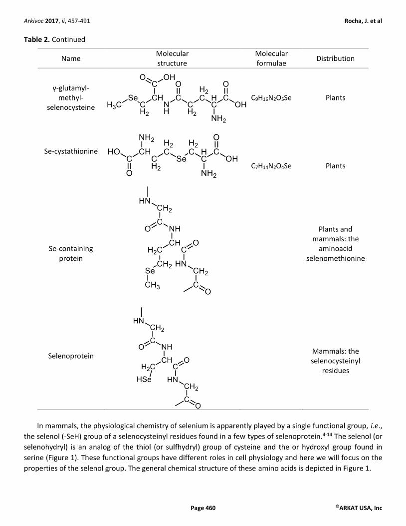
compounds in accelerating the decomposition of peroxides.135-137 The basic mechanism of peroxide
decomposition by organoselenium compounds (e.g. Ebselen and diselenides, Table 6), which forms selenol
intermediates,101,104 is supposed to be similar to that of the native GPx isoforms (Scheme 1). Other selenium
compounds that are not metabolized to selenol intermediates (for instance, tetrahydroselenophenes, Table 6)
can also mimic the activity of GPx by forming a selenoxide intermediate after decomposing peroxides.81
Scheme 1. Reduction of Ebselen and diphenyl diselenide to selenol intermediates. Reaction 1 is catalyzed by
thioredoxin reductase (TrxR) and uses NAPDH as an electron donor. Reaction 2 uses glutathione (GSH) as an
electron donor.
Ebselen and diphenyl diselenides can be reduced to selenol intermediates both after reacting directly with
thiol-containing molecules or after being reduced by TrxR138-140 (Scheme 1). Literature studies have also
indicated that organoselenium compounds can interact with specific thiol-containing proteins.141,142 Ebselen
and diphenyl diselenide have been reported to inhibit the activity of important metabolic enzymes in vitro, for
instance, Na+,K+-ATPase,101,104,108 but there is no evidence that they can target it after in vivo administration.
The inhibition of such a crucial enzyme can have unpredictable toxicological effects, particularly to the central
nervous system. The specific interaction of Ebselen with inositol monophosphatase (IMPase) has been

Arkivoc 2017, ii, 457-491 Rocha, J. et al
Page 469 ©ARKAT USA, Inc
suggested to be involved in the lithium mimetic activity of Ebselen143 and can be involved in the antidepressive
activity of Ebselen in experimental models.144 In contrast, diphenyl diselenide, which exhibited similar
antidepressive activity in rodent models, is not an inhibitor of IMPase.143 The oxidation of thiol-containing
proteins by organoselenium compounds can be involved both in their pharmacological and toxicological
effects. The chemical mechanism of oxidation of thiol-containing proteins by organochalcogens is similar to
that presented in Table 4 (reaction 1), where the thiol group of the protein substitute GSH as reducing agent.
Theoretically, the oxidation of a therapeutically important protein by an organochalcogen can both cause a
specific physiological effect and can generate the selenol intermediates. The selenol intermediate can then
decompose peroxides and exhibit antioxidant properties.
Of particular importance for the pharmacological properties of Ebselen and diphenyl diselenide, the
oxidation of antioxidant response elements (AREs), particularly the Nrf-2145-149 (a transcript factor regulated by
Keap 1) by organoselenium compounds have been suggested to participate in the antioxidant effects of
Ebselen and diphenyl diselenide.150-157 For instance, the oxidation of Keap-1 by organoselenium compounds
can activate the Nrf-2-transcript factor that stimulates the expression of antioxidant proteins. In summary, the
pharmacological or toxicological effects of organoselenium compounds include the oxidation of thiol groups
and from the chemical point of view, the basic mechanisms of reaction are similar to those presented by the
selenoproteins.
3. Biological Interest in Selenium: a Brief Historical Account
3.1 Early history of inorganic selenium toxicology
From a historical point of view, the interest in the chemistry and toxicology of inorganic selenium (salts,
oxides, acids) led to the first and vague connection between selenium as a toxic agent in mammals and the
beginning of organoselenium compound synthesis.158-162 Some years after the isolation of selenium by
Berzelius in 1817, the scientific community started to investigate both the synthesis of organoselenium
compounds and the toxicity of inorganic selenium forms to living organisms.158-162
Although citations found in the English literature have some incongruent data about the studies published
in German3,159,161,165 the investigations performed between 1820 to 1950 (both with mammals and bacteria)
were instrumental in demonstrating that living cells can reduce selenate (Se(VI)) and selenite (Se(IV)) to
elemental selenium and some volatile form of organic selenium.159-165 According to Charles Jones,159 the first
author to study the toxicity of selenium salts in animals was C. G. Gmelin. However, Moxon and Morris,3 in
their classical review on “selenium poisoning”, argued that they could not find experiments related to the
toxicity of selenium in Gmelin’s book. Gmelin studied the toxicity of tellurium salts160 and the source of
confusion can be derived from the similar chemical and biochemical behavior of selenite and tellurite in
bacteria and mammals, i.e., both salts can be reduced to elemental forms.156,159,164,165 Jones stated that Hasen
(1853) described selenium granules deposited in the organs of animals injected with selenite and identified a
garlic-like odor in the breath of poisoned animals; but apparently Hansen injected tellurite and not selenite.
The chemical nature of the volatile compound(s) with garlic odor was supposed to be either dimethyl selenide
or diethyl selenide159 (in fact, dimethyl telluride or diethyl telluride). In 1909, Jones demonstrated the
deposition of a brick-red precipitate in the organs of animals intoxicated with selenite, but he concluded that
the formation of elemental selenium was not the cause of the death of intoxicated animals,159 because he
could observe comparable selenium granules in animals treated with high but non-lethal doses of selenite.
The participation of hydrogen selenide (i.e., H2Se) as a toxic metabolite of Se(IV) or Se(VI) was not considered

Arkivoc 2017, ii, 457-491 Rocha, J. et al
Page 470 ©ARKAT USA, Inc
by Jones, though hydrogen selenide was known to cause some signs of intoxication similar to those caused by
lethal intoxication with selenite.158,159
Jones also observed a garlic-like odor in the breath of poisoned mammals. The emanation of garlic-like
odor could also be detected in the organs isolated from animals poisoned with Na2SeO3. The proposal that the
garlic-like odor present in the breath of selenium-poisoned animals was produced by dimethyl selenide was
made by Hofmeister.159,165 He injected sodium tellurite and detected the odor similar to dimethyl sulfide and
postulated that exhaled compound after administration of tellurite was dimethyl telluride. Based on the
chemical similarity between selenium and tellurium, Hofmeister speculated that the garlic-like odor detected
after selenite intoxication was produced by dimethyl selenide.165 However, a conclusive demonstration that
inorganic selenium exposure was associated with dimethyl selenide exhalation was made by McConnell and
collaborator.165 The research work concerning the toxicity of selenium and tellurium salts carried out in the
second half of the 19th century were crucial to establish the first metabolic pathway of selenium in mammals
(Scheme 2). The elucidation of the chemical nature of the first metabolically produced organoselenium
molecule was confirmed only at the end of the second half of the 20th century.164,165 The complete reductive
pathway of Se(IV) and Se(VI) to selenide (Se2-) was studied in different laboratories.159,161,164-168 Of particular
importance to the physiological chemistry of selenium, selenide (Se2-) is the unique chemical form of selenium
metabolized to selenophosphate (HPSeO32- or selenophosphoric acid, H3PSeO3), which can then be incorpor-
ated into the backbone of selenocysteine.5
Se(IV)
selenite
Se(VI)
selenate
Se(0)
elementalselenium
H2Se
hydrogenselenide
HSe
Se2
selenide
(CH3)2Se
dimethyl selenide
Garlic-like odor
Scheme 2. Formation of Se(0) and volatile organic selenium after reduction of selenate or selenite in living
cells.
The formation of elemental selenium was evidenced by the formation of brick-red selenium precipitate in
the organs of animals intoxicated with selenium salts. The formation of organic selenium (dimethylselenide)
was hypothesized by analogy with tellurite metabolism and by sensorial identification of a garlic-like odor in
the breath of living intoxicated animals or by the release of garlic odor from their isolated organs. The
formation of dimethylselenide was dissociated from the formation of Se(0) by Jones.159 He demonstrated that
heating the organs up to 60 oC suppressed the formation of the garlic odor [(CH3)2Se], but did not stop the
formation of elemental Se. Jones concluded that carbohydrates, for instance, glucose reduced the selenite to
elemental selenium. However, nowadays we know that the thiol group from glutathione and proteins are
responsible by the chemical reduction of selenite to elemental selenium.
Arkivoc 2017, ii, 457-491 Rocha, J. et al
Page 471 ©ARKAT USA, Inc
3.2 Oxidation of thiol groups and toxicity of inorganic selenium forms
Our understanding of the toxicology of inorganic selenium is still elusive, however the oxidation of thiol-
containing molecules by cationic selenium ions is involved in the process of toxicity as macroscopically
demonstrated by the early toxicologists.158,159 The reduction of Se(IV) and Se(VI) to elemental selenium by
isolated organs from animals poisoned with selenite, forming brick-red precipitates, was possible even at high
temperatures.159 This observation strongly indicates that selenate and selenite can be chemically reduced to
elemental selenium (i.e., without the participation of heat-sensitive enzymes). Accordingly, selenite can
oxidize GSH169 and the depletion of this important antioxidant molecule may cause toxic consequences to
cells.170,171 The oxidation of GSH by Se(IV) and Se(VI) produces a disulfide bond, for instance oxidized
glutathione (GSSG), and an intermediate containing -S-Se-S- interactions.172 Furthermore, during the oxidation
of thiol groups by inorganic salts of selenium, reactive oxygen species can be formed and an excessive
formation of these species can be deleterious to cells.173-175 Selenite and selenate can also oxidize the thiol
groups of proteins, disrupting various cellular processes.173,174,176-180
3.3 The therapeutic use of inorganic selenium
After the demonstration of selenite and selenate toxicological properties, elemental selenium was used in the
treatment of inoperable carcinoma.181-183 According to Watson-Williams,181 the rationale for administering
selenium suspensions to human was based on the previous successful use of the element in animal models of
cancer. Additionally, Watson-Williams181 stated that “injections of colloidal selenium had been used in human
patients with inoperable carcinoma” in France. Selenium either alone or in combination with other
therapeutic agents exhibited beneficial effects in a great proportion of patients with inoperable tumour.181-183
Nonetheless, the use of colloidal selenium was abandoned, possibly because its efficacy was not confirmed
and/or in view of the increasing awareness of the toxicology of selenium at those times (see for instance refs 3
and 133). The fear of the element, which had escalated around the '30s to '60s, was so prevalent that Frost
and Olso133 coined the term "selenophobia" to express the negative view of selenium.
The therapeutic use of elemental selenium in patients indicated that Se(0) was well tolerated by humans
after systemic administration. Contrasting with the results obtained with Se(IV) and Se(VI), which indicated
extreme toxicity in mammals,159 elemental selenium seemed to be relatively inert in humans. More recently,
the use of synthetic and naturally occurring organoselenium compounds has been reconsidered as nutritional
anti-cancer agents.184-189 However, the main results of an extensive study, in which selenomethionine and
vitamin E were used as nutritional supplements, did not indicate a protective role for selenium against
cancer.190,191 In contrast, some meta-analysis studies have supported a protective role of selenium in some
types of cancer.192,193 The discrepancies between studies can be related to important factors, such as the basal
level of selenium in the sample populations, genetic factors, the chemical form of selenium used in the studies
with selenium supplementation, amongst others.191,194-196 Literature data have indicated that intake of
selenium beyond the required nutritional level (here interpreted as that needed to support the ideal
selenoprotein synthesis) can increase the incidence of some types of cancer and other chronic degenerative
diseases, such as type 2 diabetes and amyotrophic lateral sclerosis (ALS).197-200 However, there are also
indications that high levels of toenail Se is associated with a lowered risk of developing type 2 diabetes.201,202

Arkivoc 2017, ii, 457-491 Rocha, J. et al
Page 472 ©ARKAT USA, Inc
3.4 The toxicology of naturally occurring organoselenium compounds: selenium as a causative factor of
livestock poisoning
At the same time that elemental selenium was being used clinically to relieve the malady associated with
different types of inoperable carcinomas,181-183 researchers in the USA were revealing the central role of
“naturally-occurring selenium” as the causative factor of alkali disease in livestock.3,48,203-206 The studies of
Franke and collaborators demonstrated that grains grown in regions previously associated with alkali disease
had an elevated level of selenium and showed that the extraction of selenium from the plants abolished their
toxicity.203-206 Franke and collaborators were also the pioneers in demonstrating that selenite could reproduce
the majority of toxic symptoms caused by the ingestion of plants containing high levels of selenium.
Additionally, Franke and Painter193 207-211 found that the removal of selenium from toxic protein hydrolysates
abolished the toxicity of wheat protein. Some important chemical forms of naturally occurring Se are
presented in Tables 1 and 2. In the case of the Franke and Painter studies,209-210 the main toxic form can
retrospectively be ascribed to an organic form of selenium, for instance, selenomethionine and
methylselenocysteine.212,213 Selenium accumulator plants can synthesize different organoselenium
compounds, which can have toxicological properties, but little is known about the metabolism and toxicity of
different combination of organoselenium compounds found in grain and vegetables.
The toxicity of naturally occurring selenium in livestock (alkali disease) has been associated with changes
in the structure of hair, loss of long hair, and softening of hooves.3,48,203-206 Chronic ingestion of a high level of
selenium by humans can also cause similar modification in hair (including hair loss or alopecia) and in toenail
structure.214-218 Here we should point out that keratin proteins, which are the key structural components of
hair, hooves and nails, have a high cysteine content.219 The disruption of keratin-containing hair, hooves and
nails by selenium intoxication demonstrate a tropism of selenium metabolites in cells that synthesize proteins
rich in cysteine. The incorporation of selenium in secretions from keranocytes can be attested by the high
content of the element in hair and nail.220,221
Although the toxicity of naturally occurring organoselenium compounds is not coincident with that of the
inorganic salts, the oxidation of low and high molecular mass thiol molecules can also be involved in their
toxicity. However, our knowledge about the selenium metabolites involved in the oxidative toxicity of
different naturally occurring selenium compounds is still elusive. As observed with inorganic selenium forms
and synthetic organoselenium compounds,222-226 the interaction of naturally occurring selenium compounds
with endogenous thiol can generate reactive oxygen species (ROS) by mechanisms not well-defined at the
molecular level.174-176
4. The Biological and Pharmacological Interest in Synthetic Organoselenium Compounds: an Overview
4.1 The early pharmacological and nutritional use of synthetic organoselenium compounds
Interest in the potential biological, pharmacological and, optimistically, therapeutic exploitation of synthetic
organoselenium compounds started several decades ago, when organic forms of selenium were tested as
potential anticancer agents. In 1941, diselenane-diacetic acid (HO2CCH2-Se-Se-CH2CO2H), diselenane-n-
dibutyric acid [HO2C(CH2)3-Se-Se-(CH2)3CO2H], and mercurybis(4-selenobutyrate) [HO2C(CH2)3-Se-Hg-Se-
(CH2)3CO2H] were tested in tumor-bearing mice (Table 7).227 The authors stated that selenium compounds
appeared interesting to be tested in view of their relationship with sulfur metabolism; however, the authors
assessed the distribution of selenium, but did not present data about the potential anti-tumor effect of the

Arkivoc 2017, ii, 457-491 Rocha, J. et al
Page 473 ©ARKAT USA, Inc
different compounds tested. In more recent years organoselenocyanate compounds have been extensively
studied as anticancer agents.228-231 Though this class of compound has been shown to exhibit beneficial effects
in vitro and in vivo against cancer (Table 7), they were not put through clinical trials.
Table 7. Organoselenium compounds with anti-tumorogenic potential
Name Molecular structure
Molecular formula
Diselenanediacetic acid
C4H6O4Se2
Diselenanedi(n-butyric acid)
C8H14O4Se2
mercurybis-(4-selenobutyric acid)
C8H14HgO4Se2
1,4-phenylenebis-(methylselenocyanate)
C10H8N2Se2
In addition to the potential anticancer properties, organoselenium compounds have been shown to
exhibit interesting pharmacological properties in different experimental models of human pathologies. Of
note, they can have antioxidant, neuroprotective, anti-inflammatory, antidepressive, imunomudulatory,
antimicrobial activity against pathogenic bacteria, fungi and viruses, among others14,64-91,232-239.
The effectiveness of some organoselenium compounds as beneficial agents in quite different acute or
chronic models of human degenerative disease can be explained by their anti-inflammatory and antioxidant
properties. The molecular processes involved in a variety of human pathologies have in common the
production of inflammatory mediators (for instance, diabetes, Alzheimer disease, Parkinson disease,
ischaemia, atherosclerosis).239-249 The inflammatory processes stimulate the over production of reactive
species (e.g. reactive oxygen (ROS) and nitrogen species(RNS)) and these will further stimulate the release of
pro-inflamatory molecules (Scheme 3).

Arkivoc 2017, ii, 457-491 Rocha, J. et al
Page 474 ©ARKAT USA, Inc
Scheme 3. Interplay between inflammation, oxidative stress and human diseases.
Although Scheme 3 is an oversimplification of a rather complicated processes, the modulation of peroxide
tonus by organoselenium compounds can decrease the release of pro-inflammatory mediators indirectly. A
direct anti-inflammatory role of some organochalcogens is also possible plausible, but this has not yet been
investigated in detail. The fact that organoselenium compounds exhibit protective effects in a variety of
models of diseases with different etiologies argues in favor of quite general mechanism of action. The GPx-like
or thiol peroxidase-like activity can explain the general beneficial effects of organoselenium compounds. For
the case of Ebselen and diphenyl diselenide, the GPx-like activity is thought to be mediated by the formation
of selenol intermediates. In view of the simple structure of the organoselenium compounds cited above we
can predict that the selenol intermediates formed will not present selectivity. The selenol intermediates will
be able to mimic the physiological chemistry of selenoproteins in general, and not only of GPx isoforms.
However, we have only a vague idea about the efficacy of organochalcogens to mimic specific selenoproteins.
Another important mechanism that seems to be involved in the beneficial effect of organochalcogens is their
capacity to function as weak electrophiles. Various laboratories have provided data supporting these
assumptions.150-157
The mechanism of action of the most studied organoselenium compounds is thought to involve
antioxidant and anti-inflammatory properties and the ability of activate the expression of antioxidant
genes.100-110,150-157 Here we discuss one important mechanism that has been neglected in the recent literature
about the beneficial effects of some synthetic organoselenium compounds. The studies in question were
published about 60 years ago and just one year after the appearance of the most well-known paper about the
biological importance of selenium.250,251 The proximity of the two publications of Schwartz and Foltz obscured
the importance of the second study. In this classical study from 1958, Schwarz and Foltz clearly demonstrated
that different mono- and diselenides prevented dietary liver necrosis in rats fed with a vitamin E deficient
diet.250 The new data extended the results obtained by the same authors one year before, in which they
demonstrated that factor 3 (a nutritional factor isolated from the yeast or the kidneys of pig) had selenium as
the active principle.251-253 Factor 3, which was identified as an organoselenium compound, and selenite
prevented liver degeneration induced by a diet deficient in vitamin E in rodents.250-253 These studies provided

Arkivoc 2017, ii, 457-491 Rocha, J. et al
Page 475 ©ARKAT USA, Inc
the first experimental demonstration that both inorganic and organic selenium compounds had
hepatoprotective effects in rodents. Taken together with the study of Adams and collaborators,254 which
showed the release of selenium from the organic moiety of diphenyl diselenide, the studies of Schwartz and
collaborators250,255 indicated that simple organoselenium compounds such as aliphatic diselenides, diphenyl
diselenides and diphenyl selenides can furnish selenium to the inorganic pool of selenium. The inorganic
selenide released from aromatic and aliphatic selenides can be used in the synthesis of selenoproteins. Thus,
in part, the beneficial effects of organoselenides can be related to modulation of selenoprotein synthesis.
The clinical trials with Ebselen in the '90s have indeed powered the field of synthetic organoselenium
compounds, despite the fact that the Japanese authorities did not approve its use in the treatment of brain
ischaemia.125-127,129,130 Ebselen is now included in the chemical library of the National Institutes of Health
Clinical Collection as a safe drug,143 but without a target pathology to treat. The interest in finding a target for
Ebselen has increased considerably in the last ten years and it is now being studied as a potential lithium
mimetic in humans,143,256-259 experimentally as an antimicrobial agent,260-264 among others.265 The clinical
approval of Ebselen has also stimulated the synthesis of Ebselen derivatives.70,266-273
A brief historical account of the biological timeline of selenium is presented below (Table 8).
Table 8. Timeline of selenium in science
Year Important fact Cited by
reference
1295 Perhaps the first description of chronic selenosis in animals, but
not proved. 274
1560 Perhaps the first description of chronic selenosis in humans, but
not proved. 274
1817 Discovery of selenium by Berzelius. 1
1836 Synthesis of diethylselenide reported by Löwing. 70 1847 Wöhler and Siemens synthetize selenomercaptans. 122, 123
1860 Madison describes the poisoning of cavalry horse by plants,
which later were shown to have high selenium content. 3
1894 Hofmeister identifies dimethylselenide as the cause of garlic-like
odor in the breath. 165
1909 Jones demonstrates brick red precipitate in organs of animal
intoxicated with selenite. 144
1919 Therapeutic use of colloidal selenium against cancer. 181
1924 Lesser and Weiss synthesize Ebselen. 128
1934 Franke and collaborators demonstrate that grain containing
elevated levels of selenium was directly associated with regions that presented alkali disease in cattle.
203-206
1954 Selenite was shown to be essential for formate dehydrogenase
synthesis in bacteria. 275
1957 Selenium discovered as an integral part of factor 3, which
prevents liver degeneration caused by vitamin E deficiency. 251
1958 Organoselenium compounds prevented liver degeneration
caused by a vitamin E deficient diet. 250
1973 Presence of selenium in mammalian glutathione peroxidase. 131, 132

Arkivoc 2017, ii, 457-491 Rocha, J. et al
Page 476 ©ARKAT USA, Inc
Table 8. Continued
Year Important fact Cited by
reference nos
1976 Occurrence of selenocysteine in catalytic site of glycine
reductase from bacteria. 37
1978 Occurrence of selenocysteine in catalytic site of glutathione
peroxidase from mammalian. 4
1986 Discovery of UGA as the selenocysteine codon in bacteria and
eukaryotes. 276, 277
1989 Discovery of selenocysteinyl tRNA. 278 1992 Discovery of bacterial SECIS mRNA. 279
1998 Clinical trials with Ebselen. 125-127, 129, 130
5. Concluding Remarks and Perspectives
The biological interest in selenium has paralleled that of organoselenium compounds before and after the
recognition that selenium has physiological roles in some forms of life. Nowadays, the great challenge of
organoseleno synthetic chemists is to create molecular moieties capable of imitating the physiological
chemistry of selenium (i.e., selenoproteins) without being toxic to the living cells. Optimistically, the new
compounds will need to have selectivity and catalyze specific reactions. This will be an advance over
contemporary organoselenium compounds that can imitate non-specifically the reactions catalyzed by
different isoforms of selenoenzymes. Here it is also important to emphasize that the development of
selenium-containing molecules that can selectively interact with specific thiol-containing proteins will also be
of great therapeutic value. The development of molecules that could delivery selenium preferentially to
specific tissues or groups of cells can also be important to the treatment of cancer and other pathologies. The
tasks are hard, but the field is slowly moving forward. The bottlenecks here are: 1) the absence of in silico
methods to predict the chemical, biochemical and toxicological behavior of new molecules; 2) the limitation of
in vitro methods to study the toxicological and biochemical properties of a large number of molecules (i.e., the
available methods have limited capacity of predicting either the toxicity or the beneficial effects of new
organoselenium compounds and 3) the limited number of compounds that have been studied in some detail
from the biological point of view. Thus, it seems more reasonable to select some promising classes of
compounds and test them mechanistically than to test a vast number of molecules in complex biological
systems empirically. The probability of finding a target for an organoselenium compound by chance is very
small. The use of available in silico methods can increase the chance of successful discovery of drugs with real
therapeutic potential. Consequently, the in depth study of existing in silico methodologies and their
improvement will be decisive for the future of the field of organoselenium compounds.
References
1. Comasseto, J. V. J. Braz. Chem. Soc. 2010, 21, 2027-2031.
http://dx.doi.org/10.1590/S0103-50532010001100003
2. Arnér, E. S. Exp. Cell Res. 2010, 316, 1296-1303.

Arkivoc 2017, ii, 457-491 Rocha, J. et al
Page 477 ©ARKAT USA, Inc
http://dx.doi.org/10.1016/j.yexcr.2010.02.032
3. Moxon, A. L.; Morris R. Physiol. Rev. 1943, 23, 305-337.
4. Forstrom, J. W.; Zakowski, J. J.; Tappel, A. L. Biochemistry 1978, 17, 2639-2644.
http://dx.doi.org/10.1021/bi00606a028
5. Hatfield, D. L.; Tsuji, P. A.; Carlson, B. A.; Gladyshev, V. N. Trends Biochem. Sci. 2014, 39, 112-120.
http://dx.doi.org/10.1016/j.tibs.2013.12.007
6. Lobanov, A. V.; Hatfield, D. L.; Gladyshev, V. N. Biochim. Biophys. Acta 2009, 1790, 1424-1428.
http://dx.doi.org/10.1016/j.bbagen.2009.05.014
7. Wrobel, J. K.; Power, R.; Toborek, M. IUBMB Life 2016, 68, 97-105.
http://dx.doi.org/10.1002/iub.1466
8. Cardoso, B. R.; Roberts, B.R.; Bush, A. I.; Hare, D. J. Metallomics 2015, 7, 1213-28.
http://dx.doi.org/10.1039/C5MT00075K
9. Gladyshev, V. N.; Hatfield, D. L. J. Biomed. Sci. 1999, 6, 151-160.
http://dx.doi.org/10.1007/BF02255899
10. Gromer, S.; Eubel, J. K.; Lee, B. L.; Jacob, J. Cell. Mol. Life Sci. 2005, 62, 2414-2437.
http://dx.doi.org/10.1007/s00018-005-5143-y
11. Kryukov, G. V.; Castellano, S.; Novoselov, S. V.; Lobanov, A. V.; Zehtab, O.; Guigó, R.; Gladyshev, V. N.
Science 2003, 300, 1439-1443.
http://dx.doi.org/10.1126/science.1083516
12. Alshaikh, B.; Schall, J. I.; Maqbool, A.; Mascarenhas, M.; Bennett, M.J.; Stallings, V.A. Nutr. Res. 2016, 36,
418-429.
http://dx.doi.org/10.1016/j.nutres.2015.12.014
13. Johansson, L.; Gafvelin, G.; Arnér, E. S. J. Biochim. Biophys. Acta 2005, 1726, 1-13.
http://dx.doi.org/10.1016/j.bbagen.2005.05.010
14. Hassan, W. S.; Oliveira C. S.; Noreen H. P.; Kamdem J. Nogueira C. W.; Rocha J. B. T. Curr. Org. Chem.
2016, 1, 218-231.
15. Poole, L. B. Free Radic. Biol. Med. 2015, 80, 148-157.
http://dx.doi.org/10.1016/j.freeradbiomed.2014.11.013
16. Devarie-Baez, N. O.; Silva Lopez, E. I.; Furdui, C. M. Free Radic. Res. 2016, 50, 172-194.
http://dx.doi.org/10.3109/10715762.2015.1090571
17. Boronat, S.; García-Santamarina, S.; Hidalgo, E. Free Radic. Res. 2015, 49, 494-510.
http://dx.doi.org/10.3109/10715762.2015.1009053
18. Roos, G.; Messens, J. Free Radic. Biol. Med. 2011, 51, 314-326.
http://dx.doi.org/10.1016/j.freeradbiomed.2011.04.031
19. Singh, A.; Ling, G.; Suhasini, A. N.; Zhang, P.; Yamamoto, M.; Navas-Acien, A.; Cosgrove, G.; Tuder, R. M.;
Kensler, T. W.; Watson, W. H.; Biswal, S. Free Radic. Biol. Med. 2009, 46, 376-386.
http://dx.doi.org/10.1016/j.freeradbiomed.2008.10.026
20. Devarie-Baez, N. O.; Silva Lopez, E. I.; Furdui, C. M. Free Radic. Res. 2016, 50, 172-194.
http://dx.doi.org/10.3109/10715762.2015.1090571
21. Kalinina, E. V.; Chernov, N. N.; Novichkova, M. D. Biochem. (Moskva) 2014, 79, 1562-1583.
http://dx.doi.org/10.1134/S0006297914130082
22. Radzishevsky, I.; Sason, H.; Wolosker, H. Curr. Opin. Clin. Nutr. Metab. Care 2013, 16, 72-75.
http://dx.doi.org/10.1097/MCO.0b013e32835a3466
Arkivoc 2017, ii, 457-491 Rocha, J. et al
Page 478 ©ARKAT USA, Inc
23. Wolosker, H.; Panizzutti, R.; De Miranda, J. Neurochem. Int. 2002, 41, 327-332.
http://dx.doi.org/10.1016/S0197-0186(02)00055-4
24. Wolosker, H.; Radzishevsky, I. Biochem. Soc. Trans. 2013, 41, 1546-1550.
http://dx.doi.org/10.1042/BST20130220
25. Roman, M.; Jitaru, P.; Barbante, C. Metallomics 2014, 6, 25-54.
http://dx.doi.org/10.1039/C3MT00185G
26. Labunskyy, V. M.; Hatfield, D. L.; Gladyshev, V. N. Physiol. Rev. 2014, 94, 739-777.
http://dx.doi.org/10.1152/physrev.00039.2013
27. Lu, J.; Holmgren, A. J. Biol. Chem. 2009, 284, 717-721.
http://dx.doi.org/10.1074/jbc.R800055200
28. Sarma, B. K.; Mugesh, G. Org. Biomol. Chem. 2008, 6, 965-974.
http://dx.doi.org/10.1039/b716239a
29. Achilli, C.; Ciana, A.; Minetti, G. Biofactors 2015, 41, 135-152.
http://dx.doi.org/10.1002/biof.1214
30. Hatfield, D. L.; Gladyshev, V. N. Mol. Cell. Biol. 2002, 22, 3565-3576.
http://dx.doi.org/10.1128/MCB.22.11.3565-3576.2002
31. Böck, A.; Forchhammer, K.; Heider, J.; Leinfelder, W.; Sawers, G.; Veprek, B.; Zinoni, F. Mol. Microbiol.
1991, 5, 515-520.
http://dx.doi.org/10.1111/j.1365-2958.1991.tb00722.x
32. Low, S. C.; Berry, M. J. Trends Biochem. Sci. 1996, 21, 203-208.
http://dx.doi.org/10.1016/S0968-0004(96)80016-8
33. Stadtman, T. C. Annu. Rev. Biochem. 1996, 65, 83-100.
http://dx.doi.org/10.1146/annurev.bi.65.070196.000503
34. Goto, K.; Sonoda, D.; Shimada, K.; Sase, S.; Kawashima, T. Angew. Chem. Int. Ed. 2010, 49, 545-547.
http://dx.doi.org/10.1002/anie.200905796
35. Berry, M. J.; Banu, L.; Larsen, P. R. Nature 1991, 349, 438-440.
http://dx.doi.org/10.1038/349438a0
36. Behne, D.; Kyriakopoulos, A.; Meinhold, H.; Köhrle, J. Biochem. Biophys. Res. Commun. 1990, 173, 1143-
1149.
http://dx.doi.org/10.1016/S0006-291X(05)80905-2
37. Cone, J. E.; Martin Del Rio, R.; Davis, J. N.; Stadtman, T. C. Proc. Natl. Acad. Sci. U. S. A. 1976, 73, 2659-
2663.
http://dx.doi.org/10.1073/pnas.73.8.2659
38. Kim, H. Y.; Fomenko, D. E.; Yoon, Y. E.; Gladyshev, V. N. Biochemistry 2006, 45, 13697-13704.
http://dx.doi.org/10.1021/bi0611614
39. Kozak, M. Microbiol. Rev. 1983, 47–455.
40. Jacob, C.; Giles, G. I.; Giles, N. M.; Sies, H. Angew. Chem. Int. 2003, 42, 4742-4758.
http://dx.doi.org/10.1002/anie.200300573
41. Fomenko, D. E.; Gladyshev, V. N. Antioxid. Redox. Signal. 2012, 16, 193-201.
http://dx.doi.org/10.1089/ars.2011.3980
42. Fomenko, D. E.; Marino, S. M.; Gladyshev, V. N. Mol. Cells 2008, 26, 228-235.
43. Fomenko, D. E.; Xing, W.; Adair, B. M.; Thomas, D. J.; Gladyshev, V. N. Science 2007, 315, 387-389.
http://dx.doi.org/10.1126/science.1133114
Arkivoc 2017, ii, 457-491 Rocha, J. et al
Page 479 ©ARKAT USA, Inc
44. Miseta, A.; Csutora, P. Mol. Biol. Evol. 2000, 1232-1239.
http://dx.doi.org/10.1093/oxfordjournals.molbev.a026406
45. Araie, H.; Shiraiwa, Y. Molecules 2009, 14, 4880-4891.
http://dx.doi.org/10.3390/molecules14124880
46. Huber, R. E.; Criddle, R. S. Arch. Biochem. Biophys. 1967, 122, 164-173.
http://dx.doi.org/10.1016/0003-9861(67)90136-1
47. Dickson, R. C.; Tappel, A. L. Arch. Biochem. Biophys. 1969, 130, 547-550.
http://dx.doi.org/10.1016/0003-9861(69)90068-X
48. Painter, E. P. Chem. Rev. 1941, 28, 179-213.
http://dx.doi.org/10.1021/cr60090a001
49. Behne, D.; Hilmert, H.; Scheid, S.; Gessner, H.; Elger, W. Biochim. Biophys. Acta 1988, 966, 12-21.
http://dx.doi.org/10.1016/0304-4165(88)90123-7
50. Schomburg, L.; Schweizer, U. Biochim. Biophys. Acta 2009, 1790, 1453-1462.
51. Burk, R. F.; Hill, K. E. Annu. Rev. Nutr. 2015, 35, 109-134.
http://dx.doi.org/10.1146/annurev-nutr-071714-034250
52. Chen, J.; Berry, M. J. J. Neurochem. 2003, 86, 1-12.
http://dx.doi.org/10.1046/j.1471-4159.2003.01854.x
53. Solovyev, N. D. J. Inorg. Biochem. 2015, 153, 1-12.
http://dx.doi.org/10.1016/j.jinorgbio.2015.09.003
54. Pavlidou, E.; Salpietro, V.; Phadke, R.; Hargreaves, I. P.; Batten, L.; McElreavy, K.; Pitt, M.; Mankad, K.;
Wilson, C.; Cutrupi, M. C.; Ruggieri, M.; McCormick, D.; Saggar, A.; Kinali, M. Eur. J. Paediatr. Neurol.
2016, 20, 483-488.
http://dx.doi.org/10.1016/j.ejpn.2015.12.016
55. Boukhzar, L.; Hamieh, A.; Cartier, D.; Tanguy, Y.; Alsharif, I.; Castex, M.; Arabo, A.; Hajji, S. E.; Bonnet, J. J.;
Errami, M.; Falluel-Morel, A.; Chagraoui, A.; Lihrmann, I.; Anouar, Y. Antioxid Redox Signal. 2016, 24, 557-
574.
http://dx.doi.org/10.1089/ars.2015.6478
56. Dominiak, A.; Wilkaniec, A.; Wroczyński, P.; Adamczyk, A. Curr. Neuropharmacol. 2016, 14, 282-299.
http://dx.doi.org/10.2174/1570159X14666151223100011
57. Seeher, S.; Carlson, B. A.; Miniard, A. C.; Wirth, E. K.; Mahdi, Y.; Hatfield, D. L.; Driscoll, D. M.; Schweizer,
U. Biochem. J. 2014, 462, 67-75.
http://dx.doi.org/10.1042/BJ20140423
58. Byrns, C. N.; Pitts, M. W.; Gilman, C. A.; Hashimoto, A. C.; Berry, M. J. J. Biol. Chem. 2014, 289, 9662-
9674.
http://dx.doi.org/10.1074/jbc.M113.540682
59. Pillai, R.; Uyehara-Lock, J. H.; Bellinger, F. P. IUBMB Life 2014, 66, 229-239.
http://dx.doi.org/10.1002/iub.1262
60. Pitts, M. W.; Byrns, C. N.; Ogawa-Wong, A. N.; Kremer, P.; Berry, M. J. Biol. Trace Elem. Res. 2014, 161,
231-245.
http://dx.doi.org/10.1007/s12011-014-0060-2
61. Schweizer, U.; Dehina, N.; Schomburg, L. Curr. Opin. Pediatr. 2011, 23, 429-435.
http://dx.doi.org/10.1097/MOP.0b013e32834877da
62. Reich, H. J.; Hondal, R. J. ACS Chem. Biol. 2016, 11, 821-841.

Arkivoc 2017, ii, 457-491 Rocha, J. et al
Page 480 ©ARKAT USA, Inc
http://dx.doi.org/10.1021/acschembio.6b00031
63. Lehninger, A. L. Biochemistry: The Molecular Basis of Cell Structure and Function, 2nd Edition; Worth
Pub; 2nd edition 1, 1978
64. Li, F.; Lutz, P.B.; Pepelyayeva, Y.; Arnér, E. S. J.; Bayse, C. A.; Rozovsky, S. Proc. Natl. Acad. Sci. U. S. A.
2014, 111, 6976-6981.
http://dx.doi.org/10.1073/pnas.1319022111
65. Piętka-Ottlik, M.; Potaczek, P.; Piasecki, E.; Mlochowski, J. Molecules 2010, 15, 8214-8228.
http://dx.doi.org/10.3390/molecules15118214
66. Billack, B.; Piętka-Ottlik, M.; Santoro, M.; Nicholson, S.; Młochowski, J.; Lau-Cam, C. J. Enzyme Inhib. Med.
Chem. 2010, 25, 312-317.
http://dx.doi.org/10.3109/14756360903179419
67. Piętka-Ottlik, M.; Wójtowicz-Młochowska, H.; Kołodziejczyk, K.; Piasecki, E.; Młochowski, J. Chem. Pharm.
Bull. 2008, 56, 1423-1427.
http://dx.doi.org/10.1248/cpb.56.1423
68. Wójtowicz, H.; Kloc, K.; Maliszewska, I.; Młochowski, J.; Piętka, M.; Piasecki, E., Farmaco 2004, 59, 863-
868.
http://dx.doi.org/10.1016/j.farmac.2004.07.003
69. Palus, J.; Dabrowska, E.; Pietka-Ottlik, M.; Piasecki, E.; Młochowski, J. Pol. J. Chem. 2008, 82, 1015-1022.
70. Młochowski, J.; Kloc, K.; Lisiak, R.; Potaczek, P.; Wójtowicz, H. Arkivoc. 2007, (vi) 14-46.
71. Palus, J.; Chojnacka, M.; Piasecki, E.; Zboińska, E.; Młochowski, J. Pol. J. Chem. 2004, 78, 2117-2126.
72. Bien, M.; Blaszczyk, B.; Kalinowska, K.; Mlochowski, J.; Inglot, A. D. Arch. Immunol. Ther. Exp. 1999, 47,
185-193.
73. Młochowski, J.; Gryglewski, R. J.; Inglot, A. D.; Jakubowski, A.; Juchniewicz, L.; Kloc, K. Liebigs Ann. 1996,
1751-1755.
http://dx.doi.org/10.1002/jlac.199619961108
74. Inglot, A. D.; Młochowski, J.; Zielińska-Jenczylik, J.; Piasecki, E.; Ledwoń, T. K.; Kloc, K. Arch. Immunol.
Ther. Exp. 1996, 44, 67-75.
75. Blaszczyk, B.; Inglot, A. D.; Kowalczyk-Bronisz, S. H.; Szymaniec, S.; Mlochowski, J. Arch. Immunol. Ther.
Exp. 1995, 43, 305-311.
76. Blaszczyk, B.; Inglot, A. D.; Toivanen, P.; Mlochowski, J.; Szymaniec, S. Arch. Immunol. Ther. Exp. 1995, 43,
299-303.
77. Hatchett, R. J.; Gryglewski, R. J.; Mlochowski, J.; Zembowicz, A.; Radziszewski, W. J. Physiol. Pharmacol.
1994, 45, 55-67.
78. Piasecki, E.; Inglot, A.D.; Zielinska-Jenczylik, J.; Mlochowski, J.; Syper, L. Arch. Immunol. Ther. Exp. 1992,
40, 229-234.
79. Mlochowski, J.; Kloc, K.; Syper, L.; Inglot, A. D. Phosphorus Sulfur Silicon Relat. Elem. 1991, 59, 267-270.
http://dx.doi.org/10.1080/10426509108045739
80. Inglot, A. D.; Zielińska-Jenczylik, J.; Piasecki, E.; Syper, L.; Młochowski, J. Experientia 1990, 46, 308-311.
81. http://dx.doi.org/10.1007/BF01951774
82. Arai, K.; Iwaoka, M. Curr. Org. Chem. 2016, 20, 155-165.
http://dx.doi.org/10.2174/1385272819666150724233306
83. Palomba, M.; Bagnoli, L.; Marini, F.; Santi, C.; Sancineto, L. Phosphorus Sulfur Silicon Relat. Elem. 2016,
191, 235-244.
Arkivoc 2017, ii, 457-491 Rocha, J. et al
Page 481 ©ARKAT USA, Inc
http://dx.doi.org/10.1080/10426507.2015.1067212
84. Sancineto, L.; Palomba, M.; Bagnoli, L.; Marini, F.; Santi, C. Curr. Org. Chem. 2016, 20, 122-135.
http://dx.doi.org/10.2174/1385272819666150724233204
85. Santoro, S.; Azeredo, J. B.; Nascimento, V.; Sancineto, L.; Braga, A. L.; Santi, C. RSC Adv. 2014, 4, 31521-
31535.
http://dx.doi.org/10.1039/C4RA04493B
86. Rafique, J.; Canto, R. F. S.; Saba, S.; Barbosa, F. A. R.; Braga, A. L. Curr. Org. Chem. 2016, 20, 166-188.
http://dx.doi.org/10.2174/1385272819666150810222057
87. Garud, D. R.; Koketsu, M.; Ishihara, H. Molecules 2007, 12, 504-535.
http://dx.doi.org/10.3390/12030504
88. Santi, C.; Tidei, C.; Scalera, C.; Piroddi, M.; Galli, F. Curr. Chem. Biol. 2013, 7, 25-36.
http://dx.doi.org/10.2174/2212796811307010003
89. Elsherbini, M.; Hamama, W. S.; Zoorob, H. H. Coordin. Chem. Rev. 2016, 312, 149-177.
http://dx.doi.org/10.1016/j.ccr.2016.01.003
90. Sashida, H. Yakugaku Zasshi. 2016, 136, 841-871.
http://dx.doi.org/10.1248/yakushi.15-00208
91. Młochowski, J.; Wójtowicz-Młochowska, H. Molecules 2015, 20, 10205-10243.
http://dx.doi.org/10.3390/molecules200610205
92. Kedarnath, G.; Jain, V. K. Coordin. Chem. Rev. 2013, 257, 1409-1435.
http://dx.doi.org/10.1016/j.ccr.2013.01.003
93. Kumar, A.; Rao, G. K.; Saleem, F.; Singh, A. K. Dalton Trans. 2012, 41, 11949-11977.
http://dx.doi.org/10.1039/c2dt31198d
94. Rhoden, C. R. B.; Zeni, G. Org. Biomol. Chem. 2011, 9, 1301-1313.
http://dx.doi.org/10.1039/c0ob00557f
95. Alberto, E. E.; Nascimento, V. D.; Braga, A. L. J. Braz. Chem. Soc. 2010, 21, 2032-2041.
http://dx.doi.org/10.1590/S0103-50532010001100004
96. Perin, G.; Lenardão, E. J.; Jacob, R. G. Panatieri, R. B. Chem. Rev. 2009, 109, 1177-1301.
97. Elsherbini, M.; Hamama, W. S.; Zoorob, H. H. Coordin. Chem. Rev. 2016, 312, 149-177.
http://dx.doi.org/10.1016/j.ccr.2016.01.003
98. Iwaoka, M.; Arai, K. Curr. Chem. Biol. 2013, 7, 2-24.
http://dx.doi.org/10.2174/2212796811307010002
99. Wirth, T. Angew. Chem. Int. Ed. 2000, 39, 3741-3749.
100. Freudendahl, D. M.; Shahzad, S. A.; Wirth, T. Eur. J. Org. Chem. 2009, 11, 1649-1664.
http://dx.doi.org/10.1002/ejoc.200801171
101. Mugesh, G.; Du Mont, W. W.; Sies, H. Chem. Rev. 2001, 101, 2125-2179.
http://dx.doi.org/10.1021/cr000426w
102. Nogueira, C. W.; Zeni, G.; Rocha, J. B. T. Chem. Rev. 2004, 104, 6255-6285.
http://dx.doi.org/10.1021/cr0406559
103. Mugesh, G.; Singh, H. B. Chem. Soc. Rev. 2000, 29, 347-357.
http://dx.doi.org/10.1039/a908114c
104. Bhabak, K. P.; Mugesh, G. A. Chem. Res. 2010, 43, 1408-1419.
http://dx.doi.org/10.1021/ar100059g
105. Nogueira, C. W.; Rocha, J. B. T. Arch. Toxicol. 2011, 85, 1313-1359.

Arkivoc 2017, ii, 457-491 Rocha, J. et al
Page 482 ©ARKAT USA, Inc
http://dx.doi.org/10.1007/s00204-011-0720-3
106. May, S. W. Top. Med. Chem. 2016, 17, 87-118.
http://dx.doi.org/10.1007/7355_2015_86
107. Tapiero, H. ; Townsend, D. M. ; Tew, K. D. Biomed. Pharmacother. 2003, 57, 134-144.
http://dx.doi.org/10.1016/S0753-3322(03)00035-0
108. Azad, G. K.; Tomar, R. S. Mol. Biol. Rep. 2014, 41, 4865-4879.
http://dx.doi.org/10.1007/s11033-014-3417-x
109. Nogueira, C. W.; Rocha, J. B. T. J. Brazil. Chem. Soc. 2010, 21, 2055-2071.
http://dx.doi.org/10.1590/S0103-50532010001100006
110. Sanmartín, C.; Plano, D.; Sharma, A. K.; Palop, J. A. Int. J. Mol. Sci. 2012, 13, 9649-9672.
http://dx.doi.org/10.3390/ijms13089649
111. López, Ó.; Merino-Montiel, P.; Fernández-Bolaños, J. G. Food Nutr. Components Focus. 2015, 40-64.
112. Orian, L; Toppo, S. Free Rad. Biol. Med. 2014 , 66, 65-74.
http://dx.doi.org/10.1016/j.freeradbiomed.2013.03.006
113. Lee, J. I.; Nian, H.; Cooper, A. J.; Sinha, R.; Dai, J.; Bisson, W. H.; Dashwood, R. H.; Pinto, J. T. Cancer Prev.
Res. 2009, 2, 683-693.
http://dx.doi.org/10.1158/1940-6207.CAPR-09-0047
114. Rizvi, M. A.; Guru, S.; Naqvi, T.; Kumar, M.; Kumbhar, N.; Akhoon, S.; Banday, S.; Singh, S. K.; Bhushan, S.;
Peerzada, G. M.; Shah, B. A. Bioorg. Med. Chem. Lett. 2014, 24, 3440-3446.
http://dx.doi.org/10.1016/j.bmcl.2014.05.075
115. Shimada, T.; Murayama, N.; Tanaka, K.; Takenaka, S.; Guengerich, F. P.; Yamazaki, H.; Komori, M. Chem.
Res. Toxicol. 2011, 24, 1327-1337.
http://dx.doi.org/10.1021/tx200218u
116. Rescifina, A.; Zagni, C.; Varrica M. G.; Pistarà V.; Corsaro A. Eur. J. Med. Chem. 2014, 74, 95-115.
http://dx.doi.org/10.1016/j.ejmech.2013.11.029
117. Canto, R. F.; Barbosa, F. A.; Nascimento V.; de Oliveira, A. S.; Brighente, I. M.; Braga, A. L. Org. Biomol.
Chem. 2014, 12, 3470-3477.
http://dx.doi.org/10.1039/c4ob00598h
118. Wolters, L.; Orian, L. Curr. Org. Chem. 2016, 20, 189-197.
http://dx.doi.org/10.2174/1385272819666150724233655
119. Zaccaria, F.; Wolters, L. P.; Fonseca Guerra, C.; Orian, L. J. Comput. Chem. 2016, 37, 1672-1680.
http://dx.doi.org/10.1002/jcc.24383
120. Martinez-Ramos, F.; Fonseca-Sabater, Y.; Soriano-Ursúa, M. A.; Torres, E.; Rosales-Hernández, M. C.;
Trujillo-Ferrara, J. G.; Tolentino-Lopez, L. E.; Ian, I. F.; Correa-Basurto, J. Protein Pept. Lett. 2013, 20, 705-
14.
http://dx.doi.org/10.2174/0929866511320060009
121. Shaaban, S.; Negm, A.; Ashmawy, A. M.; Ahmed, D. M.; Wessjohann, L. A. Eur. J. Med. Chem. 2016, 122,
55-71.
http://dx.doi.org/10.1016/j.ejmech.2016.06.005
122. Smith, S. M.; Min, J.; Ganesh, T.; Diebold, B.; Kawahara, T.; Zhu, Y.; McCoy, J.; Sun, A.; Snyder, J. P.; Fu, H.;
Du, Y. Chem. Biol. 2012, 19,752-763.
http://dx.doi.org/10.1016/j.chembiol.2012.04.015
123. Fredga, A. Ann. N. Y. Acad. Sci. 1972, 192, 1-9.

Arkivoc 2017, ii, 457-491 Rocha, J. et al
Page 483 ©ARKAT USA, Inc
http://dx.doi.org/10.1111/j.1749-6632.1972.tb52571.x
124. Shaw Jr, E. H.; Reid, E. E. J. Am. Chem. Soc. 1926, 48, 520-528.
http://dx.doi.org/10.1021/ja01413a033
125. The organic compounds of tellurium and selenium belonging to the alcohol series, Dean, J. 1855.
126. Saito, I.; Asano, T.; Sano, K.; Takakura, K.; Abe, H.; Yoshimoto, T.; Kikuchi, H., Ishibashi, S. Neurosurgery
1998, 42, 269-278.
http://dx.doi.org/10.1097/00006123-199802000-00038
127. Yamaguchi, T.; Sano, K.; Takakura, K.; Saito, I.; Shinohara, Y.; Asano, T.; Yasuhara, H. Stroke 1998, 29, 12-
17.
http://dx.doi.org/10.1161/01.STR.29.1.12
128. Ogawa, A.; Yoshimoto, T.; Kikuchi, H.; Sano, K.; Saito, I.; Yamaguchi, T.; Yasuhara, H. Cerebrovasc. Dis.
1999, 9, 112-118.
http://dx.doi.org/10.1159/000015908
129. Lesser, R.; Weiss, R. Ber. Dtsch. Chem. Ges. 1924, 57, 1077-1082.
http://dx.doi.org/10.1002/cber.19240570703
130. Parnham, M. J.; Sies, H. Biochem. Pharmacol. 2013, 86, 1248-1253.
http://dx.doi.org/10.1016/j.bcp.2013.08.028
131. Noguchi, N. Arch. Biochem. Biophys. 2016, 595, 109-112.
http://dx.doi.org/10.1016/j.abb.2015.10.024
132. Flohe, L.; Günzler, W. A.; Schock, H. H. FEBS Lett. 1973, 32, 132-134.
http://dx.doi.org/10.1016/0014-5793(73)80755-0
133. Rotruck, J. T.; Pope, A. L.; Ganther, H. E.; Swanson, A. B.; Hafeman, D. G.; Hoekstra, W. Science. 1973,
179, 588-590.
http://dx.doi.org/10.1126/science.179.4073.588
134. Frost D. V.; Olson O. E. CRC Crit. Rev. Tox. 1972, 1, 467-514.
http://dx.doi.org/10.3109/10408447209103467
135. Morgenstern, R.; Cotgreave, I. A.; Engman, L. Chem. Biol. Interact. 1992, 84, 77-84.
http://dx.doi.org/10.1016/0009-2797(92)90122-2
136. Wilson, S. R.; Zucker, P. A.; Huang, R. R. C.; Spector, A. J. Am. Chem. Soc. 1989, 111, 5936-5939.
http://dx.doi.org/10.1021/ja00197a065
137. Haenen, G. R.; De Rooij, B. M.; Vermeulen, N. P.; Bast, A. A. L. T. Mol. Pharmacol. 1990, 37, 412-422.
138. Back, T. G.; Moussa, Z. J. Am. Chem. Soc. 2003, 125, 13455-13460.
http://dx.doi.org/10.1021/ja0357588
139. Zhao, R.; Holmgren, A. J. Biol. Chem. 2002, 277, 39456-39462.
http://dx.doi.org/10.1074/jbc.M206452200
140. Zhao, R.; Masayasu, H.; Holmgren, A. Proc. Natl. Acad. Sci. 2002, 99, 8579-8584.
http://dx.doi.org/10.1073/pnas.122061399
141. de Freitas, A. S.; Prestes, A. S.; Wagner, C., Sudati, J. H.; Alves, D.; Porciúncula, L. O.; Kade, I. J.; Rocha, J.
B. T. Molecules. 2010, 15, 7699-7714.
http://dx.doi.org/10.3390/molecules15117699
142. Kade, I. J.; Balogun, B. D.; Rocha, J. B. T. Chem. Biol. Interact. 2013, 206, 27-36.
http://dx.doi.org/10.1016/j.cbi.2013.07.014
143. Rocha, J. B. T.; Saraiva, R. A.; Garcia, S. C.; Gravina, F. S.; Nogueira, C. W. Toxicol. Res. 2012, 1, 85-102.

Arkivoc 2017, ii, 457-491 Rocha, J. et al
Page 484 ©ARKAT USA, Inc
http://dx.doi.org/10.1039/c2tx20014g
144. Singh, N.; Halliday, A. C.; Thomas, J. M.; Kuznetsova, O.; Baldwin, R.; Woon, E. C. Y.; Aley, P. K.;
Antoniadou, I.; Sharp, T.; Vasudevan, S. R.; Churchill, G. C. Nat. Commun. 2013, 4, 2320.
145. Posser, T.; Kaster, M. P.; Baraúna, S. C., Rocha, J. B.T.; Rodrigues, A. L. S.; Leal, R. B. Eur. J. Pharmacol.
2009, 602, 85-91.
http://dx.doi.org/10.1016/j.ejphar.2008.10.055
146. Kobayashi, A.; Ohta, T.; Yamamoto, M. Method. Enzymol. 2004, 378, 273-286.
http://dx.doi.org/10.1016/S0076-6879(04)78021-0
147. Kansanen, E.; Kuosmanen, S. M.; Leinonen, H.; Levonenn, A. L. Redox Biol. 2013, 1, 45-49.
http://dx.doi.org/10.1016/j.redox.2012.10.001
148. Sobočanec, S.; Filić, V.; Matovina, M.; Majhen, D.; Šafranko, T. M.; Hadžija, M. P.; Krsnik, Z.; Kurilj, A. G.;
Šarić, A.; Abramić, M.; Balog, T. Redox Biol. 2016, 8, 149-159.
http://dx.doi.org/10.1016/j.redox.2016.01.003
149. Abolaji, A. O.; Kamdem, J. P.; Lugokenski, T. H.; Farombi, E. O.; Souza, D. O.; da Silva Loreto, T. L.; Rocha,
J. B. T. Redox Biol. 2015, 5, 328-339.
150. http://dx.doi.org/10.1016/j.redox.2015.06.001
151. Cheng, T.; Wang, W.; Li, Q.; Han, X.; Xing, J.; Qi, C.; Lan, X.; Wan, J.; Potts, A.; Guan, F.; Wang, J. Free
Radic. Biol. Med. 2016, 92, 15-28.
http://dx.doi.org/10.1016/j.freeradbiomed.2015.12.027
152. Zhang, G.; Nitteranon, V.; Guo, S.; Qiu, P.; Wu, X.; Li, F.; Xiao, H.; Hu, Q.; Parkin, K. L. Chem. Res. Toxicol.
2013, 26, 456-464.
http://dx.doi.org/10.1021/tx300515j
153. Sakurai, T.; Kanayama, M.; Shibata, T.; Itoh, K.; Kobayashi, A. Chem. Res. Toxicol. 2006, 19, 1196-1204.
http://dx.doi.org/10.1021/tx0601105
154. Tamasi, V.; Jeffries, J. M.; Arteel, G. E.; Falkner, K. C. Arch. Biochem. Bioph. 2004, 431, 161-168.
http://dx.doi.org/10.1016/j.abb.2004.07.030
155. Wood-Allum, C. A.; Barber, S. C.; Kirby, J.; Heath, P.; Holden, H.; Mead, R.; Higginbottom, A.; Allen, S.;
Beaujeux, T.; Alexson, S. E.; Ince, P. G.; Shaw, P. J. Brain 2006, 129, 1693-1709.
http://dx.doi.org/10.1093/brain/awl118
156. De Bem, A. F.; Fiuza, B.; Calcerrada, P.; Brito, P. M.; Peluffo, G.; Dinis, T. C. P.; Trujillo, M.; Rocha, J. B. T.;
Radi, R.; Almeida, L. M. Nitric Oxide Biol. Chem. 2013, 31, 20-30.
157. Fiuza, B.; Subelzú, N.; Calcerrada, P.; Straliotto, M. R.; Piacenza, L.; Cassina, A.; Rocha, J. B.; Radi, R.; De
Bem, A. F.; Peluffo, G. Free Radic. Res. 2015, 49, 122-132.
http://dx.doi.org/10.3109/10715762.2014.983096
158. Glaser, V.; Moritz, B.; Schmitz, A.; Dafré, A. L.; Nazari, E. M.; Müller, Y. M. R.; Feksa, L.; Straliottoa, M. R.;
De Bem, A. F.; Farina, M.; Rocha, J. B. T. Chem. Biol. Interac. 2013, 206, 18-26.
http://dx.doi.org/10.1016/j.cbi.2013.08.002
159. Dias, G. R. M.; Golombieski, R. M.; Portella, R. L.; Amaral, G. P.; Soares, F. A.; Rocha, J. B. T.; Nogueira, C.
W.; Barbosa, N. B. Neuroendocrinology 2014, 100, 45-59.
http://dx.doi.org/10.1159/000365515
160. Bruère, M. A. J. Anat. Physiol. 1891, 26, 62-75.
161. Jones, C. O. Biochem J. 1909, 4, 405-419.
http://dx.doi.org/10.1042/bj0040405
Arkivoc 2017, ii, 457-491 Rocha, J. et al
Page 485 ©ARKAT USA, Inc
162. Gmelin, C. G. Tubingen : H. Laupp'sche Buchhandlung 1824, 4, 1792-1860.
163. Levine, V. E. J. Bacteriol. 1925, 10, 217-262.
164. Blake, J. Am. J. Med. Sci. 1848, 29, 63-75.
165. Crookes, W. Q. J. Chem. Soc. London. 1852, 4, 12-20.
http://dx.doi.org/10.1039/QJ8520400012
166. Challenger, F. Chem. Rev. 1945, 36, 315-361.
http://dx.doi.org/10.1021/cr60115a003
167. McConnell, K. P.; Portman, O. W. J. Biol. Chem. 1952, 195, 277-282.
168. Daniels, L. A. Biol. Trace Elem. Res. 1996, 54, 185-199.
http://dx.doi.org/10.1007/BF02784430
169. Hsieh, H. S.; Ganther, H. E. Biochemistry 1975, 14, 1632-1636.
http://dx.doi.org/10.1021/bi00679a014
170. Hsieh, H. S.; Ganther, H. E.; Biochem. Biophys. Acta 1977, 497, 205-217.
http://dx.doi.org/10.1016/0304-4165(77)90153-2
171. Tsen C. C.; Tappel, A. L. J. Biol. Chem. 1958, 233, 1230-1232.
172. Nonavinakere, V. K.; Potmis, R. A.; Rasekh, H. R.; Bell, R. R.; Early, J. L. Toxicol. Lett. 1993, 66, 273-279.
http://dx.doi.org/10.1016/0378-4274(93)90008-L
173. Early, J. L.; Schnell, R. C. Toxicol. Lett. 1982, 11, 253-257.
http://dx.doi.org/10.1016/0378-4274(82)90158-8
174. Ganther H. E. Biochemistry 1968, 7, 2898-2905.
http://dx.doi.org/10.1021/bi00848a029
175. Kim, T. S.; Jeong, D. W.; Byung, Y. Y.; Ick; Y. K. Biochem. Biophys. Res. Commun. 2002, 294, 1130-1137.
http://dx.doi.org/10.1016/S0006-291X(02)00612-5
176. Stewart, M. S.; Spallholz, J. E.; Neldner, K. H.; Pence, B. C. Free Radic. Biol. Med. 1999, 26, 42-48.
http://dx.doi.org/10.1016/S0891-5849(98)00147-6
177. Kim, T. S.; Yun, B. Y.; Kim, I. Y. Biochem. Pharmacol. 2003, 66, 2301-2311.
http://dx.doi.org/10.1016/j.bcp.2003.08.021
178. Lee, K. H.; Jeong, D. Mol. Med. Rep. 2012, 5, 299-304.
179. Nilsonne, G.; Sun, X.; Nyström, C.; Rundlöf, A. K.; Fernandes, A. P.; Björnstedt, M.; Dobra, K. Free Rad.
Biol. Med. 2006, 41, 874-885
http://dx.doi.org/10.1016/j.freeradbiomed.2006.04.031
180. Jacques‐Silva, M. C.; Nogueira, C. W.; Broch, L. C.; Flores, E. M.; Rocha, J. B. T. Pharmacol. Toxicol. 2001,
88, 119-125.
http://dx.doi.org/10.1034/j.1600-0773.2001.d01-92.x
181. Barbosa, N. B. V.; Rocha, J. B. T.; Zeni, G.; Emanuelli, T.; Beque, M. M.; Braga, A. L. Toxicol. Appl. Pharm.
1998, 149, 243-253.
http://dx.doi.org/10.1006/taap.1998.8373
182. Barron, E. G.; Kalnitsky, G. Biochem. J. 1947, 41, 346-351.
http://dx.doi.org/10.1042/bj0410346
183. Watson-Williams. E. Br. Med. J. 1919, 11, 463-469.
184. Norbury, L. E. Proc. R. Soc. Med. 1923, 16, 67.
185. Hernaman-Johnson F. Proc. R. Soc. Med. 1935, 28, 1182.
186. Patterson, B. H.; Levander, O. A. Cancer Epidemiol. Biomarkers Prev. 1997, 6, 63-69.

Arkivoc 2017, ii, 457-491 Rocha, J. et al
Page 486 ©ARKAT USA, Inc
187. Ip, C. J. Nutr. 1998, 128, 1845-1854.
188. Weekley, C. M.; Harris, H. H. Chem. Soc. Rev. 2013, 42, 8870-8894.
http://dx.doi.org/10.1039/c3cs60272a
189. Misra, S.; Boylan, M.; Selvam, A.; Spallholz, J. E.; Björnstedt, M. Nutrients 2015, 7, 3536-3556.
http://dx.doi.org/10.3390/nu7053536
190. Kieliszek, M.; Błazejak, S. Molecules 2016, 21, 609.
http://dx.doi.org/10.3390/molecules21050609
191. Lü, J.; Zhang, J.; Jiang, C.; Deng, Y.; Özten, N.; Bosland, M. C. Nutr. Cancer 2016, 68, 1-17.
http://dx.doi.org/10.1080/01635581.2016.1105267
192. Lippman, S. M.; Klein, E. A.; Goodman, P. J.; Lucia, M. S.; Thompson, I. M.; Ford, L. G.; Parnes, H. L.;
Minasian, L. M.; Gaziano, J. M.; Hartline, J. A; Parsons, J. K. JAMA. 2009, 301, 39-51.
http://dx.doi.org/10.1001/jama.2008.864
193. Klein, E. A.; Thompson, I. M.; Tangen, C. M.; Crowley, J. J.; Lucia, M. S.; Goodman, P. J.; Minasian, L. M.;
Ford, L. G.; Parnes, H. L.; Gaziano, J. M.; Karp, D. D. JAMA. 2011, 306, 1549-1556.
http://dx.doi.org/10.1001/jama.2011.1437
194. Cai, X.; Wang, C.; Yu, W.; Fan, W.; Wang, S.; Shen, N.; Wu, P.; Li, X.; Wang, F. Sci. Rep. 2016, 6, 19213.
http://dx.doi.org/10.1038/srep19213
195. Maasland, D. H. E.; Schouten, L. J.; Kremer, B.; Van Den Brandt, P. A. Eur. J. Cancer 2016, 60, 83-92.
http://dx.doi.org/10.1016/j.ejca.2016.03.003
196. Lippman, S. M.; Goodman, P. J.; Klein, E. A.; Parnes, H. L.; Thompson, I. M.; Kristal, A. R.; Santella, R. M.;
Probstfield, J.L.; Moinpour, C. M.; Albanes, D.; Taylor, P. R. J. Natl. Cancer Inst. 2005, 97, 94-102.
http://dx.doi.org/10.1093/jnci/dji009
197. Duffield-Lillico, A. J.; Reid, M. E.; Turnbull, B. W.; Combs, G. F.; Slate, E. H.; Fischbach, L. A.; Marshall, J. R.;
Clark, L. C. Cancer Epidemiol. Biomarkers Prev. 2002, 11, 630-639.
198. Larsen, E. H.; Hansen, M.; Paulin, H.; Moesgaard, S.; Reid, M.; Rayman, M. J. AOAC Int. 2004, 87, 225-232.
199. Vinceti, M. Neurotoxicology 2013, 38, 25–32.
http://dx.doi.org/10.1016/j.neuro.2013.05.016
200. Vinceti, M.; Rothman, K. J.; Bergomi, M.; Borciani, N.; Serra, L.; Vivoli, G. 1998. Cancer Epidemiol.
Biomarkers Prev. 1998, 7, 853-856.
201. Vinceti, M.; Ballotari, P.; Steinmaus, C.; Malagoli, C.; Luberto, F.; Malavolti, M.; Rossi, P. G. Environ. Res.
2016, 150, 348-356.
http://dx.doi.org/10.1016/j.envres.2016.06.009
202. Stranges, S.; Sieri, S.; Vinceti, M.; Grioni, S.; Guallar, E.; Laclaustra, M.; Muti, P.; Berrino, F.; Krogh, V. BMC
Public Health 2010, 10, 564.
http://dx.doi.org/10.1186/1471-2458-10-564
203. Bleys, J.; Navas-Acien A.; Guallar E. Diabetes Care. 2007, 30, 829–834.
http://dx.doi.org/10.2337/dc06-1726
204. Park, K.; Rimm, E. B.; Siscovick, D. S.; Spiegelman, D.; Manson, J. E.; Morris, J. S.; Hu, F. B.; Mozaffarian, D.
Diabetes Care. 2012, 35, 1544-1551.
http://dx.doi.org/10.2337/dc11-2136
205. Oldfield, J. E. Anim. Sci. Online Supplement 2002, 1-4.
206. Smith, M. I.; Franke, K. W.; Westfall, B. B. Public Health Rep. 1936, 51, 1496-1505.
http://dx.doi.org/10.2307/4581989

Arkivoc 2017, ii, 457-491 Rocha, J. et al
Page 487 ©ARKAT USA, Inc
207. Olson, O. E. Int. J. Toxicol. 1986, 5, 45-70.
http://dx.doi.org/10.3109/10915818609140736
208. Koller, L. D.; Exon, J. H. Can. J. Vet. Res. 1986, 50, 297-306.
209. Franke, K. W. J. Nutr. 1934, 8, 597-608.
210. Franke, K. W.; Potter, V. R. A. J. Nutr. 1935, 10, 213-221.
211. Franke, K. W.; Painter, E. P. J. Nutr. 1935, 10, 599-611.
212. Painter, E. P.; Franke, K. W. J. Biol. Chem. 1935, 111, 643-651.
213. Franke, K. W. J. Nutr. 1934, 8, 609-612.
214. Li, H. F.; McGrath, S. P.; Zhao, F. J. New Phytol. 2008, 178, 92-102.
http://dx.doi.org/10.1111/j.1469-8137.2007.02343.x
215. Dumont, E.; Vanhaecke, F.; Cornelis, R. Anal. Bioanal. Chem. 2006, 385, 1304-1323.
http://dx.doi.org/10.1007/s00216-006-0529-8
216. Yang, G. Q.; Wang, S. Z.; Zhou, R. H.; Sun, S. Z. Am. J. Clin. Nutr. 1983, 37, 872-881.
217. Müller, D.; Desel, H. Hum. Exp. Toxicol. 2010, 29, 431-434.
http://dx.doi.org/10.1177/0960327109360046
218. MacFarquhar, J. K.; Broussard, D. L.; Melstrom, P.; Hutchinson, R.; Wolkin, A.; Martin, C.; Burk, R. F.;
Dunn, J. R.; Green, A. L.; Hammond, R.; Schaffner, W. Arch. Int. Med. 2010, 170, 256-261.
http://dx.doi.org/10.1001/archinternmed.2009.495
219. Aldosary, B. M.; Sutter, M. E.; Schwartz, M.; Morgan, B. W. Clin. Toxicol. 2012, 50, 57-64.
http://dx.doi.org/10.3109/15563650.2011.641560
220. Vinceti, M.; Wei, E. T.; Malagoli, C.; Bergomi, M.; Vivoli, G. 2001. Rev. Environ. Health 2001, 16, 233-252.
http://dx.doi.org/10.1515/REVEH.2001.16.4.233
221. Block, R. J.; Bolling, D. J. Biol. Chem. 1939, 127, 685-693.
222. Priya, M. D .L.; Geetha, A. Biol. Trace Elem. Res. 2011, 142, 148-158.
http://dx.doi.org/10.1007/s12011-010-8766-2
223. Morris, J. S.; Stampfer, M. J.; Willett, W. Biol. Trace Elem. Res. 1983, 5, 529-537.
http://dx.doi.org/10.1007/BF02988944
224. Puntel, R. L.; Roos, D. H.; Seeger, R. L.; Aschner, M.; Rocha, J. B. T. Neurotox. Res. 2013, 24, 109-118.
http://dx.doi.org/10.1007/s12640-012-9365-0
225. Santofimia-Castaño, P.; Salido, G. M.; González, A. DNA Cell Biol. 2013, 32, 147-155.
http://dx.doi.org/10.1089/dna.2012.1939
226. Zadehvakili, B.; McNeill, S. M.; Fawcett, J. P.; Giles, G. I. Biochem. Pharmacol. 2016, 104, 19-28.
http://dx.doi.org/10.1016/j.bcp.2016.01.012
227. Puntel, R. L.; Ávila, D. S.; Roos, D. H.; Pinton, S. Curr. Org. Chem. 2016, 20, 198-210.
http://dx.doi.org/10.2174/1385272819666150724234948
228. Plano, D. Baquedano, Y.; Ibánez, E.; Jiménez, I.; Palop, J. A.; Spallholz, J. E.; Sanmartín, C. Molecules 2010,
15, 7292-7312.
http://dx.doi.org/10.3390/molecules15107292
229. Gusberg, S. B.; Zamecnik, P.; Aub, J. C. J. Pharm. Exp. Ther. 1941, 71, 239-245.
230. El-Bayoumy, K.; Chae, Y. H.; Upadhyaya, P.; Meschter, C.; Cohen, L. A.; Reddy, B. S. Cancer Res. 1992, 52,
2402-2407.
231. El-Bayoumy, K.; Sinha, R. Mutat. Res. Fund. Mol. Mech. Mut. 2004, 551, 181-197.
http://dx.doi.org/10.1016/j.mrfmmm.2004.02.023
Arkivoc 2017, ii, 457-491 Rocha, J. et al
Page 488 ©ARKAT USA, Inc
232. Ip, C.; El-bayoumy, K.; Upadhyaya, P.; Ganther, H.; Vadhanavikit, S.; Thompson, H. Carcinogenesis 1994,
15, 187-192.
http://dx.doi.org/10.1093/carcin/15.2.187
233. Nayini, J.; El-bayoumy, K.; Sugie, S.; Cohen, L. A.; Reddy, B. S. Carcinogenesis 1989, 10, 509-512.
http://dx.doi.org/10.1093/carcin/10.3.509
234. Rosseti, I. B.; Wagner, C.; Fachinetto, R.; Taube Junior, P.; Costa, M. S. Mycoses 2011, 54, 506-513.
http://dx.doi.org/10.1111/j.1439-0507.2010.01888.x
235. Loreto, É. S.; Nunes Mario, D. A.; Santurio, J. M.; Alves, S. H.; Nogueira, C. W.; Zeni, G. Mycoses 2011, 54,
572-576.
http://dx.doi.org/10.1111/j.1439-0507.2010.01994.x
236. Rosseti, I. B.; Rocha, J. B. T.; Costa, M. S. J. Trace Elem. Med. Biol. 2015, 29, 289-295.
http://dx.doi.org/10.1016/j.jtemb.2014.08.001
237. Dąbrowska, E.; Pįętka-Ottlik, M.; Palus, J. Phosphorus Sulfur Silicon. 2008, 183, 1082-1086.
http://dx.doi.org/10.1080/10426500801901194
238. Rosseti, I. B.; Taube Junior, P.; de Campos, C. B. L., Rocha, J. B. T.; Costa, M. S. Curr. Drug Discov.Technol.
2014, 11, 234-238.
http://dx.doi.org/10.2174/1570163811666140924121758
239. Venturini, T. P.; Chassot, F.; Loreto, É. S.; Keller, J. T.; Azevedo, M. I.; Zeni, G.; Santurio, J. M.; Alves, S. H.
Med. Mycol. 2016, 54, 550-555.
http://dx.doi.org/10.1093/mmy/myv120
240. Saluk, J.; Bijak, M.; Nowak, P.; Wachowicz, B. Bioorg. Chem. 2013, 50, 26-33.
http://dx.doi.org/10.1016/j.bioorg.2013.07.003
241. Carroll, L.; Davie Rolo, A. P.; Teodoro, J. S.; Palmeira, C. M. Free Radic. Biol. Med. 2012, 52, 59-69.
http://dx.doi.org/10.1016/j.freeradbiomed.2011.10.003
242. Rubio-Perez, J. M.; Morillas-Ruiz, J. M. Sci. World J. 2012, doi:10.1100/2012/756357
http://dx.doi.org/10.1100/2012/756357
243. Trinchieri, G. Ann. Rev. Immunol. 2012, 30, 677-706.
http://dx.doi.org/10.1146/annurev-immunol-020711-075008
244. Alfadda, A. A.; Sallam, R. M. Reactive oxygen species in health and disease (2012) J. Biomed. Biotechnol.
2012, doi:10.1155/2012/936486.
http://dx.doi.org/10.1155/2012/936486
245. Maes, M.; Fišar, Z.; Medina, M.; Scapagnini, G.; Nowak, G.; Berk, M. Inflammopharmacology 2012, 20,
127-150.
http://dx.doi.org/10.1007/s10787-011-0111-7
246. Franceschi, C.; Campisi, J. J. Gerontol.A Biol. Sci. Med. Sci. 2014, 69, S4-S9.
http://dx.doi.org/10.1093/gerona/glu057
247. Radi, R. J. Biol. Chem. 2013, 288, 26464-26472.
http://dx.doi.org/10.1074/jbc.R113.472936
248. Zhu, H.; Li, Y. R. Exp. Biol. Med. 2012, 237, 474-480.
http://dx.doi.org/10.1258/ebm.2011.011358
249. Hassan, W.; Noreen, H.; Castro-Gomes, V.; Mohammadzai, I.; Rocha, J. B. T.; Landeira-Fernandez, J. Curr.
Pharm. Des. 2016, 22, 2960-2964.
http://dx.doi.org/10.2174/1381612822666160307145931

Arkivoc 2017, ii, 457-491 Rocha, J. et al
Page 489 ©ARKAT USA, Inc
250. Dias, V.; Junn, E.; Mouradian, M. M. J. Parkinsons Dis. 2013, 3, 461-491.
251. Pattison, D. I. Free Radic. Res. 2015, 49, 750-767.
http://dx.doi.org/10.3109/10715762.2015.1018247
252. Schwarz, K.; Foltz, C. M. J. Biol. Chem. 1958, 233, 245-251.
253. Schwarz, K.; Foltz, C. M. J. Am. Chem. Soc. 1957, 79, 3292-3293.
http://dx.doi.org/10.1021/ja01569a087
254. Schwarz, K. A. Exp. Biol. Med. 1951, 78, 852-856.
http://dx.doi.org/10.3181/00379727-78-19240
255. DeWitt, W. B.; Schwarz, K. Experientia 1958, 14, 28-30.
http://dx.doi.org/10.1007/BF02159659
256. Adams, W. Jr.; Kocsis, J. J.; Snyder, R. Toxicol. Lett. 1989, 48, 301-310.
http://dx.doi.org/10.1016/0378-4274(89)90057-X
257. Schwarz, K.; Fredga, A. J. Biol. Chem. 1969, 244, 2103-2110.
258. Singh, N.; Sharpley, A. L.; Emir, U. E.; Masaki, C.; Herzallah, M. M.; Gluck, M.A.; Sharp, T.; Harmer, C. J.;
Vasudevan, S. R.; Cowen, P. J.; Churchill, G. C. Neuropsychopharmacology 2016, 41, 1768-1778.
http://dx.doi.org/10.1038/npp.2015.343
259. Masaki, C.; Sharpley, A. L.; Godlewska, B. R.; Berrington, A.; Hashimoto, T.; Singh, N.; Vasudevan, S. R.;
Emir, U. E.; Churchill, G. C.; Cowen, P. J. Psychopharmacology 2016, 233, 1097-1104.
http://dx.doi.org/10.1007/s00213-015-4189-2
260. Masaki, C.; Sharpley, A. L.; Cooper, C. M.; Godlewska, B. R.; Singh, N.; Vasudevan, S. R.; Harmer, C. J.;
Churchill, G. C.; Sharp, T.; Rogers, R. D.; Cowen, P. J. Psychopharmacology 2016, 233, 2655-2661.
http://dx.doi.org/10.1007/s00213-016-4319-5
261. Churchill, G. C.; Singh, N.; Vasudevan, S. Biochemist 2013, 35, 16-19.
262. Thangamani, S.; Younis, W.; Seleem, M. N. Sci. Rep. 2015, 5, 11596.
http://dx.doi.org/10.1038/srep11596
263. Gustafsson, T. N.; Osman, H.; Werngren, J.; Hoffner, S.; Engman, L.; Holmgren, A. Biochem. Biophys. Acta
2016, 1860, 1265-1271.
http://dx.doi.org/10.1016/j.bbagen.2016.03.013
264. Thenin-Houssier, S.; De Vera, I. M. S.; Pedro-Rosa, L.; Brady, A.; Richard, A.; Konnick, B.; Opp, S.; Buffone,
C.; Fuhrmann, J.; Kota, S.; Billack, B.; Pietka-Ottlik, M.; Tellinghuisen, T., Choe, H., Spicer, T., Scampavia,
L., Diaz-Griffero, F., Kojetin, D. J., Valentea, S. T. Antimicrob. Agents Chemother. 2016, 60, 2195-2208.
http://dx.doi.org/10.1128/AAC.02574-15
265. Lu, J.; Vlamis-Gardikas, A.; Kandasamy, K.; Zhao, R.; Gustafsson, T. N.; Engstrand, L., Hoffner; S., Engman,
L.; Holmgren, A. FASEB J. 2013, 27, 1394-1403.
http://dx.doi.org/10.1096/fj.12-223305
266. Bender, K. O.; Garland, M.; Ferreyra, J. A.; Hryckowian, A. J.; Child, M. A.; Puri, A. W.; Solow-Cordero, D.
E.; Higginbottom, S. K.; Segal, E.; Banaei, N.; Shen, A. Sci. Transl. Med. 2015, doi:
10.1126/scitranslmed.aac9103
http://dx.doi.org/10.1126/scitranslmed.aac9103
267. Baek, J. M.; Kim, J. Y.; Yoon, K. H.; Oh, J.; Lee, M. S. Int. J. Biol. Sci. 2016, 12, 478-488.
http://dx.doi.org/10.7150/ijbs.13815
268. Wirth, T. Angew. Chem. Int. Ed. 2015, 54, 10074-10076.
http://dx.doi.org/10.1002/anie.201505056

Arkivoc 2017, ii, 457-491 Rocha, J. et al
Page 490 ©ARKAT USA, Inc
269. Elsherbini, M.; Hamama, W. S.; Zoorob, H. H.; Bhowmick, D.; Mugesh, G.; Wirth, T. Heteroatom Chem.
2014, 25, 320-325.
http://dx.doi.org/10.1002/hc.21164
270. Selvakumar, K.; Shah, P.; Singh, H. B.; Butcher, R. J. Chem. Eur. J. 2011, 17, 12741-12755.
http://dx.doi.org/10.1002/chem.201100930
271. Satheeshkumar, K.; Mugesh, G. Chem. Eur. J. 2011, 17, 4849-4857.
http://dx.doi.org/10.1002/chem.201003417
272. Pino, M. A.; Pietka-Ottlik, M.; Billack, B. Cutan. Ocul. Toxicol. 2014, 33, 32-41.
http://dx.doi.org/10.3109/15569527.2013.794818
273. Luo, Z.; Liang, L.; Sheng, J.; Pang, Y.; Li, J.; Huang, L.; Li, X. Bioorg. Med. Chem., 2014, 22, 1355-1361.
http://dx.doi.org/10.1016/j.bmc.2013.12.066
274. Mao, F.; Chen, J.; Zhou, Q.; Luo, Z.; Huang, L.; Li, X. Bioorg. Med. Chem. Lett. 2013, 23, 6737-6742.
http://dx.doi.org/10.1016/j.bmcl.2013.10.034
275. Wang, Z.; Li, W.; Wang, Y.; Li, X.; Huang, L.; Li, X. RSC Adv. 2016, 6, 7139-7158.
http://dx.doi.org/10.1039/C5RA26797H
276. Olson, O. E. J. Am. Coll. Toxicol. 1986, 5, 45-70.
http://dx.doi.org/10.3109/10915818609140736
277. Pinsent, J. Biochem. J. 1954, 57, 10-16.
278. Chambers, I.; Frampton, J.; Goldfarb, P.; Affara, N.; McBain, W.; Harrison, P. R. EMBO J. 1986, 5, 1221-
1227.
279. Zinoni, F.; Birkmann, A.; Stadtman, T. C.; Bock, A. Proc. Natl. Acad. Sci. U. S. A. 1986, 83, 4650-4654.
http://dx.doi.org/10.1073/pnas.83.13.4650
280. Lee, B. J.; Worland, P. J.; Davis, J. N.; Stadtman, T. C.; Halfield, D. J. J. Biol. Chem. 1989, 264, 9724-9727.
281. Heider, J.; Baron, C.; Böck, A. EMBO J. 1992, 11, 3759-3766.
Authors’ Biographies
João B. T. Rocha was born in Rio de Janeiro, Brazil, in 1964. He received his degree in Biological Sciences at the Universidade Federal do Rio Grande do Sul, Porto Alegre, Brazil, in 1987. Dr. Rocha received his PhD degree working under the direction of Prof. Diogo Onofre Souza in 1996. In 1989, he joined the Biochemistry

Arkivoc 2017, ii, 457-491 Rocha, J. et al
Page 491 ©ARKAT USA, Inc
Department at Universidade Federal de Santa Maria, RS, Brazil, where he is presently University Professor of Biochemistry. In 1997, he received a CNPq Postdoctoral Fellowship to work with Prof. L. De Meis at the Federal University of Rio de Janeiro. His current research interests include the development of new in vitro and in vivo methods to test the toxicity and potential therapeutic use of simple organoselenium compounds. The toxicity studies are centered on the reactivity of thiol-containing low mass molecular compounds and proteins, such as ALA-D and LDH with organoselenium compounds. The interaction of selenol intermediates with methylmercury is also under investigation in his laboratory.
Bruna C. Piccoli was born in Uruguaiana, Brazil, in 1992. She is a graduate in Nutrition at Centro Universitário Franciscano. She is doing a Master's degree in Universidade Federal de Santa Maria under the supervision of Prof. João Batista Teixeira da Rocha. Her Master's degree thesis concerns the environmental toxicants methylmercury and 4-vinylcyclohexene and their relationship with oxidative stress and protection by organoselenium compounds.
Cláudia S. Oliveira was born in Venâncio Aires, Brazil, in 1987. She is was an undergraduate in Biological Science at Universidade Federal de Santa Maria and completed her PhD degree in Toxicological Biochemistry under the direction of Prof. Maria Ester Pereira in 2015 at the same University. Now she has a postdoctoral position with Prof. João Batista Teixeira da Rocha. Her scientific interest has been concentrated on the toxic effects of inorganic and organic mercury in the early stages of development. Recently, she started to work on the benefits of organoselenium compounds against the toxic effects of mercury.


![[320] Web 3: Selenium · for Selenium Java module for Selenium Ruby module for Selenium JavaScript mod for Selenium Chrome Driver Firefox Driver Edge Driver. Examples. Starter Code](https://static.fdocuments.in/doc/165x107/5eadce82cc4f0d7405687f01/320-web-3-selenium-for-selenium-java-module-for-selenium-ruby-module-for-selenium.jpg)