Biofilms: Survival Mechanisms of Clinically Relevant ...
27
CLINICAL MICROBIOLOGY REVIEWS, Apr. 2002, p. 167–193 Vol. 15, No. 2 0893-8512/02/$04.000 DOI: 10.1128/CMR.15.2.167–193.2002 Copyright © 2002, American Society for Microbiology. All Rights Reserved. REVIEWS Biofilms: Survival Mechanisms of Clinically Relevant Microorganisms Rodney M. Donlan 1 * and J. William Costerton 2 Centers for Disease Control and Prevention, Atlanta, Georgia 30333, 1 and Center for Biofilm Engineering, Montana State University, Bozeman, Montana 59717 2 INTRODUCTION .......................................................................................................................................................167 BIOFILMS DEFINED................................................................................................................................................168 HOW MICROORGANISMS FORM BIOFILMS ..................................................................................................168 BIOFILM EXAMINATION AND MEASUREMENT ............................................................................................169 BIOFILM ULTRASTRUCTURE ..............................................................................................................................170 RESISTANCE TO ANTIMICROBIAL AGENTS ...................................................................................................172 Delayed Penetration of the Antimicrobial Agent................................................................................................172 Altered Growth Rate of Biofilm Organisms........................................................................................................174 Other Physiological Changes Due to Biofilm Mode of Growth .......................................................................174 HUMAN INFECTIONS INVOLVING BIOFILMS ................................................................................................175 Native Valve Endocarditis .....................................................................................................................................175 Otitis Media.............................................................................................................................................................176 Chronic Bacterial Prostatitis ................................................................................................................................177 Cystic Fibrosis .........................................................................................................................................................177 Periodontitis ............................................................................................................................................................179 BIOFILMS ON MEDICAL DEVICES.....................................................................................................................180 Prosthetic Heart Valves .........................................................................................................................................180 Central Venous Catheters......................................................................................................................................181 Urinary Catheters ...................................................................................................................................................181 Contact Lenses ........................................................................................................................................................183 Intrauterine Devices ...............................................................................................................................................184 Dental Unit Water Lines .......................................................................................................................................184 RELATIONSHIP BETWEEN BIOFILM FORMATION AND DISEASE ..........................................................185 Detachment of Cells or Cell Aggregates ..............................................................................................................185 Production of Endotoxins ......................................................................................................................................185 Resistance to the Host Immune System ..............................................................................................................185 Provision of a Niche for the Generation of Resistant Organisms ...................................................................185 INTERVENTION STRATEGIES ..............................................................................................................................186 Prosthetic Heart Valves .........................................................................................................................................186 Central Venous Catheters......................................................................................................................................186 Urinary Catheters ...................................................................................................................................................186 Contact Lenses ........................................................................................................................................................187 Dental Unit Water Lines .......................................................................................................................................187 Novel and Unproven Strategies ............................................................................................................................188 CONCLUSIONS .........................................................................................................................................................189 ACKNOWLEDGMENTS ...........................................................................................................................................189 REFERENCES ............................................................................................................................................................189 INTRODUCTION Biofilms have been described in many systems since Van Leeuwenhoek examined the “animalcules” in the plaque on his own teeth in the seventeenth century, but the general theory of biofilm predominance was not promulgated until 1978 (37). This theory states that the majority of bacteria grow in matrix- enclosed biofilms adherent to surfaces in all nutrient-sufficient aquatic ecosystems and that these sessile bacterial cells differ profoundly from their planktonic (floating) counterparts (37). The data on which this theory is predicated came mostly from natural aquatic ecosystems, in which direct microscopic obser- vations and direct quantitative recovery techniques showed unequivocally that more than 99.9% of the bacteria grow in biofilms on a wide variety of surfaces. This predominance of biofilms was established in all natural ecosystems except deep groundwater and abyssal oceans, and we now realize that these sessile populations account for most physiological processes in these ecosystems (40). * Corresponding author. Mailing address: Biofilm Laboratory, Epi- demiology and Laboratory Branch, Centers for Disease Control and Prevention, 1600 Clifton Road, N.E., Mail Stop C-16, Atlanta, GA 30333. Phone: (404) 639-2322. Fax: (404) 639-3822. E-mail: rld8@cdc .gov. 167 at UCLA BIOMEDICAL LIB/SERIALS on April 8, 2009 cmr.asm.org Downloaded from
Transcript of Biofilms: Survival Mechanisms of Clinically Relevant ...
CLINICAL MICROBIOLOGY REVIEWS, Apr. 2002, p. 167–193 Vol. 15, No. 20893-8512/02/$04.00�0 DOI: 10.1128/CMR.15.2.167–193.2002Copyright © 2002, American Society for Microbiology. All Rights Reserved.
REVIEWS
Biofilms: Survival Mechanisms of Clinically Relevant MicroorganismsRodney M. Donlan1* and J. William Costerton2
Centers for Disease Control and Prevention, Atlanta, Georgia 30333,1 and Center for Biofilm Engineering,Montana State University, Bozeman, Montana 597172
INTRODUCTION .......................................................................................................................................................167BIOFILMS DEFINED................................................................................................................................................168HOW MICROORGANISMS FORM BIOFILMS ..................................................................................................168BIOFILM EXAMINATION AND MEASUREMENT ............................................................................................169BIOFILM ULTRASTRUCTURE ..............................................................................................................................170RESISTANCE TO ANTIMICROBIAL AGENTS ...................................................................................................172
Delayed Penetration of the Antimicrobial Agent................................................................................................172Altered Growth Rate of Biofilm Organisms........................................................................................................174Other Physiological Changes Due to Biofilm Mode of Growth .......................................................................174
HUMAN INFECTIONS INVOLVING BIOFILMS ................................................................................................175Native Valve Endocarditis .....................................................................................................................................175Otitis Media.............................................................................................................................................................176Chronic Bacterial Prostatitis ................................................................................................................................177Cystic Fibrosis.........................................................................................................................................................177Periodontitis ............................................................................................................................................................179
BIOFILMS ON MEDICAL DEVICES.....................................................................................................................180Prosthetic Heart Valves .........................................................................................................................................180Central Venous Catheters......................................................................................................................................181Urinary Catheters ...................................................................................................................................................181Contact Lenses ........................................................................................................................................................183Intrauterine Devices ...............................................................................................................................................184Dental Unit Water Lines .......................................................................................................................................184
RELATIONSHIP BETWEEN BIOFILM FORMATION AND DISEASE ..........................................................185Detachment of Cells or Cell Aggregates..............................................................................................................185Production of Endotoxins ......................................................................................................................................185Resistance to the Host Immune System ..............................................................................................................185Provision of a Niche for the Generation of Resistant Organisms...................................................................185
INTERVENTION STRATEGIES..............................................................................................................................186Prosthetic Heart Valves .........................................................................................................................................186Central Venous Catheters......................................................................................................................................186Urinary Catheters ...................................................................................................................................................186Contact Lenses ........................................................................................................................................................187Dental Unit Water Lines .......................................................................................................................................187Novel and Unproven Strategies ............................................................................................................................188
CONCLUSIONS .........................................................................................................................................................189ACKNOWLEDGMENTS ...........................................................................................................................................189REFERENCES ............................................................................................................................................................189
INTRODUCTION
Biofilms have been described in many systems since VanLeeuwenhoek examined the “animalcules” in the plaque on hisown teeth in the seventeenth century, but the general theory ofbiofilm predominance was not promulgated until 1978 (37).This theory states that the majority of bacteria grow in matrix-
enclosed biofilms adherent to surfaces in all nutrient-sufficientaquatic ecosystems and that these sessile bacterial cells differprofoundly from their planktonic (floating) counterparts (37).The data on which this theory is predicated came mostly fromnatural aquatic ecosystems, in which direct microscopic obser-vations and direct quantitative recovery techniques showedunequivocally that more than 99.9% of the bacteria grow inbiofilms on a wide variety of surfaces. This predominance ofbiofilms was established in all natural ecosystems except deepgroundwater and abyssal oceans, and we now realize that thesesessile populations account for most physiological processes inthese ecosystems (40).
* Corresponding author. Mailing address: Biofilm Laboratory, Epi-demiology and Laboratory Branch, Centers for Disease Control andPrevention, 1600 Clifton Road, N.E., Mail Stop C-16, Atlanta, GA30333. Phone: (404) 639-2322. Fax: (404) 639-3822. E-mail: [email protected].
167
at UC
LA B
IOM
ED
ICA
L LIB/S
ER
IALS
on April 8, 2009
cmr.asm
.orgD
ownloaded from
Because bacterial biofilms cause very serious problems in in-dustrial water systems, the people who manage these systemshave been the first to develop methods to sample sessile bacteriaand develop strategies to control their costly depredations. Bio-film samplers, which are fitted into the walls of industrial pipesand vessels, are now widely used in industrial systems, and thebiocides used to protect industrial installations are routinelytested for their efficacy in killing sessile bacteria.
This consensus that bacteria grow preferentially in matrix-enclosed biofilms in natural and industrial systems was notimmediately accepted in the medical and dental areas in spiteof the universal acceptance of dental plaque as a type of bio-film. However, new methods for the direct examination ofbiofilms soon showed that the organisms that cause many de-vice-related and other chronic infections actually grow in bio-films in or on these devices (39). Gradually, important intel-lectual syntheses began to be made.
Once we concede that bacteria lack a complex nervous sys-tem that could enable them to determine their location vis-a-vis the animal body, we deduce that they have certain basicsurvival strategies that they employ wherever they are. In nat-ural and industrial systems, they form biofilms, within whichthey are protected from antibacterial chemicals (including nat-ural antibiotics), environmental bacteriophages, and phago-cytic amoebae. For these reasons, it should come as no surprisethat chronic biofilm infections resist antibiotic therapy and arephenomenally resistant to host clearance mechanisms such asantibodies and phagocytes.
For many centuries humans have suffered from acute bac-terial infections (e.g., plague), in which planktonic cells ofspecialized pathogens mounted life-threatening attacks on ourbodies. We have countered with vaccines and antibiotics, andthese acute diseases are now largely under some measure ofcontrol. However, organisms that have been successful formillions of years in the environment (e.g., Pseudomonas andLegionella spp.) are now mounting successful attacks on ourhealth care facilities. Obviously, they make full use of thebiofilm strategy that has protected them so well in their nativehabitats. Compromised individuals, who might not have sur-vived in earlier times, are especially susceptible to this newcohort of “environmental” pathogens that have invaded ourhomes and schools just as they have invaded our hospitals.
BIOFILMS DEFINED
Our definition of biofilm has evolved over the last 25 years.Marshall in 1976 (129) noted the involvement of “very fineextracellular polymer fibrils” that anchored bacteria to sur-faces. Costerton et al. (37) observed that communities of at-tached bacteria in aquatic systems were found to be encased ina “glycocalyx” matrix that was found to be polysaccharide innature, and this matrix material was shown to mediate adhe-sion. Costerton et al., in 1987 (41), stated that biofilm consistsof single cells and microcolonies, all embedded in a highlyhydrated, predominantly anionic exopolymer matrix. Chara-cklis and Marshall in 1990 (28) went on to describe otherdefining aspects of biofilms, such as the characteristics of spa-tial and temporal heterogeneity and involvement of inorganicor abiotic substances held together in the biofilm matrix.
Costerton et al., in 1995 (40), emphasized that biofilms could
adhere to surfaces and interfaces and to each other, including inthe definition microbial aggregates and floccules and adherentpopulations within pore spaces of porous media. Costerton andLappin-Scott (38) at the same time stated that adhesion triggeredexpression of genes controlling production of bacterial compo-nents necessary for adhesion and biofilm formation, emphasizingthat the process of biofilm formation was regulated by specificgenes transcribed during initial cell attachment. For example, instudies of Pseudomonas aeruginosa, Davies and Geesey (47) haveshown that the gene (algC) controlling phosphomannomutase,involved in alginate (exopolysaccharide) synthesis, is upregulatedwithin minutes of adhesion to a solid surface. Recent studies haveshown that algD, algU, rpoS, and the genes controlling polypho-sphokinase synthesis are all upregulated in biofilm formation andthat as many as 45 genes differ in expression between sessile cellsand their planktonic counterparts (E. Pulcini, J. Costerton, and K.Sauer, personal communication).
A new definition for biofilm must therefore take into con-sideration not only readily observable characteristics, i.e., cellsirreversibly attached to a surface or interface, embedded in amatrix of extracellular polymeric substances which these cellshave produced, and including the noncellular or abiotic com-ponents, but also other physiological attributes of these organ-isms, including such characteristics as altered growth rate andthe fact that biofilm organisms transcribe genes that planktonicorganisms do not.
The new definition of a biofilm is a microbially derivedsessile community characterized by cells that are irreversiblyattached to a substratum or interface or to each other, areembedded in a matrix of extracellular polymeric substancesthat they have produced, and exhibit an altered phenotype withrespect to growth rate and gene transcription. This definitionwill be useful, because some bacterial populations that fulfilledthe earlier criteria of a biofilm, which involved matrix forma-tion and growth at a surface, did not actually assume thebiofilm phenotype. These “nonbiofilm” populations, which in-clude colonies of bacteria growing on the surface of agar,behave like planktonic cells “stranded” on a surface and ex-hibit none of the inherent resistance characteristics of truebiofilms. We can now speak of biofilm cells within matrix-enclosed fragments that have broken off from a biofilm on acolonized medical device and now circulate in body fluids withall the resistance characteristics of the parent community.
HOW MICROORGANISMS FORM BIOFILMS
Now that we concede that bacteria form biofilms in essen-tially the same manner in whatever ecosystem they inhabit, it isimportant that we take full advantage of the elegant studies ofthis process that fill the environmental and industrial microbi-ology literature. The scientific and engineering community hasalready examined biofilm formation in some detail and haspublished a couple of books (30, 113) on this subject. Manyaspects of biofilm formation are counterintuitive, and it may beuseful to summarize these issues, so that the medical commu-nity does not repeat this work.
Perhaps the first surprise, for the medical community, is thatbacteria form biofilms preferentially in very high shear environ-ments (i.e., rapidly flowing milieus). Planktonic bacteria can ad-here to surfaces and initiate biofilm formation in the presence of
168 DONLAN AND COSTERTON CLIN. MICROBIOL. REV.
at UC
LA B
IOM
ED
ICA
L LIB/S
ER
IALS
on April 8, 2009
cmr.asm
.orgD
ownloaded from

shear forces that dwarf those of heart valves and exceed Reynoldsnumbers of 5,000 (30). The Reynolds number is a dimensionlessnumber describing the turbulent flow of a liquid; if this number ishigh, turbulent flow exists; if it is low, laminar flow conditionsprevail. Engineers speculate that turbulent flow enhances bacte-rial adhesion and biofilm formation by impinging the planktoniccells on the surface, but whatever the mechanism, biofilms formpreferentially at high-shear locations in natural and industrialsystems.
Studies of bacterial adhesion with laboratory strains of bac-teria, many of which had been transferred thousands of timesand lost their ability to adhere, first indicated that very smoothsurfaces might escape bacterial colonization. Subsequent stud-ies with “wild” and fully adherent bacterial strains showed thatsmooth surfaces are colonized as easily as rough surfaces andthat the physical characteristics of a surface influence bacterialadhesion to only a minor extent (40). Once a biofilm hasformed and the exopolysaccharide matrix has been secreted bythe sessile cells, the resultant structure is highly viscoelasticand behaves in a rubbery manner (197). When biofilms areformed in low-shear environments, they have a low tensilestrength and break easily, but biofilms formed at high shear areremarkably strong and resistant to mechanical breakage.
BIOFILM EXAMINATION AND MEASUREMENT
Our understanding of biofilms has developed as the methodsfor biofilm examination and characterization have evolved. Muchof the early investigative work on biofilms relied heavily on thescanning electron microscope. This technique utilizes graded sol-vents (alcohol, acetone, and xylene) to gradually dehydrate thespecimen prior to examination, since water of hydration is notcompatible with the vacuum used with the electron beam. Thisdehydration process results in significant sample distortion andartifacts; the extracellular polymeric substances, which are ap-proximately 95% water (28), will appear more as fibers than as athick gelatinous matrix surrounding the cells.
The use of transmission electron microscopy and specific poly-saccharide stains like ruthenium red allowed researchers both toidentify the nature of these extracellular fibers in biofilms and tobetter elucidate their association with the cells. Electron micros-copy has been used for the examination and characterization ofbiofilms on medical devices (160, 187) and in human infections(66, 147). Because of its excellent resolution properties, the elec-tron microscope will, in spite of its limitations, continue to be animportant tool for the biofilm scientist. Figure 1 shows a typicalscanning electron microscope image of a biofilm.
FIG. 1. Scanning electron micrograph of a biofilm on a metal surface from an industrial water system.
VOL. 15, 2002 BIOFILMS 169
at UC
LA B
IOM
ED
ICA
L LIB/S
ER
IALS
on April 8, 2009
cmr.asm
.orgD
ownloaded from

The development of the confocal laser scanning microscope(CLSM) in the 1980s provided researchers with the ability toexamine biofilms in situ without the limitations encounteredwith the scanning electron microscope, albeit at lower magni-fications. The trade-off in resolution was more than offset bythe ability to examine the biofilm matrix unaltered and intact.
The use of both CLSM and epifluorescence microscopy re-quires that the organisms in the biofilms be stained with fluores-cent stains. These stains are designed to emit light at specificwavelengths and can be used to probe specific cellular functions.For example, nucleic acid stains such as DAPI (4�,6�-diamidino-2-phenylindole), acridine orange, and Syto 9 will stain the DNAand RNA of all cells regardless of their viability. Other stains havebeen developed for probing cell viability. Propidium iodide istaken up only by cells with damaged cytoplasmic membranes, and5-cyano-2,3-ditolyl tetrazolium chloride is taken up and reducedto 5-cyano-2,3-ditolyl tetrazolium chloride-formazan only by cellsthat have a functioning cytochrome system. Using a suite of suchstains allows the biofilm researcher to quantify all the cells anddetermine which ones are viable.
Fluorescent antisera and fluorescent in situ hybridizationprobes may enable us to identify specific organisms within amixed biofilm community. Green fluorescent protein, a consti-tutively produced, plasmid-mediated molecule, can allow bio-films to be examined noninvasively, without fixation or staining(18). A confocal laser scanning microscopic image of a biofilmis shown in Fig. 2.
In more common use are techniques that rely on removal ofthe biofilms or biofilm-associated organisms from the substra-tum by some type of mechanical force, such as vortexing orsonication, prior to examination and measurement. The mostcommonly used procedure for measurement of biofilms is theviable plate count procedure, in which the resuspended anddispersed biofilm cells are plated onto a solid microbiologicalmedium, incubated, and counted.
Table 1 lists several of the methods that have been used byclinical microbiologists for the recovery and measurement of clin-ically relevant biofilms on indwelling medical devices. For most ofthese techniques, a determination of the recovery efficiency of themethod (i.e., the percentage of cells that are actually recoveredfrom the biofilm) is needed. Methods that allow a determinationof biofilm cell count in the implanted device without necessitatingdevice removal, such as the endoluminal brush technique, couldprovide a distinct advantage for the clinical practitioner, poten-tially alleviating the need for device removal when the device isfound not to contain intraluminal biofilms. These methods all relyon the quantification of biofilm cells as a measurement of totalbiofilm accumulation. Other methods have been used by biofilmresearchers for measuring biofilms, including total protein (139),absorbance at either 550 nm (88) or 950 nm (201), tryptophanfluorescence (4), endotoxin (164), and total ATP (R. W. Walterand L. M. Cooke, paper no. 410, presented at the National As-sociation of Corrosion Engineers Annual Conference, 1997). Anyof these methods could be investigated for the measurement ofclinically relevant biofilms.
It should be obvious to the reader at this point that any methodthat sets out to estimate the efficacy of a treatment against bio-films should use biofilms and not planktonic cells to do so. Stan-dard NCCLS broth microdilution methods for susceptibility test-ing cannot accurately estimate antimicrobial efficacy against
biofilms, because these techniques are based on the exposure ofplanktonic organisms to the antimicrobial agent. However, anumber of apparatuses have been developed for this purpose, asshown in Table 2. All of the model systems presented have beenshown to provide useful information on biofilm processes, andseveral of these systems have been used to determine the efficacyof various antimicrobial agents against biofilm-associated organ-isms. Key parameters that may affect the rate and extent of bio-film formation in a model system, and which therefore should beconsidered in model system design, are given in Table 3.
BIOFILM ULTRASTRUCTURE
Biofilms were perceived as unstructured accretions of bacterialcells, surrounded by the cells’ exopolysaccharide matrices, for thefirst decade (1978 to 1990) following the discovery of the impor-tance and ubiquity of biofilms. These perceptions were based onflawed techniques for direct observation, in that electron micros-copy required complete dehydration of the highly hydrated bio-film matrices and in that light microscopy was badly distorted byout-of-focus effects. CLSM was invented in the 1950s, but it wasnever used to study bacteria because the whole field was fixatedon the planktonic phenotype. CLSM produces optical slices ofcomplex structures, so that out-of-focus effects are removed, andit requires no sample preparation, so that living organisms can beobserved if fluorescence can be introduced in order to visualizethe cells. The first examination of living biofilms using CLSMproduced a whole series of revelations that are the basis of cur-rent biofilm concepts.
Foremost has been the observation that developed biofilmsare not structurally homogeneous monolayers of microbialcells on a surface. Rather, they can be described as heteroge-neous in both time and space (116). The basic building block orstructural unit of the biofilm is the microcolony, and an eluci-dation of basic biofilm processes, such as quorum sensing,antimicrobial resistance, and detachment, may hinge on anunderstanding of the physiological interactions of microcolo-nies within a developed biofilm.
Figure 3 shows a mixed-species biofilm grown on a metalsurface in a laboratory potable-water reactor system. Noteboth the heterogeneous nature and the presence of individualmicrocolonies within this biofilm. Living, fully hydrated bio-films are composed of cells (�15% by volume) and of matrixmaterial (�85% by volume), and the cells are located in ma-trix-enclosed “towers” and “mushrooms” (Fig. 4). Open waterchannels are interspersed between the microcolonies that con-tain the sessile cells (115), and physical techniques have shownthat the bulk water of these systems enters these channels toproduce convective flow (50).
With CLSM, direct observations of living biofilms, rangingfrom single-species laboratory biofilms to complex multispeciescommunities growing in natural ecosystems, have shown that thisbasic community structure is universal, with some minor varia-tions. It is difficult to illustrate the dynamic dimensions that arevery important in biofilms by using printed work and two-dimen-sional figures, but we can use the image of a forest of rubberytowers, each of which is attached to the colonized surface. Thedirect examination of biofilms in high-shear environments (197)has shown that each microcolony is deformed by these forces, toform a tadpole shape that oscillates in the bulk fluid.
170 DONLAN AND COSTERTON CLIN. MICROBIOL. REV.
at UC
LA B
IOM
ED
ICA
L LIB/S
ER
IALS
on April 8, 2009
cmr.asm
.orgD
ownloaded from
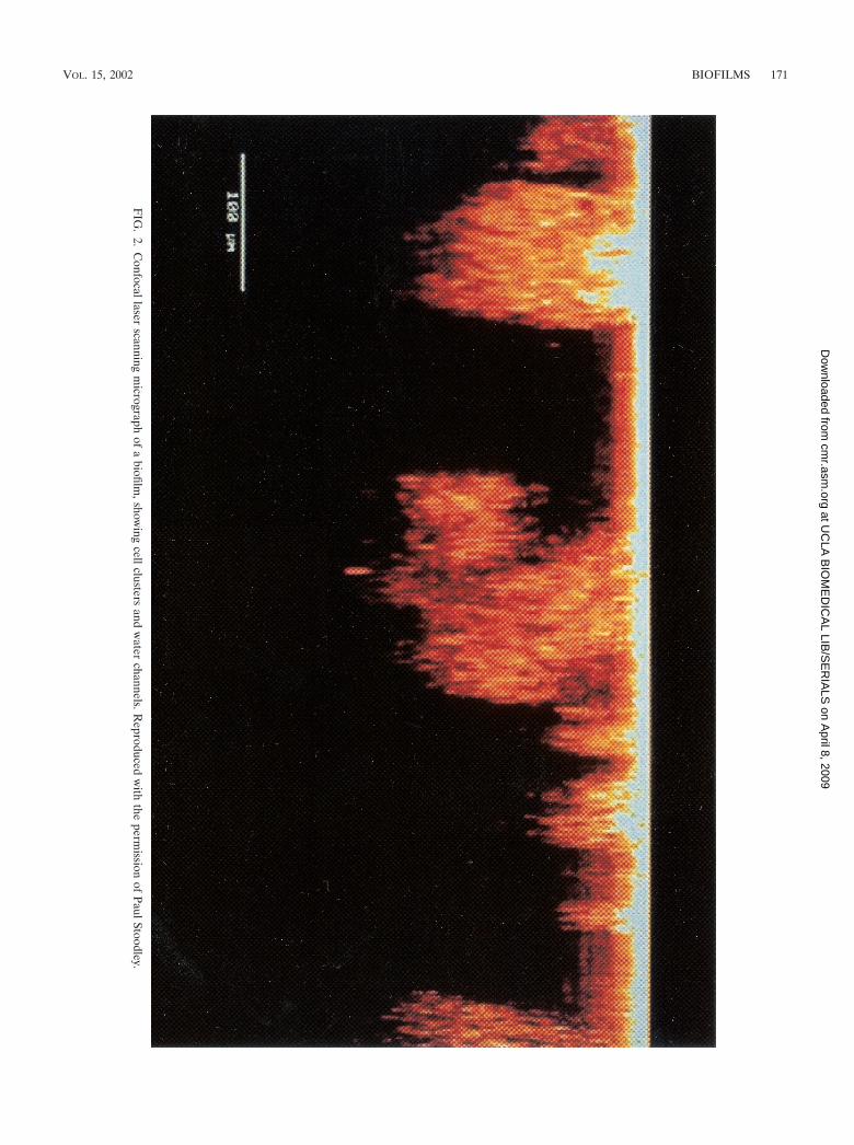
FIG
.2.
Confocallaser
scanningm
icrographof
abiofilm
,showing
cellclustersand
water
channels.Reproduced
with
theperm
issionof
PaulStoodley.
VOL. 15, 2002 BIOFILMS 171
at UC
LA B
IOM
ED
ICA
L LIB/S
ER
IALS
on April 8, 2009
cmr.asm
.orgD
ownloaded from

The structural characteristic of biofilms that has the greatestimpact on the outcome of chronic bacterial infections, such asnative valve endocarditis, is the tendency of individual micro-colonies to break off and/or detach when their tensile strengthis exceeded. This detachment of preformed microcolonies con-taining sessile cells in the antibiotic-resistant biofilm pheno-type poses a very serious risk of infective emboli in the firstcapillary bed that is encountered. This shedding of microcolo-nies from preformed biofilms on heart valves can lead to strokeor to severe pulmonary sequelae, and its consequences are wellrecognized by the clinical community.
RESISTANCE TO ANTIMICROBIAL AGENTS
The nature of biofilm structure and the physiological at-tributes of biofilm organisms confer an inherent resistance toantimicrobial agents, whether these antimicrobial agents areantibiotics, disinfectants, or germicides. Table 4 shows the dra-matic differences in susceptibility of planktonic and biofilmorganisms to antimicrobial agents. Mechanisms responsible for
resistance may be one or more of the following: (i) delayedpenetration of the antimicrobial agent through the biofilmmatrix, (ii) altered growth rate of biofilm organisms, and (iii)other physiological changes due to the biofilm mode of growth.
Delayed Penetration of the Antimicrobial Agent
Antimicrobial molecules must diffuse through the biofilm ma-trix in order to inactivate the encased cells. The extracellularpolymeric substances constituting this matrix present a diffusionalbarrier for these molecules by influencing either the rate of trans-port of the molecule to the biofilm interior or the reaction of theantimicrobial material with the matrix material. Suci et al. (198)demonstrated a delayed penetration of ciprofloxacin into Pseudo-monas aeruginosa biofilms; what normally required 40 s for asterile surface required 21 min for a biofilm-containing surface.Hoyle et al. (83) found that dispersed bacterial cells were 15 timesmore susceptible to tobramycin than were cells in intact biofilms.DuGuid et al. (57) examined Staphylococcus epidermidis suscep-tibility to tobramycin and concluded that the organization of cells
TABLE 1. Methods that have been used for measurement of biofilms on catheters
Method Basic protocola Advantage Limitation(s) Reference
Roll-plate Roll the catheter tip over the surfaceof a blood agar plate
Easy to use Examines only catheter outersurface, inaccurate
126
Vortex, then viable count Catheter section in PBS is vortexedthen cultured on different media
Measures intraluminal andextraluminal biofilm
Recovery efficiency unknown 202
Sonicate, vortex, thenviable count
Catheter section in TSB, sonicate thenvortex, then culture on blood agar
Measures intraluminal andextraluminal biofilm
Recovery efficiency unknown 178
Sonicate, vortex, homog-enize, then viablecount
Catheter section in PBS,sonicate/vortex repeatedly, thenhomogenize and culture on bloodagar
Recovery efficiencydetermined
Measures intraluminalbiofilm only
53
Acridine orange directstaining
Following roll-plate method, cathetersection is stained with acridineorange
Allows direct examinationof catheter
Method does not allowquantification
224
Endoluminal brush Brush is introduced into the implantedcatheter, removed, placed into PBS,sonicated, and plated
Allows examination ofindwelling catheter
Effect of procedure onpatient and recoveryefficiency unknown
102
Alginate swab Swab introduced into the implantedcatheter, removed, then streakedover a blood agar plate
Allows examination ofindwelling catheter
Effect of procedure onpatient and recoveryefficiency unknown
25
a PBS, phosphate-buffered saline; TSB, Trypticase soy broth.
TABLE 2. Apparatuses that have been used for growing and testing biofilms
Apparatus Organism(s) tested Flow dynamics Substratum Method for removing andquantifying biofilm Reference
Modified Robbins device Pseudomonas pseudomallei Batch/mixing Silastic disks Method of removal not given;viable count
208
Calgary biofilm device P. aeruginosa, S. aureus,E. coli
Batch/mixing Plastic pegs Sonicate peg, then viable count 26
Disk reactor Gram-negative bacteria Batch/mixing Teflon coupons Sonicate, vortex, homogenize,then viable or direct count
54
CDC biofilm reactor Gram-negative bacteria Continuous/opensystem
Needlelessconnectors(plastic)
Sonicate, vortex, homogenize,then viable or direct count
144
Perfused biofilmfermentor
Candida albicans Continuous/opensystem
Cellulose-acetatefilters
Shake in sterile water, thenviable count
11
Model bladder Gram-negative bacteria Continuous/opensystem
Urinary catheters Direct examination by SEM orTEMa or by chemicalanalysis
195
a SEM, scanning electron microscopy; TEM, transmission electron microscopy.
172 DONLAN AND COSTERTON CLIN. MICROBIOL. REV.
at UC
LA B
IOM
ED
ICA
L LIB/S
ER
IALS
on April 8, 2009
cmr.asm
.orgD
ownloaded from

within biofilms could in part explain the resistance of this organ-ism to this antimicrobial agent.
Other studies have examined antimicrobial agent penetrationand interaction with the extracellular polymeric substance mate-rial of biofilms. Hatch and Schiller (79) showed that a 2% sus-pension of alginate isolated from P. aeruginosa inhibited diffusionof gentamicin and tobramycin, and this effect was reversed byusing alginate lyase. Souli and Giamarellou (181) demonstratedthe ability of S. epidermidis slime to hinder the antimicrobialsusceptibility of Bacillus subtilis to a large number of agents. Notall antimicrobial agents were equally affected; glycopeptides such
FIG. 3. Mixed-species heterotrophic biofilm grown on stainless steel in a potable-water biofilm reactor containing Pseudomonas aeruginosa,Klebsiella pneumoniae, and Flavobacterium spp. This image of a biofilm was obtained, after staining with 4�,6�-diamidino-2-phenylindole, with aZeiss Axioskop 2 epifluorescence microscope and the Zeiss deconvolution system.
TABLE 3. Factors to consider in the developmentof a model biofilm system
Medium Inoculum Hydrodynamics Substratum
Composition,temperature,presence ofantimicrobialagents
Identity oforganism,no. of cells
Flow rate, presenceof shear, batchvs. open system,retention time
Roughness,chemistry,conditioningfilms
VOL. 15, 2002 BIOFILMS 173
at UC
LA B
IOM
ED
ICA
L LIB/S
ER
IALS
on April 8, 2009
cmr.asm
.orgD
ownloaded from

as vancomycin and teicoplanin were significantly affected,whereas agents such as rifampin, clindamycin, and the macrolideswere either unaffected or minimally affected. Another study (74)examined the diffusion of several antimicrobial agents (ceftazi-dime, cefsulodin, piperacillin, gentamicin, and tobramycin)through synthetic and naturally produced alginate gels and foundthat beta-lactam antibiotics diffused into the matrix more rapidlythan did aminoglycosides. Aminoglycosides were found to initiallybind to the alginates, but diffusion increased after an 80- to 100-min lag period.
Altered Growth Rate of Biofilm Organisms
Another proposed mechanism for biofilm resistance to antimi-crobial agents is that biofilm-associated cells grow significantlymore slowly than planktonic cells and, as a result, take up anti-microbial agents more slowly. Using a method of cell culturedesigned to determine the effect of growth rate apart from otherbiofilm processes, Evans et al. (63) found that the slowest growingEscherichia coli cells (in biofilms) were the most resistant to cet-rimide. At growth rates higher than 0.3 per h, biofilm and plank-tonic cells were equally susceptible. Another study showed that S.
epidermidis biofilm growth rates strongly influenced susceptibility;the faster the rate of cell growth, the more rapid the rate ofinactivation by ciprofloxacin (56). Anwar et al. (5) found thatolder (10-day-old) chemostat-grown P. aeruginosa biofilms weresignificantly more resistant to tobramycin and piperacillin thanwere younger (2-day-old) biofilms. A dosage of 500 �g of piper-acillin plus 5 �g of tobramycin per ml completely inactivated bothplanktonic and young (2-day-old) biofilm cells. Older (10-day-old) biofilm cell counts were reduced only approximately 20% byexposure to this dose. Similar results have been observed withseveral different combinations of bacteria and antimicrobialagents (2, 32, 51).
Other Physiological Changes Due toBiofilm Mode of Growth
Gram-negative bacteria respond to nutrient limitation andother environmental stresses by synthesizing sigma factors. In E.coli, those sigma factors that are under the control of the rpoSregulon regulate the transcription of genes whose products miti-gate the effects of stress. By studying E. coli biofilms formed bystrains with and without the rpoS gene, Adams and McLean (1)
FIG. 4. Biofilm structure cartoon. Copyright Center for Biofilm Engineering, Montana State University, Bozeman, Mont. Reprinted withpermission.
TABLE 4. Susceptibility of planktonic and biofilm bacteria to selected antibiotics
Reference Organism Antibiotic MIC or MBC ofplanktonic phenotype (�g/ml)
Concn effective againstbiofilm phenotype (�g/ml)
215 S. aureus NCTC 8325-4 Vancomycin 2 (MBC) 20a
26 Pseudomonas aeruginosa ATCC 27853 Imipenem 1 (MIC) �1,024b
26 E. coli ATCC 25922 Ampicillin 2 (MIC) 512b
208 P. pseudomallei Ceftazidime 8 (MBC) 800c
114 Streptococcus sanguis 804 Doxycycline 0.063 (MIC) 3.15d
a Concentration required for 99% reduction.b Minimal biofilm eradication concentration.c Concentration required for �99% reduction.d Concentration required for �99.9% reduction.
174 DONLAN AND COSTERTON CLIN. MICROBIOL. REV.
at UC
LA B
IOM
ED
ICA
L LIB/S
ER
IALS
on April 8, 2009
cmr.asm
.orgD
ownloaded from

found that the rpoS� E. coli biofilms had higher densities and ahigher number of viable organisms. Since rpoS is activated duringslow growth of this organism, it appears that conditions that elicitthe slowing of bacterial growth, such as nutrient limitation orbuild-up of toxic metabolites, favor the formation of biofilms.Nutrient limitation and increases in toxic metabolite concentra-tions might be particularly acute within the depths of establishedbiofilms. Tresse et al. (203) found that agar-entrapped E. coli cellswere more resistant to an aminoglycoside as oxygen tensions weredecreased. They suggested that the effect was due to lowereduptake of the antibiotic by the oxygen-starved cells. Dagostino etal. (42) proposed that initial bacterial association with a surfacemay result in the repression or induction of genes, which in turnresults in a number of physiological responses.
HUMAN INFECTIONS INVOLVING BIOFILMS
Koch’s postulates state that (i) the organism is regularlyfound in the lesions of the disease, (ii) it can be isolated in pureculture on artificial media, (iii) inoculation of this culture pro-duces a similar disease in experimental animals, and (iv) theorganism can be recovered from the lesions of these animals(49). The question of whether biofilms are etiological agents ofdisease in many cases cannot be proven according to Koch’spostulates. Nickel and Costerton (147) studied coagulase-neg-ative staphylococci (CoNS) in chronic prostatitis and were ableto detect these organisms in biopsies from infected individuals.Nevertheless, they concluded that it was not possible to statedefinitively that these organisms were the cause of the infec-tion. All that could be stated was that there was an associationbetween the presence of the organisms and the disease. Forseveral of the diseases discussed in this section, such as peri-odontitis, native valve endocarditis, and cystic fibrosis, thatassociation is stronger. For others, such as otitis media, theassociation is less well established. A discussion of severalnoted infectious diseases for which the biofilm link has beensuggested follows.
Native Valve Endocarditis
Native valve endocarditis (NVE) is a condition that resultsfrom the interaction between the vascular endothelium, gen-erally of the mitral, aortic, tricuspid, and pulmonic valves of theheart, and bacteria or fungi circulating in the bloodstream(118). The diversity of organisms causing NVE is quite exten-sive. Tunkel and Mandell (204) noted that of 2,345 cases ofinfective endocarditis, 56% were caused by streptococci (in-cluding viridans streptococci, enterococci, pneumococci, andStreptococcus bovis), 25% by staphylococci (19% coagulasepositive and 6% CoNS), and the balance by gram-negativebacteria and fungi (Candida and Aspergillus spp.). These or-ganisms gain access to the bloodstream primarily via the oro-pharynx, gastrointestinal tract, and genitourinary tract.
Normally, microorganisms adhere poorly to intact endothe-lium. However, when the endothelium is damaged, nonbacte-rial thrombotic endocarditis (NBTE), in which the thrombus isan accumulation of platelets, fibrin, and occasionally red bloodcells, will develop at the point of injury. Durack (59) inducedNBTE formation in rabbits by leaving a polyethylene catheterin place in contact with the aortic valve. Fibronectin, secreted
by endothelial cells, platelets, and fibroblasts in response to avascular injury, has been identified in thrombotic lesions ofheart valves. Fibronectin can simultaneously bind to fibrin,collagen, human cells, and bacteria (118).
Several bacteria have fibronectin receptors, including Staph-ylococcus aureus and several species of Streptococcus (118).Lowrance et al. (119, 120) showed in an animal model thatStreptococcus sanguis binds to the fibronectin molecule andthat low-fibronectin-binding mutants of S. sanguis are less vir-ulent than the high-binding strains. Several of the streptococcialso produce high-molecular-weight dextrans that promote ad-herence to the surface of the thrombus in NBTE (166). Dall etal. (43) showed that dextranase blocked microbial adhesion inexperimental animals. Inoculum size may also be important,depending on the species. Gram-negative bacteria do not ad-here as well as gram-positive organisms, and induction of en-docarditis in laboratory animals requires a much higher inoc-ulum of gram-negative bacteria than of gram-positiveorganisms (96).
Early work by Durack showed that bacteria would localize insites of NBTE within 30 min of injection into a rabbit contain-ing a polyethylene catheter (59). Though most of the bacteriawere ingested by white blood cells that were stuck to the edgesof the NBTE, some bacteria were not ingested and adhered tothe edge of the vegetation. Within hours these bacteria hadbegun to multiply. Bacterial microcolonies developed in theplatelet-fibrin matrix, primarily where there were few whiteblood cells. Several bacterial colonies eventually (after 24 h)developed fibrin capsules and were thus protected from thewhite blood cells. It appeared to the authors that the move-ment of the white blood cells was hindered by the fibrin. Du-rack and Beeson (58) also showed that most of the metabolicactivity of the biofilm bacteria was on the surface; coloniesdeeper in the thrombus were inactive. Also, they observed thatthe majority of bacteria in a vegetation enter a resting statewithin 2 days of infection.
Biofilms on native heart valves may result in valve tissuedamage or production of emboli. Ferguson et al. (66), in stud-ies of rabbits infected with staphylococci, found that bacteriapenetrated into the connective tissue of the aortic valve, struc-turally damaging it. Release of cells or clumps of cells andNBTE components into the bloodstream may also occur as aresult of NVE biofilms. These emboli may cause serious com-plications throughout the body. Fungi, because they producebulky, friable vegetations, more frequently produce emboli.Stiles and Friesinger (196) noted that fungal biofilms mayexceed 2 cm in diameter and the rate of clinically apparentemboli was higher in fungi than in bacteria. Rohmann et al.(168) found that embolic events were more common in pa-tients with vegetations larger than 10 mm in diameter.
NVE may be detected either indirectly, by a combination ofclinical symptoms and identification of organisms in the blood-stream, or by observing the vegetations via imaging techniques.One such imaging technique in common use is echocardiogra-phy. However, though it may be a good technique for docu-menting the presence or absence of biofilms, the use of echo-cardiography as a routine method for establishing diagnosis isnot recommended. Approximately half of patients with clinicalcriteria examined in a study by Stewart et al. (185) demon-strated vegetative lesions by echocardiography. These findings
VOL. 15, 2002 BIOFILMS 175
at UC
LA B
IOM
ED
ICA
L LIB/S
ER
IALS
on April 8, 2009
cmr.asm
.orgD
ownloaded from

were confirmed by others (22, 121). Berger et al. (15) notedthat the limit of detection for biofilms on infected valves is adiameter of approximately 3 mm. However, Rohmann et al.(168) found that monitoring vegetation size with transesopha-geal cardiography, particularly in culture-negative patients,may help to assess the efficacy of antimicrobial treatment.
Most medical practitioners recommend prophylactic antibi-otics when patients with a high risk of endocarditis undergodental and other invasive procedures. This treatment consistsof 3 g of amoxicillin taken orally 1 h before a procedure andthen 1.5 g 6 h later (166). This treatment would be expected tokill planktonic organisms in the bloodstream prior to attach-ment. Once the biofilm is established on the heart valves,treatment is much less effective due to a combination of masstransfer limitations and inherent resistance of biofilm organ-isms.
Depending on the organism involved, various antibiotictherapies have been used. Penicillin is the normal treatmentfor streptococcal endocarditis, and it may be supplementedwith gentamicin to produce synergistic killing. Treatment maybe increased when complications such as large vegetation sizeoccur. Other antibiotics or combinations of antibiotics are usedfor other organisms. Dall et al. (43) found that addition ofdextranase as an adjuvant to penicillin prevented microbialadhesion and facilitated penicillin sterilization of infectedvalves in experimental animals. Joly et al. (96) found thatantibiotic treatment was more successful when serum antibi-otic levels were held at least 10-fold higher than the minimalbactericidal concentration (MBC) through the entire dosingregimen. Sandoe et al. (172) successfully treated Staphylococ-cus capitus endocarditis with vancomycin and rifampin for pro-longed treatment. Perrotta and Fiore (156) found that Strep-tococcus bovis endocarditis was successfully resolved by usingpenicillin G together with streptomycin (6 days), followed byimipenem (4 days).
Candida endocarditis has been treated successfully with flu-conazole (212). Rohmann et al. (168) investigated the effect ofantibiotic treatment on vegetation size using transesophagealechocardiography in 183 patients monitored over a 76-weekperiod. The reduction in vegetation size as a result of treat-ment was as follows: vancomycin, 45%; ampicillin, 19%; andpenicillin, 5%. Penicillinase-resistant drugs resulted in a 15%increase, and cephalosporin resulted in a 40% increase. Theseresults underline the importance of closely monitoring thebiofilm size over the course of the treatment, especially sinceembolic events are more common for larger vegetations. An-other treatment approach is to surgically remove the vegeta-tion from the infected valve, a procedure termed vegetectomy(86).
Clearly, the formation of biofilms on native heart valves(termed vegetations by the medical community) is a well-doc-umented biofilm process. However, there are still importantquestions that must be addressed. What threshold number ofmicroorganisms in the bloodstream is required to develop abiofilm? Could in vitro studies be developed that will moreaccurately predict the efficacy of antimicrobial agents in vivo?Can bacteria that are ingested by leukocytes survive to colonizea sterile NBTE site?
Otitis Media
Otitis media (OM) is a disease of the middle ear that involvesthe inflammation of the mucoperiosteal lining. OM is a verycommon childhood disease, may be acute or chronic, and iscaused by a number of different organisms, including Streptococ-cus pneumoniae, Haemophilus influenzae, Moraxella catarrhalis,group A beta-hemolytic streptococci, enteric bacteria, Staphylo-coccus aureus, Staphylococcus epidermidis, Pseudomonas aerugi-nosa, and other organisms (65). Mixed cultures may also be iso-lated (73). Stenfors and Raisanen (184) quantified the bacteria inmiddle ear effusions collected from patients with OM. They foundcounts ranging between 105 and 109 per ml of effusion material. Incertain cases of chronic OM, the middle ear may contain a highlyviscous fluid (OM with effusion) (73). Under these conditions, theimplantation of tympanostomy tubes is performed to alleviatepressure build-up and hearing loss.
Tympanostomy tubes are subject to contamination, and bio-films will build up on their inner surfaces. Biedlingmaier et al.(16) investigated the colonization of Armstrong-style silicone,fluoroplastic, ionized modified silicone and silver oxide-coatedArmstrong-style silicone tubes by Pseudomonas aeruginosa,Staphylococcus aureus, and Staphylococcus epidermidis in Tryp-ticase soy broth. They found that all three organisms devel-oped biofilms on the Armstrong silicone and the silver oxide-coated Armstrong-style silicone tubes. P. aeruginosa alsodeveloped biofilms on the fluoroplastic tubes. Only the ionizedsilicone tubes remained free of contamination and biofilms.
Saidi et al. (170) investigated biofilm formation on tubesimplanted into the ears of guinea pigs inoculated with S. au-reus. In this study, the tube materials investigated includedsilicone, silver oxide-impregnated silicone, fluorplastic, silveroxide-impregnated fluorplastic, and ion-bombarded silicone.The tubes were left in place for 10 days, fixed, and examined byscanning electron microscopy. The results of this study showedthat all of the materials contained attached bacteria, thoughthe ion-bombarded silicone had fewer cells, which did notappear to have formed a biofilm.
Gourin and Hubbell (75) investigated the efficacy of silveroxide-impregnated silastic tympanostomy tubes inserted intothe ears of 630 patients with chronic OM in preventing post-operative otorrhea (drainage from the ear) in a prospectivenonrandomized clinical study. They found that the use of thetreated tympanostomy tubes resulted in a lower incidence ofpostoperative otorrhea after the first postoperative week. Theauthors opined that the silver oxide prevented adherence andcolonization of selected bacteria to the tube but probably hadno effect on the established infection in the middle ear.
The fact that biofilm organisms are significantly more resistantto antimicrobial agents has already been discussed. An additionalconsideration in the case of biofilms of otitis media is that there isvery low penetration of antibiotics into the middle ear fluid.Krause et al. (107) compared concentrations of amoxicillin, cefa-clor, erythromycin-sulfisoxazole, and trimethoprim-sulfamethox-azole in middle ear fluid and serum of children with serous OM.For samples collected 15 to 240 min after administration of asingle oral dose, levels of antibiotic in the middle ear fluid werealways significantly lower than those in the serum. Also, certainantibiotics, such as erythromycin, were never detected at all in themiddle ear fluid.
176 DONLAN AND COSTERTON CLIN. MICROBIOL. REV.
at UC
LA B
IOM
ED
ICA
L LIB/S
ER
IALS
on April 8, 2009
cmr.asm
.orgD
ownloaded from

Kondoh and Hashiba (106) evaluated the efficacy of severalmacrolide antibiotics, i.e., clarithromycin, erythromycin, andmidacamycin, against biofilms of P. aeruginosa growing on Tef-lon in a minimal medium for a 7-day exposure period. Bothclarithromycin and erythromycin inhibited biofilm formation,as evidenced by decreases in total protein, alginate, and hexoseon Teflon beads. However, the planktonic bacterial levels wereunaffected by the treatments, and the authors proposed thatthe inhibitory effects were due to factors other than bacteri-cidal activity. Both clarithromycin and erythromycin inhibitedbiofilm formation at 1/20 of the MIC. Since this concentrationcan be achieved in sputum and nasal discharges, there is a goodprobability that these antimicrobial agents would be effectiveagainst biofilm diseases caused by P. aeruginosa, including OM.
With the exception of a single report by Hayes et al. (J. D.Hayes, R. Veeh, X. Wang, J. W. Costerton, J. C. Post, andG. D. Ehrlich, abstr. 186, Am. Soc. Microbiol. Biofilm 2000Conf., 2000), there is very little evidence for the developmentof biofilms on mucosal surfaces of the middle ear in OM. Inthis study, the authors used scanning electron microscopy toprovide evidence of H. influenzae biofilms on the middle earmucosal surfaces of chinchillas that had been injected with aculture of this organism. Recent unpublished work with thechinchilla model of OM, in collaboration with Ehrlich andPost, clearly shows biofilm formation by both scanning electronmicroscopy and CLSM.
Chronic Bacterial Prostatitis
The prostate gland may become infected by bacteria thathave ascended from the urethra or by reflux of infected urineinto the prostatic ducts emptying into the posterior urethra(52). Once the bacteria enter the prostatic duct and ascini, theymultiply rapidly and elicit a host response. As long as theinfection is in the early acute stages, the bacteria can easily beeradicated with antibiotic therapy (146). If these bacteria per-sist, they can form sporadic microcolonies and biofilms thatadhere to the epithelial cells of the duct system. Organismsisolated in cases of chronic bacterial prostatitis include E. coli(most common isolate), Klebsiella, enterobacteria, Proteus, Ser-ratia, Pseudomonas aeruginosa, CoNS, coryneforms, and En-terococcus faecalis (52). In another study, Nickel and Costerton(151) isolated E. coli, P. aeruginosa, Bacteroides spp., Gard-nerella spp., Corynebacterium spp., and CoNS.
Much of our understanding of the probable role of biofilmsin chronic bacterial prostatitis has come either from studiesemploying animal models (148, 150) or from biopsies collectedfrom men with prostatitis (147, 151). Nickel et al. (150) inoc-ulated the prostates of rats with a culture of 108 E. coli organ-isms per ml by means of a sterile catheter. Rats were sacrificedafter 1, 3, and 7 days and weekly for 8 weeks, and biopsysamples of prostates were collected. These samples were ex-amined by either scanning electron microscopy or transmissionelectron microscopy. Samples were also sonicated and platedonto MacConkey agar. They demonstrated that bacteria werepresent in glycocalyx-encased microcolonies and appeared tobe firmly adherent to the ductal and acinar mucosal layers.
Nickel and Costerton (151) evaluated 20 men with a history ofchronic bacterial prostatitis. Biopsies were collected from infectedprostates, processed aseptically, and plated onto nutrient agar.
Histological specimens were also examined by scanning electronmicroscopy and transmission electron microscopy. The authorsshowed evidence of bacterial attachment to the ductal walls, es-pecially for P. aeruginosa. Nickel and Costerton (147) were alsoable to demonstrate, using needle biopsies, sporadic microcolo-nies of CoNS in the intraductal space. The microcolonies wereenveloped in a dehydrated slime matrix. Transmission electronmicroscopy portrayed bacterial biofilms very clearly, as shown inFig. 5.
Domingue and Hellstrom (52) state that treatment failures arecommon in prostatitis, probably as a result of the local environ-ment surrounding the infecting organisms and the fact that theseorganisms have produced a biofilm. Once bacteria infect the pros-tate, they produce a glycocalyx and become inactive. With thischange in metabolism, the cells can become more resistant toantimicrobial agents (146). Nickel and Costerton (151) presenteda study of chronic bacterial prostatitis in 20 men whose symptomsdid not resolve with long-term courses of antibiotic therapy. Thedosage regimens of these antibiotics had been determined byculture and sensitivity testing in the laboratory. They found that ittook significantly longer (96 h) to grow bacteria from sonicatedtissue biopsy samples than to grow bacteria cultured from patientswith cystitis. This observation lends support to the conclusion thatorganisms growing in the tissues as biofilms have an altered me-tabolism.
In light of the fact that prostatitis is apparently caused bybiofilm-associated organisms, Nickel et al. (146) have sug-gested that a recommended treatment regimen might be todeliver higher antibiotic concentrations directly to the biofilmwithin the prostatic ducts.
Cystic Fibrosis
Cystic fibrosis (CF), a chronic disease of the lower respira-tory system, is the most common inherited disease. In thiscondition, the normal mucociliary clearance system thatcleanses the bronchopulmonary epithelium of inhaled particlesdepends on an upward directional flow of a mucus layer on thetips of cilia that move freely in the underlying watery layer. InCF there is a net deficiency of water, which hinders the upwardflow of the mucus layer. Decreased secretion and increasedabsorption of electrolytes lead to dehydration and thickeningof secretions covering the respiratory epithelium (104).
According to May et al. (131), 70% of patients with CF aredefective in the cystic fibrosis transmembrane conductance regu-lator protein (CFTR), which results in altered secretions in thesecretory epithelia. The hyperviscous mucus that is produced isthought to increase the incidence of bacterial lung infections inCF patients. According to Govan and Deretic (76), the CF gene,which encodes the CFTR, has been identified. The CFTR func-tions as a chloride ion channel protein. Chloride ion transport isseverely impaired when the CFTR is defective in CF patients.Staphylococcus aureus is usually the first pulmonary isolate fromthese patients (131). It can normally be controlled by antibiotics.S. aureus and H. influenzae infections usually predispose the CF-affected lung to colonization with P. aeruginosa. Burkholderia ce-pacia has also been shown to infect the lungs of CF patients withlethal consequences, but it has never attained the 80% coloniza-tion rate of P. aeruginosa (76).
The exact mechanism of P. aeruginosa colonization of the
VOL. 15, 2002 BIOFILMS 177
at UC
LA B
IOM
ED
ICA
L LIB/S
ER
IALS
on April 8, 2009
cmr.asm
.orgD
ownloaded from

lungs of patients with CF is not known. There is evidence thatenhanced pseudomonal receptors on the respiratory epitheliamay be responsible; impaired mucociliary clearance is anotherpossibility (76). During initial colonization, the organisms arenonmucoid. Persistence of the organism in the lungs of pa-tients with CF ultimately will result in a mucoid phenotype(104). There is no clear interval between the initial coloniza-tion by P. aeruginosa and conversion to mucoid forms; it maytake several months to years. The variable timing of the emer-gence indicates that this is caused by random mutations, fol-lowed by selection of mucoid strains in the lungs of patientswith CF (76).
This mucoid phenotype was first observed by Lam et al. (110)in postmortem specimens of infected lung tissue and bronchos-copy material from infected patients. The mucoid material wasshown to be a polysaccharide material, later identified as alginate.The conditions that trigger the conversion to the mucoid pheno-
type have been investigated. Hoyle et al. (84) demonstrated, usinga chemostat and modified Robbins device, that mucoid exopo-lysaccharide was transiently produced following adherence of P.aeruginosa. May et al. (131) noted that several in vitro conditions,such as nutrient limitation, the addition of surfactants, and sub-optimal levels of antibiotics, may result in mucoidy. Mucoidy iseven elicited by addition of ethanol to the medium, indicating thatthis phenotype may be a response to dehydration.
Mathee et al. (130) showed that biofilms of P. aeruginosa chal-lenged with either activated human peripheral blood polymor-phonuclear leukocytes (PMNs) or hydrogen peroxide (a productreleased in low levels by PMNs) yielded about 0.1% mucoidcolonies, while unchallenged biofilms produced none. Alginatewas overproduced by all the mucoid colonies. They hypothesizedthat activated PMNs and the release of toxic products such ashydrogen peroxide could play a role in the generation of mucoidorganisms during the inflammatory response.
FIG. 5. Transmission electron micrograph of a prostatic duct in an area of focal chronic inflammation from a patient with an E. coli chronicprostatitis. Arrows point to bacterial microcolonies amid inflammatory cells and debris. These bacteria were cultured from both expressed prostaticsecretions and tissue biopsies obtained 4 weeks after antibiotics were discontinued. Bar, 1 �m. Reprinted from reference 151 with permission ofWiley-Liss, Inc., a subsidiary of John Wiley & Sons, Inc.
178 DONLAN AND COSTERTON CLIN. MICROBIOL. REV.
at UC
LA B
IOM
ED
ICA
L LIB/S
ER
IALS
on April 8, 2009
cmr.asm
.orgD
ownloaded from

The sputum from the lungs of patients with CF is usuallyfilled with large numbers of PMNs, and the inflammatory de-fense mechanisms in the lungs of patients with CF againstmucoid P. aeruginosa are usually dominated by PMNs andantibodies (130). In contrast to P. aeruginosa, B. cepacia doesnot generally produce alginate-like compounds, though someinvestigators have reported the production of other exo-polysaccharides. Mucoid colonial morphology in B. cepacia israre in both environmental and clinical strains. The presenceof biofilms or microcolonies of Burkholderia has not been re-ported for patients colonized solely by this organism (76).
A question posed by a number of investigators is why mu-coid P. aeruginosa infections are so recalcitrant and resistant toimmune system clearance. Koch and Hoiby (104) stated thatthe biofilm mode of growth protects the organisms from anti-microbial agents and host defenses. The alginate layer of mu-coid strains appears to prevent antibody coatings and blocksthe immunological determinants required for opsonic phago-cytosis (90, 91, 131, 135). Mucoid strains are apparently moreresistant to nonopsonic phagocytosis than are nonmucoidstrains (90, 131). There is evidence that the alginate may pro-mote adherence of the mucoid strains to epithelial cells in thepulmonary tract, thereby inhibiting clearance. In vivo experi-ments with infected rats confirmed this; mucoid P. aeruginosastrains were less rapidly removed from the pulmonary tractthan were nonmucoid strains (131).
Another mechanism for persistence and survival was pro-posed by Cochrane et al. (33). Using rats that had been arti-ficially infected with agar beads containing P. aeruginosa, theyfound that the bacteria within these beads produced elevatedlevels of high-molecular-weight iron-regulated membrane pro-teins that can function as receptors for iron-siderophore com-plexes. These molecules aid in the scavenging of low levels ofiron from the bloodstream. A host defense mechanism againstpathogenic organisms is to restrict available iron in order tolimit this essential bacterial nutrient. By producing iron-scav-enging compounds, the organisms are better able to survive inthe host.
Anwar et al. (6) also suggested that biofilm age was a criticalfactor in P. aeruginosa survival. In their experimental system,older biofilm cells of this organism were less susceptible toeither whole blood or serum than were either younger biofilmsor planktonic organisms.
The possibilities for successful treatment of CF may ulti-mately hinge on early antimicrobial treatment to prevent ordelay chronic infection with P. aeruginosa. Koch and Hoiby(104) noted that early treatment with oral ciprofloxacin andinhaled colistin could postpone chronic infection with P.aeruginosa for several years. They also suggested that a vaccineagainst this organism might be effective in preventing initialcolonization of the lungs of patients with CF.
Periodontitis
Periodontal diseases, infections involving the supporting tis-sues of teeth, range from mild and reversible inflammations ofthe gums (gingiva) to chronic destruction of periodontal tissues(gingiva, periodontal ligament, and alveolar bone). Chronicperiodontitis may lead to exfoliation of the teeth (112). Thechannel between the tooth root and the gingiva (gum), termed
the subgingival crevice, is the primary site of periodontal in-fection and will deepen into a periodontal pocket with theprogression of the disease (112).
Moore et al. (140) characterized the organisms isolated frompatients with moderate periodontal disease and found thatFusobacterium nucleatum, Peptostreptococcus micros, Eubacte-rium timidum, Eubacterium brachy, Lactobacillus spp., Actino-myces naeslundii, Pseudomonas anaerobius, Eubacterium sp.strain D8, Bacteroides intermedius, Fusobacterium sp., Seleno-monas sputigena, Eubacterium sp. strain D6, Bacteroides pneu-mosintes, and Haemophilus aphrophilus were all positively cor-related with gingivitis. They concluded that the predominantorganisms in the subgingival areas of patients with moderateperiodontitis are not found in healthy patients.
Lamont and Jenkinson (112) and Socransky and Haffajee(180) noted that Porphyromonas gingivalis is the primary agentresponsible for periodontitis. Omar et al. (154) examined sub-gingival plaque in adult patients with periodontitis and showedthat spirochetes and cocci tended to increase in these areas.Dzink et al. (60) found that the predominant microflorae ofactive lesions in subgingival areas were Fusobacterium nuclea-tum, Wolinella recta, Bacteroides intermedius, Bacteroides for-sythus, and Bacteroides gingivalis (Porphyromonas gingivalis).Marsh (128) noted that the predominant flora, even betweensites in the same subject, is highly diverse, though periodontitisis clearly a polymicrobic infection.
Proteinaceous conditioning films, called acquired pellicle, de-velop on the exposed surfaces of enamel almost immediately aftercleaning of the tooth surface within the oral cavity. The pelliclecomprises albumin, lysozyme, glycoproteins, phosphoproteins,lipids, and gingival crevice fluid (128). Within hours of pellicleformation, single cells of primarily gram-positive cocci and rod-shaped bacteria from the normal oral flora colonize these sur-faces. The pioneer species are predominantly streptococci, acti-nomycetes, and smaller numbers of Haemophilus (128). Theseorganisms have the ability to bind directly to the pellicle throughthe production of extracellular glucans (105). After several days,actinomycetes predominate, and the characteristic polysaccharidematrix of a biofilm begins to develop (128).
Organisms associating with and attaching to cells in thisearly biofilm do so by a process called coaggregation. Coag-gregation is cell-to-cell recognition whereby organisms in thebiofilm can recognize and adhere to genetically distinct bacte-ria by means of adhesins. These adhesins recognize protein,glycoprotein, or polysaccharide receptors on oral surfaces, in-cluding other cell types (105). A climax biofilm community,termed plaque, will develop within 2 to 3 weeks if the plaque isleft undisturbed, with 50- to 100-�m-thick biofilms developing(112). In addition to matrix polysaccharides, there will be poly-mers of salivary origin (128).
Plaque that becomes mineralized with calcium and phos-phate ions is termed calculus or tartar (176). In addition todevelopment on the tooth surfaces (within fissures), plaque candevelop more extensively in protected areas, including approxi-mal areas (between the teeth) and the gingival crevice (be-tween the tooth and gum). As the plaque mass increases inthese protected areas, the beneficial buffering and antimicro-bial properties of the saliva are less able to penetrate andprotect the tooth enamel, leading to dental caries or periodon-tal disease (128). In support of this, Corbet and Davies (35)
VOL. 15, 2002 BIOFILMS 179
at UC
LA B
IOM
ED
ICA
L LIB/S
ER
IALS
on April 8, 2009
cmr.asm
.orgD
ownloaded from

reviewed data showing that control of supragingival plaque byprofessional tooth cleaning and personal efforts would preventgingival inflammation and adult periodontitis.
Within the subgingival crevice, the primary source of nutri-ents for the developing biofilm is gingival crevice fluid, a serumexudate that bathes the gingival crevice. This fluid providesproteins, glycoproteins, and other nutrients. Bacterial nutrientsmay also originate from saliva and the host diet (especiallyfermentable carbohydrates) (128). Though there is a constantflow of air through the oral cavity, the tooth surface rapidlybecomes anaerobic on colonization with microorganisms.Marsh (128) noted that redox potential (Eh) fell from ��200mV to �30 mV within 2 days of colonization and to ��150mV after 7 days. The Eh of the gingival crevice is usually lowerthan that of other sites around a healthy tooth. Bradshaw et al.(20) used a model system oral biofilm and demonstrated thatanaerobes increased in proportion to aerobes with increasingbiofilm age. They showed that mixed cultures can protect ob-ligate anaerobes in the biofilms from the toxic effects of oxy-gen.
As the organisms develop biofilms in the subgingival crevice,they produce proteolytic enzymes that damage tissue directlyor interfere with host defenses (128). Collagenase and hyal-uronidase are also present and capable of degrading collagen.Breakdown of the fiber barrier system may occur, and thelesion may then progress to one that may attack the supportingstructures of the tooth (176). Gram-negative organisms alsoproduce endotoxins that may result in inflammation (176).Lamont et al. (111) demonstrated that Porphyromonas gingiva-lis was capable of invading epithelium cells in a laboratoryassay, eliciting invasion mechanisms similar to those of otherpathogens. In their assay, none of the serum concentrationsused affected the invasive ability of the organism. Serum wasused to simulate crevicular fluid.
The control of periodontitis is rooted in the removal ofestablished biofilms (plaque) from the subgingival areas, incombination with supplemental antimicrobial agents. Qui-rynen et al. (159) found that chlorhexidine rinses after me-chanical cleaning significantly improved gum health, as mea-sured by a reduction in probing depth of the gingival crevice.Kinniment et al. (101) found that pathogens such as P. gingi-valis and F. nucleatum were inhibited within laboratory oralbiofilms by treatment with chlorhexidine, in support of thefindings by Quirynen. Reynolds et al. (163) found that subgin-gival irrigation with chlorhexidine during ultrasonic scalingprovided a significant improvement in probing depth com-pared to that of the untreated control group. Jeong et al. (92)found that root planing plus a mixture of tetracycline and citricacid-containing gel was most effective in decreasing pocketdepth. In this case, the root planing consisted of mechanicallyremoving plaque and calculus from the exposed root surfaces.Citric acid acted as a chelating agent to remove mineral de-posits on the root surfaces.
Clearly, there is an association between the occurrence ofbiofilms and infection in certain human diseases. The organ-isms responsible, the extracellular components of the biofilm,the nature of the required conditioning film, and the mode ofpathogenicity vary from one disease condition to the next. Inevery case discussed, however, there are certain underlyingprocesses that are unchanging: production of an extracellular
matrix polymer, resistance to antimicrobial agents that in-creases with biofilm age, and resistance to immune systemclearance.
BIOFILMS ON MEDICAL DEVICES
Because the criteria for the biofilm mode of growth are quitebroad, as has been discussed, the environments suitable formicroorganisms to colonize and establish biofilms are practi-cally limitless. Costerton et al. (39) provided a partial listing ofmedical devices that have been shown to become colonized bybiofilms. Biofilms of various medical devices have been studiedextensively over the last 20 years, though much of the pub-lished research used very basic tools, such as viable culturetechniques and scanning electron microscopy, to characterizethe microbial diversity and visualize the biofilms. For certaindevices, such as urinary catheters and contact lenses, researchhas also elucidated the susceptibility of various materials tobacterial adhesion and biofilm formation.
A description follows of the biofilms on specific devices:prosthetic heart valves, central venous catheters, urinary(Foley) catheters, contact lenses, intrauterine devices, anddental unit water lines.
Prosthetic Heart Valves
Two major groups of prosthetic heart valves are currentlyused, mechanical valves and bioprostheses (tissue valves) (21).The rates of prosthetic valve endocarditis (PVE), or microbialinfection of the valve and surrounding tissues of the heart, aresimilar for both types of valves (21). Estimates of the rate ofPVE range from 0.5% (77) to 1 and 4% (55). The surgicalimplantation of the prosthetic valve results in tissue damage,leading to the accumulation of platelets and fibrin at the suturesite and on the device. As is the case with NVE, there is agreater susceptibility for initial microbial colonization in theselocations (55).
Illingworth et al. (87) noted that PVE is predominantlycaused by colonization of the sewing cuff fabric of the pros-thetic valve by microorganisms. Karchmer and Gibbons (98)added that the microorganisms will commonly invade the valveannulus into which the prosthetic valve has been sewn, poten-tially leading to a separation between the valve and the tissueand resulting in leakage.
Though the etiologic agents of PVE are generally identifiedby blood culture, transesophageal echocardiography is alsoused to detect biofilms (55). Organisms responsible for PVEdiffer depending on whether the infection can be classified asearly or late. CNS are the predominant early colonizers (77,98), probably resulting from initial contamination of the sur-gical site during the procedure. For late PVE, which by defi-nition is from 12 months onward following the valve replace-ment, the organisms responsible may be streptococci, CoNS,enterococci, S. aureus, gram-negative coccobacilli, or fungi(98). Hancock (77) also noted that viridans group streptococciwere the most common organism isolated during late PVE.
There still remain important questions to be answered re-garding PVE, such as rate of colonization in vivo, rate ofdetachment, and physiology of biofilm organisms in the nutri-tionally rich environment of the heart. Techniques that could
180 DONLAN AND COSTERTON CLIN. MICROBIOL. REV.
at UC
LA B
IOM
ED
ICA
L LIB/S
ER
IALS
on April 8, 2009
cmr.asm
.orgD
ownloaded from

enable investigators to visualize and quantify biofilms on valveseither in vivo or following removal and model systems that canbe used to grow biofilms on mechanical valves are needed.
Central Venous Catheters
Maki (123) noted that central venous catheters (CVCs) posea greater risk of device-related infection than does any otherindwelling medical device, with infection rates of 3 to 5%.Catheters may be inserted for administration of fluids, bloodproducts, medications, nutritional solutions, and hemodynamicmonitoring (68). Biofilms have been shown by scanning elec-tron microscopy and transmission electron microscopy to beuniversally present on CVCs and may be associated with eitherthe outside of the catheter or the inner lumen (160). Organ-isms that colonize the CVC originate either from the skininsertion site, migrating along the external surface of the de-vice, or from the hub, due to manipulation by health careworkers, migrating along the inner lumen (62, 162). Becausethe device is in direct contact with the bloodstream, the surfacebecomes coated with platelets, plasma, and tissue proteins suchas albumin, fibrinogen, fibronectin, and laminin (162). Thesematerials act as conditioning films; S. aureus adheres to pro-teins such as fibronectin, fibrinogen, and laminin, and S. epi-dermidis adheres only to fibronectin (162). The organisms mayalso produce adhesins.
Rupp et al. (169) investigated the role of S. epidermidis-produced adhesins in an animal model. The adhesins examinedwere polysaccharide intercellular adhesin and hemagglutinin.They found that wild-type organisms adhered in greater num-bers to CVCs and produced higher rates of infection than didpolysaccharide intercellular adhesin and hemagglutinin knock-out strains. Murga et al. (144) showed that gram-negative or-ganisms also adhered in vitro more extensively to materialsthat had been conditioned with freshly drawn human blood.
Colonization and biofilm formation may occur within 3 daysof catheterization (3). Raad et al. (160) also showed that cath-eters in place for less than 10 days tended to have more ex-tensive biofilm formation on the external surface of the cath-eter; for longer-term catheters (up to 30 days), biofilms weremore extensive on the internal lumen. Organisms colonizingCVCs include CoNS, S. aureus, P. aeruginosa, Klebsiella pneu-moniae, Enterococcus faecalis, and Candida albicans (62, 162).
Biofilms on CVCs have routinely been detected by a semi-quantitative procedure termed the roll-plate technique, inwhich the distal tip of the catheter is removed aseptically androlled over the surface of a nonselective medium. Quantifica-tion of the biofilm on the catheter tip is dependent on thenumber of organisms that are recovered by contact with theagar surface. A number of investigators have used this proce-dure to quantify biofilms and determine the relationship be-tween biofilm formation and bloodstream infection (3, 9, 36,124). However, this technique will not detect organisms on theinner lumen of the catheter and is unable to detect more than1,000 CFU per tip.
Raad et al. (161) observed that the roll-plate technique hasa low diagnostic sensitivity and low predictive value for cath-eter-related bacteremia. They attempted to enhance biofilmquantification by using sonication plus vortexing of cathetertips and found that a level of 104 CFU per tip was predictive of
a catheter-related septicemia. Anaissie et al. (3) studied cath-eters collected from patients and quantified the biofilms usingeither the roll-plate, sonication, or scanning electron micro-scope method. The biofilms were quantified by scanning elec-tron microscopy by measuring the total area of the outer andinner luminal surfaces covered by biofilms. They defined col-onization as either �15 CFU/tip in catheters by the roll-platetechnique or �100 CFU/tip in catheters by the sonicationtechnique.
Zufferey et al. (224) directly stained catheter tips with acri-dine orange after the tips had been processed by the roll-platetechnique. The cells in the biofilm were not quantified by thisprocedure; a positive or negative result was reported. Theyfound good agreement between the two techniques, and theacridine orange staining technique provided more rapid re-sults.
Regardless of the technique used to quantify biofilms, anyattempt to relate the occurrence of biofilms with infectionshould take into consideration the method of blood sampling.Duplicate blood samples should ideally be drawn peripherally(from a vein rather than through the CVC) to ascertain thatthe organisms in the blood sample have not originated fromthe device biofilms during sampling (162).
Urinary Catheters
Urinary catheters are tubular, latex, or silicone devices thatare inserted through the urethra into the bladder to measureurine output, collect urine during surgery, prevent urinary re-tention, or control urinary incontinence (99). One study (222)found that the percentage of patients undergoing indwellingurinary catheterization was 13.2% for hospital patients, 4.9%for nursing homes, and 3.9% for patients receiving home care.The Foley catheter has an inflatable balloon near the tip thatholds the catheter in place in the bladder, and the catheter,once installed, is connected to a drainage tube and collectionbag (189).
Catheter systems may be open or closed systems. In opensystems, the catheter drains into an open collection container;in closed systems, the catheter empties into a securely fastenedplastic collecting bag (99). In open systems, catheters quicklybecome contaminated, and patients commonly develop urinarytract infections within 4 days (99). Patients with closed systemsare much less susceptible to urinary tract infections, and theurine from the patient can remain sterile for 10 to 14 days inapproximately half the patients (99). Regardless of whether thesystem is open or closed, Stickler noted that 10 to 50% ofpatients undergoing short-term catheterization (up to 7 days)develop infections, whereas essentially all patients undergoinglong-term catheterization (greater than 28 days) will developurinary tract infections (189). McLean et al. further noted thatthe risk of catheter-associated infection increases by approxi-mately 10% for each day the catheter is in place (134).
The organisms that attach to the catheter and develop thebiofilm originate from one of several sources: (i) organisms areintroduced into the urethra or bladder as the catheter is in-serted, (ii) organisms gain entry through the sheath of exudatethat surrounds the catheter, or (iii) organisms travel intralu-minally from the inside of the tubing or collection bag (99).Rogers et al. (167) used a model bladder system to determine
VOL. 15, 2002 BIOFILMS 181
at UC
LA B
IOM
ED
ICA
L LIB/S
ER
IALS
on April 8, 2009
cmr.asm
.orgD
ownloaded from
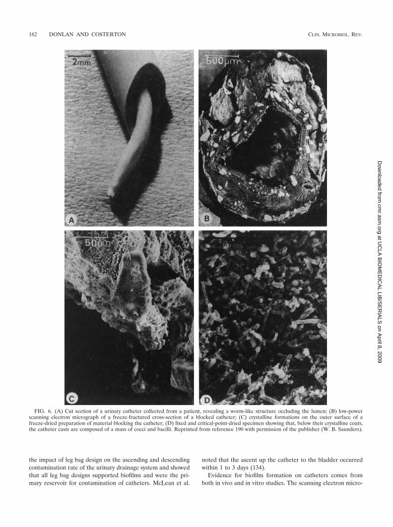
the impact of leg bag design on the ascending and descendingcontamination rate of the urinary drainage system and showedthat all leg bag designs supported biofilms and were the pri-mary reservoir for contamination of catheters. McLean et al.
noted that the ascent up the catheter to the bladder occurredwithin 1 to 3 days (134).
Evidence for biofilm formation on catheters comes fromboth in vivo and in vitro studies. The scanning electron micro-
FIG. 6. (A) Cut section of a urinary catheter collected from a patient, revealing a worm-like structure occluding the lumen; (B) low-powerscanning electron micrograph of a freeze-fractured cross-section of a blocked catheter; (C) crystalline formations on the outer surface of afreeze-dried preparation of material blocking the catheter; (D) fixed and critical-point-dried specimen showing that, below their crystalline coats,the catheter casts are composed of a mass of cocci and bacilli. Reprinted from reference 190 with permission of the publisher (W. B. Saunders).
182 DONLAN AND COSTERTON CLIN. MICROBIOL. REV.
at UC
LA B
IOM
ED
ICA
L LIB/S
ER
IALS
on April 8, 2009
cmr.asm
.orgD
ownloaded from

scope and transmission electron microscope have been used todocument biofilms on urinary catheters removed from patients(152, 189, 194). Figure 6 shows a well-developed urinary cath-eter biofilm. Stickler (189) noted one study that measured 109
P. aeruginosa cells per cm2 of luminal catheter surface.Ganderton et al. (71) measured biofilm thickness on silicone
and silicone-coated Foley catheters collected from patientsundergoing long-term catheterization and found that thick-nesses ranged up to approximately 200 �m, with an occasionalcatheter containing biofilms between 200 and 500 �m. Thatstudy also measured biofilm plate counts as high as 108 per cm2
in long-term catheters. However, they found that biofilm thick-ness and plate counts were quite variable and that there was noclear relationship between duration of catheter use and extentof biofilm formation. For example, the thickest biofilm ob-served (490 �m) was from a catheter in place for 42 days;biofilm organisms isolated were E. coli (6.5 107 per cm2) andK. pneumoniae (4.6 106 per cm2). One of the thinnest bio-films observed was from a 41-day catheter colonized by Mor-ganella morganii (2.4 107 per cm2) and diphtheroids (2.8 105 per cm2). The average maximal biofilm thickness measuredwas only 10 �m and was quite patchy.
Ladd et al. (109) proposed a rapid method for the detectionof biofilms on Foley catheters based on malachite green stain-ing of acridine orange-prestained specimens and validated themethod using P. aeruginosa-colonized catheters. They foundthat the malachite green stain minimized the autofluorescenceof the latex catheter surfaces and allowed more reliable count-ing. These investigators also found that there was no significantdifference between catheter biofilms counted directly on thecatheter surface and biofilms quantified by sonication and vi-able plating. Evidence has also been provided that, at least inthe case of P. aeruginosa, urinary catheter biofilms producequorum-sensing molecules in situ and in vitro, providing fur-ther evidence for developed biofilm communities in these sys-tems (193).
Initially, catheters are colonized by single species, such as S.epidermidis, Enterococcus faecalis, E. coli, or Proteus mirabilis.As the catheter remains in place, the number and diversity oforganisms increase. Mixed communities develop, containingsuch organisms as Providencia stuartii, P. aeruginosa, Proteusmirabilis, and Klebsiella pneumoniae (189). Other organismsisolated from urinary catheter biofilms include M. morganii,Acinetobacter calcoaceticus (194), and Enterobacter aerogenes(192). Nickel et al. (152) also noted that it appeared that onlya small percentage of the different morphological types ob-served by scanning electron microscopy and transmission elec-tron microscopy could be grown by culturing. It is possible thatat least a percentage of the organisms in these biofilms may notbe culturable or cannot compete with the more rapidly growingorganisms commonly isolated on complex media.
Urinary catheter biofilms are unique in that certain of thecomponent organisms may alter the local pH through the pro-duction of urease, which hydrolyzes the urea of the urine toform free ammonia. The ammonia, in turn, will raise the localpH and allow precipitation of minerals such as calcium phos-phate (hydroxyapatite) and magnesium ammonium phosphate(struvite). These minerals will then deposit in the catheterbiofilms (205), forming what is termed a mineral encrustation.Stickler et al. (194) presented a case study of a person whose
urinary catheter was completely blocked within 4 to 5 days;X-ray microanalysis of the biofilms in this catheter showed thatit contained elevated levels of calcium, magnesium, and phos-phorus. The primary urease-producing organisms in urinarycatheters are P. mirabilis, M. morganii, P. aeruginosa, K. pneu-moniae, and Proteus vulgaris (194, 205). Studies have shownfurther that mineral encrustations are observed only in cathe-ters containing these organisms (192, 187).
Contact Lenses
Contact lenses have been classified according to material ofconstruction, design, wear schedule, and frequency of disposal.Soft contact lenses are made of either hydrogel or silicone andare designed to allow oxygen to diffuse through the lens ma-terial to provide oxygen to the cornea. Hard contact lenses areconstructed of polymethylmethacrylate and move with eachblink, allowing oxygen-containing tears to flow underneath thelens (46). Bacteria adhere readily to both types of lenses (46,137, 182, 183).
Miller and Ahearn (137) examined initial attachment of P.aeruginosa to hydrophilic contact lenses (hydrogels) and foundthat the rate of adherence varied depending on water contentand polymer composition. Though these were initial adhesionstudies, only 2 h in duration, they observed extracellular matrixpolymers by transmission electron microscopy and rutheniumred staining. The degree of attachment was found to dependon a number of factors, including the nature of the substrate,pH, electrolyte concentration, ionic charge of the polymer, andbacterial strain tested. Their results showed that there wasgreater adherence to hydrophobic surfaces and to lenses com-posed of nonionic polymers.
Stapleton et al. (183) also observed greater adhesion of P.aeruginosa to low-water-content nonionic lenses than to ioniclenses. They found that maximal adhesion occurred after 45min and did not increase for contact periods as long as 24 h.Miller et al. (136) also showed that P. aeruginosa adhesion wasenhanced by mucin, lactoferrin, lysozyme, immunoglobulin A,bovine serum albumin, and mixtures of these molecules,though exposure to human tears resulted in both an increaseand decrease in adherence depending on the lens formulationtested. These investigators noted that the data would not allowan accurate prediction of how these molecules would performunder in situ conditions.
Organisms that have been shown to adhere to contact lensesinclude P. aeruginosa, S. aureus, S. epidermidis, Serratia spp., E.coli, Proteus spp., and Candida spp. (46). An established bio-film (extensive exopolymer matrix) was demonstrated by scan-ning electron microscopy of a lens removed from a patient withkeratitis caused by P. aeruginosa (182). McLaughlin-Borlace etal. (133) also provided evidence of biofilms on the surfaces of20 contact lens samples collected from patients with a clinicaldiagnosis of microbial keratitis. In several cases the biofilmscontained multiple species of bacteria or bacteria and fungi.
Biofilms have also been shown to develop on contact lensstorage cases (46, 133, 218). In fact, the lens case has beenimplicated as the primary source of organisms for contami-nated lens disinfectant solutions and lenses (133). One studyfound that 80% of asymptomatic lens users had contaminatedstorage cases (133). These investigators found that bacterial
VOL. 15, 2002 BIOFILMS 183
at UC
LA B
IOM
ED
ICA
L LIB/S
ER
IALS
on April 8, 2009
cmr.asm
.orgD
ownloaded from

biofilms were present on 17 of 20 storage cases examined, asignificantly greater percentage than the percentage of lensescontaining biofilms. They also isolated the identical organismfrom the lens case and the corneas of infected patients for 9 of12 samples examined. Additionally, studies have found that theprotozoan Acanthamoeba may be a component of these bio-films (46, 133) These organisms feed on the biofilm bacteriaand may also be a cause of microbial keratitis.
Intrauterine Devices
Two types of intrauterine devices (IUDs) are commonlyused: IUDs made of a nonabsorbable material, such as poly-ethylene, impregnated with barium sulfate, and IUDs that re-lease a chemically active substance, such as copper or a pro-gestational agent. IUDs generally have a tail that facilitateslocating the device for removal. These tails are composed of aplastic monofilament surrounded by a nylon sheath. One par-ticular model, the Dalkon Shield, has a tail composed of abundle of 200 to 400 such monofilaments surrounded by asheath (31).
IUD use has been shown to result in pelvic inflammatorydisease (31, 117, 219). IUDs removed from asymptomaticwomen have been shown to be heavily contaminated with S.epidermidis, enterococci, and anaerobic lactobacilli (219). Mar-rie and Costerton (127) also isolated Lactobacillus plantarum,S. epidermidis, Corynebacterium spp., group B streptococci, Mi-crococcus spp., Candida albicans, S. aureus, and Enterococcusspp. In addition, IUDs removed from women with pelvic in-flammatory disease may also contain beta-hemolytic strepto-cocci, S. aureus, E. coli, and some anaerobic bacteria (219).
Evidence for biofilms on IUDs has been demonstrated byscanning electron microscopy and transmission electron mi-croscopy (12, 89, 127) and by culture on complex media (127,200, 219). Using the scanning electron microscope, Marrie andCosterton also demonstrated the presence of human leuko-cytes and cellular debris in the biofilms (127).
The tail portion of the IUD may be a primary source ofcontamination. One study found that approximately half of theIUD samples that had no tail protruding into the cervix weresterile. Another study determined that contamination washeaviest on the distal portions of the tail, which is directlyexposed to the vaginal flora (12). Tatum et al. (200) proposedthat the tail of the Dalkon Shield IUD could act as a wick toallow bacteria to travel by capillary action and enter the endo-metrial cavity. They showed, using dye uptake and bacterialcultures in vitro, that this could happen. Also, after disinfectingthe outer surface of Dalkon Shield IUDs collected from pa-tients, they found that 86% were positive for culturable bacteria.
An interesting finding of a study by Bank and Williamson(12) was that no PMNs were observed within multifilamenttails collected from asymptomatic women. PMNs will rapidlyattach to nylon fibers and quickly become immobilized, signif-icantly limiting their ability to travel up the interfibrillar spacesof the Dalkon Shield tail. The implication is that the bacteria,once inside the tail, would be relatively protected from attackby phagocytes, which normally maintain sterility in the uterus.This may explain why organisms readily populate IUD tails,even in asymptomatic patients. However, Jaques et al. (89)presented evidence using a rabbit model that the tail does not
influence the rate of colonization of the IUD. Devices insertedthrough the vagina were colonized rapidly (within 2 weeks),while those inserted surgically remained uncolonized even af-ter 8 weeks.
Development of a reproducible nonanimal model system forgrowing and evaluating IUD biofilms might allow a clearerunderstanding of the rate of biofilm formation and the impor-tance of different materials, contaminating organisms, andtreatments which could control the process.
Dental Unit Water Lines
Unlike other medical devices discussed previously, dentalwater systems are not indwelling devices, and the public healthsignificance of biofilms in these systems is unclear. However,because dental procedures may expose patients and dentalprofessionals to opportunistic and pathogenic organisms orig-inating from the various components of the dental unit, theremay be potential for human impact. Hence, we include a dis-cussion of biofilms in these systems.
Dental units are equipped with small-bore flexible plastictubing that supplies water to different hand pieces, such as theair-water syringe, the ultrasonic scaler, and the high-speedhand piece. Units may be supplied with municipal water orfrom separate reservoirs containing either distilled or sterilewater (13). Elevated bacterial counts have been found in watercollected from these systems (13, 70, 132, 217). For example,Furuhashi and Miyamae (70) found that bacterial counts in-creased from usually less than 40 per ml in the incomingmunicipal water supply to between 103 and 105 per ml in watercollected from the three-way syringe. They noted that the air-turbine hand piece and cup water filler also had elevatedcounts. Mayo et al. (132) found that water from stagnant lines(unused for 48 h) contained counts as high as 107 per ml.Organisms generally isolated from these units include Pseudo-monas spp., Flavobacterium spp., Acinetobacter spp., Moraxellaspp., Achromobacter spp. (199), Methylobacterium spp. (213),Rhodotorula spp., hyphomycetes (Cladosporium spp., Aspergil-lus spp., and Penicillium spp.), Bacillus spp., Streptococcus spp.,CoNS, Micrococcus spp., and Corynebacterium spp. (138). Le-gionella pneumophila has also been isolated from these systems(8, 23, 155).
Evidence for biofilms in these systems has come from anumber of studies, using both scanning electron microscopy(143, 173, 213) and viable plating of organisms isolated fromthe dental unit components (199). Whitehouse et al. (213)observed a variety of bacteria embedded in an apparent poly-saccharide matrix. They also cultured the same organisms fromboth tubing biofilm samples and water samples, and numberswere similar in both types of samples. Santiago et al. (173) alsoobserved amebic trophozoites and cysts and, in one biofilmsample, nematodes by transmission electron microscopy. Usingplate count techniques following mechanical removal of bio-films from tubing sections, Tall et al. (199) isolated greaterthan 104 CFU per cm2 of tubing. They also found a positivecorrelation between biofilm and water counts. They showedthat by 180 days of exposure, the entire surface of the dentalunit water line was covered by a thick, multiple layer of extra-cellular polymeric substances.
Dental suction systems such as saliva ejectors have also been
184 DONLAN AND COSTERTON CLIN. MICROBIOL. REV.
at UC
LA B
IOM
ED
ICA
L LIB/S
ER
IALS
on April 8, 2009
cmr.asm
.orgD
ownloaded from
shown to harbor biofilms containing both mixed skin flora andaquatic bacteria. In one study (14), 25% of 35 dental unitsuction systems were culture positive; deposits on the innerlumen of evacuation lines were composed of bacterial micro-colonies and an extensive extracellular matrix containing col-lagen and fibrin.
Fortunately, water-borne outbreaks as a result of contami-nation have been very few. One exception was a case cited inwhich two cancer patients undergoing dental work contracteda P. aeruginosa infection 3 to 5 days following the dental pro-cedures. In both cases, infection (swelling) occurred in the areawhere the matrix band had been used. Pyocin typing demon-strated that the organisms cultured from the patient and waterlines were identical. However, the authors noted the possibilitythat the organisms of concern may have originated from thepatient and not the water system (177).
Important issues remain, such as survival and transmissionof pathogens (other than Legionella, e.g., nontuberculous My-cobacterium spp., E. coli O157:H7, and Cryptosporidium sp.) indental unit systems and the effect of other disinfectants, such asmonochloramine and ozone, in both preventing and control-ling biofilms.
RELATIONSHIP BETWEEN BIOFILMFORMATION AND DISEASE
It is clear from epidemiologic evidence that biofilms have arole in infectious diseases, both for specific conditions such ascystic fibrosis and periodontitis and in bloodstream and urinarytract infections as a result of indwelling medical devices. Theprocess may be particularly relevant for immunocompromisedpatients, who lack the ability to combat invading organisms.Beyond the evidence, however, the exact processes by whichbiofilm-associated organisms elicit disease in the human hostare only poorly understood at best. Suggested mechanismsinclude the following: (i) detachment of cells or cell aggregatesfrom indwelling medical device biofilms, resulting in blood-stream or urinary tract infections, (ii) production of endotox-ins, (iii) resistance to the host immune system, and (iv) provi-sion of a niche for the generation of resistant organisms(through resistance plasmid exchange). A basis for each ofthese proposed mechanisms follows.
Detachment of Cells or Cell Aggregates
Cells may detach individually from biofilms as a result of cellgrowth and division within the biofilms, or cell aggregates orclusters may detach or be sloughed from the biofilm. Thoughdetachment has not been well characterized for medical devicebiofilms, some aspects of the process can be considered uni-versal for all biofilms. Laboratory studies have shown that anincrease in shear stress, as would occur during changes indirection or rate of flow, will result in an increase in the rate ofcell erosion from the biofilm (29). It has also been shown thatdetachment of cells or aggregates may be related to changes insubstrate concentration (27). Davies et al. (48) also showedthat acyl-homoserine lactone molecules could mediate bothbiofilm architecture and detachment. Regardless of the reason,detached cells could conceivably cause an infection. Blood-
stream and urinary tract infections could conceivably resultfrom very small numbers of bacteria.
Production of Endotoxins
In addition to the direct effects of cell detachment or anti-microbial resistance, gram-negative bacteria within biofilms ofindwelling medical devices will produce endotoxins, which mayin turn elicit an immune response in the patient. Several stud-ies have measured endotoxin levels of biofilms (82, 164, 207).Vincent et al. (207) showed that bacterial counts within bio-films on hemodialyzer tubing correlated with endotoxin levels.However, none of these studies documented the levels or ki-netics of endotoxin release from the biofilms.
Resistance to the Host Immune System
Shiau and Wu (179) found that extracellular slime producedby S. epidermidis interfered with macrophage phagocytic activ-ity. Meluleni et al. (135) showed that the opsonic antibodiesmade by patients with chronic cystic fibrosis were ineffective inmediating phagocytosis and elimination of bacterial cells grow-ing in biofilm microcolonies. Ward et al. (211) used a rabbitmodel to show that bacterial growth within a biofilm on animplanted peritoneal device was unaffected by the vaccinatedanimal’s immune system (in terms of phagocytosis). The vac-cinated animals had a 1,000-fold-higher titer of the antibody,but it appears that the antibodies did not reach the surface ofbacterial cells within the biofilms. Yasuda et al. (220) foundthat E. coli cells grown in a biofilm and then resuspended wereas sensitive to phagocytosis as normal (nonbiofilm) bacteriabut were less sensitive to the killing activity of the humanpolymorphonuclear leukocytes in vitro. They hypothesized thatthe increase in resistance was the result of an increase inresistance of the biofilm bacteria to killing by active oxygenspecies in the polymorphonuclear leukocytes. These resultslead to the conclusion that organisms detaching from a biofilmon a medical device or other infection could overcome theimmune system more readily to cause an infection.
Provision of a Niche for the Generation ofResistant Organisms
It has been shown that bacteria can exchange plasmids byconjugation within biofilms, and resistance factors may be car-ried on a plasmid. Hausner and Wuertz (80) showed plasmidtransfer between E. coli and Alcaligenes eutrophus in laborato-ry-grown biofilms. Roberts et al. (165) also demonstrated con-jugation between different genera in an oral biofilm. The phys-ical proximity of cells within microcolonies in biofilms would beexpected to favor conjugation over the same process amongsuspended (planktonic) organisms. This was in fact demon-strated by Ehlers and Bouwer (61), who showed that conjuga-tion rates between different species of Pseudomonas were sig-nificantly higher in biofilms (transconjugant/recipient ratio,approximately 10�2) than for the same organisms under plank-tonic conditions (transconjugant/recipient ratio, approximately10�7). This could be especially relevant in the case of indwell-ing medical device biofilms, where resistant organisms could
VOL. 15, 2002 BIOFILMS 185
at UC
LA B
IOM
ED
ICA
L LIB/S
ER
IALS
on April 8, 2009
cmr.asm
.orgD
ownloaded from

be spread from patient to patient on the hands of health careworkers.
INTERVENTION STRATEGIES
When the inherent resistance of biofilms to industrial bio-cides was first discovered, this property was attributed to alimitation in mass transfer conferred by the matrix material(41). However, it was soon revealed that the matrix of a biofilmlimits diffusion only when the diffusing molecule actually reactswith the matrix material (186). The biofilm phenotype is re-markably resistant to antibacterial agents, including antibiotics(149), and biofilm cells are also remarkably resistant to thebactericidal effects of metal ions, including copper and silver.Wild strains of many different species of bacteria colonize thesurfaces of these metals very avidly, and some of the thickestand most luxuriant clinically relevant biofilms have formednaturally on the copper wires of IUDs (127).
Extensive attempts to control biofilm formation in industrialsystems by manipulation of the metallurgy and the surfacecharacteristics of pipes and vessels have all failed. We canexpect an equal lack of success if we take this approach withmedical devices. Industry currently relies on mechanical clean-ing and oxidative biocides; the former removes biofilms, andthe latter gradually dissolves the biofilm matrix material andeventually kills the sessile cells. As Winston Churchill is sup-posed to have said, “Those who do not understand history aredoomed to repeat it!”
That being said, intervention strategies currently used forbiofilm control will either (i) prevent initial device contamina-tion, (ii) minimize initial microbial cell attachment to the de-vice, (iii) penetrate the biofilm matrix and kill the biofilm-associated cells, or (iv) remove the device. The followingspecific treatments have been proposed for several of the med-ical devices already discussed.
Prosthetic Heart Valves
Generally, antibiotics are prescribed for prolonged periods(up to 8 weeks, depending on the antimicrobial agent pre-scribed and organism to be treated) (55), though it has beennoted that relatively few patients can be cured by antimicrobialtherapy alone (77). Illingworth et al. (87) described a silver-coated sewing cuff from a St. Jude mechanical heart valvewhich was designed to prevent microorganism attachment andcolonization. The coating (termed silzone) was a dense layer ofmetallic silver deposited on the individual fibrils designed toinhibit attachment. These authors implanted this fabric mate-rial into a guinea pig artificially infected with Staphylococcusepidermidis. By measuring inflammation, they showed that thesilzone-coated fabric produced less inflammation than un-coated fabric.
To document the efficacy of this approach, Carrel et al. (24)described both in vitro studies with a number of organisms anda case study of a patient who received a silver-coated St. Judevalve. However, Kjaergard et al. (103) found that prostheticvalve endocarditis could not be prevented by implantation ofthe St. Jude silver-coated valves and ultimately replaced thepatient’s infected valve with an aortic homograph. They notedthat the St. Jude silver-coated valve may be effective in vitro
but that it does not protect the tissues surrounding the pros-thesis; this position was supported by Cook et al. (34).
Central Venous Catheters
Several strategies for controlling biofilms on CVCs havebeen suggested, including using topical antimicrobial oint-ments, minimizing the length of catheterization, using in-linefiltration of intravenous fluids, using a surgically implanted cuffto the catheter, coating the inner lumen with antimicrobialagents, and (as a last resort) removing the device (123). Makiand Band (122) found that topical antimicrobial agents pro-vided only modest protection against catheter-related infec-tions, and this protective effect was primarily for peripheralvenous catheters in place for more than 4 days.
Freeman and Gould (69) found that 0.05% sodium met-abisulfite added to the introphic agents delivered to the leftatrial system with a catheter acted as an intravenous antisepticand eliminated left atrial colonization and endocarditis. Thesame basic approach was used by Wiernikowski et al. (214),except that sterile saline was used as the locking agent; thetime to infection was increased twofold by use of this treat-ment. A subcutaneous collagen cuff impregnated with silverhas been tested and found in some studies to prevent blood-stream infection (68, 125, 162), though Raad (162) noted thatthis treatment was ineffective for catheters in place for morethan 10 days. The silver acts as a biocidal agent to prevent theattachment and growth of bacteria.
Another approach for controlling biofilms on CVCs hasbeen to impregnate the catheter with either silver salts orantibiotics. Table 5 compares a number of these catheters andtheir efficacy in preventing biofilm formation and bloodstreaminfection. Veenstra et al. (206) reviewed the results of 13 dif-ferent clinical studies (2,830 catheters) in which antibiotic-impregnated catheters were compared with untreated cathe-ters. They concluded that central venous cathetersimpregnated with chlorhexidine combined with silver sulfadi-azine were effective in reducing the incidence of catheter col-onization and catheter-related bloodstream infections in pa-tients at high risk for catheter-related infections. Darouiche(44) reviewed the various CVC treatments incorporating silverand found that silver-chelated collagen cuffs were threefoldless likely to be colonized and fourfold less likely to causebloodstream infection than uncuffed catheters, that CVCscoated with silver alone were clinically ineffective, that CVCscoated with chlorhexidine and silver sulfadiazine provideshort-lived protection, since the internal lumen of the catheteris not treated, and that silver ionophoretic CVCs have beenshown to be protective against Staphylococcus aureus in a rab-bit model system, though clinical studies have yet to be done.
Urinary Catheters
Control strategies that have been used to inhibit biofilmformation on urinary catheters include antimicrobial oint-ments and lubricants, bladder instillation or irrigation, antimi-crobial agents in the collection bags, impregnating the catheterwith antimicrobial agents (silver oxide), and using systemicantibiotics for prophylaxis in catheterized patients (99). Sedorand Mulholland (175) also noted that the material of catheter
186 DONLAN AND COSTERTON CLIN. MICROBIOL. REV.
at UC
LA B
IOM
ED
ICA
L LIB/S
ER
IALS
on April 8, 2009
cmr.asm
.orgD
ownloaded from

construction might also be important; silicone catheters ob-struct less often than latex, Teflon, or silicone-coated latex inpatients prone to catheter encrustation.
Darouiche (44) reviewed the efficacy of various types ofsilver-coated indwelling medical devices. Two categories oftreated bladder (urinary) catheters were discussed, thosecoated with silver oxide and those coated with silver hydrogel.With the silver oxide-coated catheters, there have been mixedresults in human clinical trials. With the silver hydrogel-coatedcatheters, one prospective study indicated that the incidence ofbacteriuria was reduced by 30% for patients who received thetreated catheters, and this effect apparently was due to protec-tion from gram-positive bacteria or yeasts. Saint and Lipsky(171) reviewed eight different randomized, controlled trialsand found that silver alloy catheters were significantly betterthan untreated catheters, while silver oxide catheters were not.They opined that silver alloy catheters could be considered forpatients at highest risk for developing serious consequencesfrom a urinary tract infection, though other investigators havequestioned their conclusions (174).
Table 6 provides a list of treatments which have been eval-uated against biofilms on urinary catheter biofilms. Figure 7also shows the effect of the urease inhibitor acetohydroxamicacid on Proteus mirabilis encrustations on silicone catheters.
Contact Lenses
Several studies have compared the efficacy of contact lensstorage and cleaning solutions against bacterial biofilms onlens storage cases. Wilson et al. (218) compared quaternaryammonium compounds, chlorhexidine-gluconate, and hydro-gen peroxide and found that 3% hydrogen peroxide was mosteffective in inactivating bacterial biofilm organisms that were24 h old (Serratia marcescens, P. aeruginosa, S. epidermidis, or
Streptococcus pyogenes). Biofilms of C. albicans, on the con-trary, were highly resistant to all treatments, including hydro-gen peroxide. Another study found that sodium salicylate waseffective in decreasing initial bacterial adherence to lenses andcases (64). However, one study found that biofilms could bedetected on contact lenses removed from patients with micro-bial keratitis whose lens storage cases were treated accordingto the manufacturer’s instructions with disinfectants such ashydrogen peroxide and chlorine release systems (133). Gandhiet al. (72) showed that Serratia marcescens could grow in chlor-hexidine disinfectant solutions.
Further research is needed to determine the efficacy of dis-infectant solutions against model system biofilms and naturalbiofilms on contact lenses that have been removed from pa-tients with an active infection.
Dental Unit Water Lines
Flushing has been suggested as one treatment for reducingthe planktonic bacterial load that originates from the tubingbiofilms (177); however, several studies have shown that flush-ing alone is ineffective in significantly decreasing bacterial con-tamination (132, 173, 216). Mills et al. (138) suggested thatpovidone iodine be used to reduce microbial contamination.They demonstrated that treated tubing samples contained be-tween 4 and 5 logs fewer bacteria per ml initially followingtreatment with povidine iodine, but the levels returned to pre-treatment levels within 22 days. Fiehn and Henriksen (67)showed that treatment with 0.5 to 1 ppm free chlorine for 10min each day reduced normal bacterial counts by about 2 logsfrom pretreatment levels. When chlorination was discontin-ued, the counts continued to increase. However, Murdoch-Kinch et al. (143) found that chlorination (1:10 bleach) of
TABLE 5. Antimicrobial coating treatments that have been evaluated for the control of biofilms on central venous catheters (CVCs)
Reference Treatmenta Evaluation criteriab Resultsc
45 MR compared to CS Patient study, 356 MR-CVC compared to382 CS-CVC, avg D 8.4, roll tip orsonication
MR, 7.9% colonized, 0.3% BSI; CS, 22.8%colonized, 3.4% BSI; MR significantlymore effective
124 CS compared tountreated control
Patient study, 208 CS-CVC compared to195 untreated control-CVC, avg D 6, roll tip
CS, 13.5% colonized, 1.0% BSI; control,24.1% colonized, 4.7% BSI; CSsignificantly more effective
97 TC compared tountreated control
Patient study, 97 TC-CVC compared to81 untreated control-CVC, avg D 7,roll tip
TC, 2.06% colonized, no BSI; control,13.6% colonized, no BSI; TCsignificantly more effective
10 Silver ions compared tountreated control
Patient study, 34 SI-CVC compared to 33untreated control-CVC, avg D 4.49(SI) and 4.06 (control), sonication
SI, 52.9% colonized; control, 57.6%colonized; no significant difference
19 Silver(s) compared tountreated control
Patient study, 86 S-CVC compared to 79untreated control-CVC, median D 9(S) and 8 (control), roll tip
S, 14% colonized, 4.65% CAI; control,22.8% colonized, 16.5% CAI; Ssignificantly more effective
7 Heparin compared tountreated control
Patient study, 13 H-CVC compared to 19untreated control-CVC, D not given,semiquantitative and quantitativemethod
H, 31% colonized, no BSI; control, 74%colonized, 26.3% BSI; H significantlymore effective
—d Ciprofloxacin comparedto untreated control
Laboratory study, P. aeruginosa in a flowcell, D 300 min
�50% reduction in attachment comparedto control
a MR, minocycline plus rifampin; CS, chlorhexidine plus silver sulfadiazine; TC, tridecylmethylammonium chloride plus cephalosporin.b D, duration of catheter insertion (in days); SI, silver ion; S, silver; H, heparin. Roll tip method or sonication method used for quantification of catheter colonization.c BSI, bloodstream infection; CAI, catheter-associated infection.d Reference: C. Kwok, S. Hendricks, C. Wan, J. D. Bryers, B. D. Ratner, and T. Horbett, Proc. 23rd Int. Symp. Controlled Release Bioactive Mat., p. 230–231, 1996.
VOL. 15, 2002 BIOFILMS 187
at UC
LA B
IOM
ED
ICA
L LIB/S
ER
IALS
on April 8, 2009
cmr.asm
.orgD
ownloaded from

systems already contaminated with biofilms was ineffective inremoving them.
The problem may lie in the fact that dental unit water linesare very small in diameter, present a very high surface-to-volume ratio and relatively low flow rates, and are ideal forcolonization with aquatic bacteria, leading to biofilm forma-
tion. Other methods of treatment, such as use of separate,sterile water supplies and filtration, have also been suggested(143).
Novel and Unproven Strategies
Numerous biofilm control strategies have been proposed.Because of concerns with device compatibility or effects on thepatient, many such treatments cannot be considered for med-ical devices. Nevertheless, several merit further investigation.Zips et al. (223) showed that ultrasound treatment removed upto 95% of Pseudomonas diminuta attached to ultrafiltrationmembranes. Huang et al. (85) demonstrated the efficacy ofultrasound against P. aeruginosa biofilms on steel and alsoshowed that this treatment improved the efficacy of gentamicinagainst the same biofilms. Blenkinsopp et al. (17) found thatlow-strength electrical fields (plus or minus 12 V cm�1) com-bined with a low current density (plus or minus 2.1 mA cm�2)enhanced the efficacy of several commercial biocides at levelsbelow the threshold for efficacy against planktonic cells, asshown in Fig. 8.
In light of the fact that biofilms comprise both cells andextracellular polymeric substances, treatments that eithereradicate or penetrate the extracellular polymeric substancesmight also be effective. For example, Johansen et al. (93)showed that a mixture of enzymes was effective in eradicatinglaboratory-grown biofilms of several different organisms.Though the extracellular polymeric substance matrix of bio-films may be highly variable, especially between different or-ganisms, it might be possible to identify the polysaccharides for
TABLE 6. Examples of selected urinary catheter biofilm control treatments
Reference Treatment Results
190 Systemic ciprofloxacin therapy in catheterized patient Inhibited catheter plugging for 10 wk191 Latex catheter coated with either silicone, hydrogel, or
silver-hydrogel in in vitro assayRate of swarming by Proteus mirabilis over catheter surface
slowest (most effective) on silicone, highest (leasteffective) on hydrogel
142 Catheters coated with either hydrogel, silver, silicone, orTeflon in model bladder system containing Proteusmirabilis
Silicone provided longest time to blockage (most effective);hydrogel-silver had the shortest time to blockage (leasteffective); none completely resisted colonization
141 Urease inhibitors (acetohydroxamic acid [1 mg/ml] andfluorofamide [1 mg/ml]) in model bladder systemcontaining Proteus mirabilis
Both treatments lowered calcium and magnesiumconcentrations on catheter surface and reduced urinepH from 9.1 to 7.6
188 Chlorhexidine (200 �g/ml); mandelic acid (1%); lacticacid (1%); mandelic acid � lactic acid (0.5% each); andprovidone iodine (1%) in model bladder systemcontaining Citrobacter diversus, P. aeruginosa, andEnterobacter faecalis
Mandelic acid and mandelic acid � lactic acid reducedbiofilms for all three organisms
145 Tobramycin (200 �g/ml) in model system containing P.aeruginosa
Treatment prolonged lag phase and slowed biofilmformation rate
149 Tobramycin (1,000 �g/ml) in model system containingsilko-latex catheter discs and P. aeruginosa
Treatment resulted in 3-log reduction of biofilm;planktonic cells completely inactivated at 50 �g/ml
153 Systemic amdinocillin at increasing concns in catheterizedrabbit model system
Ineffective at levels below 400 mg/kg
157 Ciprofloxacin-loaded liposomes in hydrogel coatings onsilicone catheters in a catheterized rabbit modelinoculated with E. coli
Treatment increased time to urinary tract infection (50%)compared to untreated hydrogel-coated catheter
95 Various gram-positive and gram-negative bacteria exposedto catheter segments containing a nitrofurazone matrixcoating in an in vitro assay
All strains except P. aeruginosa inhibited to some extent bythe treatment
94 Various multidrug-resistant gram-positive and gram-negative bacteria exposed to catheter segmentscontaining a nitrofurazone coating in an in vitro assay
All strains except vancomycin-resistant enterococciinhibited by the treatment
FIG. 7. Effect of acetohydroxamic acid, a urease inhibitor, on theencrustation of silicone catheters by Proteus mirabilis biofilms. Eachvalue is the mean calculated from three replicated experiments. ��,significant difference (P � 0.01) from the control values (analysis ofvariance). The mean values for the log of the number of viable cells permilliliter of urine at 24 h were 8.02 (control), 8.16 (0.01 mg of aceto-hydroxamic acid per ml), 8.20 (0.5 mg/ml), and 8.09 (1.0 mg/ml).Reprinted from reference 141 with permission of Springer-Verlag Co.
188 DONLAN AND COSTERTON CLIN. MICROBIOL. REV.
at UC
LA B
IOM
ED
ICA
L LIB/S
ER
IALS
on April 8, 2009
cmr.asm
.orgD
ownloaded from

a specific organism in a biofilm and treat the biofilm with thatenzyme. Hatch and Schiller (79) showed that alginate lyaseallowed more effective diffusion of gentamicin and tobramycinthrough alginate, the biofilm polysaccharide of P. aeruginosa.
Davies et al. (48) showed that signaling molecules (acyl-homoserine lactones) were involved in biofilm architecture anddetachment, and it has been suggested that novel treatmentsmight be based on disruption of these quorum-sensing systems(48, 78, 193). In addition, since younger biofilms are moresusceptible to antimicrobial agents than are older biofilms ofthe same organism (as already discussed), development of non-invasive techniques that detect early biofilm formation mightresult in greater success in their treatment and removal. Anumber of laboratories are currently attempting to elucidatethe genes that are activated or repressed during initial biofilm
formation. In the future, treatments that inhibit the transcrip-tion of these genes might be able to completely inhibit biofilms.
CONCLUSIONS
Bacterial cells have grown in the biofilm phenotype for bil-lions of years, as a part of their successful strategy to colonizemost of this planet and most of its life forms. We have onlyrecognized this distinct phenotype as the predominant mode ofbacterial growth for the last two decades. Initially, biofilmsattracted our attention because we could detect them by directmethods, and our approach to understanding them was neces-sarily descriptive and somewhat academic. During the past 5years, studies of this distinct phenotype have provided a ratio-nal explanation for this pattern of gene expression, and bio-films can now be defined in genetic terms.
The medical importance of these scientific and engineeringstudies now resides not in scholarly interest, but in our abilityto explain the characteristics of device-related and otherchronic infections and to design strategies to counter theirrefractory nature. All of the chronic infections described in thisreview share the fundamental characteristics of all bacterialbiofilms. Their distinct phenotype makes them resistant toantibacterial agents, and their matrix makes them resistant tothe antibacterial molecules and cells mobilized by the host.While many biofilm infections are “stealthy,” in that they de-velop slowly and initially produce few symptoms, they may bevery damaging because they promote immune complex se-quelae and act as reservoirs for acute exacerbations.
In surveying the extent of this current invasion of our healthcare facilities by “environmental” pathogens, we have hereto-fore depended on the direct demonstration of matrix-enclosedbiofilms on the colonized device or in the infected tissue. Thisdiagnostic feature is unequivocal and useful, but we shouldnow begin to examine any infection that is refractory to anti-biotic therapy and to host defenses in terms of the genes thatare expressed to produce the refractory bacterial phenotype.Furthermore, we must begin to use the biofilm phenotype ofeach chronic pathogen in the development of new vaccines andantibiotics aimed at biofilm-specific targets and in studies ofthe causes and means of controlling this burgeoning group ofdiseases.
ACKNOWLEDGMENTS
We express our appreciation to J. Michael Miller for helpful sug-gestions and encouragment and to the following individuals for pro-viding articles or figures for this paper: Kieth Broome, John Dart,Harvey Holmes, Shannon Mills, Marc Mittelman, Curtis Nickel, DavidStickler, and Paul Stoodley. Elizabeth White of the CDC also providedassistance with the Zeiss deconvolution system.
REFERENCES
1. Adams, J. L., and R. J. C. McLean. 1999. Impact of rpoS deletion onEscherichia coli biofilms. Appl. Environ. Microbiol. 65:4285–4287.
2. Amorena, B., E. Gracia, M. Monzon, J. Leiva, C. Oteiza, M. Perez, J.-L.Alabart, and J. Hernandez-Yago. 1999. Antibiotic susceptibility assay forStaphylococcus aureus in biofilms developed in vitro. J. Antimicrob. Che-mother. 44:43–55.
3. Anaissie, E., G. Samonis, D. Kontoyiannis, J. Costerton, U. Sabharwal, G.Bodey, and I. Raad. 1995. Role of catheter colonization and infrequenthematogenous seeding in catheter-related infections. Eur. J. Clin. Micro-biol. Infect. Dis. 14:135–137.
4. Angell, P., A. A. Arrage, M. W. Mittelman, and D. C. White. 1993. On line,non-destructive biomass determination of bacterial biofilms by fluorometry.J. Microbiol. Methods 18:317–327.
FIG. 8. (A) Effect of a low-strength electric field with a low currentdensity followed by biocide application (arrows) on P. aeruginosa col-onization (mean, n 2). At 24 h, glutaraldehyde (5 ppm) (open andsolid squares) or kathon (1 ppm) (open and solid triangles) was ap-plied to both electrified and control devices. (B) Effect of biocides onan established (24-h) P. aeruginosa biofilm in the presence and absenceof a low-strength electric field with a low current density. Glutaralde-hyde (5 ppm) (open and solid squares) or quaternary ammoniumcompound (10 ppm) (open and solid diamonds) was supplied to bothelectrified and control devices for 24 h (mean, n 2). The electrifieddevices are represented by solid symbols. Reprinted from reference 17with permission of the American Society for Microbiology.
VOL. 15, 2002 BIOFILMS 189
at UC
LA B
IOM
ED
ICA
L LIB/S
ER
IALS
on April 8, 2009
cmr.asm
.orgD
ownloaded from
5. Anwar, H., J. L. Strap, K. Chen, and J. W. Costerton. 1992. Dynamicinteractions of biofilms of mucoid Pseudomonas aeruginosa with tobramycinand piperacillin. Antimicrob. Agents Chemother. 36:1208–1214.
6. Anwar, H., J. L. Strap, and J. W. Costerton. 1992. Susceptibility of biofilmcells of Pseudomonas aeruginosa to bactericidal actions of whole blood andserum. FEMS Microbiol. Lett. 92:235–242.
7. Appelgren, P., U. Ransjo, L. Bindslev, and O. Larm. 1995. Does surfaceheparinisation reduce bacterial colonisation of central venous catheters?Lancet 345:130.
8. Atlas, R. M., J. F. Williams, and M. K. Huntington. 1995. Legionellacontamination of dental-unit waters. Appl. Environ. Microbiol. 61:1208–1213.
9. Aufwerber, E., S. Ringertz, and U. Ransjo. 1991. Routine semiquantitativecultures and central venous catheter-related bacteremia. APMIS 99:627–630.
10. Bach, A., E. Heinrich, A. Frick, H. Schmidt, B. Bottiger, and E. Martin.1999. Efficacy of silver-coating central venous catheters in reducing bacte-rial colonization. Crit. Care Med. 27:515–521.
11. Baillie, G. S., and J. Douglas. 1998. Effect of growth rate on resistance ofCandida albicans biofilms to antifungal agents. Antimicrob. Agents Che-mother. 42:1900–1905.
12. Bank, H. L., and H. O. Williamson. 1983. Scanning electron microscopy ofDalkon Shield tails. Fertil. Steril. 40:334–339.
13. Barbeau, J., R. Tanguay, E. Faucher, C. Avezard, L. Trudel, L. Cote, andA. P. Prevost. 1996. Multiparametric analysis of waterline contamination indental units. Appl. Environ. Microbiol. 62:3954–3959.
14. Barbeau, J., L. ten Bokum, C. Gauthier, and A. P. Prevost. 1998. Crosscontamination potential of saliva ejectors used in dentistry. J. Hosp. Infect.40:303–311.
15. Berger, M., P. E. Gallerstein, P. Benhuri, R. Balla, and E. Goldberg. 1981.Evaluation of aortic valve endocarditis by two-dimensional echocardiogra-phy. Chest 80:61–67.
16. Biedlingmaier, J., R. Samaranayake, and P. Whelan. 1998. Resistance tobiofilm formation on otologic implant materials. Otolaryngol. Head NeckSurg. 118:444–451.
17. Blenkinsopp, S. A., A. E. Khoury, and J. W. Costerton. 1992. Electricalenhancement of biocide efficacy against Pseudomonas aeruginosa biofilms.Appl. Environ. Microbiol. 58:3770–3773.
18. Bloemberg, G. V., G. A. O’Toole, B. J. J. Lugtenberg, and R. Kolter. 1997.Green fluorescent protein as a marker for Pseudomonas spp. Appl. Envi-ron. Microbiol. 63:4543–4551.
19. Boswald, M., S. Lugauer, A. Regenfus, G. G. Braun, P. Martus, C. Geis, J.Scharf, T. Bechert, J. Griel, and J.-P. Guggenbichler. 1999. Reduced ratesof catheter-associated infection by use of a new silver-impregnated centralvenous catheter. Infection 27:S56–S60.
20. Bradshaw, D. J., P. D. Marsh, C. Allison, and K. M. Schilling. 1996. Effectof oxygen, inoculum composition and flow rate on development of mixed-culture oral biofilms. Microbiology 142:623–629.
21. Braunwald, E. 1997. Valvular heart disease, p. 1007–1076. In E. Braunwald(ed.), Heart disease, 5th ed., vol. 2. W. B. Saunders Co., Philadelphia, Pa.
22. Buda, A. J., R. J. Zotz, M. S. LeMire, and D. S. Bach. 1986. Prognosticsignificance of vegetations detected by two-dimensional echocardiographyin infective endocarditis. Am. Heart J. 112:1291–1296.
23. Callacombe, S. J., and L. L. Fernandes. 1995. Detecting Legionella pneu-mophila in water systems: a comparison of various dental units. JADA126:603–608.
24. Carrel, T., T. Nguyen, B. Kipfer, and U. Althaus. 1998. Definitive cure ofrecurrent prosthetic endocarditis using silver-coated St. Jude MedicalHeart Valves: a preliminary case report. J. Heart Valve Dis. 7:531–533.
25. Cercenado, E., J. Ena, M. Rodriguez-Creixems, I. Romero, and E. Bouza.1990. A conservative procedure for the diagnosis of catheter-related infec-tions. Arch. Intern. Med. 150:1417–1420.
26. Ceri, H., M. E. Olson, C. Stremick, R. R. Read, D. Morck, and A. Buret.1999. The Calgary Biofilm Device: new technology for rapid determinationof antibiotic susceptibilities of bacterial biofilms. J. Clin. Microbiol. 37:1771–1776.
27. Characklis, W. G. 1990. Biofilm processes, p. 195–232. In W. G. Characklisand K. C. Marshall (ed.), Biofilms. John Wiley & Sons, New York, N.Y.
28. Characklis, W. G., and K. C. Marshall. 1990. Biofilms: a basis for aninterdisciplinary approach, p. 3–15. In W. G. Characklis and K. C. Marshall(ed.), Biofilms. John Wiley & Sons, New York, N.Y.
29. Characklis, W. G., G. A. McFeters, and K. C. Marshall. 1990. Physiologicalecology in biofilm systems, p. 341–394. In W. G. Characklis and K. C.Marshall (ed.), Biofilms. John Wiley and Sons, New York, N.Y.
30. Characklis, W. G., and K. C. Marshall (ed.). 1990. Biofilms. John Wiley &Sons, Inc., New York, N.Y.
31. Chesney, P. J. 1994. Infections of the female genital tract, p. 347–374. InA. L. Bisno and F. A. Waldovogel (ed.), Infections associated with indwell-ing medical devices, 2nd ed. American Society for Microbiology, Washing-ton, D.C.
32. Chuard, C., P. Vaudaux, F. A. Waldovogel, and D. P. Lew. 1993. Suscepti-
bility of Staphylococcus aureus growing on fibronectin-coated surfaces tobactericidal antibiotics. Antimicrob. Agents Chemother. 37:625–632.
33. Cochrane, D. M. G., M. R. W. Brown, H. Anwar, P. H. Weller, K. Lam, andJ. W. Costerton. 1988. Antibody response to Pseudomonas aeruginosa sur-face protein antigens in a rat model of chronic lung infection. J. Med.Microbiol. 27:255–261.
34. Cook, G., J. W. Costerton, and R. O. Darouiche. 2000. Direct confocalmicroscopy studies of the bacterial colonization in vitro of a silver-coatedheart valve sewing cuff. Int. J. Antimicrob. Agents 13:169–173.
35. Corbet, E. F., and W. I. R. Davies. 1993. The role of supragingival plaquein the control of progressive periodontal disease. J. Clin. Periodontol.20:307–313.
36. Corona, M. L., S. G. Peters, B. J. Narr, and R. L. Thompson. 1990.Subspecialty clinics: critical care medicine. Infections related to centralvenous catheters. Mayo Clin. Proc. 65:979–986.
37. Costerton, J. W., G. G. Geesey, and G. K. Cheng. 1978. How bacteria stick.Sci. Am. 238:86–95.
38. Costerton, J. W., and H. M. Lappin-Scott. 1995. Introduction to microbialbiofilms, p. 1–11. In H. M. Lappin-Scott and J. W. Costerton (ed.), Micro-bial biofilms. Cambridge University Press, Cambridge, United Kingdom.
39. Costerton, J. W., P. S. Stewart, and E. P. Greenberg. 1999. Bacterialbiofilms: a common cause of persistent infections. Science 284:1318–1322.
40. Costerton, J. W., Z. Lewandowski, D. E. Caldwell, D. R. Korber, and H. M.Lappin-Scott. 1995. Microbial biofilms. Annu. Rev. Microbiol. 49:711–745.
41. Costerton, J. W., K.-J. Cheng, G. G. Geesey, T. I. Ladd, J. C. Nickel, M.Dasgupta, and T. J. Marrie. 1987. Bacterial biofilms in nature and disease.Annu. Rev. Microbiol. 41:435–464.
42. Dagostino, L., A. E. Goodman, and K. C. Marshall. 1991. Physiologicalresponses induced in bacteria adhering to surfaces. Biofouling 4:113–119.
43. Dall, L., W. G. Barnes, J. W. Lane, and J. Mills. 1987. Enzymatic modifi-cation of glycocalyx in the treatment of experimental endocarditis due toviridins streptococci. J. Infect. Dis. 156:736–740.
44. Darouiche, R. O. 1999. Anti-infective efficacy of silver-coated medical pros-theses. Clin. Infect. Dis. 29:1371–1377.
45. Darouiche, R. O., I. I. Raad, S. O. Heard, J. I. Thornby, O. C. Wenker, A.Gabrielli, J. Berg, N. Khardori, H. Hanna, R. Hachem, R. Harris, and G.Mayhall. 1999. A comparison of two antimicrobial-impregnated centralvenous catheters. N. Engl. J. Med. 340:1–8.
46. Dart, J. K. G. 1996. Contact lens and prosthesis infections, p. 1–30. In W.Tasman and E. A. Jaeger (ed.), Duane’s foundations of clinical ophthal-mology. Lippincott-Raven, Philadelphia, Pa.
47. Davies, D. G., and G. G. Geesey. 1995. Regulation of the alginate biosyn-thesis gene algC in Pseudomonas aeruginosa during biofilm development incontinuous culture. Appl. Environ. Microbiol. 61:860–867.
48. Davies, D. G., M. R. Parsek, J. P. Pearson, B. H. Iglewski, J. W. Costerton,and E. P. Greenberg. 1998. The involvement of cell-to-cell signals in thedevelopment of a bacterial biofilm. Science 280:295–298.
49. Davis, B. D. 1980. Evolution of microbiology and of microbes, p. 7. In B. D.Davis, R. Dulbecco, H. N. Eisen, and H. S. Ginsberg (ed.), Microbiology,3rd ed. Harper and Row, Philadelphia, Pa.
50. de Beer, D., P. Stoodley, and Z. Lewandowski. 1994. Liquid flow in heter-ogeneous biofilms. Biotechnol. Bioeng. 44:636–641.
51. Desai, M., T. Buhler, P. H. Weller, M. R. W. Brown. 1998. Increasingresistance of planktonic and biofilm cultures of Burkholderia cepacia tociprofloxacin and ceftazidime during exponential growth. J. Antimicrob.Chemother. 42:153–160.
52. Domingue, G. J., and W. J. G. Hellstrom. 1998. Prostatitis. Clin. Microbiol.Rev. 11:604–613.
53. Donlan, R. M., R. Murga, M. Bell, C. M. Toscano, J. H. Carr, T. J. Novicki,C. Zuckerman, L. C. Corey, and J. M. Miller. 2001. Protocol for thedetection of biofilms on needleless connectors attached to central venouscatheters. J. Clin. Microbiol. 39:750–753.
54. Donlan, R. M., R. Murga, and L. Carson. 1999. Growing biofilms in intra-venous fluids, p. 23–29. In J. Wimpenny, P. Gilbert, J. Walker, M. Brading,and R. Bayston (ed.), Biofilms: the good, the bad, and the ugly. Bioline,Cardiff, Wales.
55. Douglas, J. L., and C. G. Cobbs. 1992. Prosthetic valve endocarditis, p.375–396. In D. Kaye (ed.), Infective endocarditis, 2nd ed. Raven Press Ltd.,New York, N.Y.
56. DuGuid, I. G., E. Evans, M. R. W. Brown, and P. Gilbert. 1990. Growth-rate-dependent killing by ciprofloxacin of biofilm-derived Staphylococcusepidermidis; evidence for cell-cycle dependency. J. Antimicrob. Chemother.30:791–802.
57. DuGuid, I. G., E. Evans, M. R. W. Brown, and P. Gilbert. 1992. Effect ofbiofilm culture on the susceptibility of Staphylococcus epidermidis to tobra-mycin. J. Antimicrob. Chemother. 30:803–810.
58. Durack, D. T., and P. B. Beeson. 1972. Experimental bacterial endocarditisII. Survival of bacteria in endocardial vegetations. Br. J. Pathol. 53:50–53.
59. Durack, D. T. 1975. Experimental bacterial endocarditis. IV. Structure andevolution of very early lesions. J. Pathol. 115:81–89.
60. Dzink, J. L., S. S. Socransky, and A. D. Haffajee. 1988. The predominant
190 DONLAN AND COSTERTON CLIN. MICROBIOL. REV.
at UC
LA B
IOM
ED
ICA
L LIB/S
ER
IALS
on April 8, 2009
cmr.asm
.orgD
ownloaded from

cultivable microbiota of active and inactive lesions of destructive periodon-tal diseases. J. Clin. Periodontol. 15:316–323.
61. Ehlers, L. J., and E. J. Bouwer. 1999. RP4 plasmid transfer among speciesof Pseudomonas in a biofilm reactor. Water Sci. Technol. 7:163–171.
62. Elliott, T. S. J., H. A. Moss, S. E. Tebbs, I. C. Wilson, R. S. Bonser, T. R.Graham, L. P. Burke, and M. H. Faroqui. 1997. Novel approach to inves-tigate a source of microbial contamination of central venous catheters. Eur.J. Clin. Microbiol. Infect. Dis. 16:210–213.
63. Evans, D. J., D. G. Allison, M. R. W. Brown, and P. Gilbert. 1990. Effect ofgrowth-rate on resistance of gram-negative biofilms to cetrimide. J. Anti-microb. Chemother. 26:473–478.
64. Farber, B. F., H. Hsi-Chia, E. D. Donnenfield, H. D. Perry, A. Epstein, andA. Wolff. 1995. A novel antibiofilm technology for contact lens solutions.Ophthalmology 102:831–836.
65. Feigin, R. D., M. W. Kline, S. R. Hyatt, and K. L. Ford III. 1992. Otitismedia, p. 174–189. In R. D. Feigin and J. D. Cherry (ed.), Textbook ofpediatric infectious diseases, 3rd ed., vol. 1. W. B. Saunders Co., Philadel-phia, Pa.
66. Ferguson, D. J. P., A. A. McColm, D. M. Ryan, and P. Acred. 1986. Amorphological study of experimental staphylococcal endocarditis and aor-titis. II. Interrelationship of bacteria, vegetation and cardiovasculature inestablished infections. Br. J. Exp. Pathol. 67:679–686.
67. Fiehn, N.-E., and K. Henriksen. 1988. Methods of disinfection of the watersystem of dental units by water chlorination. J. Dent. Res. 67:1499–1504.
68. Flowers, R. H., K. J. Schwenzer, R. F. Kopel, M. J. Fisch, S. I. Tucker, andB. M. Farr. 1989. Efficacy of an attachable subcutaneous cuff for the pre-vention of intravascular catheter-related infection. JAMA 261:878–883.
69. Freeman, R., and F. K. Gould. 1985. Infection and intravenous catheters. J.Antimicrob. Chemother. 15:258.
70. Furuhashi, M., and T. Miyamae. 1985. Prevention of bacterial contamina-tion of water in dental units. J. Hosp. Infect. 6:81–88.
71. Ganderton, L., J. Chawla, C. Winters, J. Wimpenny, and D. Stickler. 1992.Scanning electron microscopy of bacterial biofilms on indwelling bladdercatheters. Eur. J. Clin. Microbiol. Infect. Dis. 11:789–796.
72. Gandhi, P. A., A. D. Sawant, L. A. Wilson, and D. G. Ahearn. 1993.Adaptation and growth of Serratia marcescens in contact lens disinfectantsolutions containing chlorhexidine gluconate. Appl. Environ. Microbiol.59:183–188.
73. Giebink, G. S., S. K. Juhn, M. L. Weber, and C. T. Le. 1982. The bacteri-ology and cytology of chronic otitis media with effusion. Pediatric Infect.Dis. 1:98–103.
74. Gordon, C. A., N. A. Hodges, and C. Marriott. 1988. Antibiotic interactionand diffusion through alginate and exopolysaccharide of cystic fibrosis-derived Pseudomonas aeruginosa. J. Antimicrob. Chemother. 22:667–674.
75. Gourin, C. G., and R. N. Hubbell. 1999. Otorrhea after insertion of silveroxide-impregnated silastic tympanostomy tubes. Arch. Otolaryngol. HeadNeck Surg. 125:446–450.
76. Govan, J. R. W., and V. Deretic. 1996. Microbial pathogenesis in cysticfibrosis: mucoid Pseudomonas aeruginosa and Burkholderia cepacia. Micro-biol. Rev. 60:539–574.
77. Hancock, E. W. 1994. Artificial valve disease, p. 1539–1545. In R. C.Schlant, R. W. Alexander, R. A. O’Rourke, R. Roberts, and E. H. Son-nenblick (ed.), The heart arteries and veins, 8th ed., vol. 2. McGraw-Hill,Inc., New York, N.Y.
78. Hartman, G., and R. Wise. 1998. Quorum sensing: potential means oftreating gram-negative infections? Lancet 351:848–849.
79. Hatch, R. A., and N. L. Schiller. 1998. Alginate lyase promotes diffusion ofaminoglycosides through the extracellular polysaccharide of mucoidPseudomonas aeruginosa. Antimicrob. Agents Chemother. 42:974–977.
80. Hausner, M., and S. Wuertz. 1999. High rates of conjugation in bacterialbiofilms as determined by quantitative in situ analysis. Appl. Environ. Mi-crobiol. 65:3710–3713.
81. Reference deleted.82. Holland, S. P., R. G. Mathias, D. W. Morck, J. Chiu, and S. G. Slade. 2000.
Diffuse lamellar keratitis related to endotoxins released from sterilizerreservoir biofilms. Ophthalmology 107:1227–1234.
83. Hoyle, B. D., C. K. W. Wong, and J. W. Costerton. 1992. Disparate efficacyof tobramycin on Ca�2-, Mg�2-, and HEPES-treated Pseudomonas aerugi-nosa biofilms. Can. J. Microbiol. 38:1214–1218.
84. Hoyle, B. D., L. J. Williams, and J. W. Costerton. 1993. Production ofmucoid exopolysaccharide during development of Pseudomonas aeruginosabiofilms. Infect. Immun. 61:777–780.
85. Huang, C.-T., G. James, W. G. Pitt, and P. S. Stewart. 1996. Effects ofultrasonic treatment on the efficacy of gentamicin against establishedPseudomonas aeruginosa biofilms. Colloids Surfaces B Biointerfaces 6:235–242.
86. Hughes, C. F., and N. Noble. 1988. Vegetectomy: an alternative surgicaltreatment for infective endocarditis of the atrioventricular valves in drugaddicts. J. Thorac. Cardiovasc. Surg. 95:857–861.
87. Illingworth, B. L., K. Twenden, R. F. Schroeder, and J. D. Cameron. 1998.In vivo efficacy of silver-coated (silzone) infection-resistant polyester fabric
against a biofilm-producing bacteria, Staphylococcus epidermidis. J. HeartValve Dis. 7:524–530.
88. Jacobs, L., E. E. DeBruyn, and T. E. Cloete. 1996. Spectrophotometricmonitoring of biofouling. Water Sci. Technol. 34:533–540.
89. Jaques, M., M. E. Olson, and J. W. Costerton. 1986. Microbial colonizationof tailed and tailless intrauterine contraceptive devices: influence of themode of insertion in the rabbit. Am. J. Obstet. Gynecol. 154:648–655.
90. Jensen, E. T., A. Kharazmi, K. Lam, J. W. Costerton, and N. Hoiby. 1990.Human polymorphonuclear leukocyte response to Pseudomonas aeruginosagrown in biofilms. Infect. Immun. 58:2383–2385.
91. Jensen, E. T., A. Kharazmi, N. Hoiby, and J. W. Costerton. 1992. Somebacterial parameters influencing the neutrophil oxidative burst to Pseudo-monas aeruginosa biofilms. APMIS 100:727–733.
92. Jeong, S.-N., S.-B. Han, D.-W. Lee, and I. Magnusson. 1994. Effects oftetracycline-containing gel and a mixture of tetracycline and citric acid-containing gel on non-surgical periodontal therapy. J. Periodontol. 65:840–847.
93. Johansen, C., P. Falholt, and L. Gram. 1997. Enzymatic removal anddisinfection of bacterial biofilm. Appl. Environ. Microbiol. 63:3724–3728.
94. Johnson, J. R., P. Delavari, and M. Azar. 1999. Activities of a nitrofura-zone-containing urinary catheter and a silver hydrogel catheter againstmultidrug-resistant bacteria characteristic of catheter-associated urinarytract infection. Antimicrob. Agents Chemother. 43:2990–2995.
95. Johnson, J. R., T. Berggren, and A. J. Conway. 1993. Activity of a nitro-furazone matrix urinary catheter against catheter-associated uropathogens.Antimicrob. Agents Chemother. 37:2033–2036.
96. Joly, V., B. Pangon, J.-M. Vallois, L. Abel, N. Brion, A. Bure, N. P. Chau,A. Contrepois, and C. Carbon. 1987. Value of antibiotic levels in serum andcardiac vegetations for predicting antibacterial effect of ceftriaxone in ex-perimental Escherichia coli endocarditis. Antimicrob. Agents Chemother.31:1632–1639.
97. Kamal, G. D., M. A. Pfaller, L. E. Rempe, and P. J. R. Jebson. 1991.Reduced intravascular catheter infection by antibiotic bonding. A prospec-tive, randomized, controlled trial. JAMA 265:2364–2368.
98. Karchmer, A. W., and G. W. Gibbons. 1994. Infections of prosthetic heartvalves and vascular grafts, p. 213–249. In A. L. Bisno and F. A. Waldvogel(ed.), Infections associated with indwelling medical devices, 2nd ed. Amer-ican Society for Microbiology, Washington, D.C.
99. Kaye, D., and M. T. Hessen. 1994. Infections associated with foreign bodiesin the urinary tract, p. 291–307. In A. L. Bisno and F. A. Waldovogel (ed.),Infections associated with indwelling medical devices, 2nd ed. AmericanSociety for Microbiology, Washington, D.C.
100. Kharazmi, A., B. Giwercman, and N. Hoiby. 1999. Robbins Device inbiofilm research. Methods Enzymol. 310:207–215.
101. Kinniment, S. L., J. W. T. Wimpenny, D. Adams, and P. D. Marsh. 1996.The effect of chlorhexidine on defined, mixed culture oral biofilms grown ina novel model system. J. Appl. Bacteriol. 81:120–125.
102. Kite, P., B. M. Dobbins, M. H. Wilcox, W. N. Fawley, A. J. L. Kindon, D.Thomas, M. J. Tighe, and M. J. McMahon. 1997. Evaluation of a novelendoluminal brush method for in situ diagnosis of catheter related sepsis.J. Clin. Pathol. 50:270–282.
103. Kjaergard, H. K., J. Tingleff, U. Abildgaard, and G. Pettersson. 1999.Recurrent endocarditis in silver-coated heart valve prosthesis. J. HeartValve Dis. 8:140–142.
104. Koch, C., and N. Hoiby. 1993. Pathogenesis of cystic fibrosis. Lancet 341:1065–1069.
105. Kolenbrander, P. E., and J. London. 1993. Adhere today, here tomorrow:oral bacterial adherence. J. Bacteriol. 175:3247–3252.
106. Kondoh, K., and M. Hashiba. 1998. Inhibitory effect of macrolide antibi-otics on biofilm formation by Pseudomonas aeruginosa. Nippon JibiinkokaGakkai Kaiho 101:25–36.
107. Krause, P. J., N. J. Owens, C. H. Nightingale, J. J. Klimek, W. B. Lehmann,and R. Quintiliani. 1982. Penetration of amoxicillin, cefaclor, erythromy-cin-sulfisoxazole, and trimethoprim-sulfamethoxazole into the middle earfluid of patients with chronic serous otitis media. J. Infect. Dis. 145:815–821.
108. Reference deleted.109. Ladd, T. I., D. Schmiel, J. C. Nickel, and J. W. Costerton. 1985. Rapid
method for detection of adherent bacteria on Foley urinary catheters.J. Clin. Microbiol. 21:1004–1006.
110. Lam, J., R. Chan, and J. W. Costerton. 1980. Production of mucoid micro-colonies by Pseudomonas aeruginosa within infected lungs in cystic fibrosis.Infect. Immun. 28:546–556.
111. Lamont, R. J., A. Chan, C. M. Belton, K. T. Izutsu, D. Vasel, and A.Weinberg. 1995. Porphyromonas gingivalis invasion of gingival epithelialcells. Infect. Immun. 63:3878–3885.
112. Lamont, R. J., and H. F. Jenkinson. 1998. Life below gum line: pathogenicmechanisms of Porphyromonas gingivalis. Microbiol. Mol. Biol. Rev. 62:1244–1263.
113. Lappin-Scott, H. M., and J. W. Costerton (ed.). 1995. Microbial biofilms.Cambridge University Press, Cambridge, England.
VOL. 15, 2002 BIOFILMS 191
at UC
LA B
IOM
ED
ICA
L LIB/S
ER
IALS
on April 8, 2009
cmr.asm
.orgD
ownloaded from

114. Larsen, T., and N.-E. Fiehn. 1996. Resistance of Streptococcus sanguisbiofilms to antimicrobial agents. APMIS 104:280–284.
115. Lawrence, J. R., D. R. Korber, B. D. Hoyle, and J. W. Costerton. 1991.Optical sectioning of microbial biofilms. J. Bacteriol. 173:6558–6567.
116. Lewandowski, Z. 2000. Structure and function of biofilms, p. 1–17. In L. V.Evans (ed.), Biofilms: recent advances in their study and control. HarwoodAcademic Publishers, Amsterdam, The Netherlands.
117. Lewis, R. 1998. A review of bacteriological culture of removed intrauterinecontraceptive devices. Br. J. Fam. Plan. 24:95–97.
118. Livornese, L. L., and O. M. Korzeniowski. 1992. Pathogenesis of infectiveendocarditis, p. 19–35. In D. Kaye (ed.), Infective endocarditis, 2nd ed.Raven Press, New York, N.Y.
119. Lowrance, J. H., L. M. Baddour, and W. A. Simpson. 1990. The role offibronectin binding on the rate model of experimental endocarditis causedby Streptococcus sanguis. J. Clin. Investig. 86:7–13.
120. Lowrance, J. H., D. L. Hasty, and W. A. Simpson. 1988. Adherence ofStreptococcus sanguis to conformationally specific determinants in fibronec-tin. Infect. Immun. 56:2279–2285.
121. Lutas, E. M., R. B. Roberts, R. B. Devereux, and L. M. Prieto. 1986.Relation between the presence of echocardiographic vegetations and thecomplication rate in infective endocarditis. Am. Heart J. 112:107–113.
122. Maki, D. G., and J. D. Band. 1981. A comparative study of polyantibioticand iodophor ointments in prevention of vascular catheter-related infec-tion. Am. J. Med. 70:739–744.
123. Maki, D. G. 1994. Infections caused by intravascular devices used forinfusion therapy: pathogenesis, prevention, and management, p. 155–212.In A. L. Bisno and F. A. Waldovogel (ed.), Infections associated withindwelling medical devices, 2nd ed. American Society for Microbiology,Washington, D.C.
124. Maki, D. G., S. M. Stolz, S. Wheeler, and L. A. Mermel. 1997. Preventionof central venous catheter-related bloodstream infection by use of an an-tiseptic-impregnated catheter. A randomized, controlled trial. Ann. Intern.Med. 127:257–266.
125. Maki, D., L. Cobb, J. K. Garman, J. M. Shapiro, M. Ringer, and R. B.Helgerson. 1988. An attachable silver-impregnated cuff for prevention ofinfection with central venous catheters: a prospective randomized multi-center trial. Am. J. Med. 85:307–314.
126. Maki, D. G., C. E. Weise, and H. W. Sarafin. 1977. A semiquantitativeculture method for identifying intravenous-catheter-related infection. NewEngl. J. Med. 296:1305–1309.
127. Marrie, T. J., and J. W. Costerton. 1983. A scanning and transmissionelectron microscopic study of the surfaces of intrauterine contraceptivedevices. Am. J. Obstet. Gynecol. 146:384–394.
128. Marsh, P. D. 1995. Dental plaque, p. 282–300. In H. M. Lappin-Scott andJ. W. Costerton (ed.), Microbial biofilms. Cambridge University Press,Cambridge, United Kingdom,
129. Marshall, K. C. 1976. Interfaces in microbial ecology, p. 44–47. HarvardUniversity Press, Cambridge, Mass.
130. Mathee, K., O. Ciofu, C. Sternberg, P. W. Lindum, J. I. A. Campbell, P.Jensen, M. Givskov, D. E. Ohman, S. Molin, N. Hoiby, and A. Kharazmi.1999. Mucoid conversion of Pseudomonas aeruginosa by hydrogen peroxide:a mechanism for virulence activation in the cystic fibrosis lung. Microbiol-ogy 145:1349–1357.
131. May, T. B., D. Shinabarger, R. Maharaj, J. Kato, L. Chu, J. D. DeVault, S.Roychoudhury, N. A. Zielinski, A. Berry, R. K. Rothmel, T. K. Misra, andA. M. Chakrabarty. 1991. Alginate synthesis by Pseudomonas aeruginosa: akey pathogenic factor in chronic pulmonary infections of cystic fibrosispatients. Clin. Microbiol. Rev. 4:191–206.
132. Mayo, J. A., K. M. Oertling, and S. C. Andrieu. 1990. Bacterial biofilm: asource of contamination in dental air-water-syringes. Clin. Prev. Dent.12:13–20.
133. McLaughlin-Borlace, L., F. Stapleton, M. Matheson, and J. K. G. Dart.1998. Bacterial biofilm on contact lenses and lens storage cases in wearerswith microbial keratitis. J. Appl. Microbiol. 84:827–838.
134. McLean, R. J. C., J. C. Nickel, and M. E. Olson. 1995. Biofilm associatedurinary tract infections, p. 261–273. In H. M. Lappin-Scott and J. W.Costerton (ed.), Microbial biofilms. Cambridge University Press, Cam-bridge, United Kingdom.
135. Meluleni, G. J., M. Grout, D. J. Evans, and G. B. Pier. 1995. MucoidPseudomonas aeruginosa growing in a biofilm in vitro are killed by opsonicantibodies to the mucoid exopolysaccharide capsule but not by antibodiesproduced during chronic lung infection in cystic fibrosis patients. J. Immu-nol. 155:2029–2038.
136. Miller, M. J., L. A. Wilson, and D. J. Ahearn. 1988. Effects of protein,mucin, and human tears on adherence of Pseudomonas aeruginosa to hy-drophilic contact lenses. J. Clin. Microbiol. 26:513–517.
137. Miller, M. J., and D. G. Ahearn. 1987. Adherence of Pseudomonas aerugi-nosa to hydrophilic contact lenses and other substrata. J. Clin. Microbiol.25:1392–1397.
138. Mills, S. E., P. W. Lauderdale, and R. B. Mayhew. 1986. Reduction ofmicrobial contamination in dental units with povidone-iodine 10%. JADA113:280–284.
139. Mittelman, M. W., L. L. Kohring, and D. C. White. 1992. Multipurposelaminar-flow adhesion cells for the study of bacterial colonization andbiofilm formation. Biofouling 6:39–51.
140. Moore, W. E. C., L. V. Holdeman, E. P. Cato, R. M. Smibert, J. A. Bur-meister, and R. R. Ranney. 1983. Bacteriology of moderate (chronic) pe-riodontitis in mature adult humans. Infect. Immun. 42:510–515.
141. Morris, N. S., and D. J. Stickler. 1998. The effect of urease inhibitors on theencrustation of urethral catheters. Urol. Res. 26:275–279.
142. Morris, N. S., D. J. Stickler, and C. Winters. 1997. Which indwellingurethral catheters resist encrustation by Proteus mirabilis biofilms. Br. J.Urol. 80:58–63.
143. Murdoch-Kinch, C. A., N. A. Andrews, S. Atwan, R. Jude, M. J. Gleason,and J. A. Molinari. 1997. Comparison of dental water quality managementprocedures. JADA 128:1235–1243.
144. Murga, R., J. M. Miller, and R. M. Donlan. 2001. Biofilm formation bygram-negative bacteria on central venous catheter connectors: effect ofconditioning films in a laboratory model. J. Clin. Microbiol. 39:2294–2297.
145. Nickel, J. C., J. Downey, and J. W. Costerton. 1992. Movement of Pseudo-monas aeruginosa along catheter surfaces. Urology 34:93–98.
146. Nickel, J. C., J. W. Costerton, R. J. C. McLean, and M. Olson. 1994.Bacterial biofilms: influence on the pathogenesis, diagnosis, and treatmentof urinary tract infections. J. Antimicrob. Chemother. 33(Suppl. A):31–41.
147. Nickel, J. C., and J. W. Costerton. 1992. Coagulase-negative staphylococcusin chronic prostatitis. J. Urol. 147:398–401.
148. Nickel, J. C., M. E. Olson, and J. W. Costerton. 1991. Rat model ofexperimental bacterial prostatitis. Infection 19(Suppl. 3):S126–S128.
149. Nickel, J. C., I. Ruseska, J. B. Wright, and J. W. Costerton. 1985. Tobra-mycin resistance of Pseudomonas aeruginosa cells growing as a biofilm onurinary catheter material. Antimicrob. Agents Chemother. 27:619–624.
150. Nickel, J. C., M. E. Olson, A. Barabas, H. Benediktsson, M. K. Dasgupta,and J. W. Costerton. 1990. Pathogenesis of chronic bacterial prostatitis inan animal model. Br J. Urol. 66:47–54.
151. Nickel, J. C., and J. W. Costerton. 1993. Bacterial localization in antibiotic-refractory chronic bacterial prostatitis. Prostate 23:107–114.
152. Nickel, J. C., J. A. Downey, and J. W. Costerton. 1989. Ultrastructural studyof microbiologic colonization of urinary catheters. Urology 34:284–291.
153. Olson, M. E., J. C. Nickel, A. E. Khoury, D. W. Morck, R. Cleeland, andJ. W. Costerton. 1989. Amdinocillin treatment of catheter-associated bac-teriuria in rabbits. J. Infect. Dis. 159:1065–1072.
154. Omar, A. A., H. N. Newman, and J. Osborn. 1990. Darkground microscopyof subgingival plaque from the top to the bottom of the periodontal pocket.J. Clin. Periodontol. 17:364–370.
155. Pankhurst, C. L., J. N. Philpott-Howard, J. H. Hewitt, and M. W. Casewell.1990. The efficacy of chlorination and filtration in the control and eradica-tion of Legionella from dental chair water systems. J. Hosp. Infect. 16:9–18.
156. Perrotta, E., and M. Fiore. 1997. Native valve infective endocarditis causedby Streptococcus bovis. Efficacy of short-term antibiotic therapy and useful-ness of serial echocardiographic evaluation. Recent Prog. Med. 88:521–525.
157. Pugach, J. L., V. Ditizio, M. W. Mittelman, A. W. Bruce, F. DiCosmo, andA. E. Khoury. 1999. Antibiotic hydrogel coated Foley catheters for preven-tion of urinary tract infection in a rabbit model. J. Urol. 162:883–887.
158. Reference deleted.159. Quirynen, M., C. M. L. Bollen, B. N. A. Vandekerckhove, C. Dekeyser, W.
Papaioannou, and H. Eyssen. 1995. Full- vs. partial-mouth disinfection inthe treatment of periodontal infections: short-term clinical and microbio-logical observations. J. Dent. Res. 74:1459–1467.
160. Raad, I., W. Costerton, U. Sabharwal, M. Sacilowski, W. Anaissie, andG. P. Bodey. 1993. Ultrastructural analysis of indwelling vascular catheters:a quantitative relationship between luminal colonization and duration ofplacement. J. Infect. Dis. 168:400–407.
161. Raad, I. I., M. F. Sabbagh, K. H. Rand, and R. J. Sherertz. 1992. Quanti-tative tip culture methods and the diagnosis of central venous catheter-related infections. Diagn. Microbiol. Infect. Dis. 15:13–20.
162. Raad, I. 1998. Intravascular-catheter-related infections. Lancet 351:893–898.
163. Reynolds, M. A., C. K. Lavigne, G. E. Minah, and J. B. Suzuki. 1992.Clinical effects of simultaneous ultrasonic scaling and subgingival scalingand subgingival irrigation with chlorhexidine. Mediating influence of peri-odontal probing depth. J. Clin. Periodontol. 19:595–600.
164. Rioufol, C., C. Devys, G. Meunier, M. Perraud, and D. Goullet. 1999.Quantitative determination of endotoxins released by bacterial biofilms. J.Hosp. Infect. 43:203–209.
165. Roberts, A. P., J. Pratten, M. Wilson, and P. Mullany. 1999. Transfer of aconjugative transposon, Tn 5397, in a model oral biofilm. FEMS Microbiol.Lett. 177:63–66.
166. Roberts, R. B. 1992. Streptococcal endocarditis: the viridins and beta he-molytic streptococci, p. 191–208. In D. Kaye (ed.), Infective endocarditis,2nd ed. Raven Press, New York, N.Y.
167. Rogers, J., D. I. Norkett, P. Bracegirdle, A. B. Dowsett, J. T. Walker, T.Brooks, and C. W. Keevil. 1996. Examination of biofilm formation and riskof infection associated with the use of urinary catheters with leg bags. J.Hosp. Infect. 32:105–115.
192 DONLAN AND COSTERTON CLIN. MICROBIOL. REV.
at UC
LA B
IOM
ED
ICA
L LIB/S
ER
IALS
on April 8, 2009
cmr.asm
.orgD
ownloaded from

168. Rohmann, S., R. Erhel, H. Darius, T. Makowski, and J. Meyer. 1997. Effectof antibiotic treatment on vegetation size and complication rate in infectiveendocarditis. Clin. Cardiol. 20:132–140.
169. Rupp, M. E., J. S. Ulphani, P. D. Fey, and D. Mack. 1999. Characterizationof Staphylococcus epidermidis polysaccharide intercellular adhesin/hemag-glutinin in the pathogenesis of intravascular catheter-associated infection ina rat model. Infect. Immun. 67:2656–2659.
170. Saidi, I. S., J. F. Biedlingmaier, and P. Whelan. 1999. In vivo resistance tobacterial biofilm formation on tympanostomy tubes as a function of tubematerial. Otolaryngol. Head Neck Surg. 120:621–627.
171. Saint, S., and B. A. Lipsky. 1999. Preventing catheter-related bacteriuria:Should we? Can we? How? Arch. Intern. Med. 159:800–808.
172. Sandoe, J. A., K. G. Kerr, G. W. Reynolds, and S. Jain. 1999. Staphylococcuscapitus endocarditis: two cases and review of the literature. Heart Sep. 82:e1.
173. Santiago, J. I., M. K. Huntington, A. M. Johnston, R. S. Quinn, and J. F.Williams. 1994. Microbial contamination of dental unit waterlines: short-and long-term effects of flushing. Gen. Dent. 42:528–535.
174. Schierholz, J. M., J. Beuth, G. Pulverer, and D.-P. Konig. 1999. The efficacyof silver alloy-coated urinary catheters in preventing urinary tract infection.Am. J. Med. 107:534–535.
175. Sedor, J., and S. G. Mulholland. 1999. Hospital-acquired urinary tractinfections associated with the indwelling catheter. Urol. Clin. N. Am. 26:821–828.
176. Shapiro, L., and R. E. Stallard. 1977. Etiology of periodontal disease, p.74–80. In R. C. Caldwell and R. E. Stallard (ed.), A textbook of preventivedentistry. W. B. Saunders, Philadelphia, Pa.
177. Shearer, B. G. 1996. Biofilm and the dental office. J. Am. Dental Assoc.127:181–189.
178. Sherertz, R. J., I. I. Raad, A. Belani, L. C. Koo, K. H. Rand, D. L. Pickett,S. A. Straub, and L. L. Fauerbach. 1990. Three-year experience with son-icated vascular catheter cultures in a clinical microbiology laboratory.J. Clin. Microbiol. 28:76–82.
179. Shiau, A.-L., and C.-L. Wu. 1998. The inhibitory effect of Staphylococcusepidermidis slime on the phagocytosis of murine peritoneal macrophages isinterferon-independent. Microbiol. Immunol. 42:33–40.
180. Socransky, S. S., and A. D. Haffajee. 1992. The bacteriology of destructiveperiodontal disease: current concepts. J. Periodontol. 63:322–331.
181. Souli, M., and H. Giamarellou. 1998. Effects of slime produced by clinicalisolates of coagulase-negative staphylococci on activities of various antimi-crobial agents. Antimicrob. Agents Chemother. 42:939–941.
182. Stapleton, F., and J. Dart. 1995. Pseudomonas keratitis associated withbiofilm formation on a disposable soft contact lens. Br. J. Ophthalmol.79:864–865.
183. Stapleton, F., J. K. Dart, M. Matheson, and E. G. Woodward. 1993. Bac-terial adherence and glycocalyx formation on unworn hydrogel lenses. J. Br.Contact Lens Assoc. 16:113–117.
184. Stenfors, L. E., and S. Raisanen. 1988. Quantification of bacteria in middleear effusions. Acta Otolaryngol. (Stockholm) 106:435–440.
185. Stewart, J. A., D. Silimperi, P. Harris, N. K. Wise, T. D. Fraker, and J. A.Kisslo. 1980. Echocardiographic documentation of vegetative lesions ininfective endocarditis: clinical implications. Circulation 61:374–380.
186. Stewart, P. S. 1996. Theoretical aspects of antibiotic diffusion into microbialbiofilms. Antimicrob. Agents Chemother. 40:2517–2522.
187. Stickler, D., N. Morris, M.-C. Moreno, and N. Sabbuba. 1998. Studies onthe formation of crystalline bacterial biofilms on urethral catheters. Eur.J. Clin. Microbiol. Infect. Dis. 17:649–652.
188. Stickler, D., and P. Hewett. 1991. Activity of antiseptics against biofilms ofmixed bacterial species growing on silicone surfaces. Eur. J. Clin. Microbiol.Infect. Dis. 10:416–421.
189. Stickler, D. J. 1996. Bacterial biofilms and the encrustation of urethralcatheters. Biofouling 94:293–305.
190. Stickler, D. J., J. B. King, C. Winters, and N. S. Morris. 1993. Blockage ofurethral catheters by bacterial biofilms. J. Infect. 27:133–135.
191. Stickler, D., and G. Hughes. 1999. Ability of Proteus mirabilis to swarm overurethral catheters. Eur J. Clin. Microbiol. Dis. 18:206–208.
192. Stickler, D., L. Ganderton, J. King, J. Nettleton, and C. Winters. 1993.Proteus mirabilis biofilms and the encrustation of urethral catheters. Urol.Res. 21:407–411.
193. Stickler, D. J., N. S. Morris, R. J. C. McLean, and C. Fuqua. 1998. Biofilmson indwelling urethral catheters produce quorum-sensing signal moleculesin situ and in vitro. Appl. Environ. Microbiol. 64:3486–3490.
194. Stickler, D. J., J. King, J. Nettleton, and C. Winters. 1993. The structure ofurinary catheter encrusting bacterial biofilms. Cells Mater. 3:315–319.
195. Stickler, D. J., N. S. Morris, and C. Winters. 1999. Simple physical modelto study formation and physiology of biofilms on urethral catheters. Meth-ods Enzymol. 310:494–501.
196. Stiles, G. L., and G. C. Friesinger. 1980. Bacterial endocarditis with aorticregurgitation: implications of embolism. South. Med. J. 73:582–586.
197. Stoodley, P., Z. Lewandowski, J. D. Boyle, and H. M. Lappin-Scott. 1998.Oscillation characteristics of biofilm streamers in turbulent flowing water asrelated to drag and pressure drop. Biotechnol. Bioeng. 57:536–544.
198. Suci, P. A., M. W. Mittelman, F. P. Yu, and G. G. Geesey. 1994. Investiga-tion of ciprofloxacin penetration into Pseudomonas aeruginosa biofilms.Antimicrob. Agents Chemother. 38:2125–2133.
199. Tall, B. D., H. N. Williams, K. S. George, R. T. Gray, and M. Walch. 1995.Bacterial succession within a biofilm in water supply lines of dental air-water syringes. Can. J. Microbiol. 41:647–654.
200. Tatum, H. J., F. H. Schmidt, D. Phillips, M. McCarty, and W. M. O’Leary.1975. The Dalkon Shield controversy. JAMA 231:711–717.
201. Taylor, R. J. 1996. Efficacy of industrial biocides against bacterial biofilms.Ph.D. thesis. University of Birmingham, Birmingham, United Kingdom.
202. Tenney, J. H., M. R. Moody, K. A. Newman, S. C. Schimpff, J. C. Wade,J. W. Costerton, and W. P. Reed. 1986. Adherent microorganisms on lu-menal surfaces of long-term intravenous catheters. Arch. Intern. Med.146:1949–1954.
203. Tresse, O., T. Jouenne, and G.-A. Junter. 1995. The role of oxygen limita-tion in the resistance of agar-entrapped, sessile-like Escherichia coli toaminoglycoside and �-lactam antibiotics. J. Antimicrob. Chemother. 36:521–526.
204. Tunkel, A. R., and G. L. Mandell. 1992. Infecting microorganisms, p. 85–97.In D. Kaye (ed.), Infective endocarditis, 2nd ed. Raven Press, New York, N.Y.
205. Tunney, M. M., D. S. Jones, and S. P. Gorman. 1999. Biofilm and biofilm-related encrustations of urinary tract devices. Methods Enzymol. 310:558–566.
206. Veenstra, D. L., S. Saint, S. Saha, T. Lumley, and S. D. Sullivan. 1999.Efficacy of antiseptic-impregnated central venous catheters in preventingcatheter-related bloodstream infection. JAMA 281:261–267.
207. Vincent, F. C., A. R. Tibi, and J. C. Darbord. 1989. A bacterial biofilm in ahemodialysis system. Assessment of disinfection and crossing of endotoxin.ASAIO Trans. 35:310–313.
208. Vorachit, M., K. Lam, P. Jayanetra, and J. W. Costerton. 1993. Resistanceof Pseudomonas pseudomallei growing as a biofilm on silastic disks toceftazidime and cotrimoxazole. Antimicrob. Agents Chemother. 37:2000–2002.
209. Walker, J. T., A. D. Roberts, V. J. Lucas, M. A. Roper, and R. G. Brown.1999. Quantitative assessment of biocide control of biofilms including Le-gionella pneumophila using total viable counts, fluorescence microscopy,and image analysis. Methods Enzymol. 310:629–637.
210. Reference deleted.211. Ward, K. H., M. E. Olson, K. Lam, and J. W. Costerton. 1992. Mechanism
of persistent infection associated with peritoneal implants. J. Med. Micro-biol. 36:406–413.
212. Wells, C. J., G. J. Leech, A. M. Lever, and M. H. Wansbrough-Jones. 1995.Treatment of native valve Candida endocarditis with flucanazole. J. Infect.Dis. 31:233–235.
213. Whitehouse, R. L. S., E. Peters, J. Lizotte, and C. Lilge. 1991. Influence ofbiofilms on microbial contamination in dental unit water. J. Dent. 19:290–295.
214. Wiernikowski, J. T., D. Elder-Thornley, S. Dawson, M. Rothney, and S.Smith. 1991. Bacterial colonization of tunneled right atrial catheters inpediatric oncology: a comparison of sterile saline and bacteriostatic salineflush solutions. Am. J. Pediatr. Hematol. Oncol. 13:137–140.
215. Williams, I., W. A. Venables, D. Lloyd, F. Paul, and I. Critchley. 1997. Theeffects of adherence to silicone surfaces on antibiotic susceptibility in Staph-ylococcus aureus. Microbiology 143:2407–2413.
216. Williams, H. N., H. Quinby, and E. Romberg. 1994. Evaluation and use ofa low nutrient medium and reduced incubation temperature to study bac-terial contamination in the water supply of dental units. Can. J. Microbiol.40:127–131.
217. Williams, H. N., J. Kelley, D. Folineo, G. C. Williams, C. L. Hawley, and J.Sibiski. 1994. Assessing microbial contamination in clean water dental unitsand compliance with disinfection protocol. JADA 125:1205–1211.
218. Wilson, L. A., A. D. Sawant, and D. G. Ahearn. 1991. Comparative efficaciesof soft contact lens disinfectant solutions against microbial films in lenscases. Arch. Ophthalmol. 109:1155–1157.
219. Wolf, A. S., and D. Kreiger. 1986. Bacterial colonization of intrauterinedevices (IUDs). Arch. Gynecol. 239:31–37.
220. Yasuda, H., Y. Ajiki, J. Aoyama, and T. Yokota. 1994. Interaction betweenhuman polymorphonuclear leucocytes and bacteria released from in-vitrobacterial biofilm models. J. Med. Microbiol. 41:359–367.
221. Zelver, N., M. Hamilton, B. Pitts, D. Goeres, D. Walker, P. Sturman, andJ. Heersink. 1999. Measuring antimicrobial effects on biofilm bacteria: fromlaboratory to field. Methods Enzymol. 310:629–637.
222. Zimakoff, J., B. Pontoppidan, S. O. Larsen, and D. J. Stickler. 1993. Man-agement of urinary bladder function in Danish hospitals, nursing homesand home care. J. Hosp. Infect. 24:183–199.
223. Zips, A., G. Schaule, and H. C. Flemming. 1990. Ultrasound as a means ofdetaching biofilms. Biofouling 2:323–333.
224. Zufferey, J., R. Rime, P. Francioli, and J. Bille. 1988. Simple method forrapid diagnosis of catheter-associated infection by direct acridine orangestaining of catheter tips. J. Clin. Microbiol. 26:175–177.
VOL. 15, 2002 BIOFILMS 193
at UC
LA B
IOM
ED
ICA
L LIB/S
ER
IALS
on April 8, 2009
cmr.asm
.orgD
ownloaded from