Estradiol Induces Proliferation of Peroxisome-like Microbodies and ...
University of Groningen
Biogenesis and metabolic significance of microbodies in urate-utilizing yeastsVeenhuis, M.; Hoogkamer-te Niet, M.C.; Middelhoven, W.J.
Published in:Antonie van Leeuwenhoek Journal of Microbiology
DOI:10.1007/BF00444226
IMPORTANT NOTE: You are advised to consult the publisher's version (publisher's PDF) if you wish to cite fromit. Please check the document version below.
Document VersionPublisher's PDF, also known as Version of record
Publication date:1985
Link to publication in University of Groningen/UMCG research database
Citation for published version (APA):Veenhuis, M., Hoogkamer-te Niet, M. C., & Middelhoven, W. J. (1985). Biogenesis and metabolicsignificance of microbodies in urate-utilizing yeasts. Antonie van Leeuwenhoek Journal of Microbiology,51(1), 33-43. DOI: 10.1007/BF00444226
CopyrightOther than for strictly personal use, it is not permitted to download or to forward/distribute the text or part of it without the consent of theauthor(s) and/or copyright holder(s), unless the work is under an open content license (like Creative Commons).
Take-down policyIf you believe that this document breaches copyright please contact us providing details, and we will remove access to the work immediatelyand investigate your claim.
Downloaded from the University of Groningen/UMCG research database (Pure): http://www.rug.nl/research/portal. For technical reasons thenumber of authors shown on this cover page is limited to 10 maximum.
Download date: 07-04-2018
Antonie van Leeuwenhoek 51 (1985) 33-43
Biogenesis a n d me tabo l i c s ignif icance o f m ic robod ie s in
ura te -u t i l i z ing yeas ts
M. VEENHUIS 1, M. C. HOOGKAMER-TE NIET 2 a n d
W. J. MIDDELHOVEN 2
1Laboratory for Electron Microscopy, Biological Centre, University of Groningen, Kerklaan 30, 9751 NN Haren, The Netherlands
2Department of Microbiology, Agricultural University, Hesselink van Suchtelenweg 4, 6703 CT Wageningen, The Netherlands
VEENHUIS, M., HOOGKAMER-TE NIET, M. C. and MIDDELHOVEN, W. J. 1985. Bio- genesis and metabolic significance of microbodies in urate-utilizing yeasts. An- tonic van Leeuwenhoek 51: 33-43.
Growth of Candida famata and Trichosporon cutaneum on uric acid as the sole source of carbon and nitrogen was associated with the development of a number of microbodies in the cells. Cytochemical staining experiments showed that the organelles contained urate oxidase, a key enzyme of uric acid metabo- lism, and catalase. Transfer of cells, precultured on glucose or glycerol, into uric acid-containing media indicated that these microbodies originated from the organelles, originally present in the inoculum cells, by growth and division. In urate-grown C. famata the microbodies were frequently observed in large clus- ters; in both organisms they existed in close association with mitochondria and strands of ER. The organelles lacked crystalline inclusions. In freeze-fractured cells their surrounding membranes showed smooth fracture faces.
Exposure ofurate-grown cells to glucose-excess conditions led to a rapid inac- tivation of urate oxidase activity but catalase was only slightly inactivated. Glu- cose-induced enzyme inactivation was not associated with the degradation of the microbodies present in the cells. Similarly, repression of urate oxidase syn- thesis by ammonium ions also did not lead to the degradation of peroxisomes.
INTRODUCTION
The ability of yeasts to utilize uric acid as the sole source of nitrogen is well- known (LaRue and Spencer, 1968). Previous studies have demonstrated that as in microorganisms that can use uric acid as a carbon source (Vogels and Van der Drift, 1976), urate oxidase was the key enzyme involved in urate metab- olism in yeasts. In the latter organism this enzyme is present in peroxisomes
34 M. VEENHUIS, M. C. HOOGKAMER-TE NIET AND W. J. MIDDELHOVEN
which developed in small numbers during growth of various yeast species in media containing uric acid as a nitrogen source (Fukui and Tanaka, 1979; Veen- huis et al., 1983b; Veenhuis and Harder, 1985).
Recently a number of yeast strains have been isolated that are capable of growth on uric acid as the sole source of carbon and nitrogen (Middelhoven et al., 1983). These organisms were identified as strains of Candidafamata and Trichosporon cutaneum. At the subcellular level growth of these organisms at the expense of uric acid was associated with the development of large numbers of microbodies in the cells. This latter finding prompted us to investigate the metabolic significance of the microbodies. Since the number of microbodies per cell, compared with cells grown on uric acid as the nitrogen source had consider- ably increased, we have also studied the influence of cultivation conditions on the degree of microbody proliferation. In addition, the mechanisms involved in microbody biogenesis and turnover have been investigated and compared with general mechanisms which have recently been postulated to be involved in the development and turnover of microbodies (peroxisomes/glyoxysomes) in the yeasts Hansenula polymorpha and Candida utilis under different environ- mental conditions (Veenhuis et al., 1983b; Zwart, 1983; Veenhuis and Harder, 1985). The results of these studies are presented in this paper.
MATERIALS AND METHODS
Microorganism and cultivation Candida famata CBS 8109 and Trichosporon cutaneum CBS 8110 were culti-
vated at 30°C in aerated Kluyver flask cultures containing 300 ml of the mineral medium described previously (Middelhoven et al., 1983), supplemented with 0.5~ (w/v) glucose or 1.0~ (v/v) glycerol as the carbon source and 0.25~o (w/v) ammonium chloride as the nitrogen source. Cells from the mid-exponential growth phase were washed once in the mineral medium and transferred to shake flask cultures supplemented with either 0.4 or 0.5~ (w/v) uric acid as the com- bined carbon and nitrogen source, 0.4 or 0.5~o (w/v) uric acid plus 0.25~o (w/v) ammonium sulphate, or 0.3~ (w/v) glucose plus 0.1~o (w/v) uric acid. For en- zyme assays a more buffered mineral growth medium (20 g KHzPO4 • 1 i instead of 1 g. 1 1) was used. It was supplemented with 5 g glucose per litre (T. cutaneum) or 10 g glycerol per litre (C. famata). The nitrogen source was 2 g ammonium chloride per litre. Cultivation of cells and adaptation to uric acid of washed cells took place in a vigorously aerated Kluyver flask at 30°C.
Transfer experiments Cultures of C. famata and T. cutaneum in the exponential growth phase on
urate as the sole carbon and nitrogen source (judged by the degree of disappear- ance of uric acid crystals) were supplemented with 0.3~o (w/v) glucose and/or
MICROBODIES IN URATE-UTILIZING YEASTS 35
0.25~ (w/v) ammonium sulphate. In addition, stationary phase cells were trans- ferred into fresh media with glucose and ammonium sulphate. As a control cells were transferred into media lacking the carbon and the nitrogen source. Samples were taken at regular time intervals.
Enzyme assays Cell-free extracts were prepared by sonication of whole cells. Protein and al-
lantoinase were assayed as described previously (Middelhoven, 1977). Catalase was determined spectrophotometrically at 240 nm by following the enzyme-cata- lysed decomposition of H202 (4 raM) in 0.1 M potassium phosphate buffer at pH 7.0. Urate oxidase was determined spectrophotometrically by following the decrease of the absorbance at 293 nm in an assay mixture containing 0.1 mM uric acid in 0.1 M Tris-HC1 buffer pH 9.0. Enzyme assays were performed at 30°C, and the specific activities are expressed as ~tmol substrate converted • mg protein - 1.h 1
Cytochemical staining techniques The cytochemical staining experiments were performed on glutaraldehyde-
fixed cells. Catalase activity was demonstrated with diaminobenzidine (DAB) and H 2 0 z (Veenhuis et al., 1976). Urate oxidase was demonstrated using the CeC13-technique according to the methods described previously (Veenhuis et al., 1976).
Freeze-etching Cells were incubated in 10~ (v/v) glycerol for 2-5 rain, frozen in Freon and
freeze-fractured in a Balzer's freeze-etch unit according to the method described by Moor (1964).
Fixation and post-fixation techniques Whole cells - also after cytochemical staining techniques - were fixed with
1.5~o (w/v) KMnO4 for 20 rain at room temperature. After dehydration in a graded ethanol series, the material was embedded in Epon 812. Ultrathin sec- tions were cut with a diamond knife and examined in a Philips EM300 without further staining. The number and volume fraction of peroxisomes was deter- mined on thin sections by the method described previously (Veenhuis et al., 1979)•
RESULTS
Biochemical experiments After transfer of glucose- or glycerol-grown cells of C. famata or T. cutaneum
into media containing uric acid as the combined carbon and nitrogen source,

36 M. V E E N H U I S , M. C. H O O G K A M E R - T E N I E T A N D W . J . M I D D E L H O V E N
101
;5
. . . . . .
• "" !~---o E 20 . . -~
ioo~ 1.o_= ~o I- .-'~ ~ \
2 L, 6 8 10 18 0 2 ~ 6 Time ( h ) Time (h)
Figs l, 2. Growth, uric acid consumption and enzyme profiles in batch cultures of Trichosporon cutaneum (Fig. 1) and Candidafamata (Fig. 2) after transfer of cells from the exponential growth phase on glycerol (Fig. 1) or glucose (Fig. 2) into media supplemented with uric acid as the sole source of carbon and nitrogen. I1-11, uric acid in culture medium (g per 500 ml); 0 - 0 , urate oxidase; A-A, catalase; O -O , allantoinase.
the consumption of uric acid started after a lag of approximately 2 h and was associated with a rapid increase in catalase, urate oxidase and allantoinase activi- ty in the culture (Figs 1, 2). Since uric acid is a sparingly soluble substrate, the change in biomass was difficult to assess. Therefore, the decrease in the amount of uric acid present in the culture vessel was taken as a measure for growth. Judged by this criterion catalase and urate oxidase reached their maximum acti- vities at the end of growth and remained rather stable thereupon (Figs 1, 2). Similarly, in both strains allantoinase activity reached its maximum value in the stationary growth phase.
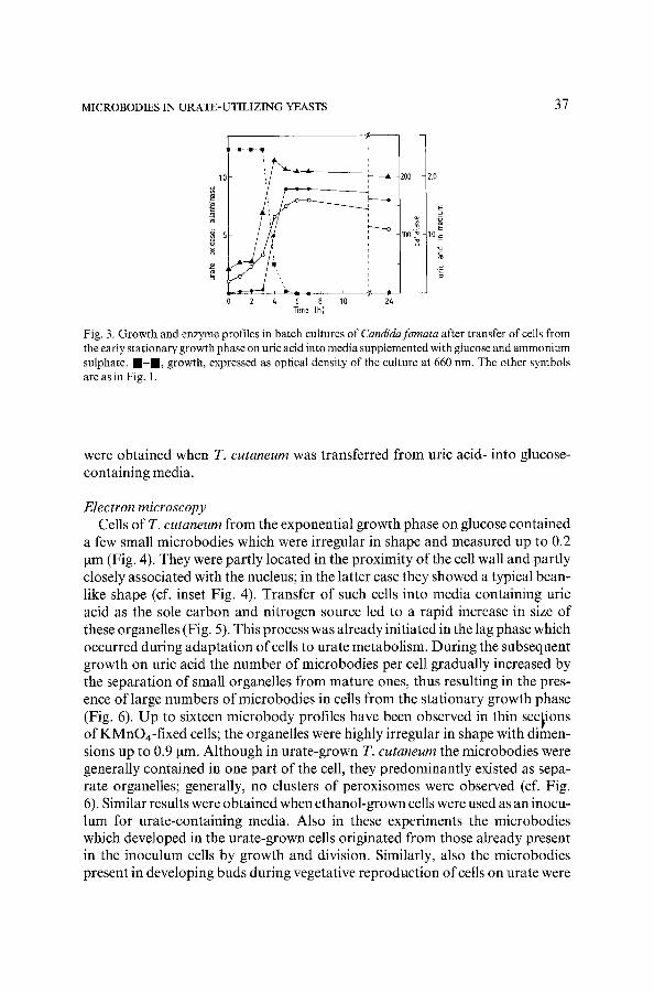
Transfer of uric acid-grown C. famata cells taken from the early stationary growth phase into media containing glucose and ammonium sulphate resulted in a rapid decrease in urate oxidase activity (Fig. 3). Catalase activity only slight- ly decreased after 2 h of cultivation, whereas the observed decrease in allantoin- ase activity could be accounted for by dilution of enzyme protein as a result of growth. Essentially similar results were obtained when glucose was added to exponentially growing cultures of C. famata growing on uric acid as the sole C- and N-source. Control experiments revealed that the observed decrease in
"urate oxidase was dependent upon the presence of glucose in the media. Transfer of cells into mineral media without any carbon or nitrogen source or media with ammonium sulphate as the N-source but lacking a carbon source did not result in inactivation of catalase, urate oxidase or allantoinase. In these experi- ments the observed decrease in enzyme activity could be explained by dilution of enzyme protein as a result of growth. Similar patterns of enzyme inactivation
MICROBODIES IN URATE-UTILIZING YEASTS 37
-m--m--~
1(
2
4 . .
6 8 10 2~ Time (h)
7
Fig. 3. Growth and enzyme profiles in batch cultures of Candidafamata after transfer of cells from the early stationary growth phase on uric acid into media supplemented with glucose and ammonium sulphate, m - m , growth, expressed as optical density of the culture at 660 nm. The other symbols are as in Fig. I.
were obtained when T. cutaneum was transferred from uric acid- into glucose- containing media.
Electron microscopy Cells of T. cutaneum from the exponential growth phase on glucose contained
a few small microbodies which were irregular in shape and measured up to 0.2 ~tm (Fig. 4). They were partly located in the proximity of the cell wall and partly closely associated with the nucleus; in the latter case they showed a typical bean- like shape (cf. inset Fig. 4). Transfer of such cells into media containing uric acid as the sole carbon and nitrogen source led to a rapid increase in size of these organelles (Fig. 5). This process was already initiated in the lag phase which occurred during adaptation of cells to urate metabolism. During the subsequent growth on uric acid the number of microbodies per cell gradually increased by the separation of small organelles from mature ones, thus resulting in the pres- ence of large numbers of microbodies in cells from the stationary growth phase (Fig. 6). Up to sixteen microbody profiles have been observed in thin sections of KMnO4-fixed cells; the organelles were highly irregular in shape with dimen- sions up to 0.9 ~tm. Although in urate-grown T. cutaneum the microbodies were generally contained in one part of the cell, they predominantly existed as sepa- rate organelles; generally, no clusters of peroxisomes were observed (cf. Fig. 6). Similar results were obtained when ethanol-grown cells were used as an inocu- lure for urate-containing media. Also in these experiments the microbodies which developed in the urate-grown cells originated from those already present in the inoculum cells by growth and division. Similarly, also the microbodies present in developing buds during vegetative reproduction of cells on urate were
38 M. VEENHUIS, M. C. HOOGKAMER-TE NIET AND W. J. MIDDELHOVEN
not synthesized 'de novo' but originated from the organelles present in the mother cell.
The mechanisms, described above for the development of microbodies in T. cutaneum during adaptation and subsequent growth on uric acid, were also oper- ative in C. famata when grown under identical conditions. The only difference observed was that the microbodies in C. famata invariably were present in large clusters, consisting of up to twelve microbodies in cells taken from the stationary phase of growth (Fig. 7).
The results of the biochemical experiments (Fig. 3) indicated that the synthesis of urate oxidase is partially repressed by glucose and ammonium ions. This is in agreement with the results of morphometrical analysis, which revealed that the number and volume density of the microbodies during growth of cells on urate were largely influenced by the composition of the growth medium. Maxi- mum values were observed under conditions in which uric acid served as the combined carbon- and nitrogen source; smaller numbers were present in cells grown on uric acid in the presence of ammonium sulphate as the nitrogen source whereas the volume density of the microbodies was lowest when uric acid served as the sole nitrogen source in media containing glucose as the carbon source (Figs 8,9; Table 1). The microbodies present in both organisms studied had a number of properties in common: they were frequently closely associated with the mitochondria and strands of endoplasmic reticulum, lacked crystalline inclu- sions whereas in freeze-etch preparations, their surrounding membranes showed smooth fracture faces (Fig. 10) similar to those described previously for H. poly- morpha and C. utilis (Veenhuis et al., 1983b; Zwart, 1983).
Cytochemical staining experiments indicated that the observed increase in mi- crobody size during growth of T. cutaneum or C. famata on uric acid was most probably due to the import of urate oxidase protein. Already in the early stages of growth on urate - 4 h after the transfer of cells - urate oxidase activity could be demonstrated cytochemically in the different microbodies present in the cells (Fig. 11), irrespective whether glucose- or ethanol-grown cells were used as the inoculum. In addition to urateoxidase also catalase activity was demonstrated in these organelles and therefore accumulation of this enzyme may add to the observed increase in microbody volume density. The cytochemical experiments indicated that, judged by the localization of the reaction products, the activities of both urate oxidase and catalase were confined to the peroxisomal matrix (Fig. 12, 13). The additional staining of the mitochondria in the DAB-based incubations occurred irrespective of the presence of substrate, was inhibited by cyanide ions and therefore was most probably due to staining of mitochondrial peroxidases (Hoffmann et al., 1970; Van Dijken et al., 1975).
The rapid inactivation ofurate oxidase in T. cutaneum and C.famata observed after exposure ofurate-grown cells to glucose-excess conditions (cf. Fig. 3), was not associated with the degradation of the microbodies present in the cells. Simi- larly, also the presence of ammonium sulphate did not lead to the degradation of the microbodies.

MICROBODIES IN URATE-UTILIZING YEASTS 39
Figs 4, 5. Thin sections of cells of Triehosporon cutaneum from the exponential growth phase on glucose, showing two microbody profiles (Fig. 4; arrows) and the increase in microbody size 6 h after transfer of cells to uric acid-containing media (Fig. 5). The inset of Fig. 4 shows a typical bean-like-shaped organelle, frequently observed in such cells in close association with the nucleus. Figs 6-9. Micrographs showing the proliferation of microbodies in cells from the early stationary growth phase on uric acid as the sole C- and N-source (Figs 6, 7), uric acid/ammonium sulphate (Fig. 8) and glucose/uric acid (Fig. 9). In Figs 8 and 9 microbodies are indicated by arrows. Figs 6, 8: Trichosporon eutaneum; Figs 7, 9: Candidafamata. All electron micrographs are taken of KMnO4-fixed cells; the marker represents 0.5 ~m. Abbrevia- tions: L, lipid droplet; M, microbody; N, nucleus; V, vacuole.

40 M. VEENHUIS, M. C. HOOGKAMER-TE NIET AND W. J. MIDDELHOVEN
Table 1. The proliferation of microbodies in Candida famata, expressed as number and volume density of the organelles in relation to different conditions of growth I
Growth conditions Number of Volume density peroxisomes of peroxisomes
C-source N-source
Glucose a m m o n i u m sulphate 0.1 0.2 Glucose uric acid 0.3 1.8 Uric acid ammon i um sulphate 2.1 6.9 Uric acid uric acid 4.2 10.1
1 Cells were grown in shake flask cultures and harvested during the late exponential growth phase. The number ofmicrobodies is expressed as average number per section, the volume density as percen- tage of the cytoplasmic volume.
DISCUSSION
Growth of yeasts on uric acid as the sole source of carbon and nitrogen is associated with the presence of increased numbers of microbodies in the cells (Middelhoven et al., 1983). The results of our present study indicate that these organelles are the sole sites ofurate oxidase activity (key enzyme in urate metab- olism) in the cells and since they also contained catalase they must be considered as peroxisomes. In fact during growth of the yeasts on uric acid these organelles are involved in the initial metabolism of the combined carbon and nitrogen source and therefore all the carbon and nitrogen required for growth flows via peroxisomes. This property of peroxisomes in urate-grown yeasts is not unique since also during growth of C. utilis on D-alanine a peroxisomal enzyme, namely D-amino acid oxidase, is involved in the primary metabolism of the carbon and nitrogen source (Zwart et al., 1983a). In addition, several examples have been described in which two different peroxisomal oxidases are involved in carbon and nitrogen metabolism, for instance during growth of cells of H. polymorpha on methanol as the C-source and methylamine, D-alanine or uric acid as the N-source (Veenhuis et al., 1981, 1983b; Zwart, 1983). Our results clearly indicate that the peroxisomes present in urate-grown cells of T. cutaneum and C. famata develop, similarly as described for other yeasts (Veenhuis et al., 1983b; Veenhuis and Harder, 1985) from pre-existing organelles present in the cells prior to their transfer to the new environment. Also in these organisms this process is indepen- dent of the former metabolic function(s) of the organelles.
Thus, peroxisomes may develop from glyoxysomes; intermediate forms may also exist, for instance, during growth of cells on ethanol as the C-source and uric acid as the N-source. Therefore, similarly as described for H. polymorpha and C. utilis (Zwart et al., 1983c; Veenhuis and Harder, 1985) the microbodies

MICROBODIES IN URATE-UTILIZING YEASTS 4l
Fig. 10. Survey of a ceil of Candidafamata, grown on uric acid, showing the smooth fracture faces (arrow) of the microbody membrane after freeze-fracturing. Figs 11-13. Cytochemical staining experiments showing the presence of urate oxidase in microbodies in Trichosporon cutaneum, 4 h after transfer of cells from glucose to uric acid-containing media (Fig. 11) and in cells of Candidafamata from the stationary growth phase on uric acid (Fig. 12) together with cataiase (Fig. 13). Urate oxidase was demonstrated with CeC13 and uric acid, catalase with DAB and H202. All electron micrographs are taken of KMnO4-fixed cells; the marker represents 0.5 lain. Abbrevia- tions: L, lipid droplet; M, microbody; N, nucleus; V, vacuole.
in T. cutaneum and C. famata const i tu te one class o f organelles; their u l t imate phys io log ica l funct ion(s) (peroxisomal , g lyoxysomal or g lyoxype rox i somal in na ture) is (are) largely prescr ibed by env i ronmenta l condi t ions . The presence o f a poss ible b iosynthe t ic funct ion, as recent ly descr ibed for mic robod ies in H. polymorpha and C. utilis (Zwar t et al., 1983b) is yet unknown.
As in prev ious studies in o ther yeasts, the n u m b e r and volume densi ty o f the mic robod ies in T. cutaneum and C.famata are largely dependen t on the compos i - t ion o f the cul t iva t ion media . This is, for instance, i l lus t ra ted by exper iments in which cells were grown in med ia con ta in ing uric acid in the absence or presence

42 M. VEENHUIS, M. C. HOOGKAMER-TE NIET AND W. J. MIDDELHOVEN
of glucose and/or ammonium sulphate. Both compounds repress urate oxidase synthesis and significantly influence the degree of proliferation of - enzymati- cally identical - microbodies (Table 1). In contrast to earlier observations on the turnover ofmicrobodies in H. polymorpha (Veenhuis et al., 1983a), exposure of urate-grown cells of T. cutaneum and C. famata to glucose- or ammonium- excess conditions did not lead to degradation of the peroxisomes present in the cells. The observed inactivation ofurate oxidase upon transfer of cells to glucose (Fig. 3) is therefore most probably due to a change in the physical state of the protein and may thus be considered a case of modification inactivation (Switzer, 1977).
As in other yeasts also the microbodies in C. famata and T. cutaneum are frequently observed in close association with strands of endoplasmic reticulum. The significance of these associations is still unclear. Most probably - as was indicated by our ultrastructural results - the endoplasmic reticulum is not in- volved in the biogenesis of the microbodies according to the classical model (De Duve and Baudhuin, 1966). In contrast, it must be expected that in yeasts urate oxidase is synthesized on free polysomes, similarly as in rat liver (Goldman and Blobel, 1978). However, the endoplasmic reticulum may be involved in the synthesis of the peroxisomal membrane. In this respect it must be stressed that, for reasons similar to those outlined for the other well-known examples of micro- body proliferation in yeasts, also urate-grown C. famata and T. cutaneum are excellent candidates for the study of various aspects ofmicrobody biogenesis.
We are indebted to Dr W. Harder for his interest and critical reading of the manuscript and to J. Zagers and K. Sjollema for their skilful assistance in prepar- ing the electron micrographs.
Received 18 April 1984
REFERENCES
D~ DuvE, C. and BAUDHUIN, P. 1966. Peroxisomes (microbodies and related particles). - - Physiol. Rev. 46: 323-357.
FUKUI, S. and TANAKA, A. 1979. Peroxisomes of alkane- and methanol-grown yeasts. Metabolic functions and practical applications. - - J. Appl. Biochem. 1: 171-201.
GOLOMAN, B. M. and BLOBEL, G. 1978. Biogenesis of peroxisomes: intracellular site of synthesis of catalase and uricase. - - Proc. Natl Acad. Sci. USA 75: 5066-5070.
HOFFMANN, H.-P., SZABO, A. and AVERS, C. J. 1970. Cytochemical localization of catalase activity in yeast peroxisomes. - - J, Bacteriol. 104:581-584.
LARUE, T. A. and SPENCER, J. F. T. 1968. The utilization of purines and pyrimidines by yeasts. - - Can. J. Microbiol. 14: 79-86.
MIDDELHOVEN, W. J. 1977. Isolation and characterization of methylammonium-resistant mutants of Saccharomyces cerevisiae with relieved nitrogen metabolite repression of allantoinase, arginase and ornithine transaminase synthesis. J. Gen. Microbiol. 100: 257-269.
MIDDELHOVEN, W. J., VAN DEN BRINK, J. A. and VEENHU~S, M. 1983. Growth of Candidafamata

MICROBODIES IN URATE-UTILIZING YEASTS 43
and Trichosporon cutaneum on uric acid as the sole source of carbon and energy, a hitherto un- known property of yeasts. - - Antonie van Leeuwenhoek 49: 361-368.
MOOR, H. 1964. Die Gefrier-Fixation lebender Zellen und ihre Anwendung in die Elektronen-Mi- kroskopie. - - Z. Zellforsch. Mikrosk. Anat. 62: 546-580.
SWlTZER, R. L. 1977. The inactivation of microbial enzymes in vivo. - - Annu. Rev. Microbiol. 31: 135-157.
VAN DDKEN, J. P., VEENHUIS, M, VERMEULEN, C. A. and HARDER, W. 1975. Cytochemical localiza- tion of catalase activity in methanol-grown Hansenula polymorpha. - - Arch. Microbiol. 105: 261 267.
VEEYI-IUIS, M., DOUMA, A., HARDER, W. and OSUMI, M. 1983a. Degradation and turnover of perox- isomes in the yeast Hansenula polymorpha induced by selective inactivation of peroxisomal en- zymes. - - Arch. Microbiol. 134: 193-203.
VEEN~IS, M. and HARDER, W. 1985. Yeast microbodies. Their substructure, biogenesis and turn- over in relation to environmental conditions. In A. H. Rose (ed.), The Yeasts, Vol. 2. - - Academic Press, London, in press.
VEENHUIS, M., KEIZER, I. and HARDER, W. 1979. Characterization of peroxisomes in glucose-grown Hansenulapolymorpha and their development after the transfer of cells into methanol-containing media. Arch. Microbiol. 120: 167-175.
VEE~UIS, M., VAN DIJKEN, J. P. and HARDER, W. 1976. Cytochemical studies on the localization of methanol oxidase and other oxidases in peroxisomes of methanol-grown Hansenula polymor- p h a . - - A r c h . Microbiol. 111: 123-135.
VEENI-IUIS, M., VAN D1JKEN, J. P. and HARDER, W. 1983b. The significance of peroxisomes in the metabolism of one-carbon compounds in yeasts. - - Adv. Microb. Physiol. 24: 1-82.
VEENHUIS, M., ZWART, K. B. and HARDER, W. 1981. Biogenesis and turnover of peroxisomes in- volved in the concurrent oxidation of methanol and methylamine in Hansenula polymorpha. - - Arch. Microbiol. 129: 35-41.
VOGELS, G. D. and VAN DER DRIFT, C. 1976. Degradation ofpurines and pyrimidines by microorgan- isms. - - Bacteriol. Rev. 40: 403-468.
ZWART, K. B. I983. Metabolic significance ofmicrobodies in the yeasts Candida utilis and Hansenula polymorpha. - - P h . D . Thesis, University of Groningen.
ZWART, K. B., OWRMARS, E. H. and HARDER, W. 1983a. The role ofperoxisomes in the metabolism of D-alanine in the yeast Candida utilis. - - FEMS Microbiol. Lett. 19:225-231.
ZWART, K. B., VEENHUIS, M. and HARDER, W. 1983b. Significance ofmicrobodies in the metabolism of L-aspartate in Candida utilis. - - FEMS Microbiol. Lett. 19: 273-279.
ZWART, K. B., VEENHUIS, M., PLAT, G. and HARDER, W. 1983c. Characterization of glyoxysomes in yeasts and their transformation into peroxisomes in response to changes in environmental condi t ions . - Arch. Microbiol. 136: 28-38.