Bioerosion by microbial euendoliths in benthic foraminifera from heavy metal-polluted coastal
14
Biogeosciences, 9, 4607–4620, 2012 www.biogeosciences.net/9/4607/2012/ doi:10.5194/bg-9-4607-2012 © Author(s) 2012. CC Attribution 3.0 License. Biogeosciences Bioerosion by microbial euendoliths in benthic foraminifera from heavy metal-polluted coastal environments of Portovesme (south-western Sardinia, Italy) A. Cherchi 1 , C. Buosi 1 , P. Zuddas 2 , and G. De Giudici 1 1 Dipartimento di Scienze Chimiche e Geologiche, Universit` a degli Studi di Cagliari, Via Trentino 51, 09127 Cagliari, Italy 2 Institut des Sciences de la Terre de Paris, Universit´ e Pierre et Marie Curie, Paris-Sorbonne, place Jussieu 4, 75252 Paris C´ edex05, France Correspondence to: A. Cherchi ([email protected]) Received: 26 June 2012 – Published in Biogeosciences Discuss.: 20 August 2012 Revised: 19 October 2012 – Accepted: 28 October 2012 – Published: 20 November 2012 Abstract. A monitoring survey of the coastal area facing the industrial area of Portoscuso-Portovesme (south-western Sardinia, Italy) revealed intense bioerosional processes. Ben- thic foraminifera collected at the same depth (about 2 m) but at different distances from the pollution source show ex- tensive microbial infestation, anomalous Mg/Ca molar ra- tios and high levels of heavy metals in the shell associated with a decrease in foraminifera richness, population density and biodiversity with the presence of morphologically ab- normal specimens. We found that carbonate dissolution in- duced by euendoliths is selective, depending on the Mg con- tent and morpho-structural types of foraminiferal taxa. This study provides evidences for a connection between heavy metal dispersion, decrease in pH of the sea-water and bio- erosional processes on foraminifera. 1 Introduction Boring microflora, constituted by cyanobacteria, algae and fungi may colonise carbonate substrates of both dead and living tissues of carbonate organisms (Tribollet, 2008). Bor- ing is evident in carbonate organisms from the Mesoprotero- zoic (Zhang and Golubic, 1987) to the present day as a con- sequence of environmental conditions (Hallock, 2005). Mi- crobial euendoliths have been described in coral reefs, mol- luscs, thalli of red algae (e.g. Golubic et al., 1975; Perkins and Tsentas, 1976; Budd and Perkins, 1980; Glaub, 1994, 2004; Chazottes et al., 1995, 2002; Le Campion-Alsumard et al., 1995; Vogel et al., 2000; Tribollet and Payri, 2001; Ghirardelli, 2002; Golubic and Schneider, 2003; Tribollet and Golubic, 2005; Tribollet, 2008; Tribollet et al., 2009), in coastal eroded limestones (Schneider and Torunski, 1983; Radtke et al., 1996), in carbonate grains (Tudhope and Risk, 1985; Al-Thukair and Golubic, 1991; Al-Thukair, 1999), as well in oil polluted marine environments (Campbell, 1983; Al-Thukair, 2002; Al-Thukair et al. 2007) and heavy-metal polluted lagoons (Succi et al., 2010). Microborings are major agents of bioerosion dissolving large quantities of calcium carbonate with a potential in buffering seawater pH, which leads to new questions on the effects of environmental factors such as eutrophication and atmospheric CO 2 increase on ocean composition evolution (Tribollet, 2008). On the other hand, present-day geochemi- cal models predict that the saturation state of surface ocean water with respect to carbonate minerals should decline dur- ing the twenty-first century. As a result calcareous organ- isms may have difficulties to calcify, leading to production of weaker skeletons and greater vulnerability to bioerosion. Organisms utilizing the more soluble form of CaCO 3 (arago- nite or high-Mg calcite) would be more adversely affected by elevated pCO 2 than those utilizing the less soluble low-Mg calcite (Morse, 1983; Morse et al., 2007). Investigations on microborings in foraminifera are docu- mented in a small body of literature (Perkins and Halsey, 1971; Alexanderson, 1972; Golubic et al., 1984; Peebles and Lewis, 1988; Shroba, 1993; Freiwald, 1995; Perry, 1998; Nielsen et al., 2003; Crevison and Hallock, 2007) and open a Published by Copernicus Publications on behalf of the European Geosciences Union.
Transcript of Bioerosion by microbial euendoliths in benthic foraminifera from heavy metal-polluted coastal
Biogeosciences, 9, 4607–4620, 2012www.biogeosciences.net/9/4607/2012/doi:10.5194/bg-9-4607-2012© Author(s) 2012. CC Attribution 3.0 License.
Biogeosciences
Bioerosion by microbial euendoliths in benthic foraminifera fromheavy metal-polluted coastal environments of Portovesme(south-western Sardinia, Italy)
A. Cherchi1, C. Buosi1, P. Zuddas2, and G. De Giudici1
1Dipartimento di Scienze Chimiche e Geologiche, Universita degli Studi di Cagliari, Via Trentino 51, 09127 Cagliari, Italy2Institut des Sciences de la Terre de Paris, Universite Pierre et Marie Curie, Paris-Sorbonne, place Jussieu 4,75252 Paris Cedex05, France
Correspondence to:A. Cherchi ([email protected])
Received: 26 June 2012 – Published in Biogeosciences Discuss.: 20 August 2012Revised: 19 October 2012 – Accepted: 28 October 2012 – Published: 20 November 2012
Abstract. A monitoring survey of the coastal area facingthe industrial area of Portoscuso-Portovesme (south-westernSardinia, Italy) revealed intense bioerosional processes. Ben-thic foraminifera collected at the same depth (about 2 m)but at different distances from the pollution source show ex-tensive microbial infestation, anomalous Mg/Ca molar ra-tios and high levels of heavy metals in the shell associatedwith a decrease in foraminifera richness, population densityand biodiversity with the presence of morphologically ab-normal specimens. We found that carbonate dissolution in-duced by euendoliths is selective, depending on the Mg con-tent and morpho-structural types of foraminiferal taxa. Thisstudy provides evidences for a connection between heavymetal dispersion, decrease in pH of the sea-water and bio-erosional processes on foraminifera.
1 Introduction
Boring microflora, constituted by cyanobacteria, algae andfungi may colonise carbonate substrates of both dead andliving tissues of carbonate organisms (Tribollet, 2008). Bor-ing is evident in carbonate organisms from the Mesoprotero-zoic (Zhang and Golubic, 1987) to the present day as a con-sequence of environmental conditions (Hallock, 2005). Mi-crobial euendoliths have been described in coral reefs, mol-luscs, thalli of red algae (e.g. Golubic et al., 1975; Perkinsand Tsentas, 1976; Budd and Perkins, 1980; Glaub, 1994,2004; Chazottes et al., 1995, 2002; Le Campion-Alsumard
et al., 1995; Vogel et al., 2000; Tribollet and Payri, 2001;Ghirardelli, 2002; Golubic and Schneider, 2003; Tribolletand Golubic, 2005; Tribollet, 2008; Tribollet et al., 2009),in coastal eroded limestones (Schneider and Torunski, 1983;Radtke et al., 1996), in carbonate grains (Tudhope and Risk,1985; Al-Thukair and Golubic, 1991; Al-Thukair, 1999), aswell in oil polluted marine environments (Campbell, 1983;Al-Thukair, 2002; Al-Thukair et al. 2007) and heavy-metalpolluted lagoons (Succi et al., 2010).
Microborings are major agents of bioerosion dissolvinglarge quantities of calcium carbonate with a potential inbuffering seawater pH, which leads to new questions on theeffects of environmental factors such as eutrophication andatmospheric CO2 increase on ocean composition evolution(Tribollet, 2008). On the other hand, present-day geochemi-cal models predict that the saturation state of surface oceanwater with respect to carbonate minerals should decline dur-ing the twenty-first century. As a result calcareous organ-isms may have difficulties to calcify, leading to productionof weaker skeletons and greater vulnerability to bioerosion.Organisms utilizing the more soluble form of CaCO3 (arago-nite or high-Mg calcite) would be more adversely affected byelevatedpCO2 than those utilizing the less soluble low-Mgcalcite (Morse, 1983; Morse et al., 2007).
Investigations on microborings in foraminifera are docu-mented in a small body of literature (Perkins and Halsey,1971; Alexanderson, 1972; Golubic et al., 1984; Peebles andLewis, 1988; Shroba, 1993; Freiwald, 1995; Perry, 1998;Nielsen et al., 2003; Crevison and Hallock, 2007) and open a
Published by Copernicus Publications on behalf of the European Geosciences Union.
4608 A. Cherchi et al.: Bioerosion by microbial euendoliths in benthic foraminifera
debate that needs to be developed. Benthic foraminifera se-creting calcium carbonate shells are considered to be verysensitive to the environmental conditions of the host sea wa-ter (e.g. Boyle, 1981; Wefer et al., 1999; Lea, 2004; Munselet al., 2010). Species distribution and population densities offoraminifera are in fact used for pollution monitoring (Alve,1991, 1995; Yanko et al., 1994, 1999; Debenay et al., 2001,2005; Coccioni et al., 2003, 2005; Armynot du Chatelet etal., 2004; Cherchi et al., 2009; Frontalini et al., 2009; Ro-mano et al., 2009; Frontalini and Coccioni, 2011; Denoyelleet al., 2012; Foster et al., 2012). Hallock (2000a, b) suggestedthat climatic change including increasing atmospheric CO2and stratospheric ozone depletion may be the cause of bor-ings developing on carbonate organisms. Indeed, effects ofelevatedpCO2 on coral reef bio-erosion have been observedand explained through a mechanism of carbonate dissolutionin a more acidic ocean (Tribollet et al., 2009). The activityof boring microflora can result in differential bioerosion de-pending on bothpCO2 partial pressure (Tribollet et al., 2009)and Mg/Ca ratio of the foraminifera tests (Peebles and Lewis,1988; Bentov and Erez, 2005).
The aim of our study is to investigate the effects of mi-crobioerosion on benthic foraminifera and trace element dy-namics in a known coastal industrial site polluted by heavymetals and industrial release of CO2 (Schintu and Degetto,1999). Field studies in the coastal area neighbouring the in-dustrial complex of Portoscuso-Portovesme (Southern Sar-dinia, Italy, Fig. 1) show that the presence of high levels ofheavy metals results in an increase in bioerosional processeson foraminifera by boring microflora. The stressed environ-mental conditions in this area have been highlighted in Cher-chi et al. (2009) through an analysis of the biotic indicesof foraminifera tests (Dominance, Shannon–Weaver, Simp-son, Evenness, Menhinick, Margalef, Equitability, Fisher-α, Berger-Parker and Q-mode Cluster Analyses – WardMethod) in this polluted environment.
2 Environmental setting
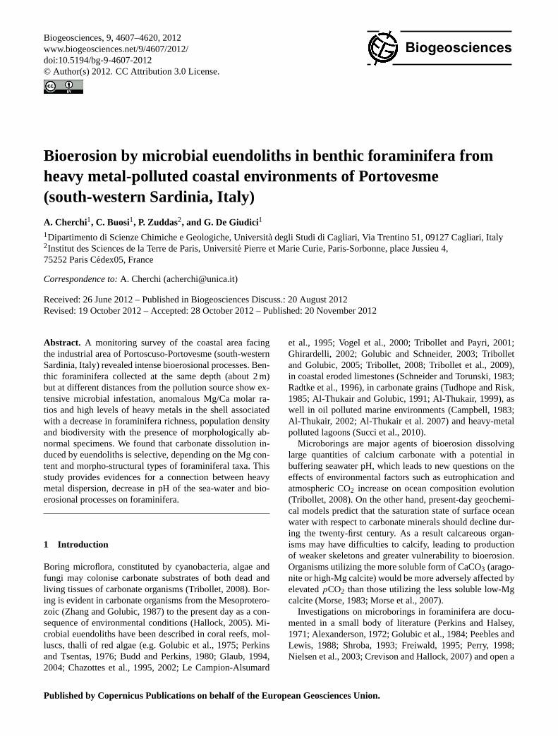
The study area is characterised by a relatively confined shal-low shelf, which slopes gently to deeper water environments(Fig. 1). Bottom sediments comprise sandy submarine beach(Pleistocene-Holocene in age), while Oligo-Miocene calc-alkaline volcanics crop out north of the sampling area. Thissouth-western coast of Sardinia has been under major an-thropogenic pressure since the 1960s, when the Portoscuso-Portovesme district began to develop and expand its activ-ities. The Portovesme Harbour, built in 1870 to ship ores(zinc blend, galena and pyrite) from the historical mines ofthe Sulcis-Iglesiente district, received major inputs from in-dustrial discharge until a few decades ago. Chemical analy-sis of marine sediments from 4 cores collected in the harbourof Portovesme facing the industrial complex revealed in theupper 2 cm high concentrations of Hg (up to 50 mg kg−1),Cd (up to 120 mg kg−1), Pb (up to 20 mg kg−1) and Zn (up
Fig. 1. Study area, location of sampling stations (Porto Pauleddu– NP, Porto Paglietto – PP, La Caletta – LC, Portovesme – PV,Punta S’Aliga – PA) and industrial complex of Portoscuso (SW Sar-dinia). 1. Submarine beach (grain size mean 0.13–0.1 mm). 2. Sub-marine beach (grain size mean 0.18–2.00 mm). 3. Alluvial deposits(Pleistocene–Holocene). 4. Volcanics (Oligo–Miocene). 5. Seabot-tom slope (%) from shoreline to 5 m isobath. 6. Longshore drift. 7.Sampling stations. 8. Red mud dumps. 9. Chimneys. 10. Lead-zincsmelter. 11. Electric power plant (1, 2, 5, 6 from Di Gregorio et al.,1996; modified).
to 70 mg kg−1) were found. Metal concentration decreaseswith an increase in distance between the sampling station andthe industrial effluent discharge point (Schintu and Degetto,1999). This large industrial development had a considerableenvironmental impact, such that this industrial district hasbeen declared an environmental hazard (D.P.C.M. – PrimeMinisterial decree, 23 April 1993) because of the several“danger centres” recognised (Agenzia Regionale per la Pro-tezione dell’ambiente della Sardegna – ARPAS, 2007).
Emissions into the atmosphere and all of the surroundingenvironments have been estimated annually at 65 000 tonsof SOx , 4000 tons of dust, 10 tons of Pb and 100 tons of Fe(Gazzetta Ufficiale Italiana, 1993). The large electric powerstations and the numerous chimneys produce also significantamounts of CO2 (Schintu and Degetto, 1999; Bettini andZanin, 2002). The industrial complex, developed on an al-luvial plain near the sea, includes factories producing alu-minium from bauxite and a lead-zinc smelter producing Pb,Zn, Cd, H2SO4 and Hg from Pb and Zn mixed sulphides. A
Biogeosciences, 9, 4607–4620, 2012 www.biogeosciences.net/9/4607/2012/

A. Cherchi et al.: Bioerosion by microbial euendoliths in benthic foraminifera 4609
Fig. 2.SEM images of surface bioerosion of porcelanaceous imper-forate foraminifera.(A) Tubular wavy tunnels parallel to substratein Pseudotriloculina rotunda(d’Orbigny) exhibiting a rectangularramification belonging toOrthogonum(cf. Form 1 in Wisshak etal., 2005).(B) Invasive infestation of microborings concentratedalong the sutures of the chamber walls producing large galleriesfrequently occupied by pennate diatoms (arrow) inQuinqueloculinasp.(C) Dense boring pattern made up of fungal filaments developedin Triloculina cf. marioniSchlumberger.(D) Colony of rod–shapedcryptoendolithic bacteria made into previous holes of larger diame-ter in the wall surface ofAdelosinasp.(E) Tubular borings, 4–5 µmin diameter, running parallel to surface of the test and penetratinginto the wall ofQuinqueloculina ungerianad’Orbigny.(F) Enlarge-ment of(E).
large dump of red muds (seawater-neutralised bauxite refin-ery residues) is located near the coast.
3 Material and methods
Sixteen surface sediment samples, coming from an areaabout 12 km2, were collected in the inner shelf facing the in-dustrial complex of Portoscuso-Portovesme where seawateris less than 2 m deep. Four stations (PP – Porto Paglietto, LC– La Caletta, PV – Portovesme, PA – Punta S’Aliga, Fig. 1)were sampled in summer. Sampled sediments came from theupper 1–2 cm below the water column. The water tempera-ture ranged between 27.7◦C and 30.6◦C. Temperature, pH,and Eh were measured for both the seawater and pore wa-ter of the sediments in every sampling station. Blank sam-ples were also collected for comparison purposes at the samebathymetry and temperature from an unpolluted coastal area(NP – Portopauleddu, Fig. 1). Benthic foraminifera assem-blages and their biodiversity indices, as well the geochemicalcharacterisation of the investigated area, was the aim of our
Fig. 3. SEM images of surface bioerosion of hyaline perforateforaminifera. (A) Rosalina bradyiCushman (umbilical side) ex-hibiting indeterminable filaments and, at right, two bunches of radi-ating tunnels (cf.Fascichnus). (B) Polymorphinasp. (oblique sideview) showing filaments of indeterminable borings in the wholetest. (C) Detail of Ammonia tepida(Cushman) spiral side inten-sively bored by long filaments of fungal hyphae with reproductiveorgans (cf.Saccomorpha clava). (D) Elphidium crispum(Linnaeus)showing in dorsal view a boring pattern of curved thin tunnels (1–2 µm in diameter) belonging to cf.Scolecia filosa, goings towardsthe primary pores of the foraminifer.(E) Densely ramified boringpattern ofIchnoreticulina elegans, characterised by dichotomousbranching goings into the primary pores ofE. crispum. (F) Invasivecolonies of indeterminable microborings (most likely bacteria, pos-sibly cyanobacteria) around and into primary pores ofE. crispum(arrows) producing a lateral enlargement of these until two–threecontiguous pores join (cf. Freiwald, 1995; Fig. 4).(G) Boring sys-tem developed among contiguous pores ofE. crispum, characterisedby large tunnels (arrows).(H) Enlargement of(G) showing largemicrobial overprint going into the pore and its transition from unis-eriate(u) to biseriate(b) cell arrangements, belonging toFascich-nus.
earlier paper, in order to assess the foraminiferal response toheavy metal pollution (Cherchi et al., 2009).
A cluster of four subsamples of constant volume (50 cm3)from every station (PP, LC, PV, PA) was collected to studyforaminifera assemblages used as environmental bioindica-tors. In laboratory, 16 subsamples were stained with RoseBengal (Walton, 1952) to differentiate living from dead spec-imens. Samples were then washed through a set of nested
www.biogeosciences.net/9/4607/2012/ Biogeosciences, 9, 4607–4620, 2012

4610 A. Cherchi et al.: Bioerosion by microbial euendoliths in benthic foraminifera
sieves (63, 125, 180, 250 µm). Foraminifera from 5 cc of the> 125 µm size fraction (a – medium grained) and 5 cc ofthe < 125 µm size fraction (b – fine-grained) were picked,counted and classified.
Considering the very low number (1–4 %) of living indi-viduals in the polluted environments (mainly at the PV andPA stations), total benthic foraminifera assemblages (deadand living) were used for statistical purposes, providing alarger database (Scott and Medioli, 1980; Samir and El-Din, 2001; Fatela and Taborda, 2002; Armynot du Chateletet al., 2004; Debenay and Guiral, 2006; Frezza and Car-boni, 2009). Total assemblages are almost identical statisti-cally and only diverge when the living/total becomes large(Murray, 1976, 1991) and biotic indices do not show signifi-cant differences between living and dead populations (Yankoet al., 1994). Occurrence of iron-oxy-hydroxides in bottomsediments, pyritized infaunal benthic foraminfera and fram-boidal pyrite in the tests from the polluted sampling stationswere occasionally found indicating reducing conditions.
Environmental scanning electron microscopy (ESEM) im-ages allow recognition of an extensive infestation by mi-crobial communities in the calcareous foraminiferal tests.The bioerosional process on foraminiferal tests was anal-ysed via ESEM (QUANTA 200, FEI, Hillsboro, Oregon, andpartly with EVOLS15, ZEISS). The epoxy resin casts offoraminifera were made at Erlangen University (Germany).Samples were gold coated before imaging, for analysis ofboth external surface and on epoxy-resin cast (Golubic etal., 1970, 1983; Wisshak et al., 2008). From ESEM images,microbial filaments and boreholes on the external surfaceshave been compared with positive epoxy resin casts, andmeasured. To estimate quantitatively the impact of the mi-crobial borings on the foraminifera and the selective bioero-sion on high-Mg and low-Mg foraminiferal tests, 300 indi-viduals were picked randomly from the fraction> 63 µm ofeach sample and were observed with a scanning electron mi-croscope. A total of 1200 foraminifera was examined. Thebioerosional features on several morpho-structural taxa havebeen quantitatively calculated. The percentage of calcareousdissolution in foraminifera, distinguishing between high-Mgand low-Mg tests, has been compared with heavy metal val-ues both in foraminiferal tests and the sediments, the pH ofpore waters and richness of specimens.
Seawater was collected at the water-sediment interface ofthe 4 polluted stations and at the reference pollution-freestation. After sampling seawater was rapidly filtered in situthrough a 0.4 µm pore-size polycarbonate filter with an all-plastic filtration assembly. Samples for cation analysis wereacidified with HNO3 suprapure acid to pH around 3. At eachsampling site, temperature, pH, and Eh were measured forboth seawater and sediment porewater. The Eh was measuredby platinum electrode and the value was corrected againstZobell’s solution (Nordstrom, 1977). The electrode used forpH potentiometric determination was calibrated against threeNIST-traceable buffer solutions (pH= 4.01, 7.00, 9.00 at
298 K). Reproducibility of pH calibrations, carried out be-fore and after measurements of a single solution, was betterthan 0.005 pH unit. However because of problems inherent tothe use of glass electrodes calibrated using NIST buffers instrong electrolyte solutions (see Dickson and Goyet, 1994),this measurement was only used to verify the solution elec-trochemical difference between the different sampled sta-tions.
Anions were determined by ion chromatography (ICDionex DX-120) and cations by ICP-AES (ARL-3520B) orICP-MS (Perkin Elmer DRC-e). Because of the complex sea-water matrix, the standard addition method, was employedfor the trace element determination (Cd, Pb and Zn) (Danzerand Currie, 1998; Cidu, 1999). The method validation wasverified by applying the same conditions to the CASS-3coastal seawater reference material. Samples and CASS-3were diluted five times with a 1 % ultrapure HNO3 solutionbefore sample spiking. Accuracy and precision were esti-mated at 10 % or better using the standard reference solu-tion and random duplicate samples. The limit of quantifica-tion (10σ value of blank solution response over time) was,respectively 0.01 µg l−1 for Cd, 0.6 µg l−1 for Pb and 0.1 forZn. The ionic balance was always in the order of ±8 %.
For digestion of samples with porcelanaceousforaminiferal tests, two portions of each sample werewashed with ultrapure water (MilliQ®) by mechanicalagitation in order to remove detrital grains. The sampleswere dried, accurately weighed and digested by slow heatingin a temperature bath at 30◦C in a Teflon beaker with 3 mlof ultrapure HNO3 (67 %). Solutions were diluted to 10 mlwith ultrapure water in volumetric flasks, and transferredto new HD-polyethylene bottles for storage (Jarvis, 1992).Metals were determined by ICP-AES and ICP-MS.
Field Emission Gun SEM (FEG-SEM) is a scanning elec-tron microscope with a high-energy beam of electrons in araster scan pattern producing information about surface to-pography and composition. The Quanta 200 FEG Environ-mental Scanning Electron Microscope (ESEM) uses a field-emission gun (FEG) electron source in an exceptionally highchamber pressure environment. It combines the advantagesof nanometer resolution to high signal to noise ratio in bothregular high vacuum and environmental (wet) modes. EDXAnalysis stands for energy-dispersive X-ray analysis; it issometimes referred to as EDS or EDAX. EDX analysis al-lows identification of the elemental composition of the spec-imen, or an area of interest thereof. The EDX analysis systemworks as an integrated feature of a scanning electron micro-scope (SEM), and can not operate on its own without thelatter.
Biogeosciences, 9, 4607–4620, 2012 www.biogeosciences.net/9/4607/2012/

A. Cherchi et al.: Bioerosion by microbial euendoliths in benthic foraminifera 4611
Table 1. Distribution on microborings at different sampling stations (PP, LC, PV and PA, see Fig. 1), taken from porcelanaceous (Po) andhyaline (Hy) foraminiferal substrates and their relative abundance (++ very common,+ common,= rare).
PP LC PV PAForaminifera substrata Po Hy Po Hy Po Hy Po Hy
Borings Trace-makers
Eurigonum nodosum Mastigocoleus testarum X X X X XX XSchmidt Lagerhein (cyanophyte)Fascichnuscf dactylus Hyella caespitosaBornet X XX X XX X(Radtke) and Flahault (cyanophyte)Scolecia filosa Plectonema terebrans X X X XX X X XRadtke Bornet and Flahault (cyanophyte)Scolecia meandria “vermicular borings” XRadtke (Zeff and Perkins, 1979) (cyanophyte)cf. Planobola macrogota cyanobacterium cf. X X XSchmidt alga cf.cf. Cavernula pediculata Gomontia polyrhiza X X XRadtke (Lagerhein) Bornet and
Flahault (chlorophyte)Ichnoreticulina elegans Ostreobium quekettii XX XX X X X(Radtke) Bornet and
Flahault (chlorophyte)Rhopalia catenata Phaeophila dendroides = X XRadtke Cronan (chlorophyte)Orthogonum fusiferum Ostracoblabe implexa X X XRadtke Bornet and Flahault (fungus)Orthogonum lineareGlaub cf. fungus = XX X X XOrthogonumisp. (Form 1 Xin Wisshak et al., 2005)Saccomorpha clava Dodgella priscus = X = XX X XX XRadtke Zebrowski (fungus)
Coccoid bacteria XX X X
4 Results and discussion
4.1 Bioerosion features
The presence of microboring traces and cavities on sampledbenthic foraminifera has been recognised using ESEM im-ages of both surface tests and epoxy resin casts. Identifiedtaxa, reported in Table 1, provide evidence of traces and cav-ities produced by phototrophic (cyanobacteria, chlorophyta)and heterotrophic (fungi) organisms. In Figs. 2 to 6, we illus-trate evidence of several endolithic traces of microbial bor-ings with morphological differences. Details of borings willbe described according to the different observational tech-niques: wall surfaces and epoxy resin casts.
Test surfaces of the porcelanaceous miliolids present theheaviest bioerosion features under the form of microbialclusters. Endolithic traces have morphological differences re-lated to biodiversity and to mode of life of the boring mi-crobial organisms. Infestation is characterised by thin indi-vidual tunnels, occasionally bifurcated, bag-shaped cavitiesand branching patterns composed by rhizoidal and short gal-leries radiating laterally from the central area. This branch-
ing pattern shows similarities withFascichnusisp. (Fig. 7a,b). From ESEM images of the traces on external surfaces offoraminiferal tests, our observations show that the taxa af-fected by higher bioerosion belong to the high-Mg porce-lanaceous group (Suborder Miliolina) and, among these, tothe genusQuinqueloculinawhich exhibits heavy microbialbioerosion. Figure 8 shows that inQuinqueloculinasp. theMg content at the bottom of a boring is one order of magni-tude lower compared to the unaltered shell surface, suggest-ing chemical reorganisation of the carbonate mineral compo-sition through a dissolution-precipitation process.
In several specimens the infestation is concentrated alongthe sutures of the chamber walls providing more easily a nu-trient source (Fig. 2b). A heavy infestation by diversifiedmicroborings in the test ofTriloculina has been recorded(Fig. 2c). Radiating traces are developed parallel to thewhole surface of foraminifers, in both porcelanaceous andhyaline types (Fig. 3 a,g,h and Fig. 7b), as previously ob-served by Glaub (2004) for “Fasciculusisp. 2”. Lined rod-shaped bacteria are sometimes visible on the later chambersof Adelosinaamong their weakly developed striae. These
www.biogeosciences.net/9/4607/2012/ Biogeosciences, 9, 4607–4620, 2012

4612 A. Cherchi et al.: Bioerosion by microbial euendoliths in benthic foraminifera
Fig. 4.SEM images of porcelanaceous foraminifera resin casts.(A)Chamber-wall of miliolid densely bored byEurigonum nodosumand globularSaccomorpha clava.(B) E. nodosumwith diagnosticlateral heterocysts (arrows) inside the thick chamber wall of mili-olid. (C) Dense assemblage of euendolithic borings in miliolid test.(D) Enlargement of(C) showingE. nodosumwith diagnostic hete-rocysts.(E) Pavement ofSaccomorpha clavainto miliolid chamberwall. (F) Colony of Scolecia meandriain miliolid chamber wall.(G) Microbial pavement in miliolid chamber wall exhibitingScole-cia filosa, E. nodosumand fungal sack-shaped cavities.(H) Diver-sified ichnotraces belonging toS. clava, cf. Planobola macrogotaand Ichnoreticulina elegansin miliolid test. (I) Dense colonies ofE. nodosumin miliolid test. (J) Colonies ofE. nodosuminside thethick chamber wall of miliolid.
cryptoendoliths bacteria (sensu Golubic et al., 1981) can gointo pre-existing holes, characterised by a larger diameter,clearly bored previously by another larger boring organism(Fig. 2d). Hyaline perforated tests (e.g.Elphidium crispum)frequently exhibit well-organised systems of microboringsbelonging to Ichnoreticulina eleganssurrounding and di-rected to pores (Fig. 3e). Bacteria infestation can produce ter-atological modifications as observed inE. crispum(Fig. 3f)where bacteria colonies induce lateral enlargement of pri-
Fig. 5. SEM images of hyaline foraminifera resin casts.(A)Euendoliths (arrows) inside the skeletal structure ofElphidiumcrispum (Linnaeus). (B) Enlargement of(A) showing indeter-minable filaments.(C) and (D) Saccomorpha clavaand indeter-minable cyanobacteria boring skeletal structure ofE. crispum. (E)Enlargement of(D) showing spherical cavities of cf.Planobolamacrogotaand sack-shaped cavity of cf.Cavernula. (F) E. crispum(vertical section) showing indeterminable filamentous (arrows) inthe complex skeletal pattern.(G) Enlargement of(F) showing fil-aments inside the skeletal of the foraminifera.(H) E. crispum(oblique section).(I) Enlargement of(H) showing tunnels (arrows)of borings in skeletal structures.
mary pores with enlargement of contiguous pores (cf. Frei-wald, 1995, inCibicides lobatulus). Superficial traces madeby endolithic boring communities can completely cover thewalls (Fig. 3a–c).
Several specimens ofE. crispumshow a boring patternmade of curved tunnels of 1–2 µm in diameter. Tunnels, afterlong runs, go towards the primary pores showing a compara-ble behaviour toI . elegansand they are tentatively attributedto Scolecia filosa. Infestation producing the primary poreconnection may be related to possible CO2 bioavailability
Biogeosciences, 9, 4607–4620, 2012 www.biogeosciences.net/9/4607/2012/

A. Cherchi et al.: Bioerosion by microbial euendoliths in benthic foraminifera 4613
Fig. 6. SEM images of hyaline foraminifera resin casts.(A) Tri-partite gallery of underteminable boring (cf.Orthogonumsp.) andglobular-shape cavities in skeletal structure ofAmmonia beccarii(Linnaeus).(B) Ichnoreticulina elegansin Ammoniasp.(C) Euen-doliths inside the peripherical wall chambers ofAmmonia tepida(Cushman).(D) Colony ofEurigonum nodosumin Ammoniasp.(E)Pavement of spherical cavities in the finely perforate test ofPoly-morphinasp. (F) and (G) Enlargement of(D) showing sphericalcavities of cf.Planobola macrogotaand sack-shaped cavities of cf.Cavernulasurrounded by foraminiferal tubules.
for phototrophic organisms from the respiratory activity ofthe heterotrophic host.
Epoxy resin casts reveal rich microbial communities,fairly diversified, both in porcelanaceous and hyaline tests(Table 1). Analysis of resin casts reveals euendoliths belong-ing to phototrophic (chlorophyta, cyanobacteria) and het-erotrophic (fungi) organisms boring both porcelanaceous andhyaline foraminifera. The wider and thicker walls of mili-olids provide a protected niche for the growth of euendoliths,as shown in Fig. 4. In porcelanaceous tests, dense pavementsof well-developed colonies of cyanobacteria and fungal spo-rangial cavities are very common. The presence of organicfilms inside the skeleton structure of these foraminifera pro-vides a source of food.
The ichnological interpretation of the euendoliths insidethe imperforate porcelanaceous tests is easier than for thoseinside the perforate hyaline hosts. The very complex inter-nal architecture of the taxa belonging to the families Elphidi-idae and Rotaliidae sometimes makes it difficult to distin-guish euendoliths from foraminifera ultrastructures (Figs. 5and 6). Even if the ichnological attribution of the euendoliths
Table 2.Seawater composition at the four sampling stations.
PP LC PV PA
pH 8.23 8.21 8.13 7.91Eh V 0.35 0.36 0.34 0.37T ◦C 27.7 27.9 29.6 30.6Ca mol l−1 0.011 0.012 0.011 0.011Mg mol l−1 0.058 0.059 0.057 0.058K mol l−1 0.012 0.012 0.012 0.012Na mol l−1 0.480 0.482 0.472 0.483SO4 mol l−1 0.037 0.040 0.043 0.037CI mol l−1 0.604 0.592 0.564 0.564HCO3 mol l−1 0.002 0.002 0.002 0.002Br mol l−1 0.001 0.001 0.001 0.001Ba mol l−1 0.053 0.052 0.058 0.076Cd mol l−1 0.002 0.002 0.002 0.004Li mol l−1 19.882 20.602 19.882 20.314Mo mol l−1 0.125 0.136 0.125 0.125Pb mol l−1 0.010 0.014 0.016 0.016Sr mol l−1 91.589 93.460 93.232 94.122Zn mol l−1 0.061 0.076 0.061 0.076
in the hyaline perforate taxa is generally uncertain, their pres-ence in the fine skeletal structure indicates that microbialcolonies can use foraminiferal canal-systems to penetrate in-side the test, and subsequently to develop boring activity inthe foraminiferal skeleton.
Our study shows that microbioerosion affects porcelana-ceous (high-Mg) imperforate miliolids (Adelosina, Pseu-dotriloculina, QuinqueloculinaandTriloculina) and penero-plids as well as hyaline (low-Mg) perforate foraminifera(Ammonia, Elphidium, Lobatula, Rosalina) (Fig. 9). Porce-lanaceous miliolids (QuinqueloculinaandTriloculina spp.)exhibit higher percentages of bioerosional features in PA sta-tion (28.72 %) and in PV station (21.75 %) while hyalineLo-batula lobatulaand Elphidium crispumreveal higher val-ues of microbial infestation in PA (16.89 %) and LC sta-tions (15.02 %), respectively. In particular,Elphidium testsshow well-developed colonies ofIchnoreticulina eleganssur-rounding and directed to primary pores. The clorophyceansin our samples seem to be more frequent in less pollutedsediments (LC). Figure 10a and b show that the numberof infested tests is 2 times higher in porcelanaceous com-pared to hyalines foraminifera and that the proportion ofinfested tests is 2–3 times higher in the lowest pH condi-tions. Bioerosion in hyaline specimens increases from 19.9 %(PP) to 59.7 % (PA), whereas in a porcelanaceous forms itincreases from 35.1 % (PP) to 80.3 % (PA). Our observationsdemonstrate that taxa affected by higher bioerosion belong tothe high-Mg porcelanaceous group (Suborder Miliolina), es-pecially Quinqueloculina. Infestation is often concentratedalong the chamber wall sutures where nutrient material ishigh (Fig. 2b). Diversified microborings on an infested testof Triloculina are illustrated in Fig. 2c.
www.biogeosciences.net/9/4607/2012/ Biogeosciences, 9, 4607–4620, 2012
4614 A. Cherchi et al.: Bioerosion by microbial euendoliths in benthic foraminifera
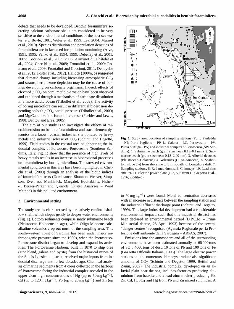
Fig. 7. SEM images of porcelanaceous foraminifera.(A) Quinqueloculinacf. berthelotianad’Orbigny intensively bored by euendoliths(black arrows) among which are colonies ofFascichnuscf. dactylus. (B) Enlargement of (A) exhibiting a well-developed colony ofF.cf. dactyluscharacterised by tunnels radiating from a central area.(C) Enlargement of(A) showing dome-shaped cavity of cf.Cavernulapediculatawith rhizoidal appendages, Zn biomineralised.(D) EDX analysis of dome-shaped cavity of cf.Cavernula pediculata.
4.2 Environmental conditions and bioerosion
Seawater composition from four sampled stations is given inTable 2. As previously reported porcelanaceous foraminiferaltests, known to be high-Mg foraminifera, are more infestedby bioerosion than hyaline forms. We found that Mg/Ca mo-lar ratio of porcelanaceous infested tests is between 0.09 and0.13, while in foraminiferal tests collected from an unpol-luted site (NP) without an apparent sign of bioerosion havea Mg/Ca value of 0.01 (Fig. 10c). Since Mg/Ca molar ratioof benthic foraminifera is normally assumed to be between0.0005 and 0.01 mol mol−1 (Lea, 1999; Toler et al., 2001;Toyofuku and Kitazato, 2005), we propose that conditions ofenvironmental stress may influence the amount of Mg incor-porated during the foraminifera’s growth. Our results are inagreement with the experimental investigation on Mg intra-shell reactions of Bentov and Erez (2005) and which are at-tributed to a kinetic mechanism played by the carbonate ionsin the calcite growth by Lopez et al. (2009). Porcelanaceousforaminiferal tests from the anthropogenically polluted la-goon of Santa Gilla (Frontalini et al., 2009) have a similarMg/Ca ratio (0.11 mol mol−1) which is greater than that col-lected at station NP, confirming the influence of the stressedenvironment on the Mg/Ca ratio of these biominerals.
Figure 10d shows that the amount of Cd, Zn and Pb in-corporated in the bioinfested shells in PP, LC, PV and PA ishigher compared to the pollution-free NP station. We foundthat the amount of heavy metals incorporated in the infestedtests is higher when the proportion of infested tests is higher.
The process of bioerosion in calcareous skeletal struc-tures can be regarded differently in high-Mg porcelanaceousimperforate tests and low-Mg bilamellar hyaline perforatetests and can also be related to the decrease of seawater pH(Fig. 10b). The porcelanaceous wall consists of a thick layerof high-Mg calcite needles with relatively large intersticesfilled with organic matter. Needles are randomly arrangedand coated inside by an inner organic lining and outside bythe outer organic layer (Towe and Cifelli, 1967; Hemlebenet al., 1986; Debenay et al., 2000a). The common infesta-tion of euendoliths in porcelanaceous tests rather than in hya-line tests can also be related to the thick calcite layer of theporcelanaceous wall, allowing an adequate erosional spacefor their growth. The perforate foraminifera are characterisedby structures of greater complexity (chamber-partitions, shellcavities, canal systems) compared with those of the porce-lanaceous group. In the bilamellar hyaline perforate group,the wall comprises carbonate layers separated by an organicmedian layer (Towe and Cifelli, 1967; Hansen and Reiss,1971; Hottinger, 1978, 2000; Hansen, 1999; Debenay et al.,2000b). Our data show that increasing heavy metal concen-trations in the sediments corresponds to an increase of mi-crobial infestation, reaching a peak at the more polluted sites(PV and PA; Fig. 10e).
Biogeosciences, 9, 4607–4620, 2012 www.biogeosciences.net/9/4607/2012/

A. Cherchi et al.: Bioerosion by microbial euendoliths in benthic foraminifera 4615
Fig. 8.SEM FEG images of porcelanaceous foraminifera (Quinque-loculinasp.). EDX analysis shows that Mg content at the bottom ofboring(A) is significantly lower than on unaltered shell surface(B).
The observed high level of microboring infestation is re-lated to the anomalous mechanism of bio-mineral construc-tion in the presence of a high level of heavy metals. In theseheavily polluted environments, euendolithic bioerosion de-velops in small shells of the biocarbonate substrates suchas calcareous foraminifera. Microbial organisms need thedevelopment of a trophic strategy related to the decreaseof micrograzers (mainly benthic foraminifera). Accordingto Mojtahid et al. (2011) some foraminifera species, suchasHaynesina germanica, Ammonia beccariiand the single-chambered speciesPsammophagasp., consume and digestlarge quantities of bacteria. The estimated grazing ratesof these three intertidal species were comparable:∼ 3.2 ngCind−1 h−1 (∼ 16 000 bacterial cells ind−1 h−1) for lowbacterial concentration (∼ 43× 106 cells) and∼ 5.7 ng Cind−1 h−1 (∼ 28 000 bacterial cells ind−1 h−1) for higherbacterial concentration (∼ 86× 106 cells). Biotic indices(Faunal Density, Species Richness, Dominance, Shannon-Weaver, Simpson, Evenness, Menhinick, Margalef, Equi-tability, Fisher-α, Berger-Parker; Cherchi et al., 2009) per-formed on foraminiferal assemblages show an abrupt de-crease with increasing heavy metal fluxes (Fig. 10a, d).Optical observations reveal an high number of abnormaland small specimens in accordance with an increase of in-dustrial pollution. Morphological abnormalities, which arecommonly found in tests from pollution-stressed benthicforaminiferal populations, may be related to incorporation of
Fig. 9. Occurrence of bioerosion given in percent in selected taxabelonging to porcelanaceous (high-Mg) imperforate miliolids andpeneroplids (*) and hyaline (low-Mg) perforate foraminifera (Am-monia, Elphidium, Lobatula, Rosalina) from sampling stations (PP,LC, PV, PA).
higher concentrations of Mg and other elements from seawa-ter into their tests (Yanko et al., 1994, 1998).
In the sandy beach part of the study area, limestone out-crops are lacking and bottom sediment is made of quartzgrains. Anomalous concentrations of euendolithic commu-nities in shallow polluted waters, question the capacity ofintensive industrial activity to produce extreme environmen-tal conditions in restricted local areas only. Mine waste con-taining toxic concentrations of heavy metals (Fe, Al, Cu, Zn,Cd, Pb, Ni, Co and Cr) discharged into aquatic systems mayproduce degradation of water quality and aquatic life (Nord-strom, 2011). Our study reveals that peculiar environmentsaffected by heavy metal fluxes from industrial processing arenot biologically dead as they are teeming with microbes, in-cluding bacteria, archaea, fungi and algae.
Global scale geochemical models that predict the carbon-ate saturation state of the surface waters in the twenty-firstcentury suggest that calcareous organisms may have diffi-culty calcifying, leading to production of weaker skeletonsand greater vulnerability to erosion. We estimated thepCO2partial pressure at equilibrium with the sampled seawater (bypH and alkalinity) using the Millero and Scheiber (1982) ionpairing model to estimate activity coefficients and found val-ues between 3–5 times higher than the average for the opensea nearby. This study of biomineralisation confirms that, atleast in shallow waters, high-Mg carbonates are altered fastercompared to low-Mg carbonates (Fig. 10b), confirming thatorganisms using the more soluble forms of CaCO3 (aragoniteand high-Mg calcite) are more adversely affected by higherpCO2. Our results are in agreement with earlier laboratoryexperiments (Morse, 1983; Morse et al., 2007) where it waspredicted that dissolution of various benthic marine organ-isms may increase under natural higherpCO2 partial pres-sure (Ries et al., 2009).
www.biogeosciences.net/9/4607/2012/ Biogeosciences, 9, 4607–4620, 2012

4616 A. Cherchi et al.: Bioerosion by microbial euendoliths in benthic foraminifera
Fig. 10. Occurrence of bioerosion given in percent in microbially infested foraminiferal tests from Portoscuso coastal environments.(A)Correlation between infested foraminifera percentages and total number of foraminiferal tests.(B) Correlation between porcelanaceoushigh-Mg and hyaline low-Mg tests and sea water pH.(C) Mg/Ca ratio in foraminiferal tests increases in more highly polluted sites.(D)Cd/Ca, Pb/Ca and Zn/Ca ratio in foraminiferal tests increase in more highly polluted sites.(E) Increase of heavy metal values in sedimentscorresponds to increase in microbial infestation.
5 Summary and conclusions
In our study, samples were collected at the same depth (about2 m) from shallow sediments affected by serious industrialcontamination in the coastal environments of Portoscuso-Portovesme. In this area, foraminiferal shells have a highcontent of heavy metals and are actively infested by micro-borings. As far as we know, these infested foraminiferal com-munities provide a state-of-the-art dataset for the understand-ing of bioerosional processes in foraminiferal hosts in pol-luted environments.
The results show (i) microbioerosion is higher in high-Mgforaminiferal tests compared to the low-Mg tests, (ii) the roleof foraminiferal skeletal architecture in the boring process,
(iii) the abundance of euendoliths is favoured by the effect ofgreater concentrations of heavy metals, especially Zn, as aninorganic nutrient.
We interpret that in the shallow water and low hydro-dynamic conditions of the Portoscuso-Portovesme lagoon,the heavy metals leached from mine tailings and industrialdischarge are not immediately dispersed in the sea waterand, thus, foraminifera can concentrate heavy metals in theirshells. This takes place in a complex biomineralisation pro-cess. In agreement with geochemical models and previousliterature, we found that high-Mg carbonate shells dissolvefaster and are deeply infested by microborings. While geo-chemical models already predict that CO2 increase is affect-ing biogenic carbonate reservoirs, this study demonstrates
Biogeosciences, 9, 4607–4620, 2012 www.biogeosciences.net/9/4607/2012/

A. Cherchi et al.: Bioerosion by microbial euendoliths in benthic foraminifera 4617
that the heavy metal dispersion process can contribute toglobal CO2 change via a complex ecological process.
Acknowledgements.The authors are very grateful to M. Wisshak(Senckenberg am Meer, Wilhelmshaven, Germany) for his helpin the epoxy resin casts preparation, in the interpretation of theichnotaxa inventory and for his useful suggestions which greatlyimproved the manuscript. We thank Rich B. Wanty (USGS,Denver) for his discussion and comments. We thank P. Meloni andG. Carcangiu (University of Cagliari, Italy) for their collaborativeassistance during electronic microscopic analysis performed withEVOLS15, ZEISS microscope. We thank E. Musu for assistancewith SEM-FEG imaging (DistrICT LAB, Sardegna Ricerche,Pula). This work was supported by the EU UMBRELLA Project(funding 226870). The authors express their sincere gratitude toAlan Lord (Forschungsinstitut Senckenberg, Frankfurt-am-Main)for the revision of the English text. The authors warmly thankErika Kothe (Jena University), Elisabetta Erba (Milano University),anonymous reviewers and the editor for their useful comments andsuggestions.
Edited by: H. Kitazato
References
Agenzia Regionale per la Protezione dell’ambiente della Sardegna(ARPAS): Determinazione dei valori di fondo nelle matrici am-bientali dell’area di Portoscuso (CI), Progetto operativo, 29 pp.,2007.
Alexanderson, E. T.: Micritization of carbonate particles: Processesof precipitation and dissolution in modern shallow-marine sed-iments, Bulletin of the Geological Institutions of the Universityof Uppsala, N.S., 3, 201–236, 1972.
Al-Thukair, A. A.: Euendolithic micro-organisms found to bore thecalcareous sediment of the Arabian Gulf, in: Proceedings of theSecond International Conference on Engineering for Calcareoussediments, Bahrain, 21–24 February 1999, 291–299, 1999.
Al-Thukair, A. A.: Effect of oil pollution on euendolithic cyanobac-teria of the Arabian Gulf, Environ. Microbiol., 4, 125–129, 2002.
Al-Thukair, A. A. and Golubic, S.: New endolithic cyanobacteriafrom the Arabian Gulf. I.Hyella immanissp. nova, J. Phycol.,27, 766–780, 1991.
Al-Thukair, A. A., Abed, R. M. M., and Mohamed, L.: Microbialcommunity of cyanobacteria mats in the intertidal zone of oil-polluted coast of Saudi Arabia, Mar. Pollut. Bull. 54, 173–179,2007.
Alve, E.: Benthic foraminifera reflecting heavy metal pollution inSørljord, Western Norway, J. Foraminifer. Res., 34, 1641–1652,1991.
Alve, E.: Benthic foraminifera response to estuarine pollution, a re-view, J. Foraminifer. Res., 25, 190–203, 1995.
Armynot du Chatelet, E., Debenay, J. P., and Saulard, R.:Foraminiferal proxies for pollution monitoring in moderatelypolluted harbors, Environ. Pollut., 127, 27–40, 2004.
Bentov, S. and Erez, I.: Novel observations on biomineralisationprocesses in foraminifera and implications for Mg/Ca ratio in theshells, Geology, 33, 841–844, 2005.
Bettini, V. and Zannin, D. (Eds.): Portoscuso, la gassificazione dellaV.I.A.-Il Gassificatore Ati-Sulcis, le problematiche ambientaliconnesse al progetto, CUEN, Napoli, Italy, 1–190, 2002.
Boyle, E. A.: Cadmium, zinc, copper, and barium in foraminiferatests, Earth Planet. Sc. Lett., 53, 11–35, 1981.
Budd, D. A. and Perkins, R. D.: Bathymetric zonation and paleoeco-logical significance of (algal) microborings in Puerto Rican shelfand slope sediments, J. Sediment. Petrol., 50, 881–904, 1980.
Campbell, S. E.: Petrochemical pollution: endolith response, in:VIes jornees d’etudes sur les pollutions marines en Mediterranee,Workshop on Pollution of the Mediterranean, Cannes, France, 2–4 December 1982, 183–189, 1983.
Chazottes, V., Le Campion-Alsumard, T., and Peyrot-Clausades,M.: Bioerosion rates on coral reefs: interactions between mac-roborers, microborers and grazers (Moorea, French Polynesia),Palaeogeogr. Palaeocl. Palaeoecol., 113, 189–198, 1995.
Chazottes, V., Le Campion-Alsumard, T., Peyrot-Clausade, M., andCuet, P.: The effects of eutrophication-related alterations to coralreef communities on agents and rates of bioerosion (Reunion Is-land, Indian Ocean), Coral Reefs, 21, 375–390, 2002.
Cherchi, A., Da Pelo, S., Ibba, A., Mana, D., Buosi, C., and Floris,N.: Benthic foraminifera response and geochemical characteri-zation of the coastal environment surrounding the polluted in-dustrial area of Portovesme (South-Western Sardinia, Italy), Mar.Pollut. Bull., 59, 281–296, 2009.
Cidu, R.: Trace elements Li, Be, B, Al, V, Cr, Co, Ni, Se, Sr, Ag,Sn, Sb, Ba and Tl, in: Handbook of Water Analysis, edited by:Nollet, L. M. L. and Dekker, M., Inc., 459–482, 1999.
Coccioni, R., Marsili, A., and Venturati, A.: Foraminiferi e stressambientale, in: Verso la Gestione integrata della Costa del MonteSan Bartolo: Risultati di un Progetto Pilota, edited by: Coccioni,R., Quaderni del Centro di Geobiologia dell’Universita degliStudi di Urbino 1, 99–118, 2003.
Coccioni, R., Frontalini, F., Marsili, A., and Troiani, F.:Foraminiferi bentonici e metalli in traccia: implicazioni ambien-tali, in: La dinamica Evolutiva della Fascia Costiera tra le foci deiFiumi Foglia e Metauro: Verso la Gestione Integrata di una Costadi Elevato Pregio Ambientale, edited by: Coccioni, R., Quadernidel Centro di Geobiologia dell’Universita degli Studi di Urbino3, 57–92, 2005.
Crevison, H. and Hallock, P.: Anomalous features observed on testsof live archaiasine foraminifers from the Florida Keys, USA, J.Foraminifer. Res., 37, 223–233, 2007.
Danzer, K. and Currie, L. A.: Guidelines for calibration in analyticalchemistry, Pure Appl. Chem., 70, 993–1014, 1998.
Debenay, J. P. and Guiral, D.: Mangrove swamp foraminifera, indi-cators of sea level or paleoclimate cf., Revue de Paleobiologie,2, 567–574, 2006.
Debenay, J. P., Guillou, J. J., Redois, F., and Geslin, E.: Distributiontrends of foraminiferal assemblages in paralic environments: abase for using foraminifera as early warning indicators of an-thropic stress, in: Environmental Micropaleontology, edited by:Martin, R., Kluwer Academic/Plenum Publishing Corporation,39–67, 2000a.
Debenay, J. P, Guillou, J. J., Geslin, E., and Lesourd, M.: Crystal-lization of calcite in foraminiferal tests, Micropaleontology, 46,87–94, 2000b.
Debenay, J. P., Geslin, E., Eichler, B. B., Duleba, W., Sylvestre,F., and Eichler, P.: Foraminiferal assemblages in a hypersaline
www.biogeosciences.net/9/4607/2012/ Biogeosciences, 9, 4607–4620, 2012
4618 A. Cherchi et al.: Bioerosion by microbial euendoliths in benthic foraminifera
lagoon Araruama (RJ) Brazil, J. Foraminifer. Res., 31, 133–151,2001.
Debenay, J. P., Millet, B., and Angelidis, M. O.: Relationships be-tween foraminiferal assemblages and hydrodynamics in the Gulfof Kalloni, Greece, J. Foraminifer. Res., 35, 327–343, 2005.
Denoyelle, M., Geslin, E., Jorissen, F. J., Cazes, L., and Galgani,F.: Innovative use of foraminifera in ecotoxicology: A marinechronic bioassay for testing potential toxicity of drilling muds,Ecol. Indic., 12, 17–25, 2012.
Di Gregorio, F., Federici, P. R., Fierro, G., and Ginesu, S.: At-lante delle spiagge della Sardegna, Foglio n. 232–233, Isola diS. Pietro-Carbonia, R.A.S, Cagliari, 1996.
Dickson, A. G. and Goyet, C. (Eds.): Handbook of methods for theanalysis of the various parameters of the carbon dioxide systemin sea water, Version 2, ORNL/CDIAC-74, 1–187, 1994.
Fatela, F. and Taborda, R.: Confidence limits of species proportionin microfossil assemblage, Mar. Micropaleontol., 45, 169–174,2002.
Foster, W. J., Armynot du Chatelet, E., and Rogerson, M.: Testingbenthic foraminiferal distributions as a contemporary quantita-tive approach to biomonitoring estuarine heavy metal pollution,Mar. Pollut. Bull., 64, 1039–1048, 2012.
Freiwald, A.: Bacteria-induced carbonate degradation: a tapho-nomic case, study ofCibicides lobatulusfrom a high-boreal car-bonate setting, Palaios, 10, 337–346, 1995.
Frezza, V. and Carboni, M. G.: Distribution of recent foraminiferalassemblages near the Ombrone River mouth (Northern Tyrrhe-nian Sea, Italy), Revue de Micropaleontologie, 52, 43–66, 2009.
Frontalini, F. and Coccioni, R.: Benthic foraminifera as bioindica-tors of pollution: A review of Italian research over the last threedecades, Revue de Micropaleontologie, 54, 115–127, 2011.
Frontalini, F., Buosi, C., Da Pelo, S., Coccioni, R., Cherchi, A., andBucci, C.: Benthic foraminifera as bio-indicators of trace ele-ment pollution in the heavily contaminated Santa Gilla lagoon(Cagliari, Italy), Mar. Pollut. Bull., 58, 858–877, 2009.
Gazzetta Ufficiale della Repubblica Italiana: Piano di disinquina-mento per il risanamento del territorio del Sulcis-Iglesiente, 190,14 August 1993.
Ghirardelli, L. A.: Endolithic Microorganisms in Live and DeadThalli of Coralline Red Algae (Corallinales, Rhodophyta) in theNorthern Adriatic Sea, Acta Geologica Hispanica, 37, 53–60,2002.
Glaub, I.: Mikrobohrspuren in ausgewahlten Ablagerungsraumendes europaischen Jura und der Unterkreide (Klassifikation undPalokologie), Cour. For. Sekenbg., 174, 1–289, 1994.
Glaub, I.: Recent and sub-recent microborings from the upwellingarea off Mauritania (West Africa) and their implications forpalaeoecology, in: The Applications of Ichology to Palaeoenvi-ronmental and Stratigraphic Analysis, edited by: Mc-Ilroy, D.,Geolog. Soc. London, Spec. Publ., 228, 63–76, 2004.
Golubic, S. and Schneider, J.: Microbial endoliths as internalbiofilms, in: Fossil and Recent biofilms, edited by: Krumbein, W.E., Dornieden, T., and Volkmann, M., Kluwer, Dordrecht, 249–263, 2003.
Golubic, S., Brent, G., and Le Campion-Alsumard, T.: Scanningelectron microscopy of endolithic algae and fungi using a mul-tipurpose casting-embedding technique, Lethaia, 3, 203–209,1970.
Golubic, S., Perkins, R. D., and Lukas, K. J.: Boring microorgan-isms and microborings in carbonate substrates, in: The Studyof Trace Fossils, edited by: Frey, R. W., Springer-Verlag, NewYork., 229–259, 1975.
Golubic, S., Friedmann, I., and Schneider, J.: The lithobiontic eco-logical niche, with special reference to microorganisms, Sedi-ment. Geol., 51, 475–478, 1981.
Golubic, S., Campbell, S. E, and Spaeth, C.: Kunstharzangusse fos-silifer Mikroben-Bohrgange, Praparator, 29, 197–200, 1983.
Golubic, S., Campbell, S. E, Drobne, K., Cameron, B., Balsam, W.L., Cimerman, F., and Dubois, L.: Microbial endoliths: a ben-thic overprint in the sedimentary record, and a paleobathymet-ric cross-reference with foraminifera, J. Paleontol., 58, 351–361,1984.
Hallock, P.: Symbiont-bearing foraminifera:harbingers of globalchange cf., Micropaleontology, 46, 95–104, 2000a.
Hallock, P.: Larger foraminifera as indicators of coral-reef vital-ity, in: Environmental Micropaleontology, edited by: Martin,R., Kluwer Academic/Plenum Publishing Corporation, 121–150,2000b.
Hallock, P.: Global change and modern coral reefs: New opportu-nities to understand shallow-water carbonate depositional pro-cesses, Sediment. Geol., 175, 19–33, 2005.
Hansen, H. J.: Shell construction in modern calcareousForaminifera, in: Modern Foraminifera, edited by: Sen Gupta,B. K., Springer, New York, 57–70, 1999.
Hansen, H. J. and Reiss, Z.: Electron microscopy of Rotaliaceanwall structures, B. Geol. Soc. Denmark, 20, 329–346, 1971.
Hemleben, C., Anderson, O. R., Berthold, W., and Spindler, M.:Calcification and chamber formation in Foraminifera – a briefoverview, in: Biomineralization in Lower Plants and Animals,edited by: Leadbeater, B. S. C. and Riding, R., Oxford, England,Clarendon Press, 237–249, 1986.
Hottinger, L.: Comparative anatomy of elementary shell structuresin selected larger foraminifera, in: Foraminifera, edited by: Hed-ley, R. H. and Adams, C. G., New York, Academic Press, 3, 203–266, 1978.
Hottinger, L.: Functional morphology of benthic foraminiferalshells, envelopes of cells beyond measure, Micropaleontology,46, 57–86, 2000.
Jarvis, I.: Sample preparation for ICPMS, in: Handbook of Induc-tively Coupled Plasma Mass Spectrometry, edited by: Jarvis, K.E., Gray, A. L., and Houk, R. S., Blackie Academic and Profes-sional, Glasgow, UK, 172–224, 1992.
Lea, D. W.: Trace elements in foraminiferal calcite, in: Modernforaminifera, edited by: Sen Gupta, B. K., Kluwer AcademicPublishers, UK, 259–277, 1999.
Lea, D. W.: Elemental and isotopic proxies of past ocean temper-atures, in: The Oceans and Marine Geochemistry, Treatise ongeochemistry volume 6, edited by: Elderfield, H., Holland, H. D.,and Turekian, K. K., Elsevier, Amsterdam, Heidelberg, 365–390,2004.
Le Campion-Alsumard, T., Golubic, S. and Hutchings, P.: Micro-bial endoliths in skeletons of live and dead corals:Porites lobata(Moorea, French Polynesia), Mar. Ecol.-Prog. Ser., 117, 149–157, 1995.
Lopez, O., Zuddas, P., and Faivre, D.: Influence of temperatureand seawater composition on calcite crystal growth mechanismsand kinetics: implications for Mg incorporation in calcite lattice,
Biogeosciences, 9, 4607–4620, 2012 www.biogeosciences.net/9/4607/2012/

A. Cherchi et al.: Bioerosion by microbial euendoliths in benthic foraminifera 4619
Geochim. Cosmochim. Acta, 73, 23370–23470, 2009.Millero, F. J. and Scheiber, D. R.: Use of the ion pairing model to
estimate activity coefficients of the ionic components of naturalwater, Am. J. Sci., 282, 1508–1540, 1982
Mojtahid, M., Zubkov, M. V., Hartmann, M., and Gooday, A. J.:Grazing of intertidal benthic foraminifera on bacteria: Assess-ment using pulse-chase radiotracing, J. Exp. Mar. Biol. Ecol.,399, 25–34, 2011.
Morse, J. W.: The kinetics of calcium carbonate dissolution andprecipitation, in: Carbonates: Mineralogy and Chemistry, editedby: Reeder, R. J., Mineralogical Society of America, WashingtonD.C., 11, 227–264, 1983.
Morse, J. W., Arvidson, R. S., and Luttge, A.: Calcium carbonateformation and dissolution, Chem. Rev., 107, 342–381, 2007.
Munsel, D., Kramar, U., Dissard, D., Nehrke, G., Berner, Z., Bijma,J., Reichart, G.-J., and Neumann, T.: Heavy metal incorporationin foraminiferal calcite: results from multi-element enrichmentculture experiments with Ammonia tepida, Biogeosciences, 7,2339–2350,doi:10.5194/bg-7-2339-2010, 2010.
Murray, J. W.: Comparative studies of living and dead benthicforaminiferal distributions, in: Foraminifera, edited by: Hedley,R. H. and Adams, C. G., Academic Press, London, 45–110, 1976.
Murray, J. W. (Ed.): Ecology and Palaeoecology of BenthicForaminifera, Longman Scientific and Technical, New York, 1–426, 1991.
Nielsen, K. S. S., Nielsen, J. K., and Bromley, R. G.: Palaeoecolog-ical and ichnological significance of microborings in Quaternaryforaminifera, Palaeontol. Electron., 6, 1–13, 2003.
Nordstrom, D. K.: Thermochemical redox equilibria of Zobell’s so-lution, Geochim. Cosmochim. Acta, 41, 1835–1841, 1977.
Nordstrom, D. K: Hydrogeochemical processes governing the ori-gin, transport and fate of major and trace elements from minewastes and mineralized rock to surface waters, Appl. Geochem.,26, 1777–1791, 2011.
Peebles, M. W. and Lewis, R. D.: Differential infestation of shallow-water benthic foraminifera by microboring organisms: possiblebiases in preservation potential, Palaios, 3, 345–351, 1988.
Perkins, R. D. and Halsey, S. D.: Geologic significance of micro-boring fungi and algae in Carolina shelf sediments, J. Sediment.Petrol., 41, 843–853, 1971.
Perkins, R. D. and Tsentas, C. I.: Microbial infestations of carbonatesubstrates planted on the St Croix shelf, West Indies, Geol. Soc.Am. Bull., 87, 1616–1628, 1976.
Perry, C. T.: Grain susceptibility to the effects of microboring: im-plications for the preservation of skeletal carbonates, Sedimen-tology, 45, 39–51, 1998.
Radtke, G., Le Campion-Alsumard, T., and Golubic, S.: Microbialassemblage of the bioerosional “notch” along tropical limestonecoasts, Algological Studies, 83, 469–482, 1996.
Ries, J. B., Cohen, A. L., and McCorkle, D. C.: Marine calcifiersexhibit mixed responses to CO2-induced ocean acidification, Ge-ology, 37, 1131–1134, 2009.
Romano, E., Bergamin, L., Ausili, A., Pierfranceschi, G., Maggi,C., Sesta, G., and Gabellini, M.: The impact of the Bagnoli in-dustrial site (Naples, Italy) on sea-bottom environment. Chemi-cal and textural features of sediments and the related response ofbenthic foraminifera, Mar. Pollut. Bull., 59, 245–256, 2009.
Samir, A. M. and El-Din, A. B.: Benthic foraminiferal assemblagesand morphological abnormalities as pollution proxies in two
Egyptian bays, Mar. Micropaleontol., 41, 193–237, 2001.Schintu, M. and Degetto, S.: Sedimentary records of heavy metals in
the industrial harbour of Portovesme, Sardinia (Italy), Sci. TotalEnviron., 241, 129–141, 1999.
Schneider, J. and Torunski, H.: Biokarst on limestone coasts, mor-phogenesis and sediment production, Mar. Ecol., 4, 45–63, 1983.
Scott, D. B. and Medioli, F. S.: Quantitative studies of marshforaminiferal distribution in Nova Scotia and comparison withthose in other parts of the world: implications for sea level stud-ies, Cushman Foundation for Foraminiferal Research, SpecialPublication, 17, 58 pp., 1980.
Shroba, C. S.: Taphonomic features of benthic foraminifera in atemperate setting: experimental and field observations on therole of abrasion, solution and microboring in the destruction offoraminiferal tests, Palaios, 8, 250–266, 1993.
Succi, M. C., Romano, E., Bergamin, L., Celia Magno, M., and Car-boni M. G.: Morphological abnormalities in benthic foraminiferafrom the highly anthropized Orbetello Lagoon (Tuscany, Italy),in: Forams 2010, International Symposium on Foraminifera,Bonn, 5–10, September 2010.
Toler, S. K., Hallock, P., and Schijf, J.: Mg/Ca ratios in stressedforaminifera,Amphistegina gibbosa, from the Florida Keys, Mar.Micropaleontol., 43, 199–206, 2001.
Towe, K. and Cifelli, R.: Wall ultrastructure in the calcareousforaminifera: crystallographic aspects and a model for calcifica-tion, J. Paleontol., 41, 742–762, 1967.
Toyofuku, T. and Kitazato, H.: Micromapping of Mg/Ca val-ues in cultured specimens of the high-magnesium ben-thic foraminifera, Geochem. Geophys. Geosyst., 6, Q11P05,doi:10.1029/2005GC000961, 2005.
Tribollet, A.: The boring microflora in modern coral reefs: areview of its roles, in: Current Developments in Bioerosion,edited by: Wisshak, M. and Tapanila, L., Springer-Verlag, Berlin-Heiderlberg, 67–94, 2008.
Tribollet, A. and Golubic, S.: Cross-shelf differences in the patternand pace of bioerosion of experimental carbonate substrates ex-posed for 3 years on the northern Great Barrier Reef, Australia,Coral Reefs, 24, 422–434, 2005.
Tribollet, A. and Payri, C.: Bioerosion of the coralline alga Hy-drolithon onkodes by microborers in the coral reefs of Moorea,French Polynesia, Oceanol. Acta, 24, 329–342, 2001.
Tribollet, A., Godinot, C., Atkinson, M., and Langdon, C.: Ef-fects of elevatedpCO2 on dissolution of coral carbonates bymicrobial euendoliths, Global Biogeochem. Cy., 23, GB3008,doi:10.1029/2008GB003286, 2009.
Tudhope, A. W. and Risk, M. J.: Rate of dissolution of carbonatesediments by microboring organisms, Davies Reef, Australia, J.Sediment. Petrol., 55, 440–447, 1985.
Vogel, K., Gektidis, M., Golubic, S., Kiene, W. E., and Radtke, G.:Experimental studies on microbial bioerosion at Lee StockingIsland, Bahamas and One Tree Island, Great Barrier Reef, Aus-tralia: implications for paleoecological reconstructions, Lethaia,33, 191–204, 2000.
Walton, W. R.: Techniques for recognition of living foraminifera.Contributions from the Cushman Foundation for ForaminiferalResearch, 3, 56–90, 1952.
Wefer, G., Berger, W. H., Bijma, J., and Fischer, G.: Clues to oceanhistory: A brief overview of proxies, in: Use of proxies in pa-leoceanography, edited by: Fischer, G. and Wefer, G., Springer,
www.biogeosciences.net/9/4607/2012/ Biogeosciences, 9, 4607–4620, 2012

4620 A. Cherchi et al.: Bioerosion by microbial euendoliths in benthic foraminifera
Berlin, Heidelberg, New York, Barcelona, Hong Kong, London,Milan, Paris, Singapore, Tokyo, 1–68, 1999.
Wisshak, M., Gektidis, M., Freiwald, A., and Lundalv, T.: Bio-erosion along a bathymetric gradient in cold-temperate setting(Kosterfjord, SW Sweden): an experimental study, Facies, 51,93–117, 2005.
Wisshak, M., Seuss, B., and Nutzel, A.: Evolutionary implicationsof an exceptionally preserved Carboniferous microboring assem-blage in the Buckhorn Asphalt Lagerstatte (Oklahoma, USA), in:Current Developments in Bioerosion, edited by: Wisshak, M. andTapanila, L., Springer-Verlag, Berlin-Heiderlberg, 21–54, 2008.
Yanko, V., Kronfeld, J., and Flexer, A.: Response of benthicforaminifera to various pollution sources: implications for pol-lution monitoring, J. Foraminifer. Res., 24, 1–17, 1994.
Yanko, V., Ahmad, M. G., and Kaminski, M.: Morphological de-formities of benthic foraminiferal tests in response to pollu-tion by heavy metals: implications for pollution monitoring, J.Foraminifer. Res., 28, 177–200, 1998.
Yanko, V., Arnold, A. J., and Parker, W. C.: Effects of marine pol-lution on benthic foraminifera, in: Modern Foraminifera, editedby: Sen Gupta, B. K., Kluwer Academic Publisher, Dordrecht,217–235, 1999.
Zhang, Y. and Golubic, S.: Endolithic microfossils (Cyanophyta)from early Proterozoic stromatolites, Hebei, China, Acta Mi-cropaleont. Sin., 4, 1–12, 1987.
Biogeosciences, 9, 4607–4620, 2012 www.biogeosciences.net/9/4607/2012/