Bioconstruction of copper nanoparticles using stem bark ...
6
Research Journal of Chemical Vol. 8(2), 10-15, February (201 International Science Community Associa Bioconstruction of copper n nitida a Department of Chemistry, Michael Okp Avai Received 12 th Novem Abstract Copper nanoparticles are involved in seve this research report, copper nanoparticl Picralima nitida. The nanoparticles wer electron microscopy (SEM) and X-ray nanoparticles synthesis from orange to go surface Plasmon peak indicating the forma investigate interactions and changes in ch copper nanoparticles and that of the stem indicates that the components of P. nitida features. The morphology of the copper n shaped structures with smooth surfaces nanoparticles with average size to be in th Pseudomonas aeruginosa which is a Gra Gram-positive bacterium. The copper nan by P. aeruginosa and S. aureus. Keywords: Copper nanoparticles, Picralim Introduction Copper is one of the most widely used mate due to its electrical, optical, catalytic, biomedi antibacterial applications 1 . Among the me copper nanoparticles are potentially attractiv due to their good optical, electrical and th superior strength, and use as sensors, c bactericidal effect as antimicrobial and because they are very reactive and their high ratio helps to interact with other materials eff is highly toxic to most microorganisms and n cells, therefore, it is considered an effective It is also considered safe for applications in fo in water treatment 4-7 . Copper and copper o have been studied as potential antimicrobi infectious organisms such as Escherichia coli Vibria cholera, Pseudomonas aeruginosa, S Staphylococcus aureus 8,9 . Copper nanoparticles have been successful radiolysis, laser irradiation, thermal deco induced reduction in supercritical wate microemulsions, reverse micelles, vap sonoelectrochemical, flame spray and chemic Sciences ______________________________________ 18) ation nanoparticles using stem bark ext and their antibacterial potency O.U. Igwe* and C.M. Ejiako para University of Agriculture, Umudike, P.M.B. 7267 Umuahia [email protected] ilable online at: www.isca.in, www.isca.me mber 2017, revised 2 nd February 2018, accepted 16 th February 20 veral applications due to the fact that they possess certai les have been synthesized by a green approach using re characterised using UV-visible spectroscopy, FT-IR diffraction (XRD) method. The change in colour olden yellow within 10 minutes confirms the formation of c ation of copper nanoparticles appeared at 213 nm. FT-IR hemical compositions of the mixtures during biosynthesis. bark extract were similar with minor differences. The sim stem bark extract got attached to the copper nanoparticle nanoparticles reveals that the particles consist of spher s. XRD analysis reveals the crystalline nature of th the range of 35-61 nm. The copper nanoparticles showed am-negative bacterium but with lesser effect on Staphylo noparticles synthesized here could be employed in the tre ma nitida, antibacterial potency, bark extract. erials in the world ical, antifungal and etal nanoparticles, ve, which may be hermal properties, catalysts, and its antifungal agents surface-to-volume fectively 2,3 . Copper non-toxic to animal bactericidal metal. ood packaging and oxide nanoparticles ial agents against i, Bacillus subtilis, Syphilis typhus and lly synthesized by omposition, thiol- er, reduction in pour deposition, cal reduction 10 . The use of toxic chemicals for the synt their applications in clinical fields clean, biocompatible, nontoxic an nanoparticles synthesis deserves me has shifted toward ‘green’ ch approach. These approaches environmental-friendly, cost-effe reducing agents for synthesis of cop Natural plant materials such as ma latex of Euphorbia nivulia have be copper nanoparticles 12 . Other ones and curcumin 2 , gum karaya 13 , Glori sp. 10 , Cassia alata 15 , Phyllanth amarus 16 and Rubia cardifolia 17 . synthesized from these plant mat activities. Amongst various natural materi construction, plants seem to be nanoparticles produced by plants various sizes and shapes, and the than in the case of microorganisms the bioconstruction of copper nano extract of Picralima nitida and antibacterial potency. _________ISSN 2231-606X Res. J. Chem. Sci. 10 tract of Picralima a, Abia State, Nigeria 018 in desirable properties. In the stem bark extract of R spectroscopy, scanning observed during copper copper nanoparticles. The R spectroscopy was used to . The FT-IR spectra of the milarity of the two spectra es retaining their essential rical, cubic and irregular e bio-synthesized copper d potent inhibition against ococcus aureus which is a eatment of diseases caused thesis of nanoparticles limits s. Therefore, development of nd eco-friendly methods for erit. The interest in this field hemistry and bio-processor focus on utilization of ective and biocompatible pper nanoparticles 11 . agnolia leaf extract and stem een used for the synthesis of s include extracts of lemon iosa superba L. 14 , Eucalyptus us embilica 11 , Phyllanthus . The copper nanoparticles terials showed antimicrobial ials used for nanoparticle e the best candidates, and s are more stable, possess rate of production is faster s 12,13 . In this study, we report particles using the stem bark d the assessment of their
Transcript of Bioconstruction of copper nanoparticles using stem bark ...
Research Journal of Chemical
Vol. 8(2), 10-15, February (201
International Science Community Association
Bioconstruction of copper nanoparticles using stem bark extract of
nitida and their antibacterial potency
Department of Chemistry, Michael Okpara
Available online at: Received 12th November
Abstract
Copper nanoparticles are involved in several applications due to the fact that they possess certain desirable properties. In
this research report, copper nanoparticles
Picralima nitida. The nanoparticles were characterised using UV
electron microscopy (SEM) and X-ray diffraction (XRD) method. The c
nanoparticles synthesis from orange to golden yellow within 10 minutes confirms the formation of copper nanoparticles
surface Plasmon peak indicating the formation of copper nanoparticles
investigate interactions and changes in chemical compositions of the mixtures during biosynthesis. The FT
copper nanoparticles and that of the stem bark extract were similar with minor d
indicates that the components of P. nitida stem bark extract got attached to the copper nanoparticles retaining their essenti
features. The morphology of the copper nanoparticles reveals that the particles co
shaped structures with smooth surfaces. XRD analysis reveals the crystalline nature of the bio
nanoparticles with average size to be in the range of 35
Pseudomonas aeruginosa which is a Gram
Gram-positive bacterium. The copper nanoparticles synthesized here could be employed in the treatment of diseases cause
by P. aeruginosa and S. aureus.
Keywords: Copper nanoparticles, Picralima nitida
Introduction
Copper is one of the most widely used materials in the world
due to its electrical, optical, catalytic, biomedical, antifungal and
antibacterial applications1. Among the metal nanoparticles,
copper nanoparticles are potentially attractive, which may be
due to their good optical, electrical and thermal properties,
superior strength, and use as sensors, catalysts, and its
bactericidal effect as antimicrobial and antifungal agents
because they are very reactive and their high surface
ratio helps to interact with other materials effectively
is highly toxic to most microorganisms and non
cells, therefore, it is considered an effective bactericidal metal.
It is also considered safe for applications in food packaging and
in water treatment4-7
. Copper and copper oxide nanoparticles
have been studied as potential antimicrobial agents against
infectious organisms such as Escherichia coli
Vibria cholera, Pseudomonas aeruginosa, Syphilis typhus
Staphylococcus aureus8,9
.
Copper nanoparticles have been successfully synthesized by
radiolysis, laser irradiation, thermal decomposition, thiol
induced reduction in supercritical water, reduction in
microemulsions, reverse micelles, vapour deposition,
sonoelectrochemical, flame spray and chemical reduction
Chemical Sciences _______________________________________
(2018)
Association
Bioconstruction of copper nanoparticles using stem bark extract of
and their antibacterial potency O.U. Igwe* and C.M. Ejiako
Department of Chemistry, Michael Okpara University of Agriculture, Umudike, P.M.B. 7267 Umuahia, Abia State, Nigeria
Available online at: www.isca.in, www.isca.me November 2017, revised 2nd February 2018, accepted 16th February 201
Copper nanoparticles are involved in several applications due to the fact that they possess certain desirable properties. In
this research report, copper nanoparticles have been synthesized by a green approach using the stem bark extract of
Picralima nitida. The nanoparticles were characterised using UV-visible spectroscopy, FT-IR spectroscopy, scanning
ray diffraction (XRD) method. The change in colour observed during copper
nanoparticles synthesis from orange to golden yellow within 10 minutes confirms the formation of copper nanoparticles
surface Plasmon peak indicating the formation of copper nanoparticles appeared at 213 nm. FT-IR spectroscopy was used to
investigate interactions and changes in chemical compositions of the mixtures during biosynthesis. The FT
copper nanoparticles and that of the stem bark extract were similar with minor differences. The similarity of the two spectra
indicates that the components of P. nitida stem bark extract got attached to the copper nanoparticles retaining their essenti
features. The morphology of the copper nanoparticles reveals that the particles consist of spherical, cubic and irregular
shaped structures with smooth surfaces. XRD analysis reveals the crystalline nature of the bio
nanoparticles with average size to be in the range of 35-61 nm. The copper nanoparticles showed potent
Pseudomonas aeruginosa which is a Gram-negative bacterium but with lesser effect on Staphylococcus aureus which is a
positive bacterium. The copper nanoparticles synthesized here could be employed in the treatment of diseases cause
Picralima nitida, antibacterial potency, bark extract.
Copper is one of the most widely used materials in the world
biomedical, antifungal and
. Among the metal nanoparticles,
are potentially attractive, which may be
due to their good optical, electrical and thermal properties,
superior strength, and use as sensors, catalysts, and its
bactericidal effect as antimicrobial and antifungal agents
their high surface-to-volume
ratio helps to interact with other materials effectively2,3
. Copper
is highly toxic to most microorganisms and non-toxic to animal
cells, therefore, it is considered an effective bactericidal metal.
or applications in food packaging and
. Copper and copper oxide nanoparticles
have been studied as potential antimicrobial agents against
Escherichia coli, Bacillus subtilis,
Syphilis typhus and
Copper nanoparticles have been successfully synthesized by
radiolysis, laser irradiation, thermal decomposition, thiol-
induced reduction in supercritical water, reduction in
celles, vapour deposition,
sonoelectrochemical, flame spray and chemical reduction10
. The
use of toxic chemicals for the synthesis of nanoparticles limits
their applications in clinical fields. Therefore, development of
clean, biocompatible, nontoxic and e
nanoparticles synthesis deserves merit. The interest in this field
has shifted toward ‘green’ chemistry and bio
approach. These approaches focus on utilization of
environmental-friendly, cost-effective and biocompatible
reducing agents for synthesis of copper nanoparticles
Natural plant materials such as magnolia leaf extract and stem
latex of Euphorbia nivulia have been used for the synthesis of
copper nanoparticles12
. Other ones include extracts of
and curcumin2, gum karaya
13, Gloriosa superba
sp.10
, Cassia alata15
, Phyllanthus embilica
amarus16
and Rubia cardifolia17
. The copper nanoparticles
synthesized from these plant materials showed antimicrobial
activities.
Amongst various natural materials used for nanoparticle
construction, plants seem to be the best candidates, and
nanoparticles produced by plants are more stable, possess
various sizes and shapes, and the rate of production is faster
than in the case of microorganisms
the bioconstruction of copper nanoparticles using the stem bark
extract of Picralima nitida and the assessment of their
antibacterial potency.
_____________ISSN 2231-606X
Res. J. Chem. Sci.
10
Bioconstruction of copper nanoparticles using stem bark extract of Picralima
University of Agriculture, Umudike, P.M.B. 7267 Umuahia, Abia State, Nigeria
2018
Copper nanoparticles are involved in several applications due to the fact that they possess certain desirable properties. In
have been synthesized by a green approach using the stem bark extract of
IR spectroscopy, scanning
hange in colour observed during copper
nanoparticles synthesis from orange to golden yellow within 10 minutes confirms the formation of copper nanoparticles. The
IR spectroscopy was used to
investigate interactions and changes in chemical compositions of the mixtures during biosynthesis. The FT-IR spectra of the
ifferences. The similarity of the two spectra
indicates that the components of P. nitida stem bark extract got attached to the copper nanoparticles retaining their essential
nsist of spherical, cubic and irregular
shaped structures with smooth surfaces. XRD analysis reveals the crystalline nature of the bio-synthesized copper
61 nm. The copper nanoparticles showed potent inhibition against
negative bacterium but with lesser effect on Staphylococcus aureus which is a
positive bacterium. The copper nanoparticles synthesized here could be employed in the treatment of diseases caused
use of toxic chemicals for the synthesis of nanoparticles limits
their applications in clinical fields. Therefore, development of
clean, biocompatible, nontoxic and eco-friendly methods for
nanoparticles synthesis deserves merit. The interest in this field
has shifted toward ‘green’ chemistry and bio-processor
approach. These approaches focus on utilization of
effective and biocompatible
ducing agents for synthesis of copper nanoparticles11
.
Natural plant materials such as magnolia leaf extract and stem
have been used for the synthesis of
. Other ones include extracts of lemon
Gloriosa superba L.14
, Eucalyptus
Phyllanthus embilica11
, Phyllanthus
. The copper nanoparticles
synthesized from these plant materials showed antimicrobial
Amongst various natural materials used for nanoparticle
construction, plants seem to be the best candidates, and
nanoparticles produced by plants are more stable, possess
various sizes and shapes, and the rate of production is faster
croorganisms12,13
. In this study, we report
the bioconstruction of copper nanoparticles using the stem bark
and the assessment of their
Research Journal of Chemical Sciences _________________________________
Vol. 8(2), 10-15, February (2018)
International Science Community Association
Materials and methods
Collection of Plant Materials: The stem bark of
collected from the tree plant located at Ubakala, Umuahia south
L.G.A., Abia State, Nigeria. The plant sample was sun
mortared and then milled into a fine powder. Meanwhile, leaves
from the plant were taken alongside the stem bark for
identification and authentication at the Taxonomy Section of
Forestry Department of Michael Okpara University.
Preparation of Aqueous Plant Extract: The powdered stem
bark material was dispersed in 200 ml of sterile distilled water
in a 500 ml glass beaker and boiled at 800C for 15 min and was
allowed to cool. After that, the solution was filtered through
Whatman No. 1 filter paper (Springfield Mill. Maidstone. Kent,
England) and the filtrate was used immediately for the synthesis
of copper nanoparticles.
Synthesis of Copper Nanoparticles: For the synthesis of
copper nanoparticles, 10 ml of the aqueous stem bark extract
was added to 90ml of 1×10⁻³
M aqueous CuSO
in a 250ml Erlenmeyer flask. Within 10 min. change in colour
was observed from orange to golden yellow indicating the
formation of copper nanoparticles. The copper nanoparticles
solution obtained was purified by repeated centrifugation at
10000 rpm for 15 min followed by re-dispersion of the pellet in
deionized water. Then the copper nanoparticles were dried in an
oven at 800C and then allowed to cool before storing in an
airtight container.
UV-visible Spectroscopy Analysis: The bioreduction process
of copper ions in aqueous solution was measured by the
sampling of 1 ml aliquot compared with 1 ml of distilled water
used as blank and subsequently measuring the UV
spectrum of the solution. UV-visible spectrum was monitored
on Cary Series UV-vis spectrophotometer Agilent Technology,
operated within the wavelength range of 200 to 800nm.
FT-IR Spectroscopy Measurement: This was carried out on
nitida stem bark extract and on the copper nanoparticles. FT
measurement of the samples was performed using FTIR
630 Fourier Transform Infrared Spectrophotometer,
Technology, in a transmittance method at a resolution of 8
in potassium bromide (KBr) pellets in the wave number range of
4000-650cm-1
.
Scanning Electron Microscopy (SEM) Analysis:
of the nanoparticles was studied using SEM analysi
Phenom ProX Scanning Element Microscope manufactured by
PhenomWorld Eindhoven, the Netherlands).
X-ray Diffraction (XRD) Analysis: XRD (PAN
Netherlands) patterns were obtained with a diffractometer
(Empyrean model, Netherlands) operated at a voltage of 45 KV
and a current of 40mA using Cu-Kα radiation in a
configuration with a wavelength (λ) of 1.541
_________________________________________________
Association
The stem bark of P. nitida was
collected from the tree plant located at Ubakala, Umuahia south
L.G.A., Abia State, Nigeria. The plant sample was sun-dried,
mortared and then milled into a fine powder. Meanwhile, leaves
he stem bark for
identification and authentication at the Taxonomy Section of
Forestry Department of Michael Okpara University.
The powdered stem
bark material was dispersed in 200 ml of sterile distilled water
C for 15 min and was
allowed to cool. After that, the solution was filtered through
Whatman No. 1 filter paper (Springfield Mill. Maidstone. Kent,
England) and the filtrate was used immediately for the synthesis
For the synthesis of
copper nanoparticles, 10 ml of the aqueous stem bark extract
aqueous CuSO4.5H2O solution
ml Erlenmeyer flask. Within 10 min. change in colour
was observed from orange to golden yellow indicating the
formation of copper nanoparticles. The copper nanoparticles
solution obtained was purified by repeated centrifugation at
dispersion of the pellet in
er. Then the copper nanoparticles were dried in an
and then allowed to cool before storing in an
The bioreduction process
of copper ions in aqueous solution was measured by the
1 ml aliquot compared with 1 ml of distilled water
used as blank and subsequently measuring the UV-visible
visible spectrum was monitored
vis spectrophotometer Agilent Technology,
th range of 200 to 800nm.
This was carried out on P.
stem bark extract and on the copper nanoparticles. FT-IR
measurement of the samples was performed using FTIR-Cary
630 Fourier Transform Infrared Spectrophotometer, Agilent
nce method at a resolution of 8cm-1
in potassium bromide (KBr) pellets in the wave number range of
Scanning Electron Microscopy (SEM) Analysis: Morphology
of the nanoparticles was studied using SEM analysis (Model -
ProX Scanning Element Microscope manufactured by
XRD (PAN analytical,
Netherlands) patterns were obtained with a diffractometer
ted at a voltage of 45 KV
α radiation in a θ-2θ
λ) of 1.541 . The sample
was made smoother and was imparted on a slide which was then
charged into the machine after adjusting the machine parameters
and was operated via a monitor.
Antibacterial Assay: The agar well diffusion assay method was
used to evaluate the antibacterial potency of the copper
nanoparticles against the test microorganisms. Concentrations of
100, 50 and 25mg/mL prepared from the nanoparticles in a 2
fold dilution process were tested against the organisms. Sterile
Mueller Hinton Agar (MHA) was poured into sterile Petri
dishes and allowed to set. Standardized concentrations (0.5
McFarland Turbidity Standard) of overnight cultures of test
isolates were swabbed aseptically on the agar plates and holes (6
mm) were made in the agar plates using a sterile metal cork
borer. 50µl of the various dilutions of the copper nanoparticles
and control standard were put in each hole under aseptic
condition, kept at room temperature for one hour to allow the
agents to diffuse into the agar medium and incubated
accordingly. Gentamycin (10µg) was used as a positive control
in the antibacterial evaluation. The MHA plates w
incubated at 370C for 24h. The diameter zones of inhibition
produced were measured and recorded. This procedure was
conducted in duplicates for each of the test organisms.
Results and discussion
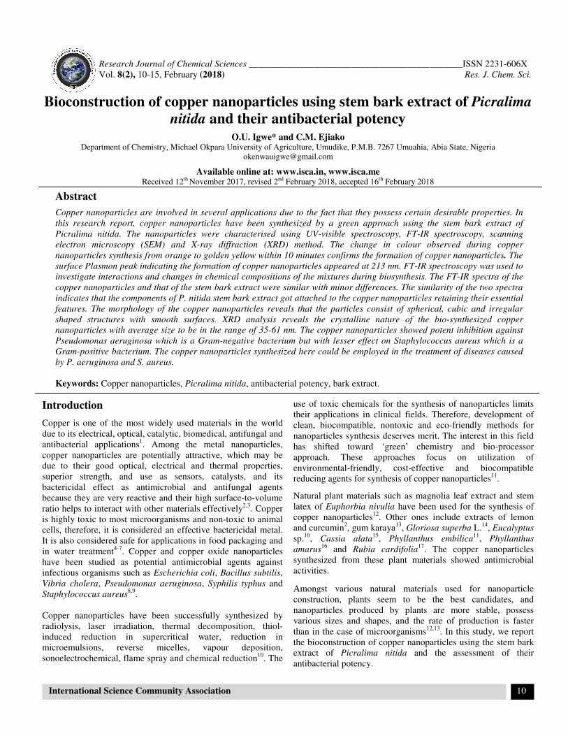
UV-visible Spectroscopy: The change in colour
during copper nanoparticles synthesis from orange to golden
yellow within 10 minutes confirms the formation of copper
nanoparticles. UV-vis spectroscopy which remains one of the
most convenient methods for the assessment of metal
nanoparticles formation and characterization was employed.
Figure-1 shows the UV-vis absorption spectrum of the
synthesized copper nanoparticles. The surface Plasmon peak
indicating the formation of copper nanoparticles appeared at 213
nm. Although many researchers have r
Plasmon peak of copper nanoparticles to appear above 500nm
Caroling et al11
reported a value of 294nm for copper
nanoparticles biosynthesized using the extract of
embilica. It is noteworthy that Plasmon absorption positi
depend on certain factors which include particle size, shape,
solvent type as well as capping and stabilizing agent, hence a
value of 213nm might be as a result of any of these factors. The
type and concentration of phytochemicals present in the ste
bark extract of P. nitida might have influenced the arrangement
of molecules around the copper particles.
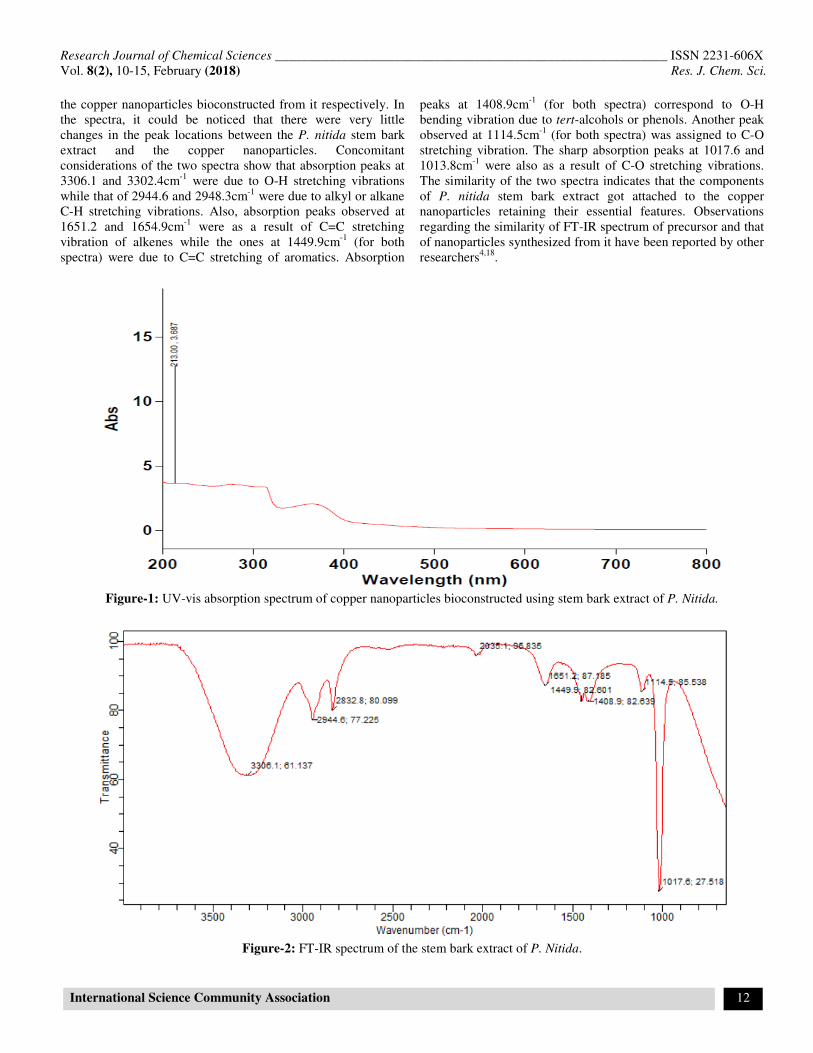
FT-IR Spectroscopy: FT-IR spectroscopy was used to
investigate the interactions between different species and
changes in chemical compositions of t
biosynthesis. FT-IR measurement was carried out on both the
plant extract and copper nanoparticles to identify the possible
functional groups responsible for the reduction of copper ions
and the capping as well as stabilization of the co
nanoparticles by P. nitida stem bark extract.
show the FT-IR spectra of the stem bark extract of
__________________________ ISSN 2231-606X
Res. J. Chem. Sci.
11
was made smoother and was imparted on a slide which was then
adjusting the machine parameters
The agar well diffusion assay method was
used to evaluate the antibacterial potency of the copper
nanoparticles against the test microorganisms. Concentrations of
0 and 25mg/mL prepared from the nanoparticles in a 2-
fold dilution process were tested against the organisms. Sterile
Mueller Hinton Agar (MHA) was poured into sterile Petri
dishes and allowed to set. Standardized concentrations (0.5
andard) of overnight cultures of test
isolates were swabbed aseptically on the agar plates and holes (6
mm) were made in the agar plates using a sterile metal cork-
l of the various dilutions of the copper nanoparticles
ut in each hole under aseptic
condition, kept at room temperature for one hour to allow the
agents to diffuse into the agar medium and incubated
) was used as a positive control
in the antibacterial evaluation. The MHA plates were then
for 24h. The diameter zones of inhibition
produced were measured and recorded. This procedure was
conducted in duplicates for each of the test organisms.
The change in colour observed
during copper nanoparticles synthesis from orange to golden
yellow within 10 minutes confirms the formation of copper
vis spectroscopy which remains one of the
most convenient methods for the assessment of metal
rmation and characterization was employed.
vis absorption spectrum of the
synthesized copper nanoparticles. The surface Plasmon peak
indicating the formation of copper nanoparticles appeared at 213
many researchers have reported the surface
Plasmon peak of copper nanoparticles to appear above 500nm3,4
,
reported a value of 294nm for copper
nanoparticles biosynthesized using the extract of Phyllanthus
It is noteworthy that Plasmon absorption position may
depend on certain factors which include particle size, shape,
solvent type as well as capping and stabilizing agent, hence a
value of 213nm might be as a result of any of these factors. The
type and concentration of phytochemicals present in the stem
might have influenced the arrangement
of molecules around the copper particles.
IR spectroscopy was used to
investigate the interactions between different species and
changes in chemical compositions of the mixtures during
IR measurement was carried out on both the
plant extract and copper nanoparticles to identify the possible
functional groups responsible for the reduction of copper ions
and the capping as well as stabilization of the copper
stem bark extract. Figures-2 and 3
IR spectra of the stem bark extract of P. nitida and
Research Journal of Chemical Sciences ___________________________________________________________ ISSN 2231-606X
Vol. 8(2), 10-15, February (2018) Res. J. Chem. Sci.
International Science Community Association 12
the copper nanoparticles bioconstructed from it respectively. In
the spectra, it could be noticed that there were very little
changes in the peak locations between the P. nitida stem bark
extract and the copper nanoparticles. Concomitant
considerations of the two spectra show that absorption peaks at
3306.1 and 3302.4cm-1
were due to O-H stretching vibrations
while that of 2944.6 and 2948.3cm-1
were due to alkyl or alkane
C-H stretching vibrations. Also, absorption peaks observed at
1651.2 and 1654.9cm-1
were as a result of C=C stretching
vibration of alkenes while the ones at 1449.9cm-1
(for both
spectra) were due to C=C stretching of aromatics. Absorption
peaks at 1408.9cm-1
(for both spectra) correspond to O-H
bending vibration due to tert-alcohols or phenols. Another peak
observed at 1114.5cm-1
(for both spectra) was assigned to C-O
stretching vibration. The sharp absorption peaks at 1017.6 and
1013.8cm-1
were also as a result of C-O stretching vibrations.
The similarity of the two spectra indicates that the components
of P. nitida stem bark extract got attached to the copper
nanoparticles retaining their essential features. Observations
regarding the similarity of FT-IR spectrum of precursor and that
of nanoparticles synthesized from it have been reported by other
researchers4,18
.
Figure-1: UV-vis absorption spectrum of copper nanoparticles bioconstructed using stem bark extract of P. Nitida.
Figure-2: FT-IR spectrum of the stem bark extract of P. Nitida.
Research Journal of Chemical Sciences ___________________________________________________________ ISSN 2231-606X
Vol. 8(2), 10-15, February (2018) Res. J. Chem. Sci.
International Science Community Association 13
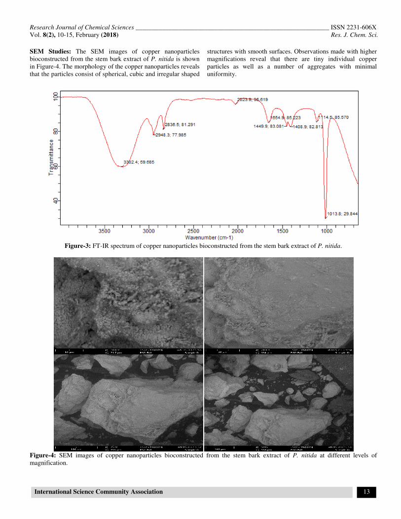
SEM Studies: The SEM images of copper nanoparticles
bioconstructed from the stem bark extract of P. nitida is shown
in Figure-4. The morphology of the copper nanoparticles reveals
that the particles consist of spherical, cubic and irregular shaped
structures with smooth surfaces. Observations made with higher
magnifications reveal that there are tiny individual copper
particles as well as a number of aggregates with minimal
uniformity.
Figure-3: FT-IR spectrum of copper nanoparticles bioconstructed from the stem bark extract of P. nitida.
Figure-4: SEM images of copper nanoparticles bioconstructed from the stem bark extract of P. nitida at different levels of
magnification.
Research Journal of Chemical Sciences _________________________________
Vol. 8(2), 10-15, February (2018)
International Science Community Association
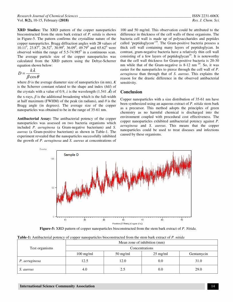
XRD Studies: The XRD pattern of the copper nanoparticles
bioconstructed from the stem bark extract of
in Figure-5. The pattern confirms the crystalline nature of the
copper nanoparticles. Bragg diffraction angles with 2
10.110, 23.87
0, 26.52
0, 30.99
0, 36.09
0, 49.79
observed within the range of 5.5-74.9850 in a continuous scan.
The average particle size of the copper nanoparticles was
calculated from the XRD pattern using the Debye
equation shown below:
cos
kD
λ
β θ=
where D is the average diameter size of nanoparticles (in nm),
is the Scherrer constant related to the shape and index (
the crystals with a value of 0.9, λ is the wavelength (1.541
the x-rays, β is the additional broadening which
at half maximum (FWHM) of the peak (in radians
Bragg angle (in degrees). The average size of the copper
nanoparticles was obtained to be in the range of 35
Antibacterial Assay: The antibacterial potency of the copper
nanoparticles was assessed on two bacteria organisms which
included P. aeruginosa (a Gram-negative bacterium) and
aureus (a Gram-positive bacterium) as shown in Table
experiment revealed that the nanoparticles successfully inhibited
the growth of P. aeruginosa and S. aureus at concentrations of
Figure-5: XRD pattern of copper nanoparticles bioconstructed from the stem bark extract of
Table-1: Antibacterial potency of copper nanoparticles bioconstructed from the stem bark extract of
Test organisms
100 mg/ml
P. aeruginosa 12.5
S. aureus 4.0
_________________________________________________
Association
The XRD pattern of the copper nanoparticles
bioconstructed from the stem bark extract of P. nitida is shown
5. The pattern confirms the crystalline nature of the
copper nanoparticles. Bragg diffraction angles with 2θ values of
, 49.790 and 65.62
0 were
in a continuous scan.
he average particle size of the copper nanoparticles was
calculated from the XRD pattern using the Debye-Scherrer
is the average diameter size of nanoparticles (in nm), K
is the Scherrer constant related to the shape and index (hkl) of
is the wavelength (1.541 ) of
is the additional broadening which is the full-width
(in radians), and θ is the
Bragg angle (in degrees). The average size of the copper
nanoparticles was obtained to be in the range of 35-61 nm.
The antibacterial potency of the copper
was assessed on two bacteria organisms which
negative bacterium) and S.
positive bacterium) as shown in Table-1. The
experiment revealed that the nanoparticles successfully inhibited
at concentrations of
100 and 50 mg/ml. This observation could be attributed to the
difference in thickness of the cell walls of these organisms. The
bacteria cell wall is made up of polysaccharides and peptides
called ‘peptidoglycon’19
. The Gram
thick cell wall containing many layers of peptidoglycan. In
contrast, gram-negative bacteria have a relatively thin cell wall
consisting of a few layers of peptidoglycan
that the cell wall thickness for Gram
nm while that of the Gram-negative is 8
easier for the nanoparticles to pierce through the cell wall of
aeruginosa than through that of S. aureus
reason for the drastic difference in the observed antibacterial
sensitivity.
Conclusion
Copper nanoparticles with a size distribution of 35
been synthesized using an aqueous extract of
as a precursor. This method adopts the principles of green
chemistry as no harmful chemical is discharged into the
environment coupled with procedural cost effectiveness. The
copper nanoparticles exhibited antibacterial potency against
aeruginosa and S. aureus. This means that the copper
nanoparticles could be used to treat diseases and infections
caused by these organisms.
XRD pattern of copper nanoparticles bioconstructed from the stem bark extract of
Antibacterial potency of copper nanoparticles bioconstructed from the stem bark extract of P. nitida
Mean zone of inhibition (mm)
Concentrations
100 mg/ml 50 mg/ml 25 mg/ml
12.5 12.0 0.0
2.5 0.0
__________________________ ISSN 2231-606X
Res. J. Chem. Sci.
14
100 and 50 mg/ml. This observation could be attributed to the
difference in thickness of the cell walls of these organisms. The
bacteria cell wall is made up of polysaccharides and peptides
Gram-positive bacteria possess a
thick cell wall containing many layers of peptidoglycan. In
negative bacteria have a relatively thin cell wall
consisting of a few layers of peptidoglycan19
. It is noteworthy
Gram-positive bacteria is 20-30
negative is 8-12 nm 20
. So, it was
easier for the nanoparticles to pierce through the cell wall of P.
S. aureus. This explains the
in the observed antibacterial
Copper nanoparticles with a size distribution of 35-61 nm have
been synthesized using an aqueous extract of P. nitida stem bark
as a precursor. This method adopts the principles of green
chemistry as no harmful chemical is discharged into the
environment coupled with procedural cost effectiveness. The
copper nanoparticles exhibited antibacterial potency against P.
. This means that the copper
nanoparticles could be used to treat diseases and infections
XRD pattern of copper nanoparticles bioconstructed from the stem bark extract of P. Nitida.
P. nitida
Gentamycin
31.0
29.0
Research Journal of Chemical Sciences ___________________________________________________________ ISSN 2231-606X
Vol. 8(2), 10-15, February (2018) Res. J. Chem. Sci.
International Science Community Association 15
Acknowledgement
The authors are grateful to Mr. I. K. Ndukwe of Plant
Taxonomy Section, Forestry Department, Michael Okpara
University of Agriculture, Umudike, for identifying and
authenticating the plant sample.
References
1. Tian K., Liu C., Yang H. and Ren X. (2012). In situ
synthesis of copper nanoparticles/polystyrene composite.
Colloids and Surfaces A: Physicochemical and Engineering
Aspects, 397, 12-15.
2. Jayandran M., Haneefa M.M. and Balasubramanian V.
(2015). Green synthesis of copper nanoparticles using
natural reducer and stabilizer and an evaluation of
antimicrobial activity. J. chem. pharm. Sci., 7(2), 251-259.
3. Subhankari I. and Nayak P.L. (2013). Antimicrobial
activity of copper nanoparticles synthesised by ginger
(Zingiber officinale) extract. World J. Nanosci. Technol., 2,
14-17.
4. Saranyaadevi K., Subha V., Ravindran R.S.E. and
Renganathan S. (2014). Synthesis and characterization of
copper nanoparticles using Capparis zeylanica leaf extract.
Int. J. Chem. Tech. Res., 6(10), 4533-4541.
5. Kalimuthu K., Babu R.S., Venkataraman D., Bilal M. and
Gurunathan S. (2008). Biosynthesis of silver nanocrystals
by Bacillus licheniformis. Colloids Surf B., 65(1), 150-153.
6. Wijnhoven S.W., Peijnenburg W.J., Herberts C.A., Hagens
W.I., Oomen A.G., Heugens E.H. and Dekkers S. (2009).
Nano-silver–a review of available data and knowledge gaps
in human and environmental risk assessment.
Nanotoxicology, 3(2), 109-138.
7. Klueh U., Wagner V., Kelly S., Johnson A. and Bryers J.D.
(2000). Efficacy of silver-coated fabric to prevent bacterial
colonization and subsequent device-based biofilm
formation. J. Biomed. Mater. Res., 53(6), 621-631.
8. Akhavan O. and Ghaderi E. (2010). Cu and CuO
nanoparticles immobilized by silica thin films as
antibacterial materials and photocatalysts. Surface and
Coatings Technol., 205(1), 219-223.
9. Hassan M.S., Amna T., Yang O.B., E1-Newehy M.H., Al-
Deyab S.S. and Khil M.S. (2012). Smart copper oxide
nanocrystals: synthesis, characterization, electrochemical
and potent antibacterial activity. Colloids Surf. B: Biointer-
faces, 97, 201-206.
10. Kolekar R.V., Bhade S.P.D., Rajiv K., Priyanka R., Rajvir
S. and Pradeepkumar K.S. (2015). Biosynthesis of copper
nanoparticles using aqueous extract of Eucalyptus sp. plant
leaves. Curr. Sci., 109(2), 255-257.
11. Caroling G., Vinodhini E., Ranjitham A.M. and Shanthi P.
(2015). Biosynthesis of copper nanoparticles using aqueous
Phyllanthus embilica (Gooseberry) extract- characterisation
and study of antimicrobial effects. Int. J. Nano. Chem.,
1(2), 53-63.
12. Iravani S. (2011). Green synthesis of metal nanoparticles
using plants. Green Chemistry, 13(10), 2638-2650.
13. Padil V.V.T. and Černík M. (2013). Green synthesis of
copper oxide nanoparticles using gum karaya as a
biotemplate and their antibacterial application. Int. J.
Nanomed., 8, 889-898.
14. Naika H.R., Lingaraju K., Manjunath K., Kumar D.,
Nagaraju G., Suresh D. and Nagabhushana H. (2015).
Green synthesis of CuO nanoparticles using Gloriosa
superba L.extract and their antibacterial activity. J. Taibah
University for Science, 9(1), 7-12.
15. Jayalakshm I. and Yogamoorthi A. (2014). Green synthesis
of copper oxide nanoparticles using aqueous extract of
flowers of Cassia alata and particles characterisation. Int. J.
Nanomater. Biostructures, 4(4), 66-71.
16. Acharyulu N.P.S., Dubey R.S., Swaminadham V., Kollu P.,
Kalyani R.L. and Pammi S.V.N. (2014). Green synthesis of
Cuo nanoparticles using Phyllanthus amarus leaf extract
and their antibacterial activity against multidrug resistance
bacteria. International Journal of Engineering, 3(4), 639-
41.
17. Mariselvam R., Ranjitsingh A.J.A., Padmalatha C. and
Selvakumar P.M. (2014). Green Synthesis of copper
quantum dots using Rubia cardifolia plant root extracts and
its antibacterial properties. J. Academia and Industrial Res.,
3(4), 191-194.
18. Xu Q., Zhao Y., Xu J.Z. and Zhu J. (2006). Preparation of
functionalized copper nanoparticles and fabrication of a
glucose sensor. Sensors and Actuators B, 114, 379-386.
19. Prakash N., Jayapradeep S. and Sudha P.N. (2009).
Investigation of antimicrobial properties of silver and
copper nanoparticles encspsulated in chitosan. Proceeding
of First International Conference on Nanostructured
Materials and Nanocomposites. Kottayam, India, 6th
-8th
April, 311-317.
20. Sagar A. (2015). Differences between gram-positive and
gram-negative bacteria. Online microbiology notes.
http://www.microbiologyinfo.com/differences-between-
gram-positive-and-gram-negative-bacteria/ retrieved 5th
May, 2017.