Biocompatibility, FDA and ISO 10993 - saliterman.umn.edu · Voskerician, G., et al.,...
54
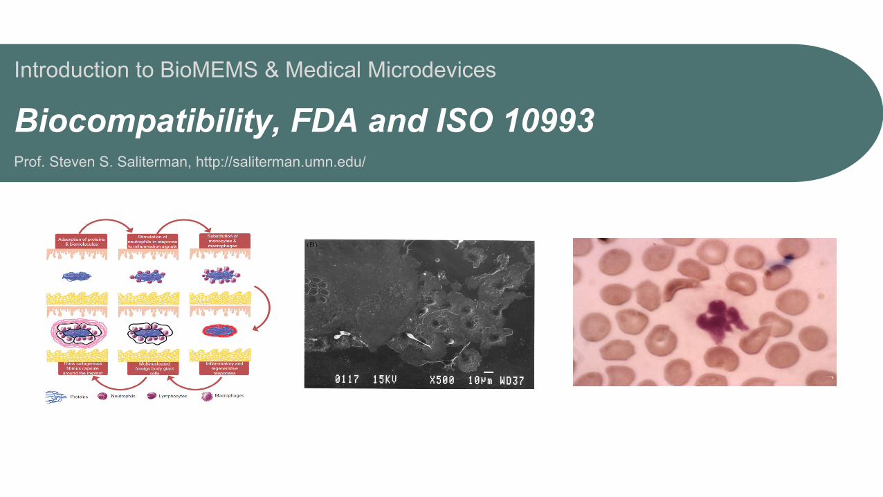
Introduction to BioMEMS & Medical Microdevices Biocompatibility, FDA and ISO 10993 Prof. Steven S. Saliterman, http://saliterman.umn.edu/
Transcript of Biocompatibility, FDA and ISO 10993 - saliterman.umn.edu · Voskerician, G., et al.,...
Introduction to BioMEMS & Medical Microdevices
Biocompatibility, FDA and ISO 10993 Prof. Steven S. Saliterman, http://saliterman.umn.edu/
Steven S. Saliterman
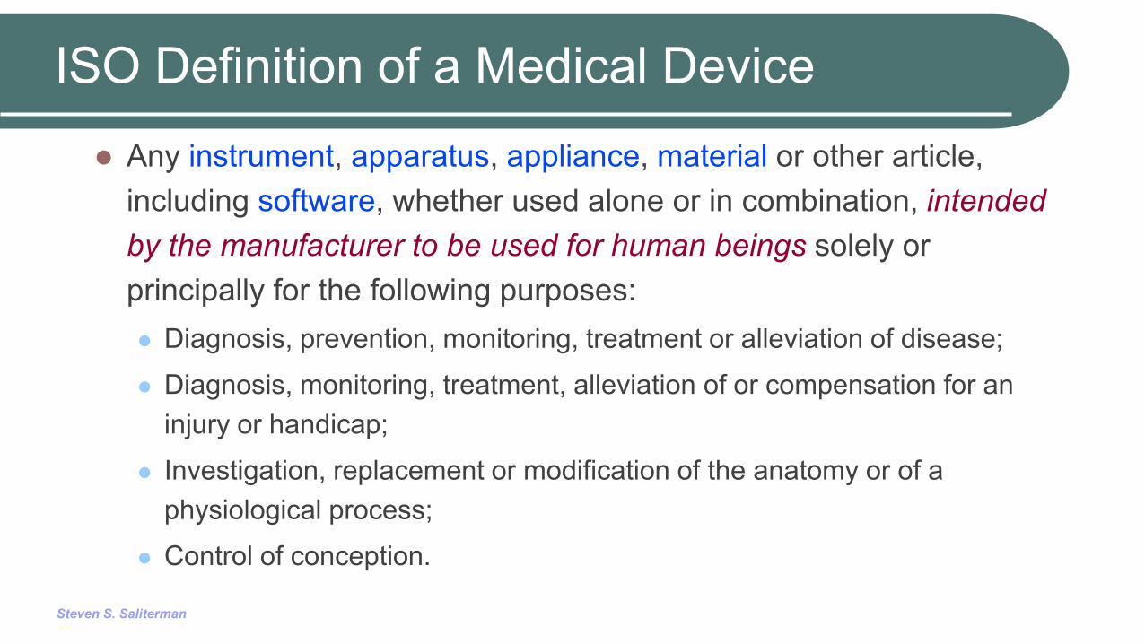
ISO Definition of a Medical Device
Any instrument, apparatus, appliance, material or other article, including software, whether used alone or in combination, intended by the manufacturer to be used for human beings solely or principally for the following purposes: Diagnosis, prevention, monitoring, treatment or alleviation of disease;
Diagnosis, monitoring, treatment, alleviation of or compensation for an injury or handicap;
Investigation, replacement or modification of the anatomy or of a physiological process;
Control of conception.
Steven S. Saliterman
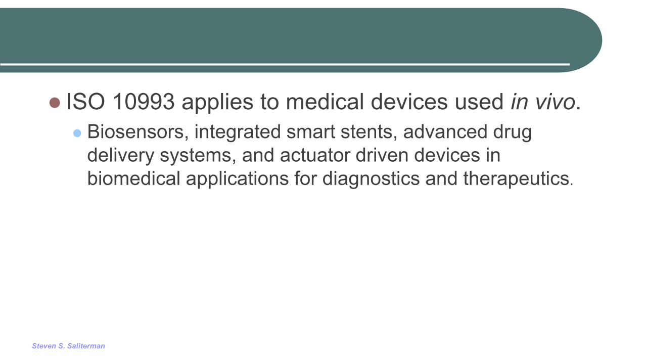
ISO 10993 applies to medical devices used in vivo. Biosensors, integrated smart stents, advanced drug
delivery systems, and actuator driven devices in biomedical applications for diagnostics and therapeutics.
Steven S. Saliterman
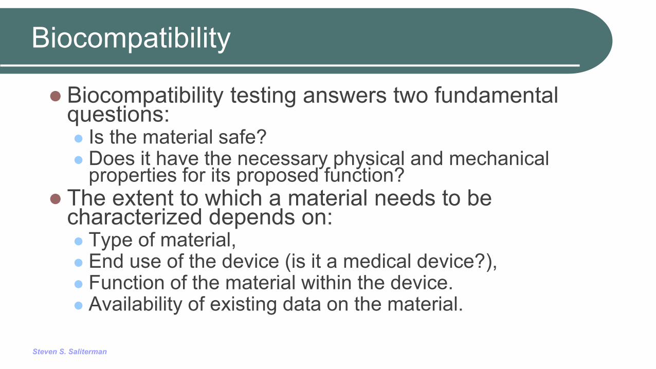
Biocompatibility
Biocompatibility testing answers two fundamental questions: Is the material safe? Does it have the necessary physical and mechanical
properties for its proposed function? The extent to which a material needs to be
characterized depends on: Type of material, End use of the device (is it a medical device?), Function of the material within the device. Availability of existing data on the material.
Steven S. Saliterman
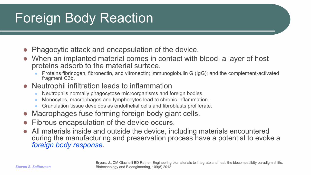
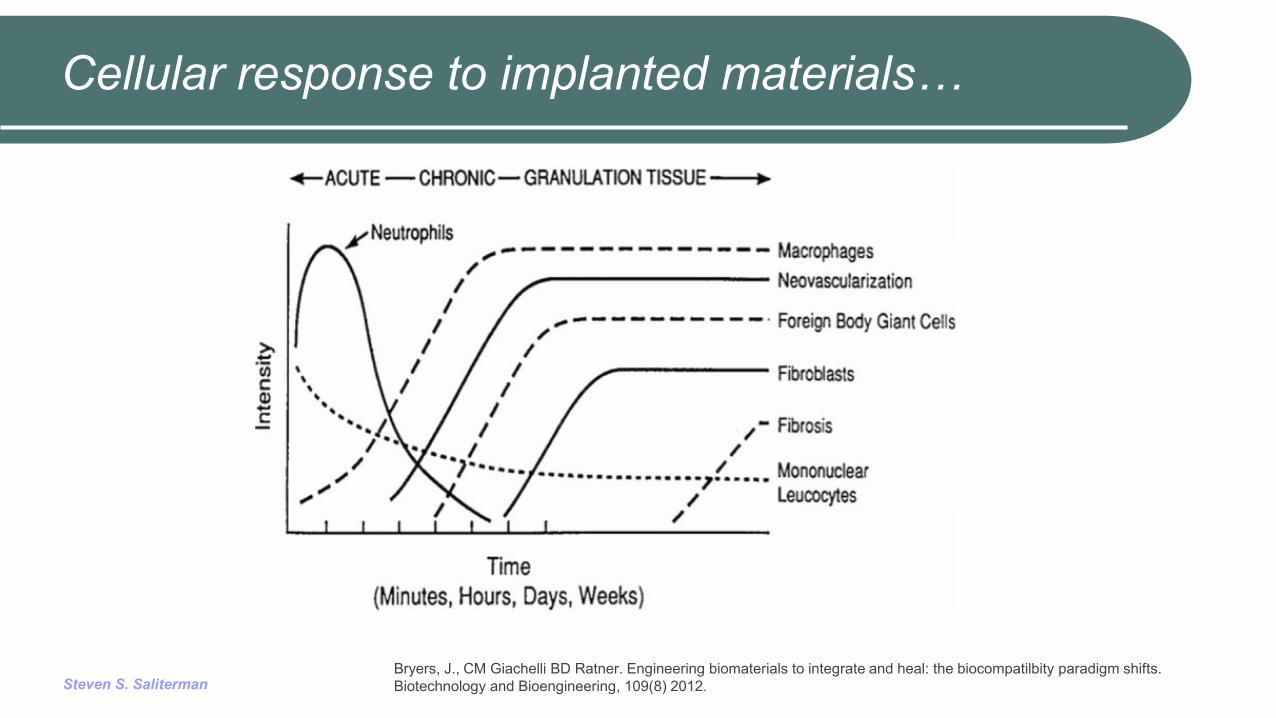
Foreign Body Reaction
Phagocytic attack and encapsulation of the device. When an implanted material comes in contact with blood, a layer of host
proteins adsorb to the material surface. Proteins fibrinogen, fibronectin, and vitronectin; immunoglobulin G (IgG); and the complement-activated
fragment C3b. Neutrophil infiltration leads to inflammation
Neutrophils normally phagocytose microorganisms and foreign bodies. Monocytes, macrophages and lymphocytes lead to chronic inflammation. Granulation tissue develops as endothelial cells and fibroblasts proliferate.
Macrophages fuse forming foreign body giant cells. Fibrous encapsulation of the device occurs. All materials inside and outside the device, including materials encountered
during the manufacturing and preservation process have a potential to evoke a foreign body response.
Bryers, J., CM Giachelli BD Ratner. Engineering biomaterials to integrate and heal: the biocompatilbity paradigm shifts. Biotechnology and Bioengineering, 109(8) 2012.

Steven S. Saliterman
Host Foreign Body Response…
Barkam, S, et al. Fabricated micro-nano devices for in vivo and in vitro biomedical applications. WIREs Nanomed Nanobiotechnol 2013, 5:544–568
Steven S. Saliterman
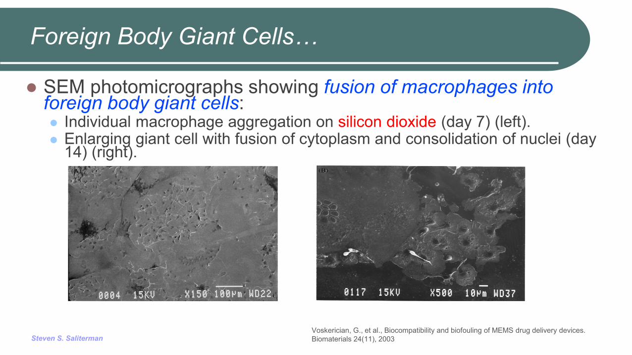
SEM photomicrographs showing fusion of macrophages into foreign body giant cells: Individual macrophage aggregation on silicon dioxide (day 7) (left). Enlarging giant cell with fusion of cytoplasm and consolidation of nuclei (day
14) (right).
Foreign Body Giant Cells…
Voskerician, G., et al., Biocompatibility and biofouling of MEMS drug delivery devices. Biomaterials 24(11), 2003
Steven S. Saliterman
Cellular response to implanted materials…
Bryers, J., CM Giachelli BD Ratner. Engineering biomaterials to integrate and heal: the biocompatilbity paradigm shifts. Biotechnology and Bioengineering, 109(8) 2012.
Steven S. Saliterman
Biofouling
Biofouling is the process whereby functioning of a medical device is interfered with by the biological response of the host.
This commonly occurs when macrophages and foreign body giant cells (FBGCs) attach to the implanted device, accumulate, grow and interfere with normal operation.
Surface coating of biomaterials seems one good approach to lessen the inflammatory response, lessen macrophage adhesion and FBGCs growth, and improve wound healing.
The foreign body response by the host is largely independent of the material’s being polymeric, ceramic or metallic; being hydrophobic or hydrophilic; or being hard or soft.
Steven S. Saliterman
The status of the proteins on a material surface is believed to determine the ultimate biocompatibility of a given polymer.
Producing a more biocompatible surface requires achieving specific responses between the polymer surface and the adjacent cells and to reduce non-specific interactions. Methods include passivating the polymer surfaces to minimize
non-specific protein interaction. Functionalizing the polymer surface with biomolecules to induce
specific protein adsorption and cell responses.
Chen, H. et al. Biocompatible polymer materials: Role of protein–surface interactions. Progress in Polymer Science 33, pp 1059-1087 (2008).

Steven S. Saliterman
Non-Fouling Surfaces…
Non-fouling (i.e., protein adsorption-resistant) polymer coatings Use of nontoxic (biocompatible) materials: Effectively inhibit in vivo biofouling, Appropriate thickness and permeability to allow analyte transport, Techniques to deposit coating onto a variety of materials and
architectures. Must be mechanically, chemically, and electrically robust to
withstand surface deposition, sterilization methods, implantation procedures, and in vivo environment.
Polyethylene glycol (PEG), HO(-CH2CH2O-)n H, is an example.
Bryers, J., CM Giachelli BD Ratner. Engineering biomaterials to integrate and heal: the biocompatilbity paradigm shifts. Biotechnology and Bioengineering, 109(8) 2012.
Steven S. Saliterman
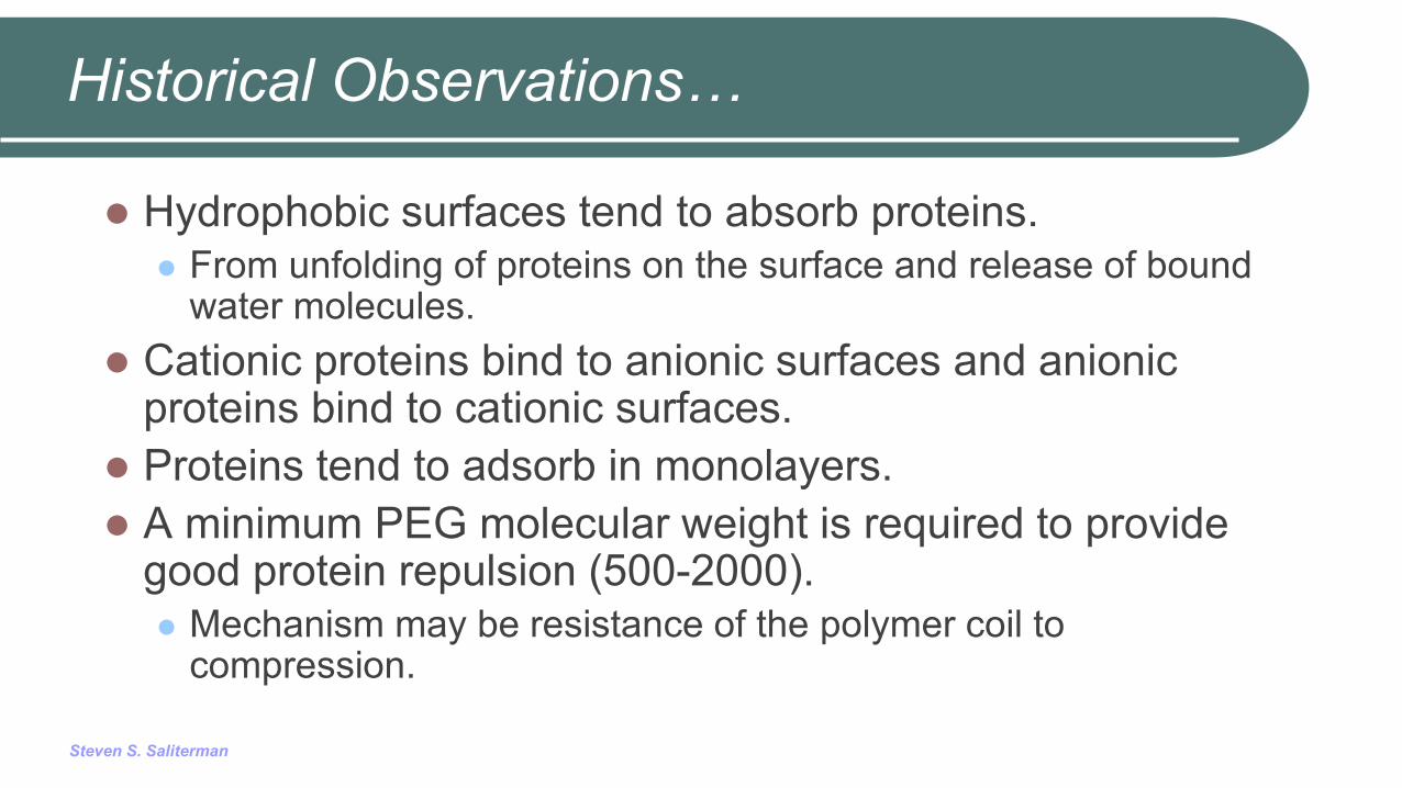
Historical Observations…
Hydrophobic surfaces tend to absorb proteins. From unfolding of proteins on the surface and release of bound
water molecules. Cationic proteins bind to anionic surfaces and anionic
proteins bind to cationic surfaces. Proteins tend to adsorb in monolayers. A minimum PEG molecular weight is required to provide
good protein repulsion (500-2000). Mechanism may be resistance of the polymer coil to
compression.
Steven S. Saliterman
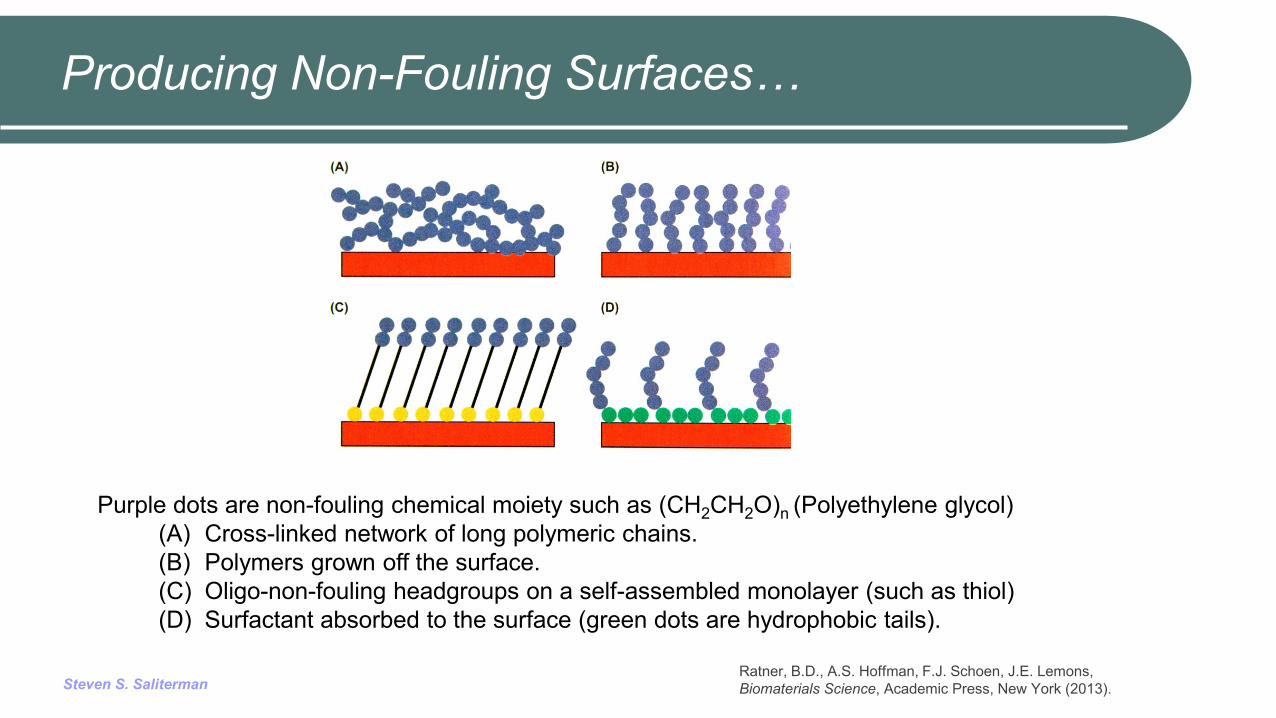
Producing Non-Fouling Surfaces…
Purple dots are non-fouling chemical moiety such as (CH2CH2O)n (Polyethylene glycol) (A) Cross-linked network of long polymeric chains. (B) Polymers grown off the surface. (C) Oligo-non-fouling headgroups on a self-assembled monolayer (such as thiol) (D) Surfactant absorbed to the surface (green dots are hydrophobic tails).
Ratner, B.D., A.S. Hoffman, F.J. Schoen, J.E. Lemons, Biomaterials Science, Academic Press, New York (2013).

Steven S. Saliterman
Surface Engineering…
Barkam, S, et al. Fabricated micro-nano devices for in vivo and in vitro biomedical applications. WIREs Nanomed Nanobiotechnol 2013, 5:544–568

Steven S. Saliterman
Flowchart for Biological Evaluation
Steven S. Saliterman
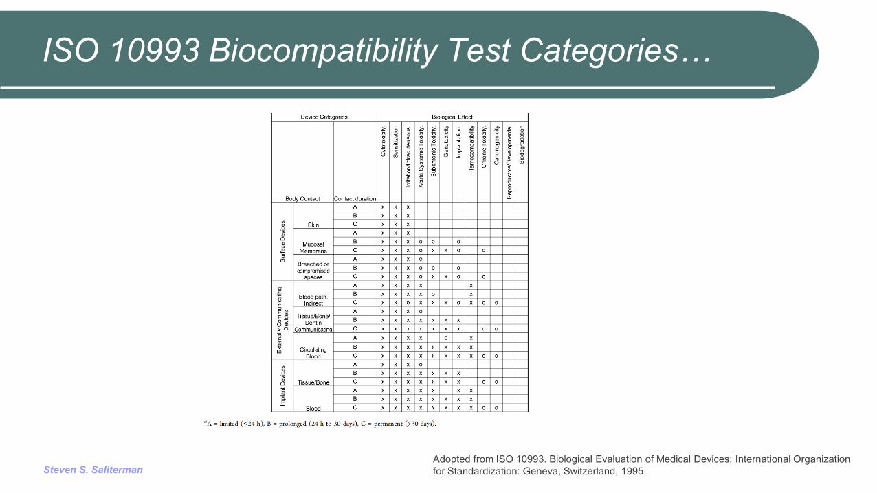
ISO 10993 Biocompatibility Test Categories…
Adopted from ISO 10993. Biological Evaluation of Medical Devices; International Organization for Standardization: Geneva, Switzerland, 1995.
Steven S. Saliterman
ISO 10993 Standard…
The ISO 10993 International Standard pertains to: Surface devices on the skin, mucosal membranes,
breached or compromised surfaces. External communicating devices with blood, tissue, bone,
dentin. Implantable devices.
Its purpose is to protect humans and to serve as a framework for selecting tests to evaluate biological responses.
In so doing consideration has been given to minimize the number and exposure of test animals.

Steven S. Saliterman
Characterization Methods
Identification of a materials constituents and: Changes of the material over time, Changes with exposure to different environments, Lot-to-lot consistency for manufacturing purposes.
Methodologies: Infrared spectral analysis (IR), Thermal analysis, Density analysis, Molecular weight distribution, Mechanical properties, Surface properties, Extract Characterization.
Steven S. Saliterman
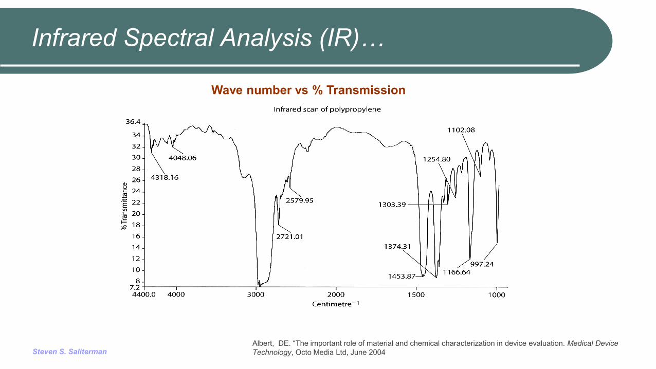
Infrared Spectral Analysis (IR)…
Albert, DE. “The important role of material and chemical characterization in device evaluation. Medical Device Technology, Octo Media Ltd, June 2004
Wave number vs % Transmission
Steven S. Saliterman
Useful in material identification and for following polymer degradation.
Molecules absorb specific frequencies that are characteristic of their structure. These absorptions are resonant frequencies, i.e. the frequency of the absorbed radiation matches the transition energy of the bond or group that vibrates.
A basic IR spectrum is essentially a graph of infrared light absorbance (or transmittance) on the vertical axis vs. frequency or wavelength on the horizontal axis.
Typical units of frequency used in IR spectra are reciprocal centimeters (sometimes called wave numbers), with the symbol cm−1
Steven S. Saliterman
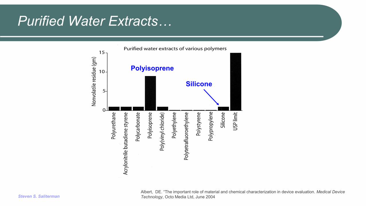
Purified Water Extracts…
Polyisoprene
Silicone
Albert, DE. “The important role of material and chemical characterization in device evaluation. Medical Device Technology, Octo Media Ltd, June 2004
Steven S. Saliterman
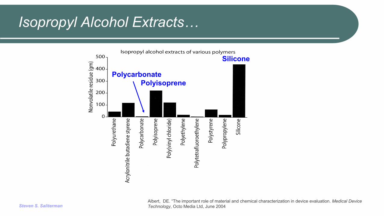
Isopropyl Alcohol Extracts…
Albert, DE. “The important role of material and chemical characterization in device evaluation. Medical Device Technology, Octo Media Ltd, June 2004
Polycarbonate Polyisoprene
Silicone
Steven S. Saliterman
Cytotoxicity
Cytotoxicity refers to cell damage caused by materials, either by direct contact or by leachable substances (extracts).
Cell damage may occur by a variety of means including activation of the complement system. The complement system involves a number of serum
factors that are activated in the presence of antigen-antibody binding, bacteria and viruses, or foreign materials.
Steven S. Saliterman
Cytotoxicity Assessment…
Determination of cytotoxicity includes: Microscopic (qualitative) evaluation
Morphology Vacuolization Detachment Cell lysis Membrane integrity
Quantitative evaluation Cell death Inhibition of cell growth Cell proliferation Cellular secretions
Steven S. Saliterman
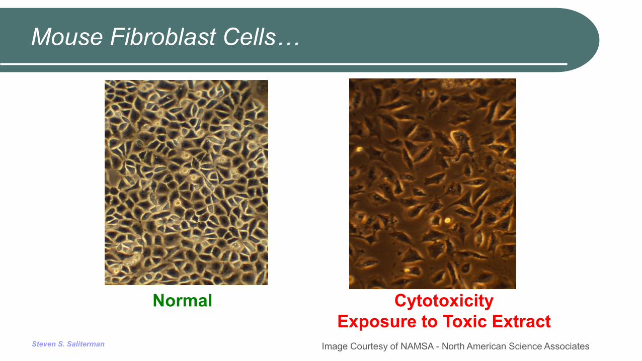
Mouse Fibroblast Cells…
Normal Cytotoxicity Exposure to Toxic Extract
Image Courtesy of NAMSA - North American Science Associates
Steven S. Saliterman
Sensitization
Sensitization refers to a materials ability to induce specific delayed-type hypersensitivity in the body upon initial exposure: Haptens, Langerhans cells and T-cell lymphocytes, Lymphokines.
Testing: Guinea pig maximization test (GPMT), Closed-patch test (Buehler test), Murine Local Lymph Node Assay.
Steven S. Saliterman
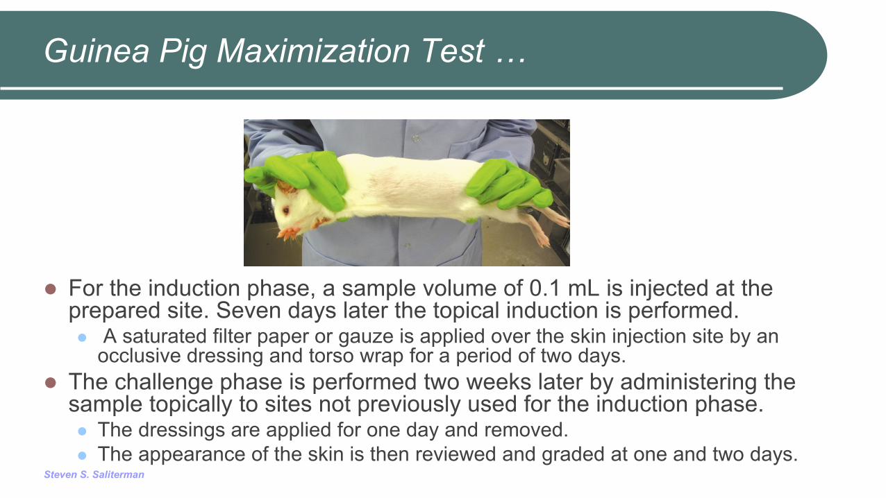
Guinea Pig Maximization Test …
For the induction phase, a sample volume of 0.1 mL is injected at the prepared site. Seven days later the topical induction is performed. A saturated filter paper or gauze is applied over the skin injection site by an
occlusive dressing and torso wrap for a period of two days. The challenge phase is performed two weeks later by administering the
sample topically to sites not previously used for the induction phase. The dressings are applied for one day and removed. The appearance of the skin is then reviewed and graded at one and two days.
Steven S. Saliterman
Irritation
Irritation refers to a non-specific inflammatory response to a single, repeated or continuous application of a material.
Areas tested: Skin, Eyes, Oral mucosa, Genitalia, Rectum.
Rabbits and human subjects are often used.
Steven S. Saliterman
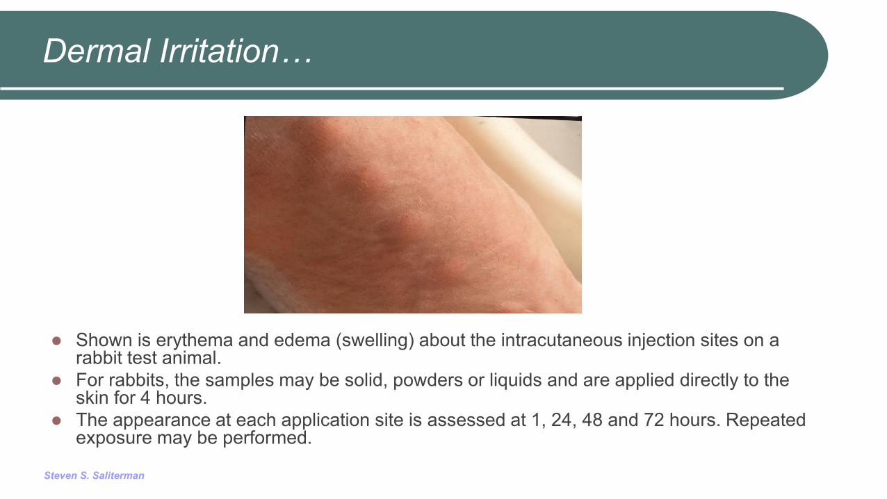
Dermal Irritation…
Shown is erythema and edema (swelling) about the intracutaneous injection sites on a rabbit test animal.
For rabbits, the samples may be solid, powders or liquids and are applied directly to the skin for 4 hours.
The appearance at each application site is assessed at 1, 24, 48 and 72 hours. Repeated exposure may be performed.
Steven S. Saliterman
Systemic Toxicity – Whole Body
Systemic toxicity (body at large): Acute toxicity - within 24 hours, Subacute toxicity - single dose or multiple doses of
a test sample during a period from 14 to 28 days, Subchronic toxicity - at 90 days, but not exceeding
10% of the life cycle of the device, Chronic toxicity - single or multiple exposures to
medical devices, materials and extracts during at least 10% of their lifespan of the test animal.
Steven S. Saliterman
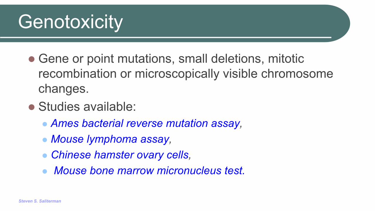
Genotoxicity
Gene or point mutations, small deletions, mitotic recombination or microscopically visible chromosome changes.
Studies available: Ames bacterial reverse mutation assay, Mouse lymphoma assay, Chinese hamster ovary cells, Mouse bone marrow micronucleus test.
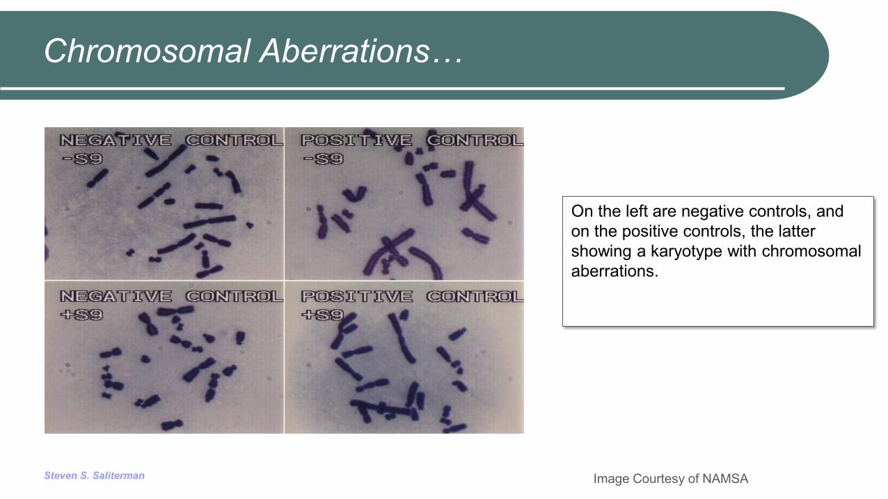
Steven S. Saliterman Image Courtesy of NAMSA
On the left are negative controls, and on the positive controls, the latter showing a karyotype with chromosomal aberrations.
Chromosomal Aberrations…
Steven S. Saliterman
Implantation
Tests for assessment of the local effects of implant material on living tissue. Comparison is made with reactions observed to medical devices whose
clinical acceptability has already been established. Short term studies of less than 12 weeks implantation, and long term studies
of greater than twelve weeks may be performed. Solid implant materials for testing must be prepared in the same manner as
they are intended for implantation, including form, density, hardness, surface finish, sterilization, and handling.
Non-solid materials such as liquids, pastes and particulates may also be used, and be contained in polyethylene, polypropylene or polytetrafluoroethylene tubes. Controls of similar size, shape of surface finish should be used.
Steven S. Saliterman
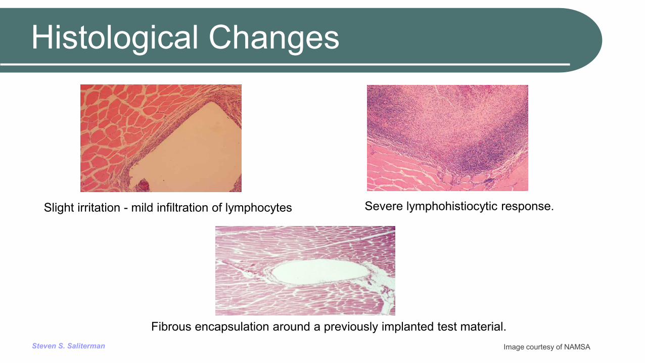
Histological Changes
Image courtesy of NAMSA
Slight irritation - mild infiltration of lymphocytes
Severe lymphohistiocytic response.
Fibrous encapsulation around a previously implanted test material.
Steven S. Saliterman
Types of Histological Findings…
The extent of fibrous capsular involvement around the device and adjoining tissue.
Tissue inflammatory changes, including polynuclear leucocytes, lymphocytes, plasma cells, eosinophils, macrophages and multinucleated cells.
Presence of tissue necrosis, capillary wall breakdown or other deterioration.
Material debris, fatty infiltration and granuloma formation. Quality and quantity of tissue ingrowth into porous
materials.
Steven S. Saliterman
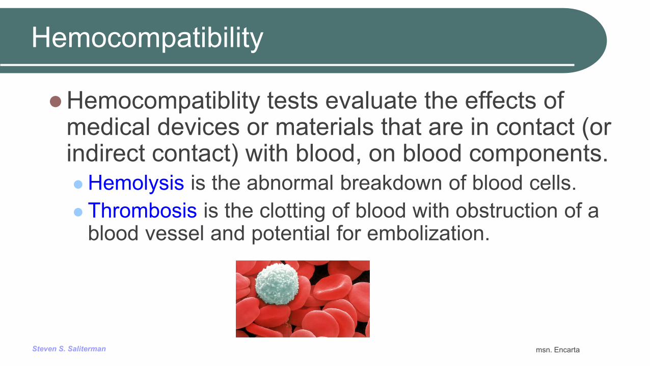
Hemocompatibility
Hemocompatiblity tests evaluate the effects of medical devices or materials that are in contact (or indirect contact) with blood, on blood components. Hemolysis is the abnormal breakdown of blood cells. Thrombosis is the clotting of blood with obstruction of a
blood vessel and potential for embolization.
msn. Encarta
Steven S. Saliterman
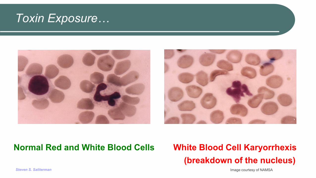
Toxin Exposure…
Normal Red and White Blood Cells White Blood Cell Karyorrhexis (breakdown of the nucleus)
Image courtesy of NAMSA

Steven S. Saliterman
Degradation
Degradation is the unwanted breakdown of implanted medical device materials.
Ideally in an implanted device all materials of degradation are ultimately removed by the body without toxicity.
Polymer degradation. Ceramic degradation. Metal and alloy electrochemical effects,
Steven S. Saliterman
Polymer Degradation…
Chemical bond scission due to hydrolytic and oxidative processes.
Enzymes, proteins and other cellular activity can alter the rate and nature of degradation.
Ultraviolet cleavage of chemical bonds. Gamma and electron radiation that cause embrittlement,
discoloration and thermal instability. Metal induced degradation from impurities, additives or
hybrid construction.

Steven S. Saliterman
Summary
Biocompatibility testing answers two fundamental questions: Is the material safe? Does it have the necessary physical and mechanical
properties for its proposed function? Biofouling is the process whereby functioning of a
medical device is interfered with by the biological response of the host.
The ISO 10993 Standard is to protect humans and to serve as a framework for selecting tests to evaluate biological responses.

Steven S. Saliterman
ISO 10993 Subparts discussed: Characterization Cytotoxicity Sensitization Irritation System Toxicity Genotoxicity Implantation Hemocompatibility Degradation
Addendum – Tested materials.
Steven S. Saliterman
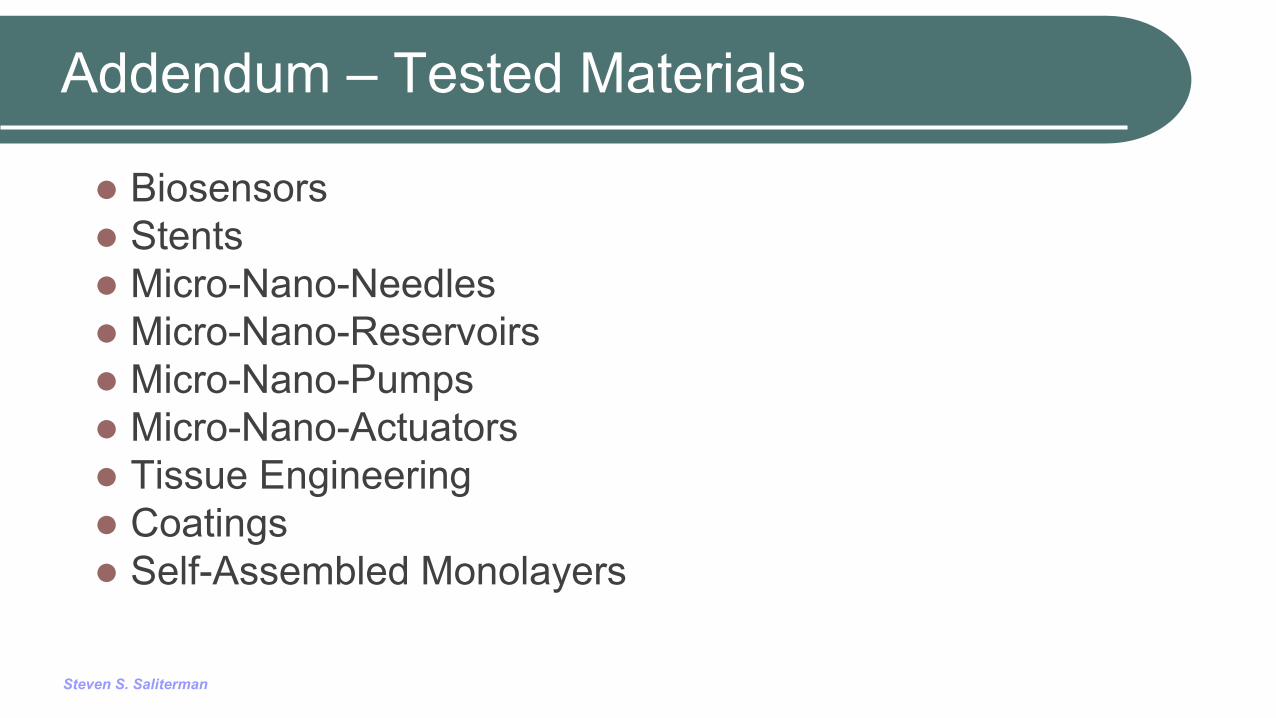
Addendum – Tested Materials
Biosensors Stents Micro-Nano-Needles Micro-Nano-Reservoirs Micro-Nano-Pumps Micro-Nano-Actuators Tissue Engineering Coatings Self-Assembled Monolayers

Steven S. Saliterman
NFS Compositions…
Ratner, B.D., A.S. Hoffman, F.J. Schoen, J.E. Lemons, Biomaterials Science, Academic Press, New York (2013).

Steven S. Saliterman
Thermodynamics of Protein Absorption…
Ratner, B.D., A.S. Hoffman, F.J. Schoen, J.E. Lemons, Biomaterials Science, Academic Press, New York (2013).
Steven S. Saliterman
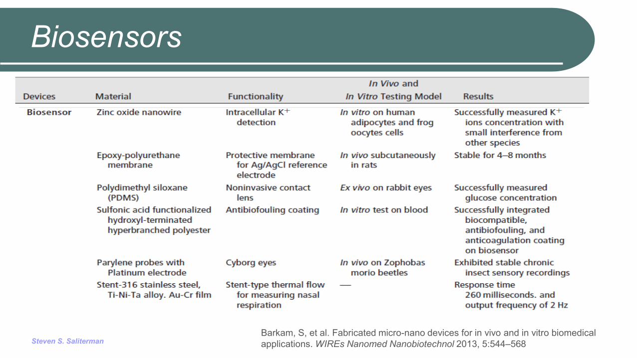
Biosensors
Barkam, S, et al. Fabricated micro-nano devices for in vivo and in vitro biomedical applications. WIREs Nanomed Nanobiotechnol 2013, 5:544–568

Steven S. Saliterman
Stents
Barkam, S, et al. Fabricated micro-nano devices for in vivo and in vitro biomedical applications. WIREs Nanomed Nanobiotechnol 2013, 5:544–568

Steven S. Saliterman
Micro-Nano Needles
Barkam, S, et al. Fabricated micro-nano devices for in vivo and in vitro biomedical applications. WIREs Nanomed Nanobiotechnol 2013, 5:544–568
Steven S. Saliterman
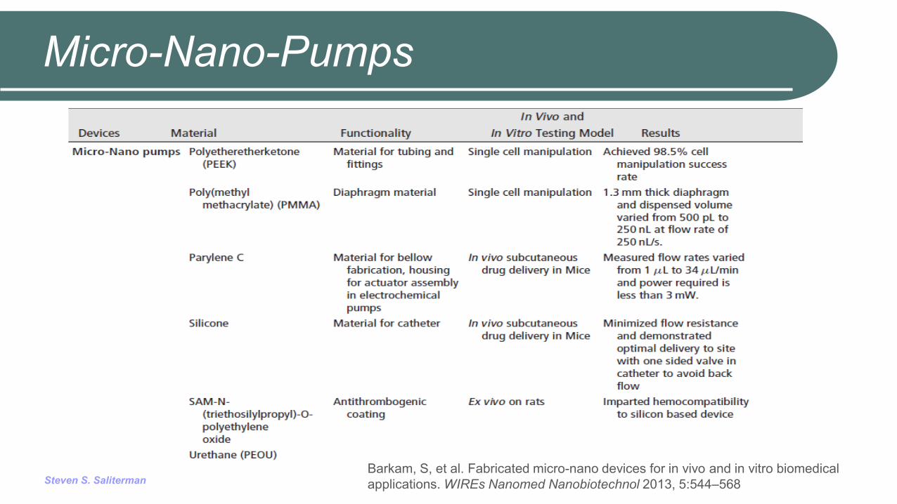
Micro-Nano-Pumps
Barkam, S, et al. Fabricated micro-nano devices for in vivo and in vitro biomedical applications. WIREs Nanomed Nanobiotechnol 2013, 5:544–568
Steven S. Saliterman
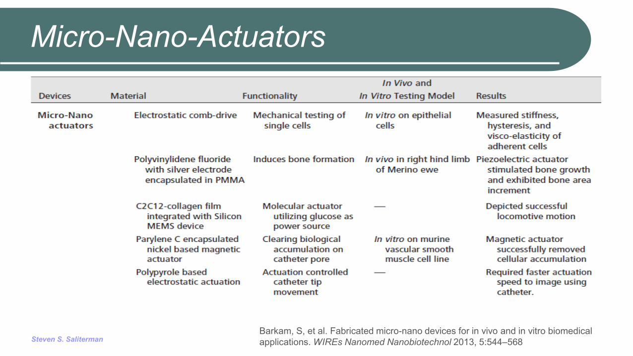
Micro-Nano-Actuators
Barkam, S, et al. Fabricated micro-nano devices for in vivo and in vitro biomedical applications. WIREs Nanomed Nanobiotechnol 2013, 5:544–568
Steven S. Saliterman
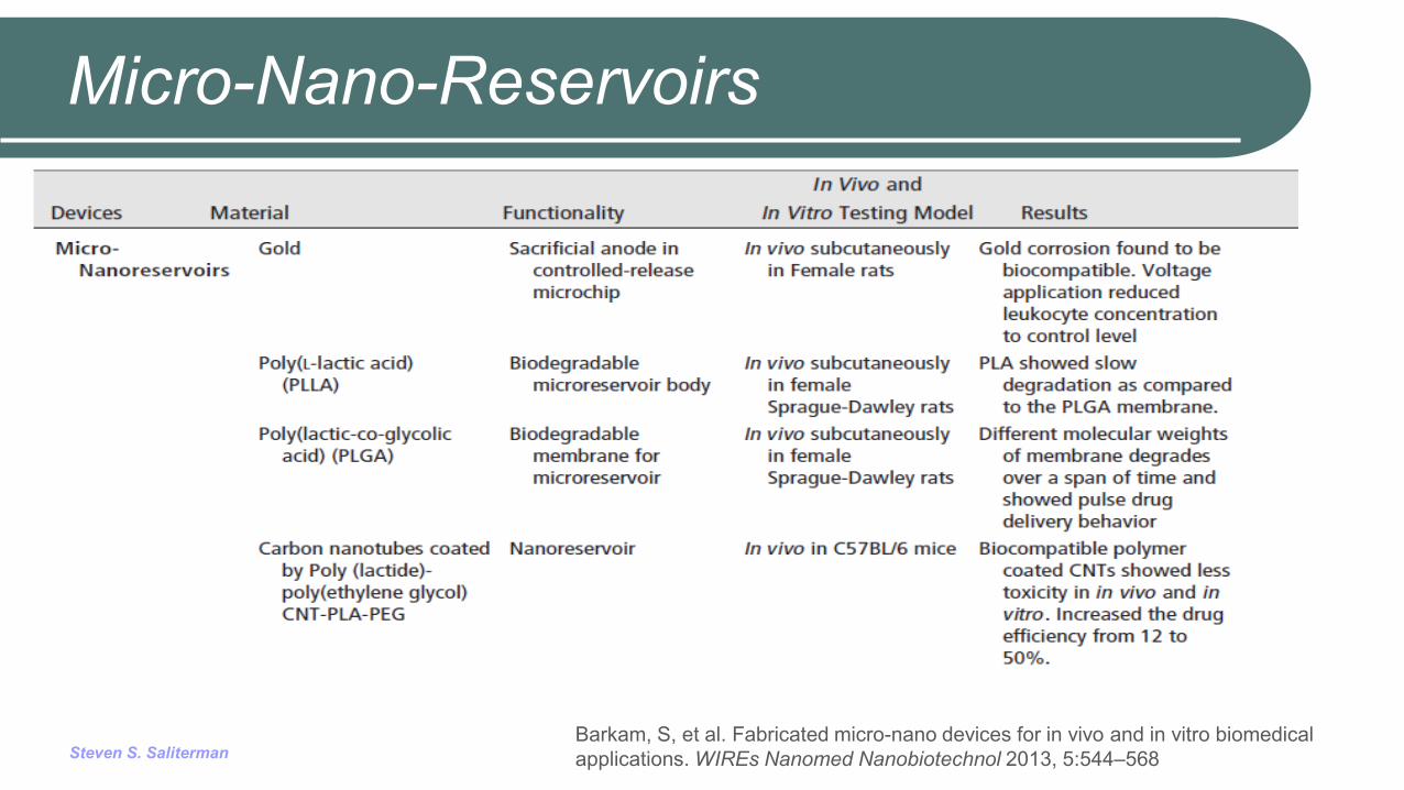
Micro-Nano-Reservoirs
Barkam, S, et al. Fabricated micro-nano devices for in vivo and in vitro biomedical applications. WIREs Nanomed Nanobiotechnol 2013, 5:544–568
Steven S. Saliterman
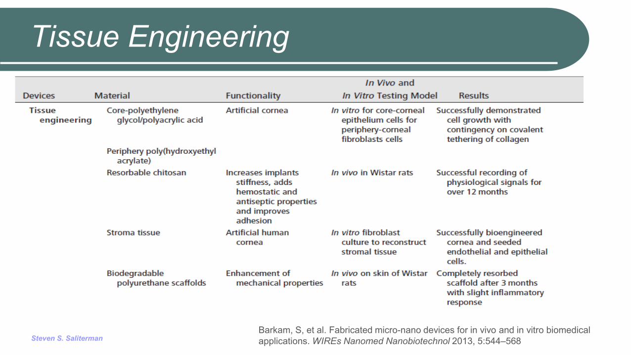
Tissue Engineering
Barkam, S, et al. Fabricated micro-nano devices for in vivo and in vitro biomedical applications. WIREs Nanomed Nanobiotechnol 2013, 5:544–568
Steven S. Saliterman
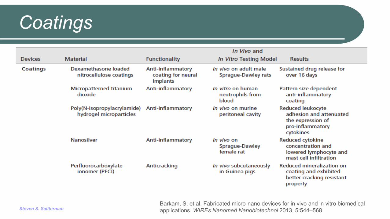
Coatings
Barkam, S, et al. Fabricated micro-nano devices for in vivo and in vitro biomedical applications. WIREs Nanomed Nanobiotechnol 2013, 5:544–568
Steven S. Saliterman
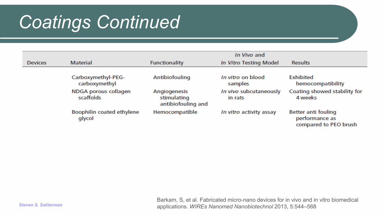
Coatings Continued
Barkam, S, et al. Fabricated micro-nano devices for in vivo and in vitro biomedical applications. WIREs Nanomed Nanobiotechnol 2013, 5:544–568
Steven S. Saliterman
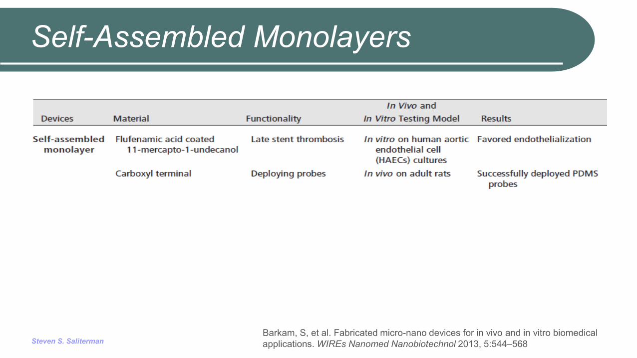
Self-Assembled Monolayers
Barkam, S, et al. Fabricated micro-nano devices for in vivo and in vitro biomedical applications. WIREs Nanomed Nanobiotechnol 2013, 5:544–568