Biochimica et Biophysica Acta - ainfo.cnptia.embrapa.br · a Unidade Multidisciplinar de Genômica...
12
A comparative proteomic analysis of Gluconacetobacter diazotrophicus PAL5 at exponential and stationary phases of cultures in the presence of high and low levels of inorganic nitrogen compound L.M.S. Lery a,c , W.M.A. von Krüger a,c, ⁎, F.C. Viana b,c , K.R.S. Teixeira b,c , P.M. Bisch a,c a Unidade Multidisciplinar de Genômica – Instituto de Biofísica Carlos Chagas Filho – Universidade Federal do Rio de Janeiro, Brazil b EMBRAPA Agrobiologia – Seropédica, Brazil c Rede de Proteômica do Estado do Rio de Janeiro, Brazil abstract article info Article history: Received 26 February 2008 Received in revised form 7 June 2008 Accepted 23 June 2008 Available online 9 July 2008 Keywords: Gluconacetobacter diazotrophicus Proteome Nitrogen-fixation Two-dimensional gel electrophoresis (2D-GE) Mass spectrometry (MS) Protein identification A proteomic view of G. diazotrophicus PAL5 at the exponential (E) and stationary phases (S) of cultures in the presence of low (L) and high levels (H) of combined nitrogen is presented. The proteomes analyzed on 2D- gels showed 131 proteins (42E + 32S + 29H + 28L) differentially expressed by G. diazotrophicus, from which 46 were identified by combining mass spectrometry and bioinformatics tools. Proteins related to cofactor, energy and DNA metabolisms and cytoplasmic pH homeostasis were differentially expressed in E growth phase, under L and H conditions, in line with the high metabolic rate of the cells and the low pH of the media. Proteins most abundant in S-phase cells were stress associated and transporters plus transferases in agreement with the general phenomenon that binding protein-dependent systems are induced under nutrient limitation as part of hunger response. Cells grown in L condition produced nitrogen-fixation accessory proteins with roles in biosynthesis and stabilization of the nitrogenase complex plus proteins for protection of the nitrogenases from O 2 -induced inactivation. Proteins of the cell wall biogenesis apparatus were also expressed under nitrogen limitation and might function in the reshaping of the nitrogen-fixing G. diazotrophicus cells previously described. Genes whose protein products were detected in our analysis were mapped onto the chromosome and, based on the tendency of functionally related bacterial genes to cluster, we identified genes of particular pathways that could be organized in operons and are co-regulated. These results showed the great potential of proteomics to describe events in G. diazotrophicus cells by looking at proteins expressed under distinct growth conditions. © 2008 Elsevier B.V. All rights reserved. 1. Introduction Although abundant in the terrestrial atmosphere, gaseous nitrogen (N 2 ) is chemically inert at ambient temperature, the nitrogenous mineral and organic reserves in the ground are limited and quickly depleted by successive cropping [1]. In tropical regions, conditions of temperature and humidity speed up the processes of decomposition of the organic substances, resulting in highly acid soils with still lower content of nitrogenous compounds [2,3]. As much as 30% of the total fertilizers needed for agricultural crops are nitrogen-derived chemicals produced under high costs of energy [4]. Consequently, decreasing dependence on chemical insumes for agricultural production is a need to reduce cost and the negative impact to the economy and environment. Biological processes, mediated by microbial action, are crucial for cycling of nutrients in the planet and have been used with success to control plagues, domestic illnesses of plants and animals [5]. Therefore, a biological approach, such as the use of N 2 -fixing bacteria to provide plants reduced N, remains as one of the best solutions for the problem. Gluconacetobacter diazotrophicus was first isolated from sugar cane [6], where it has been found as an endophyte in roots, stems and leaves [7–9]. It has also been isolated from coffee plant, sweet-potato, pineapple, cameroon grass, tea, banana, ragi, rice [4,10–14] and, even from insects that infest sugar cane [15]. G. diazotrophicus is a N 2 -fixing bacterium [6,16], well adapted to the high sucrose environment of the sugarcane plant [6,10], where it fixes nitrogen under microaerophilic conditions [6,17]. Its optimum pH for growth and nitrogen-fixation is 5.5, but in vitro it can grow and fix nitrogen at pH as low as 2.5, however, it does not grow at pH higher than 7.0 [17]. It produces the plant growth promoting hormones indole-3-acetic acid (IAA) and gibberellins A1 and A3 [9,18] and bacteriocins, and more, it is able to solubilize in vitro two essential micronutrient for plant growth, zinc and phosphorous compounds [19,20], therefore, G. diazotrophicus provides a good model to study symbiosis with non-leguminous plants [10]. Biochimica et Biophysica Acta 1784 (2008) 1578–1589 ⁎ Corresponding author. Avenida Carlos Chagas Filho, CCS, bloco G, Instituto de Biofísica Carlos Chagas Filho, sala G1-043, Ilha do Fundão, CEP 21941-902, Rio de Janeiro, RJ, Brazil. Tel.: +55 21 25626531; fax: +55 21 22808193. E-mail address: [email protected] (W.M.A. von Krüger). 1570-9639/$ – see front matter © 2008 Elsevier B.V. All rights reserved. doi:10.1016/j.bbapap.2008.06.020 Contents lists available at ScienceDirect Biochimica et Biophysica Acta journal homepage: www.elsevier.com/locate/bbapap
-
Upload
nguyenkhanh -
Category
Documents
-
view
214 -
download
0
Transcript of Biochimica et Biophysica Acta - ainfo.cnptia.embrapa.br · a Unidade Multidisciplinar de Genômica...

Biochimica et Biophysica Acta 1784 (2008) 1578–1589
Contents lists available at ScienceDirect
Biochimica et Biophysica Acta
j ourna l homepage: www.e lsev ie r.com/ locate /bbapap
A comparative proteomic analysis of Gluconacetobacter diazotrophicus PAL5 atexponential and stationary phases of cultures in the presence of high and low levelsof inorganic nitrogen compound
L.M.S. Lery a,c, W.M.A. von Krüger a,c,⁎, F.C. Viana b,c, K.R.S. Teixeira b,c, P.M. Bisch a,c
a Unidade Multidisciplinar de Genômica – Instituto de Biofísica Carlos Chagas Filho – Universidade Federal do Rio de Janeiro, Brazilb EMBRAPA Agrobiologia – Seropédica, Brazilc Rede de Proteômica do Estado do Rio de Janeiro, Brazil
⁎ Corresponding author. Avenida Carlos Chagas FilhBiofísica Carlos Chagas Filho, sala G1-043, Ilha do FuJaneiro, RJ, Brazil. Tel.: +55 21 25626531; fax: +55 21 22
E-mail address: [email protected] (W.M.A. von Krü
1570-9639/$ – see front matter © 2008 Elsevier B.V. Aldoi:10.1016/j.bbapap.2008.06.020
a b s t r a c t
a r t i c l e i n f oArticle history:
A proteomic view of G. diazo Received 26 February 2008Received in revised form 7 June 2008Accepted 23 June 2008Available online 9 July 2008Keywords:Gluconacetobacter diazotrophicusProteomeNitrogen-fixationTwo-dimensional gel electrophoresis (2D-GE)Mass spectrometry (MS)Protein identification
trophicus PAL5 at the exponential (E) and stationary phases (S) of cultures in thepresence of low (L) and high levels (H) of combined nitrogen is presented. The proteomes analyzed on 2D-gels showed 131 proteins (42E+32S+29H+28L) differentially expressed by G. diazotrophicus, from which 46were identified by combining mass spectrometry and bioinformatics tools. Proteins related to cofactor,energy and DNA metabolisms and cytoplasmic pH homeostasis were differentially expressed in E growthphase, under L and H conditions, in line with the high metabolic rate of the cells and the low pH of the media.Proteins most abundant in S-phase cells were stress associated and transporters plus transferases inagreement with the general phenomenon that binding protein-dependent systems are induced undernutrient limitation as part of hunger response. Cells grown in L condition produced nitrogen-fixationaccessory proteins with roles in biosynthesis and stabilization of the nitrogenase complex plus proteins forprotection of the nitrogenases from O2-induced inactivation. Proteins of the cell wall biogenesis apparatuswere also expressed under nitrogen limitation and might function in the reshaping of the nitrogen-fixing G.diazotrophicus cells previously described. Genes whose protein products were detected in our analysis weremapped onto the chromosome and, based on the tendency of functionally related bacterial genes to cluster,we identified genes of particular pathways that could be organized in operons and are co-regulated. Theseresults showed the great potential of proteomics to describe events in G. diazotrophicus cells by looking atproteins expressed under distinct growth conditions.
© 2008 Elsevier B.V. All rights reserved.
1. Introduction
Although abundant in the terrestrial atmosphere, gaseous nitrogen(N2) is chemically inert at ambient temperature, the nitrogenousmineral and organic reserves in the ground are limited and quicklydepleted by successive cropping [1]. In tropical regions, conditions oftemperature and humidity speed up the processes of decompositionof the organic substances, resulting in highly acid soils with still lowercontent of nitrogenous compounds [2,3].
As much as 30% of the total fertilizers needed for agricultural cropsare nitrogen-derived chemicals produced under high costs of energy[4]. Consequently, decreasing dependence on chemical insumes foragricultural production is a need to reduce cost and the negativeimpact to the economy and environment. Biological processes,mediated by microbial action, are crucial for cycling of nutrients in
o, CCS, bloco G, Instituto dendão, CEP 21941-902, Rio de808193.ger).
l rights reserved.
the planet and have been used with success to control plagues,domestic illnesses of plants and animals [5]. Therefore, a biologicalapproach, such as the use of N2-fixing bacteria to provide plantsreduced N, remains as one of the best solutions for the problem.
Gluconacetobacter diazotrophicuswas first isolated from sugar cane[6], where it has been found as an endophyte in roots, stems andleaves [7–9]. It has also been isolated from coffee plant, sweet-potato,pineapple, cameroon grass, tea, banana, ragi, rice [4,10–14] and, evenfrom insects that infest sugar cane [15]. G. diazotrophicus is a N2-fixingbacterium [6,16], well adapted to the high sucrose environment of thesugarcane plant [6,10], where it fixes nitrogen under microaerophilicconditions [6,17]. Its optimum pH for growth and nitrogen-fixation is5.5, but in vitro it can grow and fix nitrogen at pH as low as 2.5,however, it does not grow at pH higher than 7.0 [17]. It produces theplant growth promoting hormones indole-3-acetic acid (IAA) andgibberellins A1 and A3 [9,18] and bacteriocins, and more, it is able tosolubilize in vitro two essential micronutrient for plant growth, zincand phosphorous compounds [19,20], therefore, G. diazotrophicusprovides a good model to study symbiosis with non-leguminousplants [10].

1579L.M.S. Lery et al. / Biochimica et Biophysica Acta 1784 (2008) 1578–1589
G. diazotrophicus has been the focus of several studies aiming tounderstand the mechanisms behind diazotrophy. The bacteriumcontains a major cluster of nif and fix genes [21–25] and in thepresence of low levels of the ammonium it expresses both, the Mo-nitrogenase and NifA, the transcriptional regulator of all nif genes [23].G. diazotrophicus Mo-nitrogenase activity is inhibited under highintracellular oxygen concentrations at the level of its synthesis and byirreversible damage to the iron protein [26]. However, it can also beprotected due to the formation of a complex with a FeSII protein(Shethna protein) [27]. In this complex the enzyme remains inactive,but can be reactivated in the presence of an efficient respiratorysubstrate to lower intracellular levels of O2 [27–29]. Interestingly, G.diazotrophicus nitrogenase activity is more tolerant to oxygen thanthat of most diazotrophs [17], what explains the ability of its coloniesto fix nitrogen at a wide range of atmospheric pO2 [30]. G.diazotrophicus Mo-nitrogenase purified components, Gd1(MoFe) andGd2 (Fe), were found to be quite similar to Av1 and Av2, thecomponents of the Azotobacter vinelandii nitrogenase, in terms of size,composition, interactions and catalytic properties, however, theyshowed narrower pH range for efficient catalysis and highersensitivity to pH and salt [29].
G. diazotrophicus properties studied so far suggest it is a diazotrophquite distinct from other root-associated organisms [17], therefore,efforts have been done to better understand its physiology, whatmight contribute to the development of biotechnological approachesfor a more sustainable and environmentally safe crop production [31].
One way to identify metabolic pathways involved in a specificbiological process is the differential proteome analysis by two-dimensional gel electrophoresis (2D-GE) followed by mass spectro-metry (MS) and bioinformatics to characterize proteins of interest.This approach, largely used to provide broad pictures of thephysiological processes associated with a given condition in manybacterial species, including G. diazotrophicus [32–38], has been used inthe present study to identify proteins specifically expressed atexponential (E) and stationary (S) phases of liquid cultures underlow (L) and high (H) levels of a combined nitrogen compound. Manynovel proteins of G. diazotrophicus with roles in specific and in thegeneral metabolic pathways have been detected under the growthconditions used. The present data set together with others recentlypublished [38] constitute a major advance in the molecular micro-biology of this species and will contribute to a better understanding ofthe physiology of the bacterium.
2. Materials and methods
2.1. Bacterial strain and culture conditions
G. diazotrophicus, strain PAL5 (BR11281), was precultured in DYGSmedium [39] to exponential phase. The cells were harvested bycentrifugation (18,000 ×g, 10 min, 4 °C), washed under the sameconditions and resuspended at a final concentration of 2×10⁶ cells/mlin 2 l of themodified definedminimal medium LGI (LGIM) at initial pH5.5. For growth under N-limitation or N-abundance media weresupplemented with (NH4)2SO4 at 1 mM (low combined nitrogen level,L) or 10 mM (high combined nitrogen level, H) respectively, aspreviously described [17]. Bacterial growth was carried out in afermentor (Bioengineering, GAC of 2.5 l), at 30 °C and 400 rpm, undercontrolled air or air/N2 flow [17]. To avoid contamination duringgrowth, filters were used at inlets of gases and at openings. Duringgrowth levels of dissolved oxygen (dO) and pH of the cultures weremonitored continuously and concentrations of (NH4)2SO4 weremeasured as previously described [17]. Culture with H level of(NH4)2SO4 was initially saturated with O2 atmospheric to dO 80%and, during growth, air influx assured nitrogenase complex inactivity[17]. For growth with L level of (NH4)2SO4, the chamber was initiallyfluxed with a mixture of air and N2 in a proportion of 1:1 to give an
initial dO of 50%. During growth, as levels of dO decreased duerespiration, gases were injected into the chamber to keep dO at levelscompatible with cell growth and nitrogen-fixation [17]. Growth wasfollowed by cell number counts (Newbauer-counting chamber),optical density at 600 nm (spectrophotometer PE — Model Lambda11) and total protein content [40]. Samples for further analysis werecollected during exponential (E, 25–50 h) and stationary (S, 55–70 h)phases of growth.
2.2. Sample preparation and 2D gel electrophoresis (2D-GE)
Cells were harvested by centrifugation (18,000 ×g, 10min, 4 °C) andthe cellular pellet was resuspended in a lysis buffer (2% Chaps, 8 Murea, 0.13 M DTT, 0.02% (v/v) Pharmalyte 3–10, 1 mM PMSF). Cellsuspensionwas submitted to 20 cycles of freezing [N2 (l)] and thawing(37 °C) and lysis was monitored by optical microscopy. A clear lysatewas obtained by centrifugation at 50,000 ×g for 1 h at 4 °C and proteincontent was estimated by 2D Quant Kit (GE Healthcare).
For the first dimension approximately 500 µg of proteins in 320 µlof solution (2% Chaps, 8 M urea, 0.13 M DTT, 0.02% (v/v) Pharmalyte 3–10, 8 mM PMSF, traces of bromophenol blue) was used to rehydrate an18 cm strip pH 3–10 (Immobiline DryStrip; GE Healthcare) for 16 h atroom temperature. Proteins were focused according to the manufac-turer's instructions (GE Healthcare) [41]. The gel strip was equilibratedand then loaded onto a 12–14% gradient gel (SDS-ExcelGel XL) andelectrophoresed as recommended [41]. First and second dimensionruns were carried out in the Multiphor II unit (GE Healthcare).
Gels were stained with Coomassie Blue R-250 [42] scanned withthe ImageScanner LabScan v5.0 and analyzed with the ImageMaster2D Platinum v5.0 software (GE Healthcare).
2.3. Comparative analysis of gels
Proteomic analysis of G. diazotrophicus lysates of cells at E or S-phase of cultures in LGIM with starter doses of 1 or 10 mM of(NH4)2SO4, were carried out by 2D-GE. For each condition (exponen-tial/low, EL; exponential/high, EH; stationary/low, SL and stationary/high, SH) triplicate gels were run (Fig. 1). Two gels of the triplicate runfor each phase/condition derived from one culture and the third froma distinct culture. Comparison of 2D-GE protein patterns of cellscollected at the same phase of growth under similar conditions, indistinct experiments, permitted us to evaluate variability at biologicallevel (biological replicate). Analysis of the same sample in two gels, onthe other hand, allowed us to check technical variability (experimentalreplicate).
Protein spots on all gels were automatically detected with theImageMaster 2D Platinum v5.0 software which also performs auto-matematching using filtering, querying and the statistical applicationsrequired (IMageMaster™ 2D Platinum software version 5.0, UserManual, [43]). All the spots were also manually confirmed. Images ofthree replicate gels (member gels) were superposed using the samesoftware and a master gel containing all spots representative of eachculture phase and condition (E, S, L andH)was obtained. In eachmastergel protein spot position, shape and optical density were averaged andunmatched spots or those whose relative volumes differed more than20% among member gels were not considered (Fig. 1).
Relative values of isoelectric point (pIr) andmolecular mass (MWr)of the proteins of interest were determined considering lineardistributions of pH (3–10) and the protein molecular weight markers(GE Healthcare), respectively.
In order to identify proteins specific to the different culture phasesand conditions under study, intersections of the master gels wereobtained and compared. Spots common to two gels were considereddifferentially expressed if their relative volumes differed more than 3fold (Fig. 1). Relative volumes (%Vol) were normalized volumes thatremain relatively independent of irrelevant variations between gels,

1580 L.M.S. Lery et al. / Biochimica et Biophysica Acta 1784 (2008) 1578–1589
particularly caused by varying experimental conditions [43]. %Vol isexpressed as:
kVol =Vol
Pn
S = 1Vols
� 100
where, Vol is the volume of the spot of interest and Vols is the volumeof each spot S in a gel containing n spots.
2.4. Mass spectrometry analysis and protein identification
Protein spots were cut from gels and stored at −20 °C or processedimmediately. Dried gel fragments were treated with porcine trypsin(Promega, Madison, WI, USA) and peptides were extracted with 50%acetonitrile (ACN), 5% trifluoroacetic acid (TFA). Sample volume wasreduced to 10 µl (Speed Vac) and 1 µl was mixed with equal volume ofa saturated solution of α-cyano-4-hydroxycinnamic acid matrix in50% ACN, 0.1% TFA. The mix was spotted onto a MALDI-TOF sampleplate and allowed to crystallize at room temperature. Analysis wasperformed in a Voyager DE PRO BioSpectrometer Workstation(Applied Biosystem, Foster City, CA, USA). Spectra were obtained inreflectron-delayed extraction mode with high resolution for 800–4000 Da range.
MALDI-TOF-TOF analysis was performed in cases where identifica-tion by MALDI-TOF was not successful. Precursor ion fragmentation,using N2 as collision-induced dissociation gas and collision cellpressure kept at 2.8×10⁻⁶ Torr, was carried out in a 4700 ExplorerProteomics Analyzer (Applied Biosystem).
Trypsin autolysis peptide masses 842.5 and 2211.1 and calibrationmixture 1 or 2 (Sequazyme Peptide Mass Standard kit, PerSeptiveBiosystems, Foster City, CA, USA) were used, respectively, as internaland external standards in both MS procedures.
Peptide mass fingerprints were analyzed by two independentapproaches: online Protein Prospector MS-Fit interface (http://prospector.ucsf.edu) that matched the mass spectrometry data toprotein sequences in NCBI database (G. diazotrophicus databaseversion NC_010125.1, GI:162145846, with 3.778 protein sequences
Fig. 1. Experimental design for comparative two-dimensional gel analysis. Three member ggrowth [low (L) or high (H) starter concentration of (NH4)2SO4] were used to obtain a masterthe replicates. The four master gels were: EH, EL, SH and SL. Master gels EH and EL were coComparison of the master gels SH and SL generated an iS (intersection stationary) virtual geland 32 of the S-phase of growth were identified. EH and SH master gels were also comparedcomparison of EL and SL master gels generated iL (intersection low) gel with 335 spots. Cocondition of growth.
on 04/28/2008; Bacterial protein sequence database with 8.022.379protein sequences on 04/28/2008). Only proteins with hit in bothNCBIdatabases were considered identified.
For protein identification a first MOWSE score above 104 and at least a10-fold difference in MOWSE score from the second possible hit wasrequired. Additionally, a minimum of 20% of protein coverage and 4peptides of mass spectra should match the sequence of the protein in thedatabank. Congruence in the results of both approaches was an essentialrequirement and meant that the protein hit in NCBI database washomologous or similar to the ORF hit in the G. diazotrophicus database.
The MS/MS spectra data showing the daughter ions, generated bythe collision-induced dissociation and internal fragmentations (b or yions), were used to search for an ORF in the G. diazotrophicus databaseusing local MASCOT interface [44]. The criteria of identificationwere aMascot score above 70, a minimum of 20% of protein coverage and 4peptides from the mass spectra with hits in the database.
2.5. Bioinformatics analysis of proteins
A series of computational tools were used to assign or validatepotential functions to the differentially expressed proteins. TheBLASTp program [45,46] and PFAM database [47] were used,respectively, to search for sequence similarities, protein domains orconserved protein regions.
3. Results and discussion
3.1. Characterization of member and master gels
The high similarity among the protein patterns on the three 2D gelsof a set (member gels) run for each phase/condition, i.e., exponential/low, EL; exponential/high, EH; stationary/low, SL and stationary/high,SH, showed us that the technical approach used was correct and theresults are reliable.
The three member gels were compared and a master gel,containing only the spots whose relative volumes differed less than
els obtained for each culture phase [exponential (E) or stationary (S)] and condition ofgel that contained only the spots whose relative volumes differed less than 20% amongmpared and an iE (intersection exponential) virtual gel with 349 spots was generated.with 339 spots. iL and iS virtual gels were compared and 42 spots specific of the E-phaseand 336 spots common to both originated an iH virtual gel (intersection high). Similarly,mparison of iH and iL virtual gels revealed 29 spots specific to the H and 28 to the L

1581L.M.S. Lery et al. / Biochimica et Biophysica Acta 1784 (2008) 1578–1589
20% among the replicates, was obtained for EH, EL, SH and SL statuswith 381, 378, 375 and 372 reproducible spots, respectively (Fig. 1). Intotal, 307 spots common to all gels plus phase or/and condition-specific spots (42 E, 32 S, 29 H, 28 L; 3 EH, 7 SH, 1 EL, 5 SL, Fig. 1) couldbe visualized over a pH range 3–10, but largely (75%) between pH 4–7and molecular weights from 22 kDa to 97 kDa (for ca. 80% of theproteins, Fig. 2).
A remarkable feature of all G. diazotrophicus 2D profiles was a hugeprotein spot with pI around 4 and molecular weight of 45 kDa. Anotherdistinctive characteristic was clusters of spots, possibly, isoforms ofproteins. Many of these presented little or no observable mass alteration,but differed in charge (Fig. 2). Protein isoforms have been identified in avariety of organisms suggesting that they might be common and couldrepresent a new aspect of protein function regulation [32,38,48,49].
3.2. Differential protein expression analysis
Morphological and physiological changes in G. diazotrophicusduring E and S-phases of growth are largely unknown. Similarly,molecular components and cellular mechanisms of adaptation tonitrogen compounds starvation have not been extensively studied inthis bacterium. In an attempt to better understand these processes acomparative proteomic approach was used to detect changes inexpression, abundance and post-translational modifications of pro-teins. In this paper, the protein expression of G. diazotrophicus at E andS culture phases, under L and H growth conditions was analyzed.
Comparisons of master gels EL×EH and SL×SH, resulted in intersec-tion gels called iE and iS, respectively (Fig.1). Protein spots not common toiE and iS, but present only on iE gel were characteristic of E-phase,similarly, those unique to iS gel were proper of the stationary phase,independently of the initial inorganic nitrogen concentrations in the
Fig. 2. Protein expression patterns of G. diazotrophicus showing spots (outlined in blue) specifilegend of Fig. 1. Spots identified by mass spectrometry were numbered according to Tables
culturemedia. Accordingly, 42 spotswere expressed preferentially in cellsat E and 32 at S-phases of growth (Fig. 1).
Some proteinswere only observed on gels from cells of EL and SL orEH and SH phase/conditions (Fig. 1). The intersection of the mastergels EL×SL, the iL virtual gel, was compared to the intersection of themaster gels EH×SH, the iH virtual gel (Fig. 1). Some protein spots ofthe 131 proteins (42E+32S+29H+28L=131) expressed differentiallyby G. diazotrophicus 46 were identified by mass spectrometry.Twenty-nine were identified by MALDI-TOF analysis of tryptic digests.Those not identified by peptide mass fingerprint (PMFs), weresubmitted to tandem MS/MS fragmentation to generate informationon their amino acid sequences.
In all, the amount of differentially expressed proteins confidentlyidentified was: 31% (13 spots in 42) from E-phase grown cells, 34% (11 in32 spots) from S-phase grown cells, 68% (19 in 28 spots) of thoseexpressed under L condition and 10% (3 in 29 spots) of those specific tocells grown under H condition (Tables 1–4). The remaining proteins (86)could not be identified byMS orMS/MS, in some cases due to poor qualityof mass spectra derived from low amount and/or mixture of proteins inthe spots. However, in cases where good quality spectra were obtaineddata might have not matched sequence entries, most probably, due topoor representation in the protein sequence databases [50].
The 46 proteins identified in the present were only present on iL(28) or on iH virtual gel (29) and were considered specific of cellsgrown under L or H levels of combined nitrogen, respectively (Fig. 1).
3.3. Protein identification
Of the 131 proteins (42E+32S+29H+28L=131) expressed differ-entially by G. diazotrophicus 46 were identified by mass spectrometry.Twenty-nine were identified by MALDI-TOF analysis of tryptic digests.
c to the growth phase (E or S) and culture condition (L or H), detected as described in the1–4.

Table 2G. diazotrophicus proteins differentially expressed at least three fold during stationary(S) phase in comparison to exponential (E) growth phase
Spot Locus Protein Method Cob PM Score
Transferases16 1778 Glucosamine-fructose-6-phosphate
aminotransferaseMS 46 16 1.02E+07
18 2536 Glycosyl transferase MSMS 54 10 3.76E+0720 1186 Phosphoenolpyruvate-protein
phosphotransferaseMS 37 18 1.49E+07
Transporters15 463 Extracellular solute-binding protein of
ABC-type dipeptide transport systemMS 36 19 1.07E+07
24 2287 ABC transporter, ATP-binding protein MSMS 31 14 1.89E+0717 2628 Ribose transport ATP-binding protein
RbsAMS 40 18 3.50E+07
Nutritional stress25 2393 Leucyl-tRNA synthetase MS 27 20 4.94E+0719 809 tRNA(Ile)-lysidine synthase MS 50 20 7.49E+0721 3036 NADH-quinone oxidoreductase chain E MS 32 10 4.79E+07
General metabolism22 1995 N-carbamoyl-l-amino acid hydrolase MS 56 24 3.35E+07
Other23 1917 HemK-homolog protein MS 34 9 2.48E+07
Cob: % of identified protein covered by matched peptides; PM: total number of peptidesthat matched the sequence in the database.
1582 L.M.S. Lery et al. / Biochimica et Biophysica Acta 1784 (2008) 1578–1589
Those not identified by peptide mass fingerprint (PMFs), weresubmitted to tandem MS/MS fragmentation to generate informationon their amino acid sequences.
In all, the amount of differentially expressed proteins confidentlyidentified was: 31% (13 spots in 42) from E-phase grown cells, 34% (11in 32 spots) from S-phase grown cells, 68% (19 in 28 spots) of thoseexpressed under L condition and 10% (3 in 29 spots) of those specific tocells grown under H condition (Tables 1–4). The remaining proteins(86) could not be identified by MS or MS/MS, (possibly 28 spots), insome cases due to poor quality of mass spectra derived from lowamount and/or mixture of proteins in the spots. However, in caseswhere good quality spectra were obtained data might have notmatched sequence entries, most probably, due to poor representationin the protein sequence databases [50].
The 46 proteins identified in the present work are products of 38different genes; therefore, many of them represent productsexpressed from a single gene. In fact, clusters of spots constitute adistinctive characteristic of G. diazotrophicus 2D profiles (Section 3.3,[38]). Some proteins presented MWr and pIr slightly distinct from thetheoretical values (MWt and pIt) due to cleavage of signal peptidesand/or to charge alterations resulting from post-translational mod-ifications of the proteins.
3.4. Functional categorization of proteins identified
Tables 1–4 present the 46 proteins identified in the present work,grouped according to their putative functions in the cells.
3.4.1. Proteins induced during the exponential (E) and stationary (S)growth phases
The major group (4 proteins — 31%) differentially induced in G.diazotrophicus during the E growth phase was formed by those of DNAreplication, recombination and repair processes (Table 1), whereastransporters and transferases grouped the majority of the proteins (8proteins — 72%) expressed during the S-phase of the culture (Table 2).Other E-phase cell specific categories include proteins with roles in
Table 1G. diazotrophicus proteins differentially expressed at least three fold during exponential(E) phase in comparison to stationary (S) growth phase
Spot Locus Protein Method Cob PM Score
Intracellular pH (pHi) homeostasis12 1566 Sensor protein KdpD MS 41 16 3.17E+072 693 ATP synthase delta chain MS 77 12 7.19E+04
Synthesis of carbon and energy storage compounds3 1240 Acetoacetyl-CoA thiolase MSMS 44 18 3.84E+07
Cofactor metabolism8 1901 Molybdenum cofactor biosynthesis
protein AMSMS 35 11 1.79E+07
4 2953 Biotin synthase MS 34 14 2.00E+076 3616 Conserved hypothetical MS 60 16 1.63E+07
Nucleic acid metabolism9 3581 Phage integrase MSMS 65 25 1.33E+0710 29 Transposase MSMS 22 12 1.56E+0711 3613 Resolvase MS 42 9 4.10E+041 3776 Cytosine-specific methyltransferase
DdeIMS 21 5 1.46E+07
5 2014 Nucleoside diphosphate kinase MS 64 20 1.84E+07
Cell envelope biogenesis7 2522 UDP-glucuronate 5′-epimerase MSMS 46 25 1.96E+07
Secretion and motility13 2154 Signal peptidase I MS 38 11 1.33E+07
Cob: % of identified protein covered bymatched peptides; PM: total number of peptidesthat matched the sequence in the database.
cell signaling, secretion and motility, nucleotide metabolism, lipidmetabolism, and others. Proteins involved in energy and coenzymemetabolisms, on the other hand, were detected in both, E and S-phasecells (Tables 1–2). S-phase cells expressed additionally a protein oftranslation processes (Table 2).
3.4.2. Proteins induced under low (L) and high (H) levels of combinednitrogen
The vast majority (7 proteins — 37%) of proteins induceddifferentially in G. diazotrophicus under L growth condition werethose of regulatory pathways, followed by proteins of cell envelopebiogenesis, nitrogen-fixation and coenzyme metabolism (Table 3).Minor groups comprehended proteins with roles in symbiosis, energymetabolism, secondary metabolism and translation.
Only three proteins were induced specifically in cells grown underH condition, with functions in amino acid and DNA metabolism,motility and secretion (Table 4).
3.5. Putative roles for the proteins induced during the exponential (E)growth phase
Themajority of proteins expressed by G. diazotrophicus cells duringthe E growth phase were involved in DNA metabolism and energyproduction and conversion, in agreement with the high energyrequirement of this phase of growth. Putative roles for some proteinsexpressed by the cells during E-growth phase are discussed below.
3.5.1. Intracellular pH (pHi) homeostasisThe initial pH of the medium (pHo) used to grow G. diazotrophicus
in this study was 5.5, but during growth pH dropped to 2.7 and onlyrecovered slightly at the stationary phase, under both, L and H cultureconditions (results not shown). Therefore, many proteins expressedduring the E growth phase might be involved in the maintenance ofpHi.
The osmosensitive K+ channel sensor histidine kinase KdpD (spot12, Table 1, GDI1566), for instance, is a signal transduction protein,whose increased expression in E-phase cells, under the culture

Table 3G. diazotrophicus proteins differentially expressed at least three fold under low (L) incomparison to high (H) concentration of (NH4)2SO4
Spot Locus Protein Method Cob PM Score
Nitrogen-fixation35 450 Nitrogenase-stabilizing/protective
protein NifWMSMS 37 6 1.96E+04
41 430 FeMo cofactor biosynthesis protein NifB MSMS 45 10 1.87E+0444 2267 NifR3-like protein MSMS 39 10 2.01E+09
Cell wall metabolism30 1624 Glycosyl transferase MS 30 21 1.39E+0743 1778 Glucosamine-fructose-6-phosphate
aminotransferaseMSMS 30 14 1.10E+04
27 3185 Peptidoglycan synthetase FtsI MS 41 16 2.92E+0736 2185 Outer membrane lipoprotein MSMS 46 7 1.28E+0437 2185 Outer membrane lipoprotein MSMS 41 5 1.02E+0438 2185 Outer membrane lipoprotein MSMS 37 4 1.99E+0439 2185 Outer membrane lipoprotein MSMS 46 6 1.79E+04
General metabolism29 2054 Dienelactone hydrolase MS 42 13 4.38E+07
Regulators31 3301 Polyhydroxyalkanoate synthesis
regulatorsMS 44 4 6.45E+04
32 3301 Polyhydroxyalkanoate synthesisregulators
MS 52 7 9.95E+04
33 3301 Polyhydroxyalkanoate synthesisregulators
MS 48 5 7.11E+04
26 1273 Transcriptional activatory MS 65 18 1.28E+0728 682 Transcriptional regulator, GntR family MS 46 9 1.07E+07
Others42 935 50S ribosomal protein L25 MSMS 39 11 2.56E+0434 1720 Dihydrolipoyllysine-residue
acetyltransferaseMS 51 7 3.55E+04
40 2955 Putative succinoglycan MSMS 33 5 1.10E+07
Cob: % of identified protein covered bymatched peptides; PM: total number of peptidesthat matched the sequence in the database.
Table 4G. diazotrophicus proteins differentially expressed at least three fold under high (H) incomparison to low (L) concentration of (NH4)2SO4
Spot Locus Protein Method Cob PM Score
DNA metabolism45 3581 Phage integrase MS 45 25 6.03E+06
Protein metabolism46 1990 Peptidase M24 MS 22 11 1.67E+0747 2154 Signal peptidase I MS 38 11 1.33E+07
Cob: % of identified protein covered bymatched peptides; PM: total number of peptidesthat matched the sequence in the database.
1583L.M.S. Lery et al. / Biochimica et Biophysica Acta 1784 (2008) 1578–1589
condition used, could help pHi homeostasis [51]. In Escherichia coliand other bacteria, the two component system KdpD/KdpE regulatesthe expression of the kdpFABC, a system for high affinity K+ transport,induced in response to K+ limitation, osmotic upshock and low pHo.The influx of K+ via Kdp-ATPase is accompanied by extrusion of H+
with a subsequent increase in pHi [51,52]. The E. coli subunits KdpA,KdpB and KdpC are essential for Kdp-ATPase function in vivo, KdpF, onthe other hand, is important for the stability of the complex [53]. G.diazotrophicus genome also encodes kdpE (GDI1567) and the essentialKdp-ATPase genes, kdpABC (GDI1563, GDI1564, GDI1565) therefore,we suggest that G. diazotrophicus Kdp-ATPase could contribute to pHihomeostasis, under the conditions studied.
The ATP synthase delta chain (atpD, spot 2, Table 1, GDI0693) is asubunit of the multisubunit enzyme (ATP synthase) that catalyzes therespiratory synthesis of ATP coupled to the influx of protons inmitochondria and chloroplasts. But, in bacteria, both, ATP synthesisand hydrolysis are catalyzed by the ATP synthase, depending on thecell's metabolic requirements [54]. In many acid-tolerant bacterialspecies it has been observed an increased activity of the enzyme inresponse to reduced pHo [55–58]. Higher ATP synthase activity underlow pHo would cause ATP hydrolysis coupled to outward protonpumping, in order to keep constant pHi in these acid-tolerant species.We hypothesized that an increased expression of AtpD, under lowpHo, would similarly contribute, at least in part, for the high acidtolerance of G. diazotrophicus exponential-phase cells.
3.5.2. Synthesis of carbon and energy storage compoundsThe acetoacetyl-CoA thiolase (spot 3, Table 1, GDI1240) is a
ubiquitous enzyme that catalyzes the reversible thiolytic cleavage of3-ketoacyl-CoA into acyl-CoA and acetyl-CoA and can be found as
multiple isozymes in prokaryotes [59]. In many bacterial speciesacetoacetyl-CoA thiolases (phaA, EC: 2.3.1.9) are involved in thebiosynthesis of polyhydroxyalkanoic acids (PHAs), storage polyestercompounds deposited as inclusions in the cytoplasm [60]. Genescoding for the enzymes involved in the biosynthesis of PHA havebeen studied in a broad range of bacteria, including endophyticnitrogen-fixing species such as Herbaspirillum seropedicae [61], A.vinelandii [62], Bradyrhizobium japonicum and Rhizobium sp. [63] andSinorhizobium meliloti [64] that use PHA as a main carbon store.However, a relationship between PHA metabolism and the nitrogen-fixation process in these bacteria has not been established. The mostwidely distributed PHA biosynthetic pathway in bacteria includes ab-ketothiolase (phaA), which condenses two molecules of acetyl-CoAto give acetoacetyl-CoA, a acetoacetyl-CoA reductase (phaB), and aPHA synthase (phaC) [60,65,66]. Nothing is found in the literature onthe metabolism of PHAs in G. diazotrophicus, however, the differentialexpression of a putative phaA by cells at the exponential growthphase, as occur in other bacterial species [66], suggests that thebacterium might be able to store carbon and energy in the form ofPHA. Interestingly, a search in the G. diazotrophicus genome databasefor other genes required for PHA synthesis leads us to three otherloci: GDI3304, annotated as phbC for a putative poly-beta-hydro-xybutyrate polymerase, GDI3305 for a putative acetoacetyl-CoAreductase (phaB) and GDI3301, for a polyhydroxyalkanoate synthesisregulator (phaR). A putative PhaR was, in fact, identified in thepresent study as one of the proteins differentially expressed under Lgrowth condition (spots 31–33, Table 1, refer to item Generalmetabolism). Therefore, it is well possible that the acetoacetyl-CoAthiolase, identified in this work, could function as a PhaA, whichalong with a putative PhaB (GDI3305), PhaC (GDI3304) plus theputative regulator PhaR (GDI3301) would enable production of PHAsvia acetyl-CoA.
3.5.3. Cofactor metabolismMolybdenum (Mo) exhibits biological activity when in the form of
Mo-cofactor (Moco) that consists of a Mo atom covalently bound toone or two dithiolates attached to a unique tricyclic pterin moietycommonly referred to as molybdopterin (MPT) [67]. Moco-containingenzymes catalyze important redox reactions. Some of them are vitalfor nitrogen assimilation, phytohormone synthesis, purine catabolismand stress response [68]. With the exception of bacterial nitrogenases,where Mo is part of the FeMo cofactor (FeMoco), MTP or a modifiedMTP is the active site at the catalytic center of all other Mo-enzymes[68]. Some proteins of the dimethyl sulfoxy (Me2SO) reductase familyin Rodobacter capsulatus and E. coli require a MTP form of MoCocovalently bound to GMP (molybdopterin guanine dinucleotide, bis-MGD), produced via reactions catalyzed by MobA and MobB [69]. Inthe present study we managed to identify the MTP biosynthesisprotein A (moaA, spot 8, Table 1, GDI1901), which, in E. coli and otherbacterial species catalyzes the first step in molybdenum cofactorbiosynthesis, the conversion of 5¢-GTP to an oxygen-sensitivetetrahydropyranopterin. MoaA is a S-adenosyl-L-methionine (SAM)-dependent enzyme of the Radical SAM superfamily proteins, that

1584 L.M.S. Lery et al. / Biochimica et Biophysica Acta 1784 (2008) 1578–1589
generate radical species by the reductive cleavage of SAM through anunusual Fe–S center [68]. By searching the G. diazotrophicus genomedatabase, we further identified all the genes of the biosyntheticpathway of MTP, moaC, moaE, moaD, moeA, moeB and moaB/mogA(respectively loci GDI1938, GDI1822, GDI1821, GDI1924, GDI1293 andGDI0771). Additionally, the G. diazotrophicus genome contains mobBand mobA genes (loci GDI0166 and GD0167, respectively), suggestingthat bis-MGD might be required for the activity of some of thebacterium Mo-dependent reductases.
Biotin synthase (bioB, spot 4, Table 1, GDI2953), similarly to MoaA(GDI1901), is amember of the emerging Radical SAMsuperfamily due tothe presence of a conserved CX3CX2C motif (C is cysteine and X is anyamino acid), a Fe–S cluster [70]. In E. coli, BioB catalyzes the last step ofthe biosynthesis of biotin (vitamin H), which requires, additionally, BioF,BioC, BioD, BioA, BioH [71]. However, in the nitrogen-fixing symbiotic S.meliloti and other rhizobium species, BioB might perform a distinct role,since, in these organisms, several key genes of biotin biosynthesis areabsent or nonfunctional [72]. Interestingly, G. diazotrophicus, similarlyto E. coli and differently to some nitrogen-fixing symbiotic organisms,possesses, besides bioB (GDI2953), a cluster of other genes of the biotinbiosynthetic pathway, corresponding to the loci GDI1919 (bioA),GDI1920 (bioF), GD1922 (bioCD), suggesting that it might be able toproduce biotin when required.
Another protein identified among the expressed differentially in E-growth phase cells is the product of GDI3616 (spot 6, Table 1) that hasbeen annotated as an uncharacterized conserved protein of unknownfunction. However, FASTA analysis and searches in the Pfam databaseof protein domain families showed that GDI3616 product hassequence identity and motifs common to the bacterial glutathione-dependent formaldehyde-activating enzyme, Gfa (data not shown).Gfa catalyzes the condensation of formaldehyde and glutathione to S-hydroxymethylglutathione, the first step of the formaldehyde-detoxification pathway, and all known members of the family contain5 strongly conserved cysteine residues. Gfa from Paracoccus deni-trificans, one of the best studied, shows 41.9% identity and 74.2%similarity to the product of GDI3616 and shares with it the conservedcysteine cluster [73,74]. Therefore, there are reasons to believe thatthe product of GDI3616 is indeed a member of the bacterial Gfafamily.
3.5.4. Nucleic acids metabolismThree proteins specific to the E-phase grown cells (Table 1) are
known to be involved in non-homologous DNA recombinationprocesses [75]: a member of “phage integrase” family (spot 9,GDI3581), a putative resolvase (spot 11, GDI3613) and a transposase(spots 10, GD0029). These enzymes are generally encoded by genes ofchromosomally integrated plasmids, phage, transposons and insertionsequences [76], suggesting they have been acquired by horizontalgene transfer [77].
Cytosine-specific methyltransferase DdeI (spot 1, Table 1,GDI3776), which specifically methylates cytosines in DNA [78], is,most probably, a member of a restriction/modification system of G.diazotrophicus, a defense tool against foreign genetic material.
The nucleoside diphosphate kinase (ndk, spot 5, Table 1, GDI2014),on the other hand, catalyzes the last reaction of nucleoside tripho-sphate (NTP) biosynthesis, the reversible transfer of γ-phosphatesbetween nucleoside di- and triphosphate at very high efficienciesthrough a conserved histidine residue [79]. NDK shows little substratespecificity, utilizing the ribose and deoxyribose forms of both purineand pyrimidine NDPs as substrates, thus functioning as a house-keeping enzyme in the maintenance of nucleotide pools for DNA andRNA synthesis [80]. Bioinformatics analysis of G. diazotrophicus NDKmolecule shows that it shares 53.1% identity (80.4% similar) insequence with its counterpart from E. coli and has also the His117 atthe active site and other residues (Arg87, Thr93, Arg104, Asn114,Ser119 and Glu128) conserved among all known NDKs [81,82].
Therefore, the potential metabolic roles of NDK in G. diazotrophicusmight be those involved in growth and cell signaling [83], highlyactive processes in cells at the E-phase of culture.
3.5.5. Other functionsOther proteins expressed differentially during E growth phase
included: the UDP-glucuronate 5¢-epimerase (spot 7, GDI2522), anenzyme that plays roles in the cell envelope biogenesis, carbohydratetransport and metabolism, and a putative signal peptidase type I,SPaseI (lepB, spot 13, Table 1; spot 47, Table 4, GDI2154). In all thebacterial species analyzed so far, SPaseI has been shown to be essentialfor cell viability [84].
3.6. Putative roles for the proteins induced in the stationary (S) growthphase
In response to nutrient starvation, in their natural habitat or inculture, bacterial cells enter the S-growth phase, a physiological statecharacterized by a transcriptional switch, whereby many genes arerepressed and others are induced. Two major groups of proteinsspecifically induced in G. diazotrophicus cells during S-phase of growthwere the transporters and transferases. At this phase of culture cells arenot much involved in neosynthesis, but rather, with the ability torespond to nutrient starvation, due to reduced anabolic activities andthe arrest of energy generating systems. Putative roles for the proteinsexpressed by S-growth phase cells are discussed below.
3.6.1. TransferasesOne of such proteins is the glucosamine-fructose-6-phosphate
aminotransferase (GlmS, spot 16, Table 2, GDI1778), which catalyzesthe formation of glucosamine 6-phosphate from fructose 6-phosphateand glutamine, as discussed below in Section 3.7.
A glycosyl transferase (spot 18, Table 2, GDI2536) and aphosphoenolpyruvate phosphotransferase (EI protein, product ofptsI, spot 20, Table 2, GDI1186) detected specifically in cells duringthe S-growth phase are also sugar metabolism proteins. Phosphoe-nolpyruvate phosphotransferase/EI protein is a sugar phosphotrans-ferase system (PTS) protein that catalyzes the uptake and concomitantphosphorylation of numerous carbohydrates [85].
3.6.2. TransportersThree transporters of the ABC (ATP-Binding Cassette) family were
found up-regulated in S-phase cells. One of them, DppA, is theextracellular solute-binding protein of the ABC-type dipeptidetransport system (dppA, spot 15, Table 2, GDI0463), also superexpressed by Bacillus subtillis cells under ammonium starvation [86]and by Streptococcus pyogenes in response to amino acid starvation[87]. In enteric bacteria and in the N2-fixing Rhizobium leguminosarum,DppA uptakes into the cytoplasm exogenous dipeptides and also δ-aminolevulinic acid (ALA), a heme precursor structurally similar todipeptides [88,89]. In addition to its role in transport, in Rhodobactersphaeroides DppA acts as a molecular chaperone protecting theperiplasmic acid-unfolded dimethyl-sulfoxide reductase (DMSOR)from aggregation [90]. These findings suggest that, besides workingas a dipeptide transporter when amino acids are limited, G.diazotrophicus DppA might also act as a periplasmic chaperone,somehow contributing to the acid resistance of the starved S growthphase cells.
The other two ABC family proteins were putative ATP-bindingsubunits of transport systems (spots 17 and 24, Table 2, GDI2628 andGDI2287, respectively). GDI2628 is one of the four rbsA genes we havefound in the G. diazotrophicus genome database. Its product is theATP-binding protein (RbsA) of the ribose affinity ABC transportersystem, also expressed by E. coli under nutrient deficiency [91] and byMycobacterium smegmatis during the S-growth phase [92]. Therefore,the differential expression of GDI2628 gene product by stationary
1585L.M.S. Lery et al. / Biochimica et Biophysica Acta 1784 (2008) 1578–1589
phase G. diazotrophicus cells might indicate that they are able totransport sugar into the cytoplasm to cope with nutritional stress.
3.6.3. Nutritional stressG. diazotrophicus overexpression of a leucyl tRNA synthetase (leuS,
spot 25, Table 2, GDI2393) and of a tRNA (Ile)-lysidine synthase (tilS,spot 19, Table 2, GDI0809) involved, respectively, in the synthesis oftRNALeu and tRNAIle during the S-growth phase of culture is probably aresponse to amino acid depletion, as happens in E. coli and otherbacterial species under limiting supply of these nutrients [93,94].
3.6.4. General metabolismNADH-quinone oxidoreductase (nuoE, spot 21, Table 2, GDI3036),
on the other hand, is a cation-pump of the respiratory complex I (NDHI-type) that, in several bacterial species, couples the transfer ofelectrons from NADH to ubiquinone with the translocation of cationsacross the membrane [95,96]. In A. vinelandii NDH I activity isincreased in cells grown under O2-depleted conditions, the effectbeing independent of the NH3 availability [97–99]. Thus, theexpression of a subunit of this respiratory complex by stationary G.diazotrophicus cells is consistent with the low level of O2 in the culturemedia in this phase of growth. In both S. typhimurium and E. coli, thesubunits of NADH I are encoded by the genes of the nuo locus, whichincludes a total of 14 nuo genes (nuo A–N) [100,101]. Interestingly, inG. diazotrophicus a nuo gene cluster of 14 genes (GDI2459–2471) ispresent in the genome, probably as an operon, suggesting that thebacterium has the potential to use the NDH I-type respiratory complexwhen required.
Other protein identified in S-phase cells was an N-carbamoyl-L-amino acid hydrolase also known as L-N-carbamoylase (spot 22, Table2, GDI1995), which catalyzes the hydrolysis of the N-carbamoyl-L-α-amino acid to the corresponding free amino acid. Its expression bycells at the stationary phase of culture might also be a response to theamino acid depletion. L-N-carbamoylase is specific to L-amino acids,substances used by the food, pharmaceutical and cosmetic industries[102]. Therefore, L-N-carbamoylases from different sources have beenused to produce optically pure L-amino acids from D, L-5 monosub-stituted hydantoin by a process known as hydantoinase [103]. L-N-carbamoylases from many bacterial species have been purified andtheir properties were evaluated for commercial application in thehydantoinase process [104–106]. A multiple alignment includingdeduced acid sequences of GDI1195 and of well characterized L-N-carbamoylases from distinct bacterial sources showed that GDI1195product shares with them conserved motifs and the five amino acidresidues that form the zinc-binding center, the active site of theenzyme [104]. Therefore, the possibility that the L-N-carbamoylase ofG. diazotrophicus (GDI1195) could have a biotechnological applicationcannot be overlooked.
3.6.5. Other functionsG. diazotrophicus S-phase cells expressed additionally a putative
HemK-homolog, called RF MTase (hemk-homolog, spot 23, Table 2,GDI1917) that methylates both release factors, RF1 and RF2, in E. coli[107]. Analysis of deduced amino acid sequence of the putative RFMTase (GDI1917) shows it shares 37.8% identity (62.6% similar) withits homolog from E. coli K12 and presents amino acid sequence motifsconserved in other proteins of the HemK family (data not shown). Inmany bacterial species hemk gene and prfA (the gene for RF1) arenext to each other in the chromosomes of the cells. Interestingly, inG. diazotrophicus, prfA (GDI1915) and hemk (GDI1917) are veryclose, in line with the hypothesis that GDI1917 encodes indeed a RFMTase [107]. A recent study on the role of E. coli methylation of RF1and RF2 showed they are required for normal translation terminationin vivo of proteins that are important for growth under poor carbonsources. Some of such proteins are encoded by genes that may havepoor termination signals and, consequently, require efficient RFs
[108]. The expression of a RF MTase by G. diazotrophicus cells at thestationary phase of culture, might be an indication that similarmechanism operates in the bacterium to allow growth undernutritional stress.
3.7. Putative roles for the proteins induced under low (L) level of(NH4)2SO4
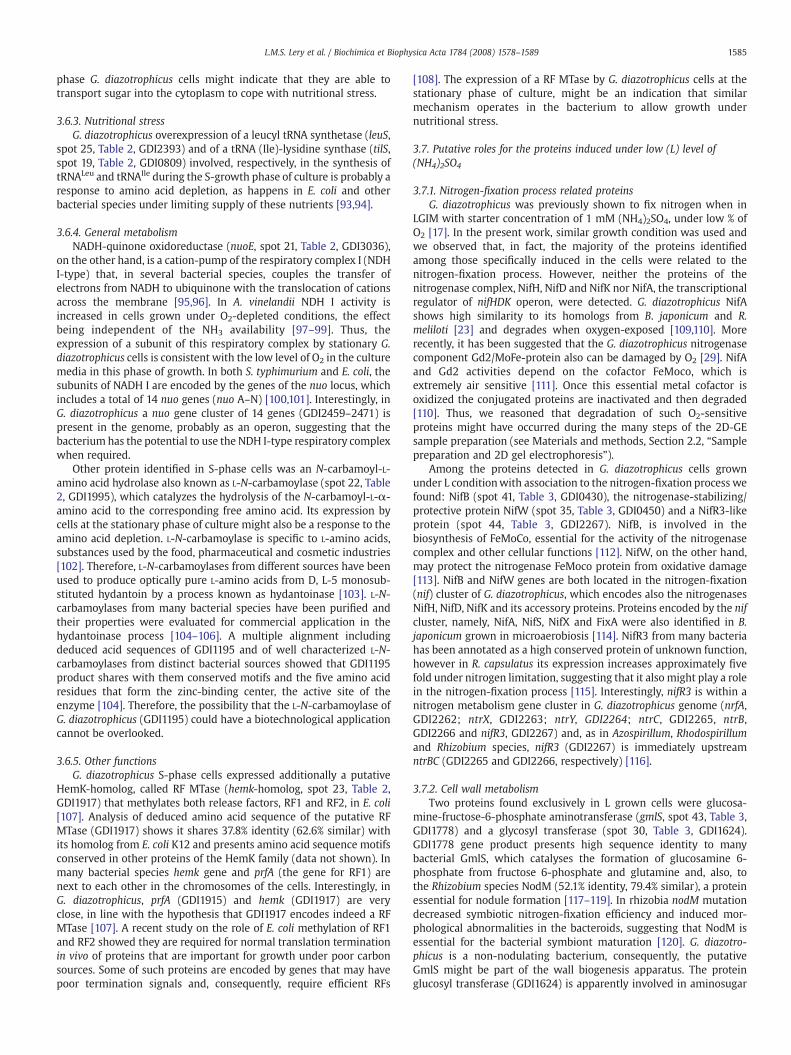
3.7.1. Nitrogen-fixation process related proteinsG. diazotrophicus was previously shown to fix nitrogen when in
LGIM with starter concentration of 1 mM (NH4)2SO4, under low % ofO2 [17]. In the present work, similar growth condition was used andwe observed that, in fact, the majority of the proteins identifiedamong those specifically induced in the cells were related to thenitrogen-fixation process. However, neither the proteins of thenitrogenase complex, NifH, NifD and NifK nor NifA, the transcriptionalregulator of nifHDK operon, were detected. G. diazotrophicus NifAshows high similarity to its homologs from B. japonicum and R.meliloti [23] and degrades when oxygen-exposed [109,110]. Morerecently, it has been suggested that the G. diazotrophicus nitrogenasecomponent Gd2/MoFe-protein also can be damaged by O2 [29]. NifAand Gd2 activities depend on the cofactor FeMoco, which isextremely air sensitive [111]. Once this essential metal cofactor isoxidized the conjugated proteins are inactivated and then degraded[110]. Thus, we reasoned that degradation of such O2-sensitiveproteins might have occurred during the many steps of the 2D-GEsample preparation (see Materials and methods, Section 2.2, “Samplepreparation and 2D gel electrophoresis”).
Among the proteins detected in G. diazotrophicus cells grownunder L conditionwith association to the nitrogen-fixation process wefound: NifB (spot 41, Table 3, GDI0430), the nitrogenase-stabilizing/protective protein NifW (spot 35, Table 3, GDI0450) and a NifR3-likeprotein (spot 44, Table 3, GDI2267). NifB, is involved in thebiosynthesis of FeMoCo, essential for the activity of the nitrogenasecomplex and other cellular functions [112]. NifW, on the other hand,may protect the nitrogenase FeMoco protein from oxidative damage[113]. NifB and NifW genes are both located in the nitrogen-fixation(nif) cluster of G. diazotrophicus, which encodes also the nitrogenasesNifH, NifD, NifK and its accessory proteins. Proteins encoded by the nifcluster, namely, NifA, NifS, NifX and FixA were also identified in B.japonicum grown in microaerobiosis [114]. NifR3 from many bacteriahas been annotated as a high conserved protein of unknown function,however in R. capsulatus its expression increases approximately fivefold under nitrogen limitation, suggesting that it also might play a rolein the nitrogen-fixation process [115]. Interestingly, nifR3 is within anitrogen metabolism gene cluster in G. diazotrophicus genome (nrfA,GDI2262; ntrX, GDI2263; ntrY, GDI2264; ntrC, GDI2265, ntrB,GDI2266 and nifR3, GDI2267) and, as in Azospirillum, Rhodospirillumand Rhizobium species, nifR3 (GDI2267) is immediately upstreamntrBC (GDI2265 and GDI2266, respectively) [116].
3.7.2. Cell wall metabolismTwo proteins found exclusively in L grown cells were glucosa-
mine-fructose-6-phosphate aminotransferase (gmlS, spot 43, Table 3,GDI1778) and a glycosyl transferase (spot 30, Table 3, GDI1624).GDI1778 gene product presents high sequence identity to manybacterial GmlS, which catalyses the formation of glucosamine 6-phosphate from fructose 6-phosphate and glutamine and, also, tothe Rhizobium species NodM (52.1% identity, 79.4% similar), a proteinessential for nodule formation [117–119]. In rhizobia nodM mutationdecreased symbiotic nitrogen-fixation efficiency and induced mor-phological abnormalities in the bacteroids, suggesting that NodM isessential for the bacterial symbiont maturation [120]. G. diazotro-phicus is a non-nodulating bacterium, consequently, the putativeGmlS might be part of the wall biogenesis apparatus. The proteinglucosyl transferase (GDI1624) is apparently involved in aminosugar

1586 L.M.S. Lery et al. / Biochimica et Biophysica Acta 1784 (2008) 1578–1589
metabolism and might, as well, play roles in the biosynthesis of thewall.
Another protein of the cell envelope of G. diazotrophicus differen-tially expressed by cells under L growth condition was a putativepeptidoglycan synthetase FtsI (spot 27, Table 3, GDI3185), also knownas penicillin-binding protein 3 (PBP3) [121,122]. PBP is a family ofproteins involved in the last stages of the synthesis of themurein [123]and E. coli PBP3 specifically catalyzes peptide cross-bridges of theseptal cell wall peptidoglycan during cell division [124]. It has beenshown that the levels of N, specially in the form of ammonium, inducemorphological changes in G. diazotrophicus cells [4]. Therefore, it ispossible that NodM/GlmS (spot 43, GDI1778), glycosyl transferase(spot 30, GDI1624) as well as, the outer membrane lipoprotein (spots36–39, Table 3, GDI2185) and FtsI/PBP3 (spot27, GDI3185) play roles inthe reshaping of the G. diazotrophicus cell wall during growth undernitrogen limited condition.
3.7.3. General metabolismA dienelactone hydrolase, DHL (spot 29, Table 3, GDI2054) was also
expressed by cells under L level of (NH4)2SO4. It is a detoxificationenzyme responsible for the degradation of chlorinated aromaticcompounds in Pseudomonas, Rhodococcus opacus and Ralstoniaeutropha [125–127]. Its differential expression in G. diazotrophicuscells under L condition suggests it might work in the detoxification ofnoxious products and that G. diazotrophicus has the potential to dealwith aromatic compounds produced in abundance by plants in thesymbiotic stage of life [128]. For the nitrogen-fixing rhizobiapolyphenols in the rhizosphere function as signaling molecules toinitiate plant–host infection, once inside the nodules the bacteroidsmetabolize these aromatic compounds to use as carbon source [114].However, no such observation has been reported for G. diazotrophicus.
3.7.4. RegulatorsOther proteins expressed by L condition grown cells were a
putative polyhydroxyalkanoate (PHA) synthesis repressor (phaR, spots31–33, Table 3, GDI3301) involved in the control of PHA biosynthesis,as discussed above (Section 3.5, “Synthesis of carbon and energystorage compounds”) and a transcriptional activator protein (spot 26,Table 3, GDI1273). Besides having a sequence-specific DNA-bindingmotif (helix–turn–helix) the GDI1273 gene product has also a putativecAMP binding site, suggesting that it might be a transcriptionregulator of the Crp family. However, it shows high sequencesimilarity to the oxygen-responsive transcriptional regulators FNR(fumarate nitrate reductase regulator), which under anoxic conditionsis in the active state. We thus hypothesized that the product ofGDI1273 is a member of the CRP-FNR superfamily [129] that is,apparently, required for growth of G. diazotrophicus cells underdeprivation of combined nitrogen and low level of O2.
A putative member of the transcriptional regulator GntR family (spot28, Table 3, GDI0682) was found in significant higher levels in G.diazotrophicus cells grown under L than in those under H condition. GntRbelongs to a family of bacterial helix–turn–helix DNA-binding transcrip-tional regulators that comprises four subfamilies, with more than 270members, distributed among the most diverse bacterial groups. A greatnumber of them are of unknown function, but others, are involved in theregulation the most various biological processes [130].
3.7.5. Other functionsThe putative dihydrolipoyllysine-residue acetyltransferase, also
called pyruvate dehydrogenase E2 component (aceF, spot 34, Table 3,GDI1720) was identified in cells deprived of combined nitrogen. AceF(GDI1720) is one of the three components of the pyruvate dehydro-genase that catalyses the oxidative decarboxylation of pyruvate toacetyl-CoA and CO2 via a series of the enzyme-bound intermediates.The enzyme has been well studied in E. coli whose AceF subunit is anoxidation-responsive factor of 66,095 kDa. However, it has been
detected as smaller components by SDS-PAGE suggesting that is liableto cleavage [131]. The putative AceF-homolog of G. diazotrophicusidentified in this work has the theoretical molecular weight of38,708 kDa, but was detected on the 2D gel as a protein of about18 kDa, an indication that, similarly to its E. coli counterpart, it hasbeen submitted to a cleavage mechanism [131]. It is already knownthat microaerobic diazotrophs possess numerous restriction strategiesto protect nitrogenases from inactivation by oxygen. Therefore, theexpression of an E. coli AceF-homolog in G. diazotrophicus nitrogen-fixing cells could be one of the protection mechanisms used by thebacterium against oxidative inactivation of the nitrogenases.
The 50S ribosomal protein L25 (spot 42, Table 3, GDI0935) was alsosuper expressed by cells under L growth condition.
3.8. Putative roles for the proteins induced under high (H) level(NH4)2SO4
3.8.1. Protein and DNA metabolismsOnly three proteins were differentially induced in G. diazotrophicus
grown under H condition, an integrase, (spot 45, Table 4, GDI3581),probably associated to DNA recombination [76] and two peptidases,namely, the peptidase M24 (spot 46, Table 4, GDI1990) and peptidasesignal I, SPaseI (spot 47, Table 4, GDI2154). M24 and SPaseI peptidasesspecifically remove the N-terminal amino acids from peptides andmight function in protein export [84]. Gram-negative bacteria typicallyhave only one chromosomal type I SPase, exceptions are P. aeruginosawith two and B. japonicum with three SPases [132]. A search in thegenomedatabase ofG. diazotrophicus leads us to two proteins annotatedas putative type I SPases, the products of GDI2154 (identified in thiswork) and of GDI3512, suggesting that the bacterium might be amongthe atypical type I peptidase signal I producers.
4. Conclusion
This study can be regarded as an additional step towardsestablishing the cytoplasmic proteome of G. diazotrophicus. Thebacterium was cultured in a bioreactor under low and highconcentrations of combined nitrogen, controlled levels of oxygenand varying pH, conditions that allowed the expression of special setsof proteins. These were analyzed by 2D-GE and a reasonable numberof protein spots were detected and used to construct master gelsrepresenting the proteomes of exponential (E) and stationary (S)phase cells under low (L) or high (H) levels of (NH4)2SO4. Comparisonof master gels showed 131 proteins (42E+32S+ 29H+28L) expresseddifferentially by G. diazotrophicus, fromwhich 46 were identified. Thegroups of proteins differentially induced during the E growth phasewere those expected to be functional in metabolically active bacterialcells. Particularly interesting was the increased expression of proteinsthat could play roles in pHi homeostasis, suggesting that the high acidtolerance of the exponential-phase G. diazotrophicus cells is, appar-ently, mediated by the products of a special set of genes. Cells from S-phase of growth, on the other hand, survived nutrient starvation andstress mainly by activating many transporters and transferases andother proteins to help the cells to cope with low levels of amino acidsand carbon sources. The vast majority of proteins induced differen-tially by cells under low (L) level of (NH4)2SO4 comprised those withroles in cell wall biosynthesis, confirming previous observation thatthe level of N, especially in the form of ammonium, affects themorphology of G. diazotrophicus cells [4]. Nitrogen-fixation relatedproteins were also identified in cells grown under L condition, as wellas proteins involved in the protection of the nitrogenases complexagainst oxidative damage, in agreement with the results of otherstudies, [4,17,131]. Moreover, the results of the present work permittedto deduce some novel metabolic pathways for G. diazotrophicus fromthe sequenced genome directly, based on the tendency of functionallyrelated bacterial genes to cluster along the chromosome. Therefore,

1587L.M.S. Lery et al. / Biochimica et Biophysica Acta 1784 (2008) 1578–1589
this study and our previous work [38] represent landmark accom-plishments in the functional proteomic analysis of G. diazotrophicus.
Acknowledgements
We thank the Riogene Genome Project Coordinator, Dr. PauloCavalcante Gomes Ferreira, and all other participants of the project forthe availability of G. diazotrophicus genome sequences and annota-tions. We are also grateful to LNLS/MCT for the use of the 4700Proteomics Analyzer TOF/TOF and to FAPERJ, CNPq and CAPES for thefinancial support.
References
[1] J. Kim, D.C. Rees, Nitrogenase and biological nitrogen fixation, Biochemistry 33(1994) 389–397.
[2] J. Six, C. Feller, K. Denef, S.M. Ogle, J.C.M. Sa, A. Albrecht, Soil organic matter, biotaand aggregation in temperate and tropical soils— effects of no-tillage, Agronomie22 (2002) 755–775.
[3] USDA, Natural Resources Conservation Service. Rangeland soil quality—organicmatter, http://soils.usda.gov/sqi (2001) in August 2006.
[4] R. Muthukumarasamy, G. Revathi, S. Seshadri, C. Lakshminarasimhan, Glucona-cetobacter diazotrophicus (syn. Acetobacter diazotrophicus), a promising diazo-trophic endophyte in tropics, Curr. Sci. 83 (2002) 137–145.
[5] A.C. Senok, A.Y. Ismaeel, G.A. Botta, Probiotics: facts and myths, Clin. Microbiol.Infect. 11 (2005) 958–966.
[6] V.A. Cavalcante, J. Döbereiner, A new acid-tolerant nitrogen-fixing bacteriumassociated with sugarcane, Plant Soil 108 (1988) 23–31.
[7] C.H. Bellone, S.D.V.C. De Bellone, R.O. Pedraza, M.A. Monzon, Cell colonizationand infection thread formation in sugar cane roots by Acetobacter diazotrophicus,Soil Biol. Biochem. 29 (1997) 965–967.
[8] Z. Dong, M. Heydrich, K. Bernard, M.E. McCully, Further evidence that the N(2)-fixing endophytic bacterium from the intercellular spaces of sugarcane stems isAcetobacter diazotrophicus, Appl. Environ. Microbiol. 61 (1995) 1843–1846.
[9] L.E. Fuentez-Ramirez, T. Jimenez-Salgado, I.R. Abarca-Ocampo, J. Caballero-Mellado, Acetobacter diazotrophicus, an indoleacetic acid producing bacteriumisolated from sugarcane cultivars of México, Plant Soil 154 (1993) 145–150.
[10] R.P. Li, I.C. MacRae, Specific association of diazotrophic acetobacters withsugarcane. Soil Biol. Biochem. 23 (1991) 999–1002.
[11] T. Jimenez-Salgado, L.E. Fuentes-Ramirez, A. Tapia-Hernandez, M.A. Mascarua-Esparza, E. Martinez-Romero, J. Caballero-Mellado, Coffea arabica L., a new hostplant for Acetobacter diazotrophicus, and isolation of other nitrogen-fixingacetobacteria, Appl. Environ. Microbiol. 63 (1997) 3676–3683.
[12] A. Tapia-Hernandez, M.R. Bustillos-Cristales, T. Jimenez-Salgado, J. Caballero-Mellado, L.E. Fuentes-Ramirez, Natural endophytic occurrence of Acetobacterdiazotrophicus in pineapple plants, Microb. Ecol. 39 (2000) 49–55.
[13] P. Loganathan, R. Sunlta, A.K. Parlda, S. Nair, Isolation and characterization of twogenetically distant groups of Acetobacter diazotrophicus from a new host plantEleusine coracana L. J. Appl. Microbiol. 87 (1999) 167–172.
[14] R. Muthukumarasamy, I. Cleenwerck, G. Revathi, M. Vadivelu, D. Janssens, B.Hoste, K.U. Gum, K.D. Park, C.Y. Son, T. Sa, J. Caballero-Mellado, Naturalassociation of Gluconacetobacter diazotrophicus and diazotrophic Acetobacterperoxydans with wetland rice, Syst. Appl. Microbiol. 28 (2005) 277–286.
[15] N.J. Ashbolt, P.A. Inkerman, Acetic acid bacterial biota of the pink sugar canemealybug, Saccharococcus sacchari, and its environs, Appl. Environ. Microbiol. 56(1990) 707–712.
[16] R.M. Boddey, O.C. de Oliveira, S. Urquiaga, V.M. Reis, F.L. Olivares, V.L.D. Baldani, J.Dobereiner, Biological nitrogen fixation associated with sugar cane and rice:contributions and prospects for improvement, Plant and Soil 90 (1995) 195–209.
[17] M.P. Stephan, M. Oliveira, K.R.S. Teixeira, G. Martinez-Drets, J. Döbereiner,Physiology and dinitrogen fixation of Acetobacter diazotrophicus, FEMSMicrobiol.Lett. 77 (1991) 67–72.
[18] F. Bastian, A. Cohen, P. Piccoli, V. Luna, R. Baraldi, R. Bottini, Production of indole-3-acetic acid and giberellins A(1) and A(3) by Gluconacetobacter diazotrophicusand Herbaspirillum seropedicae in chemically-defined culture media. PlantGrowth Regul. 24 (1998) 7–11.
[19] K.S. Maheshkumar, P.U. Krishnaraj, A.R. Alagawadi, Mineral phosphate solubiliz-ing activity of Acetobacter diazotrophicus: a bacterium associated with sugarcane,Curr. sci. 76 (1999) 874–875.
[20] V.S. Saravanan, M. Madhaiyan, M. Thangaraju, Solubilization of zinc compoundsby the diazotrophic, plant growth promoting bacterium Gluconacetobacterdiazotrophicus, Chemosphere 66 (2007) 1794–1798.
[21] M. Sevilla, D. Meletzus, K. Teixeira, S. Lee, A. Nutakki, I. Baldani, C. Kennedy,Analysis of nif and regulatory genes in Acetobacter diazotrophicus, Soil Biol.Biochem. 29 (1997) 871–874.
[22] K.R.S. Teixeira, M. Wulling, T. Morgan, R. Galler, E.M. Zellerman, I.J. Baldani, C.Kennedy, D. Meletzus, Molecular analysis of the chromosomal region encodingthe nifA and nifB genes of Acetobacter diazotrophicus, FEMS Microbiol. Lett. 71(1999) 521–530.
[23] K.R. Teixeira, T. Morgan, D. Meletzus, R. Galler, J.I. Baldani, C. Kennedy,Identification, sequencing and structural analysis of a nifA-like gene of Aceto-bacter diazotrophicus, An. Acad. Bras. Cienc. 71 (1999) 521–530.
[24] S. Lee, A. Reth, D. Meletzus, M. Sevilla, C. Kennedy, Characterization of a majorcluster of nif, fix, and associated genes in a sugarcane endophyte, Acetobacterdiazotrophicus, J. Bacteriol. 182 (2000) 7088–7091.
[25] D. Meletzus, K. Teixeira, O. Perlova, R. Nawroth, E. Zellermann, T. Morgan, V.Baldani, C. Kennedy, Characterization of genes involved in regulation of nitrogenfixation and ammonium sensing in Acetobacter diazotrophicus, an endophyte ofsugarcane, Biological Nitrogen Fixation for the 21st Century, Kluwer academicpublishers, Dordrecht, The Netherlands, 1998, pp. 125–126.
[26] V.M. Reis, J. Dobereiner, Effect of high sugar concentration on nitrogenase activityof Acetobacter diazotrophicus, Arch. Microbiol. 171 (1998) 13–18.
[27] A. Ureta, S. Nordlund, Evidence for conformational protection of nitrogenaseagainst oxygen in Gluconacetobacter diazotrophicus by a putative FeSII protein, J.Bacteriol. 184 (2002) 5805–5809.
[28] I. Goldberg, V. Nadler, A. Hochman, Mechanism of nitrogenase switch-off byoxygen, J. Bacteriol. 169 (1987) 874–879.
[29] K. Fisher, W.E. Newton, Nitrogenase proteins from Gluconacetobacter diazotro-phicus, a sugarcane-colonizing bacterium, Biochim. Biophys. Acta 1750 (2005)154–165.
[30] B. Pan, J.K. Vessey, Response of the endophytic diazotroph Gluconacetobacterdiazotrophicus on solid media to changes in atmospheric partial O(2) pressure,Appl. Environ. Microbiol. 67 (2001) 4694–4700.
[31] J.I. Baldani, V.L. Baldani, History on the biological nitrogen fixation research ingraminaceous plants: special emphasis on the Brazilian experience, An. Acad.Bras. Cienc. 77 (2005) 549–579.
[32] M.A. Djordjevic, H.C. Chen, S. Natera, G. Van Noorden, C. Menzel, S. Taylor, C.Renard, O. Geiger, G.F. Weiller, A global analysis of protein expression profiles inSinorhizobium meliloti: discovery of new genes for nodule occupancy and stressadaptation, Mol. Plant Microbe. Interact. 16 (2003) 508–524.
[33] M. Hecker, S. Engelmann, S.J. Cordwell, Proteomics of Staphylococcus aureus—current state and future challenges, J. Chromatogr. B Analyt. Technol. Biomed. LifeSci. 787 (2003) 179–195.
[34] S. Heim, M. Ferrer, H. Heuer, D. Regenhardt, M. Nimtz, K.N. Timmis, Proteomereference map of Pseudomonas putida strain KT2440 for genome expressionprofiling: distinct responses of KT2440 and Pseudomonas aeruginosa strain PAO1to iron deprivation and a new form of superoxide dismutase, Environ. Microbiol.5 (2003) 1257–1269.
[35] M.J. Han, S.Y. Lee, The Escherichia coli proteome: past, present, and futureprospects, Microbiol. Mol. Biol. Rev. 70 (2006) 362–439.
[36] A. Coelho, E. de Oliveira Santos, M.L. Faria, D.P. de Carvalho, M.R. Soares,W.M. vonKruger, P.M. Bisch, A proteome reference map for Vibrio cholerae El Tor,Proteomics 4 (2004) 1491–1504.
[37] W.M. von Kruger, L.M. Lery, M.R. Soares, F.S. de Neves-Manta, C.M. Batista e Silva, A.G. Neves-Ferreira, J. Perales, P.M. Bisch, The phosphate-starvation response in Vi-brio cholerae O1 and phoB mutant under proteomic analysis: disclosing functionsinvolved in adaptation, survival and virulence, Proteomics 6 (2006) 1495–1511.
[38] L.M.S. Lery, A. Coelho, W.M.A. von Krüger, M.S.M. Gonçalves, M.F. Santos, R.H.Valente, E.O. Santos, S.L.G. Rocha, J. Perales, G.B. Domont, K.R.S. Teixeira, P.M.Bisch, Protein expression profile of Gluconacetobacter diazotrophicus PAL5, asugarcane endophytic plant-growth promoting bacterium, Proteomics (in press).
[39] J. Rodrigues Neto, V.A. Malavolta Jr., O. Victor, Meio simples para isolamento ecultivo de Xanthomonas campestris pv. citri Tipo B. Summa Phytopathol. 12(1986) 16.
[40] O.H. Lowry, N.J. Rosebrough, A.L. Farr, R.J. Randall, Protein measurement with theFolin phenol reagent, J. Biol. Chem. 193 (1951) 265–275.
[41] T. Berkelman, T. Stenstedt, 2-D Electrophoresis using Immobilized pH Gradients:Principles and Methods, Piscataway, NJ (1998).
[42] V. Neuhoff, R. Stamm, H. Eibl, Clear background and highly sensitive proteinstaining with Coomassie Blue dyes in polyacrylamide gels: a systematic analysis.Electrophoresis 6 (1985) 427–448.
[43] GE-Healthcare, GeneBio, Swiss-Institute-of-Bioinformatics, ImageMaster 2DPlatinum software version 5.0, User Manual, GE Healthcare 2000.
[44] D.N. Perkins, D.J. Pappin, D.M. Creasy, J.S. Cottrell, Probability-based proteinidentification by searching sequence databases using mass spectrometry data,Electrophoresis 20 (1999) 3551–3567.
[45] S.F. Altschul, W. Gish, W. Miller, E.W. Myers, D.J. Lipman, Basic local alignmentsearch tool, J. Mol. Biol. 215 (1990) 403–410.
[46] S.F. Altschul, T.L. Madden, A.A. Schaffer, J. Zhang, Z. Zhang, W. Miller, D.J. Lipman,Gapped BLAST and PSI-BLAST: a new generation of protein database searchprograms, Nucleic Acids Res. 25 (1997) 3389–3402.
[47] A. Bateman, L. Coin, R. Durbin, R.D. Finn, V. Hollich, S. Griffiths-Jones, A. Khanna,M. Marshall, S. Moxon, E.L. Sonnhammer, D.J. Studholme, C. Yeats, S.R. Eddy, ThePfam protein families database, Nucleic Acids Res. 32 (2004) D138–D141.
[48] A.C. Len, S.J. Cordwell, D.W. Harty, N.A. Jacques, Cellular and extracellularproteome analysis of Streptococcus mutans grown in a chemostat, Proteomics 3(2003) 627–646.
[49] A.C. Len, D.W. Harty, N.A. Jacques, Proteome analysis of Streptococcus mutansmetabolic phenotype during acid tolerance, Microbiology 150 (2004) 1353–1366.
[50] R.S. Johnson, M.T. Davis, J.A. Taylor, S.D. Patterson, Informatics for proteinidentification bymass spectrometry, Methods (San Diego, Calif 35 (2005) 223–236.
[51] M.O. Walderhaug, J.W. Polarek, P. Voelkner, J.M. Daniel, J.E. Hesse, K. Altendorf,W. Epstein, KdpD and KdpE, proteins that control expression of the kdpABCoperon, are members of the two-component sensor-effector class of regulators,J. Bacteriol. 174 (1992) 2152–2159.
[52] A. Treuner-Lange, A. Kuhn, P. Durre, The kdp system of Clostridium acetobuty-licum: cloning, sequencing, and transcriptional regulation in response topotassium concentration, J. Bacteriol. 179 (1997) 4501–4512.

1588 L.M.S. Lery et al. / Biochimica et Biophysica Acta 1784 (2008) 1578–1589
[53] W. Epstein, The roles and regulation of potassium in bacteria, Prog. Nucleic AcidRes. Mol. Biol. 75 (2003) 293–320.
[54] A.E. Senior, J. Weber, Happymotoring with ATP synthase, Nat. Struct. Mol. Biol. 11(2004) 110–112.
[55] T. Miwa, T. Abe, S. Fukuda, S. Ohkawara, T. Hino, Regulation of H(+)-ATPasesynthesis in response to reduced pH in ruminal bacteria, Curr. Microbiol. 42(2001) 106–110.
[56] M. Matsumoto, H. Ohishi, Y. Benno, H+-ATPase activity in Bifidobacterium withspecial reference to acid tolerance, Int. J. Food Microbiol. 93 (2004) 109–113.
[57] H. Kobayashi, T. Suzuki, T. Unemoto, Streptococcal cytoplasmic pH is regulated bychanges in amount and activity of a proton-translocating ATPase, J. Biol. Chem.261 (1986) 627–630.
[58] A. Yokota, S. Amachi, S. Ishii, F. Tomita, Acid sensitivity of a mutant of Lactococcuslactis subsp. lactis C2 with reduced membrane-bound ATPase activity, Biosci.Biotechnol. Biochem. 59 (1995) 2004–2007.
[59] J.T. Davis, R.N. Moore, B. Imperiali, A.J. Pratt, K. Kobayashi, S. Masamune, A.J.Sinskey, C.T. Walsh, T. Fukui, K. Tomita, Biosynthetic thiolase from zoogloearamigera. I. Preliminary characterization and analysis of proton transfer reaction,J. Biol. Chem. 262 (1987) 82–89.
[60] B.H. Rehm, A. Steinbuchel, Biochemical and genetic analysis of PHA synthasesand other proteins required for PHA synthesis, Int. J. Biol. Macromol. 25 (1999)3–19.
[61] D.F. Chaves, P.P. Ferrer, E.M. de Souza, L.M. Gruz, R.A. Monteiro, F. de OliveiraPedrosa, A two-dimensional proteome reference map of Herbaspirillum serope-dicae proteins, Proteomics 7 (2007) 3759–3763.
[62] D. Segura, E. Vargas, G. Espin, Beta-ketothiolase genes in Azotobacter vinelandii,Gene 260 (2000) 113–120.
[63] S.A. Kim, L. Copeland, Enzymes of poly-(beta)-hydroxybutyrate metabolism insoybean and chickpea bacteroids, Appl. Environ. Microbiol. 62 (1996) 4186–4190.
[64] C. Wang, X. Sheng, R.C. Equi, M.A. Trainer, T.C. Charles, B.W. Sobral, Influence ofthe poly-3-hydroxybutyrate (PHB) granule-associated proteins (PhaP1 andPhaP2) on PHB accumulation and symbiotic nitrogen fixation in Sinorhizobiummeliloti Rm1021, J. Bacteriol. 189 (2007) 9050–9056.
[65] A. Steinbuchel, S. Hein, Biochemical and molecular basis of microbial synthesis ofpolyhydroxyalkanoates in microorganisms, Adv. Biochem. Eng. Biotechnol. 71(2001) 81–123.
[66] L.L. Madison, G.W. Huisman, Metabolic engineering of poly(3-hydroxyalkano-ates): from DNA to plastic, Microbiol. Mol. Biol. Rev. 63 (1999) 21–53.
[67] K.V. Rajagopalan, J.L. Johnson, The pterin molybdenum cofactors, J. Biol. Chem.267 (1992) 10199–10202.
[68] G. Schwarz, R.R. Mendel, Molybdenum cofactor biosynthesis and molybdenumenzymes, Annu. Rev. Plant. Biol. 57 (2006) 623–647.
[69] T. Palmer, A. Vasishta, P.W.Whitty, D.H. Boxer, Isolation of protein FA, a product ofthe mob locus required for molybdenum cofactor biosynthesis in Escherichia coli,Eur. J. Biochem. 222 (1994) 687–692.
[70] H.J. Sofia, G. Chen, B.G. Hetzler, J.F. Reyes-Spindola, N.E. Miller, Radical SAM, anovel protein superfamily linking unresolved steps in familiar biosyntheticpathways with radical mechanisms: functional characterization using newanalysis and information visualization methods, Nucleic Acids Res. 29 (2001)1097–1106.
[71] K.C. Lin, A. Campbell, D. Shiuan, Binding characteristics of Escherichia coli biotinrepressor-operator complex, Biochim. Biophys. Acta 1090 (1991) 317–325.
[72] P. Entcheva, D.A. Phillips, W.R. Streit, Functional analysis of Sinorhizobiummelilotigenes involved in biotin synthesis and transport, Appl. Environ. Microbiol. 68(2002) 2843–2848.
[73] A.M. Neculai, D. Neculai, C. Griesinger, J.A. Vorholt, S. Becker, A dynamic zincredox switch, J. Biol. Chem. 280 (2005) 2826–2830.
[74] M. Goenrich, S. Bartoschek, C.H. Hagemeier, C. Griesinger, J.A. Vorholt, Aglutathione-dependent formaldehyde-activating enzyme (Gfa) from Paracoccusdenitrificans detected and purified via two-dimensional proton exchange NMRspectroscopy, J. Biol. Chem. 277 (2002) 3069–3072.
[75] P.G. Zaphiropoulos, Non-homologous recombination mediated by Thermusaquaticus DNA polymerase I. Evidence supporting a copy choice mechanism,Nucleic Acids Res. 26 (1998) 2843–2848.
[76] M.C. Smith, H.M. Thorpe, Diversity in the serine recombinases, Mol. Microbiol. 44(2002) 299–307.
[77] J.G. Lawrence, H. Hendrickson, Lateral gene transfer: whenwill adolescence end?Mol. Microbiol. 50 (2003) 739–749.
[78] R.J. Roberts, D. Macelis, REBASE—restriction enzymes and methylases, NucleicAcids Res. 24 (1996) 223–235.
[79] N.B. Ray, C.K. Mathews, Nucleoside diphosphokinase: a functional link betweenintermediary metabolism and nucleic acid synthesis, Curr. Top. Cell. Regul. 33(1992) 343–357.
[80] G.W. Sundin, S. Shankar, S.A. Chugani, B.A. Chopade, A. Kavanaugh-Black, A.M.Chakrabarty, Nucleoside diphosphate kinase from Pseudomonas aeruginosa:characterization of the gene and its role in cellular growth and exopolysacchar-ide alginate synthesis, Mol. Microbiol. 20 (1996) 965–979.
[81] A.D. Tepper, H. Dammann, A.A. Bominaar, M. Veron, Investigation of the activesite and the conformational stability of nucleoside diphosphate kinase by site-directed mutagenesis, J. Biol. Chem. 269 (1994) 32175–32180.
[82] M.A. Bernard, N.B. Ray, M.C. Olcott, S.P. Hendricks, C.K. Mathews, Metabolicfunctions of microbial nucleoside diphosphate kinases, J. Bioenerg. Biomem-branes 32 (2000) 259–267.
[83] A.M. Chakrabarty, Nucleoside diphosphate kinase: role in bacterial growth,virulence, cell signalling and polysaccharide synthesis, Mol. Microbiol. 28 (1998)875–882.
[84] R. Tuteja, Type I signal peptidase: an overview, Arch. Biochem. Biophys. 441(2005) 107–111.
[85] J. Deutscher, C. Francke, P.W. Postma, How phosphotransferase system-relatedprotein phosphorylation regulates carbohydrate metabolism in bacteria, Micro-biol. Mol. Biol. Rev. 70 (2006) 939–1031.
[86] T. Tam le, C. Eymann, D. Albrecht, R. Sietmann, F. Schauer, M. Hecker, H.Antelmann, Differential gene expression in response to phenol and catecholreveals different metabolic activities for the degradation of aromatic compoundsin Bacillus subtilis, Environ. Microbiol. 8 (2006) 1408–1427.
[87] K. Steiner, H. Malke, relA-independent amino acid starvation response networkof Streptococcus pyogenes, J. Bacteriol. 183 (2001) 7354–7364.
[88] T. Elliott, Transport of 5-aminolevulinic acid by the dipeptide permease in Sal-monella typhimurium, J. Bacteriol. 175 (1993) 325–331.
[89] E. Verkamp, V.M. Backman, J.M. Bjornsson, D. Soll, G. Eggertsson, The periplasmicdipeptide permease system transports 5-aminolevulinic acid in Escherichia coli,J. Bacteriol. 175 (1993) 1452–1456.
[90] M. Matsuzaki, Y. Kiso, I. Yamamoto, T. Satoh, Gene disruption analysis of DppAisolated as a periplasmic molecular chaperone-like protein for folding ofdimethyl sulfoxide reductase in Rhodobacter sphaeroides f. sp. denitrificans,FEMS Microbiol. Lett. 193 (2000) 223–229.
[91] J.E. Lopilato, J.L. Garwin, S.D. Emr, T.J. Silhavy, J.R. Beckwith, D-ribose metabolismin Escherichia coli K-12: genetics, regulation, and transport, J. Bacteriol. 158(1984) 665–673.
[92] R. Wang, J.T. Prince, E.M. Marcotte, Mass spectrometry of the M. smegmatisproteome: protein expression levels correlate with function, operons, and codonbias, Genome Res. 15 (2005) 1118–1126.
[93] H. Putzer, S. Laalami, A.A. Brakhage, C. Condon, M. Grunberg-Manago,Aminoacyl-tRNA synthetase gene regulation in Bacillus subtilis: induction,repression and growth-rate regulation, Mol. Microbiol. 16 (1995) 709–718.
[94] E. McGinnis, L.S. Williams, Regulation of synthesis of the aminoacyl-transferribonucleic acid synthetases for the branched-chain amino acids of Escherichiacoli, J. Bacteriol. 108 (1971) 254–262.
[95] D. Flemming, P. Hellwig, S. Lepper, D.P. Kloer, T. Friedrich, Catalytic importance ofacidic amino acids on subunit NuoB of the Escherichia coli NADH:ubiquinoneoxidoreductase (complex I), J. Biol. Chem. 281 (2006) 24781–24789.
[96] M. Tao, K. Turk, J. Diez, M.G. Grutter, G. Fritz, J. Steuber, Crystallization of theNADH-oxidizing domain of the Na+-translocating NADH:ubiquinone oxidore-ductase from Vibrio cholerae, Acta Crystallogr., F Struct. Biol. Cryst. Commun. 62(2006) 110–112.
[97] Y.V. Bertsova, A.V. Bogachev, V.P. Skulachev, Two NADH:ubiquinone oxidor-eductases of Azotobacter vinelandii and their role in the respiratory protection,Biochim. Biophys. Acta 1363 (1998) 125–133.
[98] M.A. Smith, M. Finel, V. Korolik, G.L. Mendz, Characteristics of the aerobicrespiratory chains of the microaerophiles Campylobacter jejuni and Helicobacterpylori, Arch. Microbiol. 174 (2000) 1–10.
[99] J. Steuber, The Na+-translocating NADH:quinone oxidoreductase (NDH I) fromKlebsiella pneumoniae and Escherichia coli: implications for the mechanism ofredox-driven cation translocation by complex I, J. Bioenerg. Biomembranes 33(2001) 179–186.
[100] U. Weidner, S. Geier, A. Ptock, T. Friedrich, H. Leif, H. Weiss, The gene locus of theproton-translocating NADH: ubiquinone oxidoreductase in Escherichia coli.Organization of the 14 genes and relationship between the derived proteinsand subunits of mitochondrial complex I, J. Mol. Biol. 233 (1993) 109–122.
[101] C.D. Archer, X.Wang, T. Elliott, Mutants defective in the energy-conserving NADHdehydrogenase of Salmonella typhimurium identified by a decrease in energy-dependent proteolysis after carbon starvation, Proc. Natl. Acad. Sci. U S A 90(1993) 9877–9881.
[102] A.S. Bommarius, M. Schwarm, K. Drauz, The membrane reactor for the acylaseprocess from laboratory results to commercial scale, Chim. Oggi. 14 (1996)61–64.
[103] J. Altenbuchner, M. Siemann-Herzberg, C. Syldatk, Hydantoinases and relatedenzymes as biocatalysts for the synthesis of unnatural chiral amino acids, Curr.Opin. Biotechnol. 12 (2001) 559–563.
[104] S. Martinez-Rodriguez, F.J. Las Heras-Vazquez, L. Mingorance-Cazorla, J.M.Clemente-Jimenez, F. Rodriguez-Vico, Molecular cloning, purification, andbiochemical characterization of hydantoin racemase from the legume sym-biont Sinorhizobium meliloti CECT 4114, Appl. Environ. Microbiol. 70 (2004)625–630.
[105] T. Ohmachi, M. Narita, M. Kawata, A. Bizen, Y. Tamura, Y. Asada, A novel N-carbamoyl-L-amino acid amidohydrolase of Pseudomonas sp. strain ON-4a:purification and characterization of N-carbamoyl-L-cysteine amidohydrolaseexpressed in Escherichia coli, Appl. Microbiol. Biotechnol. 65 (2004) 686–693.
[106] H.Y. Hu, W.H. Hsu, H.R. Chien, Characterization and phylogenetic analysis of athermostable N-carbamoyl-L-amino acid amidohydrolase from Bacillus kausto-philus CCRC11223, Arch. Microbiol. 179 (2003) 250–257.
[107] V. Heurgue-Hamard, S. Champ, A. Engstrom,M. Ehrenberg, R.H. Buckingham, ThehemK gene in Escherichia coli encodes the N(5)-glutamine methyltransferasethat modifies peptide release factors, Embo J. 21 (2002) 769–778.
[108] L. Mora, V. Heurgue-Hamard, M. de Zamaroczy, S. Kervestin, R.H. Buckingham,Methylation of bacterial release factors RF1 and RF2 is required for normaltranslation termination in vivo, J. Biol. Chem. 282 (2007) 35638–35645.
[109] H.M. Fischer, Genetic regulation of nitrogen fixation in rhizobia, Microbiol. Rev.58 (1994) 352–386.
[110] E. Morett, H.M. Fischer, H. Hennecke, Influence of oxygen on DNA binding,positive control, and stability of the Bradyrhizobium japonicum NifA regulatoryprotein, J. Bacteriol. 173 (1991) 3478–3487.

1589L.M.S. Lery et al. / Biochimica et Biophysica Acta 1784 (2008) 1578–1589
[111] L.L. Martin, L.C. West, B. Wu, An extrusion strategy for the FeMo cofactor fromnitrogenase. Towards synthetic iron–sulfur proteins, Eur. J. Biochem. 268 (2001)5676–5686.
[112] H. Hennecke, Nitrogen fixation genes involved in the Bradyrhizobium japonicum-soybean symbiosis, FEBS Lett. 268 (1990) 422–426.
[113] C. Freiberg, R. Fellay, A. Bairoch,W.J. Broughton, A. Rosenthal, X. Perret, Molecularbasis of symbiosis between Rhizobium and legumes, Nature 387 (1997) 394–401.
[114] A.D. Sarma, D.W. Emerich, Global protein expression pattern of Bradyrhizobiumjaponicum bacteroids: a prelude to functional proteomics, Proteomics 5 (2005)4170–4184.
[115] P.J. Cullen, W.C. Bowman, D.F. Hartnett, S.C. Reilly, R.G. Kranz, Translationalactivation by an NtrC enhancer-binding protein, J. Mol. Biol. 278 (1998) 903–914.
[116] Y. Zhang, E.L. Pohlmann, M.C. Conrad, G.P. Roberts, The poor growth of Rhodos-pirillum rubrum mutants lacking PII proteins is due to an excess of glutaminesynthetase activity, Mol. Microbiol. 61 (2006) 497–510.
[117] M. John, J. Schmidt, U. Wieneke, H.D. Krussmann, J. Schell, Transmembraneorientation and receptor-like structure of the Rhizobium meliloti commonnodulation protein NodC, Embo J. 7 (1988) 583–588.
[118] E. Kamst, J. Pilling, L.M. Raamsdonk, B.J. Lugtenberg, H.P. Spaink, Rhizobiumnodulation protein NodC is an important determinant of chitin oligosaccharidechain length in Nod factor biosynthesis, J. Bacteriol. 179 (1997) 2103–2108.
[119] A.M. Hirsch, M.R. Lum, J.A. Downie, What makes the rhizobia-legume symbiosisso special? Plant Physiol. 127 (2001) 1484–1492.
[120] N. Baev, M. Schultze, I. Barlier, D.C. Ha, H. Virelizier, E. Kondorosi, A. Kondorosi,Rhizobium nodM and nodN genes are common nod genes: nodM encodesfunctions for efficiency of nod signal production and bacteroid maturation, J.Bacteriol. 174 (1992) 7555–7565.
[121] C. Goffin, J.M. Ghuysen, Multimodular penicillin-binding proteins: an enigmaticfamily of orthologs and paralogs, Microbiol. Mol. Biol. Rev. 62 (1998) 1079–1093.
[122] T. Den Blaauwen, M.E. Aarsman, N.O. Vischer, N. Nanninga, Penicillin-bindingprotein PBP2 of Escherichia coli localizes preferentially in the lateral wall and
at mid-cell in comparison with the old cell pole, Mol. Microbiol. 47 (2003)539–547.
[123] D.J. Scheffers, M.G. Pinho, Bacterial cell wall synthesis: new insights fromlocalization studies, Microbiol. Mol. Biol. Rev. 69 (2005) 585–607.
[124] A. Piette, C. Fraipont, T. Den Blaauwen, M.E. Aarsman, S. Pastoret, M. Nguyen-Disteche, Structural determinants required to target penicillin-binding protein 3to the septum of Escherichia coli, J. Bacteriol. 186 (2004) 6110–6117.
[125] M. Bruckmann, R. Blasco, K.N. Timmis, D.H. Pieper, Detoxification of proto-anemonin by dienelactone hydrolase, J. Bacteriol. 180 (1998) 400–402.
[126] O.V. Moiseeva, I.P. Solyanikova, S.R. Kaschabek, J. Groning, M. Thiel, L.A.Golovleva, M. Schlomann, A new modified ortho cleavage pathway of 3-chlorocatechol degradation by Rhodococcus opacus 1CP: genetic and biochemicalevidence, J. Bacteriol. 184 (2002) 5282–5292.
[127] V. Seibert, M. Thiel, I.S. Hinner, M. Schlomann, Characterization of a gene clusterencoding the maleylacetate reductase from Ralstonia eutropha 335T, an enzymerecruited for growth with 4-fluorobenzoate, Microbiology 150 (2004) 463–472.
[128] A.J. Beveridge, D.L. Ollis, A theoretical study of substrate-induced activation ofdienelactone hydrolase, Protein Eng. 8 (1995) 135–142.
[129] H. Korner, H.J. Sofia, W.G. Zumft, Phylogeny of the bacterial superfamily of Crp-Fnr transcription regulators: exploiting the metabolic spectrum by controllingalternative gene programs, FEMS Microbiol. Rev. 27 (2003) 559–592.
[130] S. Rigali, A. Derouaux, F. Giannotta, J. Dusart, Subdivision of the helix–turn–helixGntR family of bacterial regulators in the FadR, HutC, MocR, and YtrA subfamilies,J. Biol. Chem. 277 (2002) 12507–12515.
[131] P.E. Stephens, M.G. Darlison, H.M. Lewis, J.R. Guest, The pyruvate dehydro-genase complex of Escherichia coli K12. Nucleotide sequence encoding thedihydrolipoamide acetyltransferase component, Eur. J. Biochem. 133 (1983)481–489.
[132] M.L. van Roosmalen, N. Geukens, J.D. Jongbloed, H. Tjalsma, J.Y. Dubois, S. Bron,J.M. van Dijl, J. Anne, Type I signal peptidases of Gram-positive bacteria,Biochim. Biophys. Acta 1694 (2004) 279–297.