Biochemistry of nervous system, vision and sense. Metabolism of the eye. Mitochondrial disease. Jana...
28
Biochemistry of nervous system, vision and sense. Metabolism of the eye. Mitochondrial disease. Jana Švarcová
-
Upload
judith-dixon -
Category
Documents
-
view
224 -
download
2
Transcript of Biochemistry of nervous system, vision and sense. Metabolism of the eye. Mitochondrial disease. Jana...

Biochemistry of nervous system, vision and sense. Metabolism of the eye. Mitochondrial disease.
Jana Švarcová

➀ action potential reaches presynaptic membrane➁ opening of the voltage gated Ca2+-channels➂ released Ca2+ ions induce exocytosis of the neurotransmitter ➃ exocytosis of the neurotransmitter! each neuron releases only one NT➄ depolarization of the membrane → initiation of the action potential on post-synaptic memb. ➅ different action of metabotropic receptors – interaction with G-proteins
Synapse – passing the signal

3 types of neurons according to the function: Afferent neurons - transport impulses from
periphery to CNS (sensory) Efferent neurons - transport impulses from
CNS to muscles and glands (motoric) Interneurons – mainly in CNS, interconnect
neurons
Passing the signal

Action potential
= initiation and transfer of the neuron signal
neuron signal – electric signal, created by ion flux across plasma membrane of the neuron
membrane potential intracellular – high conc. of K+ (low Na+)
Na+/K+-ATPase – so called sodium pump requiring ATP Initiation of the signal – important role –
passive transport of K+ across plasma membrane

Action potential
✻ Charge reversal at membrane

Neurotransmitters
„small nitrogen-containing molecules“
glutamate GABA glycine acetylcholine dopamine noradrenalin serotonin histamine adrenalin aspartate
Neuropeptides
Small peptides synthesized in CNS endorfines Growth hormone and
TSH („thyroid-stimulating hormone“)

Neurotransmitters

Excitation AAreceptors for excitation AA: ionotropic – subtypes according to the selecktive
agonists: NMDA – (N-metyl-D-aspartic acid) AMPA – (amino-3-hydroxy-5-metyl-4-izoxazolpropionic acid) KA receptory – (kainate receptors, kainic acid)
metabotropic Stimulation of
phospholipase C inhibition of
adenylatecyclase and regulation of specific Ca2+ and K+ channels

Glutamate synapse glutamate – excitation
NT
Resorbed by neurons and changed to glutamate ~ 80 % of glutamate(alternative – synthesis from Glc or 2-oxoglutarate)storage – vesicleselimination – high affinity transporters for excitation AA – presynaptic membrane and membranes of surrounding glial cells= glutamate-glutamine cycle
synthesized directly in neurons from precursor molecules
glutamine – synthesized by glial cells

GABA – major inhibitory NT
GABA synthesis – from glutamate (precursor Glc, Pyr)
cofactor – PLP (from vit. B6) → vit. B6 insufficiency– decreased concentration of GABA in brain → loss of synaptic inhibition
! Inhibitory effect of GABA – synthesised from compound with excitation effect
Inhibitory AA
Glutamatedecarboxylase
Gluta
mate
dehyd
rogen
as
e

GABA-ergic synapse
synthesized GABA – encapsulated into vesicles by GABA vesicular transporter
After excretion – GABA resorbed from synaptic cleft (neurons or surrounding glial cells); transport requires presence of extracellular Na+
and Cl-

Acetylcholine
The first discovered NT Synthesized from acetylCoA
and choline in presynaptic part (cytosolar E-cholinacetyl transferase)
choline – from plasma; transport dependent on Na+. Required acetylCoA - from pyruvate, directly in neuron (glycolysis)
storage – synthesized acetylcholine – in vesicles
excreted – fast decomposition to inactive metabolites (acetate and choline;
E-acetylcholinesterase). Choline is transported back to presynaptic part

Acetylcholine
neurodegenerative changes of cholinergic neurons – serious pathological disorders
cholinergic pathways – mainly modulation entry to cortical and hippocampus neurons
→ drugs blocking acetylcholinesterase improve memory and learning performance and can partly reduce the consequences of lesions in cortex
Alzheimer disease – degeneration of cholinergic neurons in area of basal forebrain (= progressive loss of intellectual abilities)

Nicotin type acetylcholin receptors
Neuromuscular junctions, autonomous ganglia, adrenal medulla and CNS
ligand gated voltage channels – activation leads to influx of Na+ and Ca2+ → cell depolarization
typical characteristics – desensitisation (independent to other proteins; e.g. arestin) Rate of desensitisation – regulated by
phosphorylation of receptor subunit by proteinkinase A and C (or long term exposition of ligand)

Biochemistry of the eye, vision and senses.

Vision
Outer parts of eye – cornea and sclera
cornea – veinless , colourless, hydrated, and formed by collagen, needed continuous moisturizing
tears – nourishing covering cells of cornea and defense iris – regulating of light entering the eye lens – high protein content (α-, β-, γ-crystallines and theirs
insoluble aggregates) retina – the light detecting layer of the eye containing
photoreceptor cells rods – black and white (low light intensity) cones – colour vision
perception of light (∼400-750 nm) and its colours → resolution of contrast (black and white/colour ) and thus the contours; using the eye movement + shape of the eye

Cornea energy required for the integrity of cornea –
dependent on the glucose metabolism High percentage of ATP – aerobic glycolysis
(more than 70 % of Glc – pentose shunt) ⇒ thus, the oxygen supply is limiting for normal cornea metabolism. The usage of atmospheric oxygen had been observed in 1930 (Fischer)
hypoxia – oxygen is supplied by tears; metabolism of cornea - anaerobic → production and accumulation of lactate. Synthesis of glycogen is inhibited and glycogen supplies in epithelium decrease (example: contact lenses)
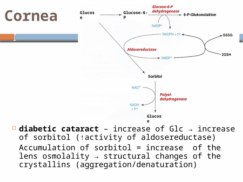
Cornea
diabetic cataract – increase of Glc → increase of sorbitol (↑activity of aldosereductase) Accumulation of sorbitol = increase of the lens osmolality → structural changes of the crystallins (aggregation/denaturation)
Glucose Glucose-6-P
Glucose-6-P dehydrogenase
Aldosereductase
Glucose
Polyol- dehydrogenase

Olfaction
olfactory receptor cells can regenerate – difference from other sensing cells (60 days)
olfactory receptors – several hundreds of different homologous olfactory receptors (vision – based on 4 different types of photoreceptor (3 cones and 1 rod) only
olfactory epithelial receptor has only one specific receptor and reacts to only one or small number of similar smells
odorants; reaching the cilia of receptors through mucilage layer, where they bind to Odorant binding proteins (OBP), and become water soluble
Transport rate – size of the complex odorant-OBP, mucilage viscosity and mechanical obstacles (tangled cilia) → olfactory signal is therefore amplified in the beginning phase
Transduction – interaction of odorant with specific receptor in cilial membrane of olfactory cell → activation of Golf-protein, stimulating adenylatecyclase and production of cAMP. cAMP activates protein kinase, which phosphorylates polypeptides of Na+-channel, opening. Opening of Na+-channel depolarizes cell membrane → generation of electric signal to the brain .

sourness – concentration of H+ ions, blocking outflux of K+ via voltage channel
saltiness = higher Na+ concentration - passive influx of these ions to cell, pumped out by ATPase
sweetness = sugars – activate membrane receptor → adenylatecyclase activation → formed cAMP blocks K+ channel → membrane depolarization
bitterness– specific protein gustducin (Ggust), which α subunit activates cAMP-phosphodiesterase → decreased level of cGMP → closure of Na+-channel and hyperpolarisation
Taste receptor – located on taste buds Taste perception – only water soluble
compound 4 basic tastes Saltiness and Sourness – change of
membrane depolarization in receptors – ions influx across plasma membrane
bitterness and sweetness. – sensing connected to activation of G-proteins
Umami – the fifth taste. Name coming from japanese (umai = tasty, delikate). Specific taste receptor - umami taste-mGluR4 (discovered in 2000) → sensing of glutamate acid(glutamates)
Taste

Mitochondrial diseases

Characteristics of the diseases
In general - mitochondria are energetic centres and mutation of Mt chromosomes cause defects in these energetic cycles ⇒ diseases manifest in organs and organ systems with high energy demand for instance CNS consumes up to 20 % of all ATP
produced by the body; this is the reason why CNS is the mostly affects organ. The others are muscles, heart liver and kidneys
mitochondrial dysfunctions play important role in more serious cell damage
some pathological states – important increase and decrease of the Mt volume ant their count (+ / -)
So called megamitochondria – found in case of alcohol liver disease or some nutrient deficiencies

genetic information in Mt – prone to mutations (in similar way as nucleolar DNA)
Mt genome exposure to the mutagens → changes in DNA
Frequency of the mutation occurrence – in mtDNA in average 10 × higher than in nDNA
reason of higher frequency of mutation: in Mt less correcting mechanism Main task of Mt - oxidative phosphorylation,
causing increased concentration of oxygen radicals; BUT: mtDNA is not shielded by histons
mtDNA replication is more frequent , P of error↑
Characteristics of the diseases

mitochondria – only maternal inheritance; (all children, no sex difference). Father suffering Mt disorder will not transfer this to his offsprings.
Predictability of Mt disease is low (during youth the energetic effectivity is sufficient)
⇒ important ratio affected to “healthy“ Mt (determined by Mt genotype) heteroplazia × homoplazia (mutation of all mtDNA) example. 20 year old – 85 % affected mtDNA ⇒
healthy appearance × close relative – affected 96% of Mt → the most serious symptoms
Characteristics of the diseases

Characteristics of the diseases
Deletion or point mutation of mtDNA – usually manifested by mitochondrial encephalomyopathy
Clinically heterogeneous group of diseases (but there are common morphologic abnormities in Mt causing different disorders in Mt metabolism: transport of the substrate from cytosol to mitochondria utilization of the substrate enzymes of citrate cycle coupling of phosphorylation with electron transport enzymes of electron transport chain
clinical manifestation - not thriving, psychomotoric retardation, symptoms of encephalopathy, myopathy, hepatopathy, hypertrophic cardiomyopathy, atrophy of nervus opticus
laboratory report – often lactate acidosis in case of defects in enzyme activity of respiratory chain complex, pyruvatedehydrogenase and ATP-synthase
Manifestation of hereditary Mt defects

Mt diseases - overview
Leber's hereditary optic neuropathy (LHON) Mitochondrial encephalomyopathy, lactic
acidosis, and stroke-like episodes (MELAS) Maternally inherited myopathy and
cardiomyopathy and others

Leber's hereditary optic neuropathy(LHON)
One of the most common mitochondrial diseases caused by point mutation in v mtDNA
Incidence is low , usually teenagers; prevalence cca 1 : 25 000
Symptoms and manifestation of the disease- eye damage with gradual blinding. Beginning – temporary visual losses ( loss of central vision and colour vision); severe optic atrophy and permanent decrease of visual acuity
Mutations in the MT-ND1, MT-ND4, MT-ND4L, and MT-ND6 genes (NADH dehydrogenase). Mutations in any of the genes disrupt this process to cause a
variety of syndromes depending on the type of mutation and other factors. It remains unclear how these genetic changes cause the death of cells in the optic nerve and lead to the specific features of Leber hereditary optic neuropathy.

Mitochondrial encephalomyopathy, lactic
acidosis, and stroke-like episodes (MELAS)
At present the most common disorder with encephalomyopathy symptoms
Appears in childhood Manifestation – low height, recurrent
headaches, loss of appetite, vomiting, and seizures with lactate acidosis. Later spasms, brain damage caused by calcification of CNS, ischemias. Repeated stroke-like episodes.
Affected neural tissue – occurrence of lesions, visible by magnetic resonance
Other symptoms - in 1.5 % of the cases the diabetes mellitus is cased probably – Langerhans islets are loosing the source
of energy and subsequently stop insulin synthesis