Biochemistry of Melanin
36
BIOCHEMISTRY OF MELANIN FORMATION AARON BUNSEN LERNER AND THOMAS B. FITZPATRICK’ From the Department of Dermatology and Sy#hilology, University of Michigan School of Medicine ANN ARBOR, MICHIGAN and the Mayo Foundation ROCHESTER, MINNESOTA ELANIN PIGMENTATION has aroused the curiosity and attention of man since the beginning of recorded history. This interest stems in part from the social, protective and cosmetic significance of pigmentation. In addi- tion, much interest has arisen concerning the possiblerole of melanin formation in the development of the melanomawhich has been described as having the most sinis- ter reputation of malignant tumors (I). As Ewing (2) has stated, “The variations in pigment offer a very delicate indication of the functional activity of the cells and a unique opportunity to trace the relation between the functional activity and the growth capacity of the tumor cells.” As a result, this subject has attracted the at- tention of workers in many fields (3), for example, the cytologist in the study of the melanin granules; the geneticist, the anthropologist and the biologist in a study of the relationship of heredity, environment and nutrition to the development of pigment; and the organic and biologic chemists in the study of the chemical mechanism under- lying melanin formation. In the past quarter of a century several comprehensive reviews on the mecha- nism of melanin formation have appeared (~-II), but in only a few of these hasthere been any consideration of the biochemical aspects of this problem. No attempt was made to connect the processes of melanin formation operating in various species. Different mechanisms were proposed for humans, lower animals and plants. Investigation of the biochemistry of melanin formation in plants, insects and marine animals had shown that the enzyme tyrosinase catalyzes the oxidation of L-tyrosine to dihydroxyphenyl L-alanine (dopa) and then the oxidation of dopa to melanin. Tyrosine Tyrosinase + Dopa -+ (Intermediates) -4 Melanin Until recently, however, the presence of tyrosinase in mammalian tissuehad not been conclusively demonstrated, and it was believed that melanin in mammalian tissue is formed by a mechanism different from that operating in other species. Histochemical evidence indicated that mammalian skin contains an enzyme, dopa- oxi&ase, which catalyzes the oxidation of dopa, but not tyrosine, to melanin. Dopa Dopa Oxidase + (Intermediates) -+ Melanin 1 Fellow in Dermatology and Syphilology, Mayo Foundation. 91
-
Upload
wekesamadzimoyo -
Category
Documents
-
view
166 -
download
15
description
Biochemistry of Melanin
Transcript of Biochemistry of Melanin

BIOCHEMISTRY OF MELANIN FORMATION
AARON BUNSEN LERNER AND THOMAS B. FITZPATRICK’
From the Department of Dermatology and Sy#hilology, University of Michigan School of Medicine
ANN ARBOR, MICHIGAN
and the Mayo Foundation
ROCHESTER, MINNESOTA
ELANIN PIGMENTATION has aroused the curiosity and attention of man since the beginning of recorded history. This interest stems in part from the social, protective and cosmetic significance of pigmentation. In addi-
tion, much interest has arisen concerning the possible role of melanin formation in the development of the melanoma which has been described as having the most sinis- ter reputation of malignant tumors (I). As Ewing (2) has stated, “The variations in pigment offer a very delicate indication of the functional activity of the cells and a unique opportunity to trace the relation between the functional activity and the growth capacity of the tumor cells.” As a result, this subject has attracted the at- tention of workers in many fields (3), for example, the cytologist in the study of the melanin granules; the geneticist, the anthropologist and the biologist in a study of the relationship of heredity, environment and nutrition to the development of pigment; and the organic and biologic chemists in the study of the chemical mechanism under- lying melanin formation.
In the past quarter of a century several comprehensive reviews on the mecha- nism of melanin formation have appeared (~-II), but in only a few of these has there been any consideration of the biochemical aspects of this problem. No attempt was made to connect the processes of melanin formation operating in various species. Different mechanisms were proposed for humans, lower animals and plants.
Investigation of the biochemistry of melanin formation in plants, insects and marine animals had shown that the enzyme tyrosinase catalyzes the oxidation of L-tyrosine to dihydroxyphenyl L-alanine (dopa) and then the oxidation of dopa to melanin.
Tyrosine Tyrosinase
+ Dopa -+ (Intermediates) -4 Melanin
Until recently, however, the presence of tyrosinase in mammalian tissue had not been conclusively demonstrated, and it was believed that melanin in mammalian tissue is formed by a mechanism different from that operating in other species. Histochemical evidence indicated that mammalian skin contains an enzyme, dopa- oxi&ase, which catalyzes the oxidation of dopa, but not tyrosine, to melanin.
Dopa Dopa Oxidase
+ (Intermediates) -+ Melanin
1 Fellow in Dermatology and Syphilology, Mayo Foundation.
91
A. B. LERNER AND T. B. FITZPATRICK 6;ol tcnte 30
Largely as a result of these beliefs, two separate hypotheses of melanogenesis evolved. Melanin formation in insects and plants was associated with tyrosinase, jTthi.le melanin production in mammalian skin was associated with dopa-oxidase.
In the present review, it is shown on the basis of recent evidence that these separate concepts can now be merged into a single hypothesis to account for melanin formation in man, lower animals, insects and plants. The properties of the enzymes concerned with melanin formation and the mechanisms of their reactions are con- sidered primarily. Attempts will be made when possible to correlate these properties and reaction mechanisms with normal and pathologic melanin pigmentation.
In view of the widespread occurrence of melanin pigment in nature and the keen interest shown in the problem of melanogenesis, it may seem surprising that there has been so much delay in clarifying the mechanism of melanin formation. Several obstacles have impeded progress in research on melanogenesis. Firstly, melanin (a darkly colored polymerized product of the oxidation of o-dihydroxyphenyl com- pounds, such as dopa, epinephrine, catechol) is of unknown chemical composition. Secondly, it is difficult to purify melanin and as a result, different chemical composi- tions have been reported by different investigators. Thirdly, epidermal extracts could not be prepared which conclusively demonstrated enzymatic activity toward tyrosine and dopa. This was due either to the small quantity of enzyme in the skin or to the fact that extracts made by grinding and homogenizing skin had not been satisfactory. Finally, the formation of melanin from its precursors is a complex process involving several steps, and the rates of the reactions depend on many surrounding chemical and physical factors.
HISTOCHEMICAL STUDIES
As early as 1901 v. Fiirth (12) advanced the hypothesis that melanin formation is the result of the action of an intracellular oxidase on aromatic or chromogen groups in certain protein molecules. Bloch, a Swiss dermatologist, stimulated by this hypoth- esis attempted to prove it by experimental methods (4). His approach was directed by two clinical observations. First, melanin pigmentation is a prominent feature of Addison’s disease, a disorder resulting from hypofunction of the adrenal glands. The possibility ,arose that there might be some relationship, albeit a paradoxical one, be- IVWX the increased pigmentation and the metabolism of epinephrine-like substances. Second, in cases of metastatic melanoma with melanuria, the urine contains significant amounts of catechol derivatives. These facts suggested to Bloch a chemical similarity between the precursor of melanin and the compounds epinephrine and catechol. He therefore selected a naturally occurring amino acid, 3,4-dihydroxyphenyl+
alanine (which he abbreviated as dopa, using the initial letters of its German name, :li-axy-phenyl-alanine) as the substrate for histochernical studies.
Bloch immersed frozen sections of pigmented human skin in a I : 1,000 solution of dopa buffered to PH 7.3 to 7.4. He noted that after 24 hours at room temperature lnelanin granules were deposited in the cytoplasm of cells located in the basal layer --
2 Dopa is found in the wing coverings of cockchafers Melolontha melotontha L. and M. hippo- cosfdtii F. (r3), and in the cocoons of one of the Satumidae, Samba cecropia L. (14). It also occurs in Vit-irt .faba (IS) and the Georgia velvet bean, Stizolobim~ deeringianum (16).

January 1950 BIOCHEMISTRY OF MEL.4NIN FORK4TION 93
of the epidermis. These specialized cells, which Bloch called melanoblasts, are located at the epidermo-dermal junction and were considered by Bloch to be the natural site of melanin formation. The intensity of the response, moreover, corresponded to the known capacity of the skin to form melanin. Melanin deposition was most intense in the areas of the skin capable of heavy pigmentation such as the basal layer of the epidermis, the germinal layer and cells of the hair matrix, and the cells of pigmented nevi. Melanin deposition did not occur in albino skin and in the skin of patients with vitiligo, a skin disease characterized by localized areas of complete loss of melanin pigment. Bloch (4, therefore, concluded that the ‘dopa reaction’ is a reliable indica- tor of the capacity of cells to form pigment.
The origin of the melanoblast has been a subject of controversy for many years. Bloch (4)) Peck (17) and others have supported the thesis that the melanoblasts are derived from ordinary palisade basal cells under certain types of stimuli. Masson (I 8) and Becker (19) , however, believe that the melanoblasts are of neuro-epithelial origin. In recent years several groups of investigators have carried out embryological studies in amphibia (20, 2 I), birds (22-24) and mice (25-27) and have shown conclusively that the melanoblasts are derived from the neural crest region.
Further investigation provided evidence that the catalytic effect of certain cells on the oxidation of dopa to melanin is due to the presence of an enzyme, which Bloch called dopa-oxidase. The evidence for the enzymatic nature of the dopa reaction included the following: I) The reaction did not occur after the tissue had been heated to IOOOC.; z) a definite pi range was required, the optimal PH being 7.35; 3) the reaction was completely inhibited by M/~,OOO hydrogen sulfide or M/~OO potassium cyanide, which are known inhibitors of some enzymes; 4) only the levorotatory form of dopa was catalytically oxidized by the cells?
To substantiate further the enzyme hypothesis Bloch and Schaaf (28) prepared extracts of skin from newborn rabbits and demonstrated by visual calorimetric methods the presence of a heat-labile, cyanide-sensitive catalyst capable of accelerat- ing the formation of melanin from dopa. Extracts of albino skin under the same con- ditions were ineffective in catalyzing the oxidation of dopa. Although Bloch’s tech- nics for measuring enzymatic activity were not detailed his data provided convincing evidence that the catalytic effect of certain epidermal cells on the oxidation of dopa is due to the presence of an enzyme.
The presence of dopa-oxidase in normal human or animal skin has not been con- clusively demonstrated by enzymatic technics such as the manometric measure- ment of oxygen uptake by skin slices or extracts in the presence of dopa. Although this has been attempted by several investigators, none has been successful because of the small quantity of enzyme present in the skin as well as the difficulty of pre- paring skin extracts and homogenates suitable for measuring enzymatic activity. It is also possible that in preparing extracts of skin the investigators neglected to take into account the possibility of a naturally occurring inhibitor which could mask the presence of the enzyme. Recently, Rothman and co-workers (29) succeeded in
8 Many other related compounds, such as ndopa, tyrosine, phenylalanine, P-hydroxyphenyl- pyruvic acid, homogentisic acid, quinol, pyrogallol, catechol, protocatechuic acid, 3,&ihydroxy- phenylacetic acid, epinephrine, trihydroxyphenylalanine, glycyl-dopa, pyrrole and tryptophane, did not form melanin granules after incubation with frozen sections of skin.

94 A. B. LERNER AND T. B. FITZPATRICK TW tme 30
demonstrating the presence in human epidermis of a water-soluble, dialyzable, heat-stable inhibitor of plant tyrosinase. This inhibitory principle was counteracted by iodoacetamide and another powerful sulfhydryl inhibitor, p-chloromercuriben- zoic acid. Later work (30) showed a direct relationship between the sulfhydryl group concentration and the inhibitory power of the epidermal extracts.
Bloch’s histochemical studies with dopa, which have been amply confirmed, have not been generally accepted as a complete explanation for the mechanism of melanogenesis since dopa has never been demonstrated in mammalian tissue. Raper (31) was able to isolate dopa in crystalline form as the first oxidation product re- sulting from the action of tyrosinase obtained from mealworms (Tenebrio molitor) on tyrosine. This strongly suggested that the reactions in the formation of melanin that are subsequent to dopa production are the same whether the initial substrate is tyrosine or dopa and emphasized the gap in the knowledge of the mechanism of the conversion of tyrosine to dopa in various tissues. Raper (IO) provided conclusive evidence that tyrosine, a naturally occurring amino acid present in all tissues includ- ing skin, is the initial substrate in the formation of melanin by plant and insect tyrosinase. As stated previously, Bloch’s dopa-oxidase reaction could not be obtained after incubation of the frozen skin sections with tyrosine under the same conditions in which dopa was effective. Since it was generally felt that tyrosine is the natural or physiologic substrate in the enzymatic formation of melanin by mammalian tissues as well as by plants and insect tissues, and since the reaction whereby tyrosine is converted to dopa in mammalian cells was not known, the mechanism of melanin formation in mammals remained a puzzling problem for many years.
Much investigation remains to be done on this problem. Bloch’s qualitative studies might well be extended by use of present-day microchemical methods for quantitative determinations and histologic localizations of small quantities of en- zymes. In addition, through the use of Cl*-labeled tyrosine it may be possible to show conclusively whether or not tyrosine is the natural substrate involved in melanin formation by melanoblasts.
BIOCHEMICAL STUDIES
Melanin Formation in Lower Autimals-the Tyrosinase Concept
The enzymatic nature of the melanin-producing reactions was first satisfactorilv investigated in plants and fungi. In 1895 Bourquelot and Bertrand (32) reported that a substance present in the mushroom, Russ&a Izigricatzs, was converted into a black pigment. This substance, later shown to be tyrosine, was acted on by an enzyme in the fungus which was named tyrosinase (33). Since that time tyrosinase has been found in a wide variety of plant and animal tissues (34-36) and a vast litera- ture has accumulated on this subject.
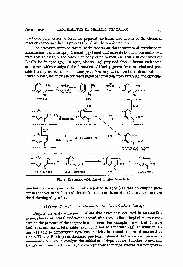
Working with tyrosinase obtained from plants and mealworms, Raper (IO) was able to determine many of the reaction mechanisms whereby tyrosine is con- verted into melanin. He showed that in the presence of tyrosinase and oxygen, tyrosine is first oxidized to dopa, and the dopa is then oxidized to dopa-quinone. Dopa-quinone is converted to an indole derivative which, after undergoing several
January 1950 BIOCHEMISTRY OF MELANIN FORMATION 95
reactions, polymerizes to form the pigment, melanin. The details of the chemical reactions concerned in this process (fig. I) will be considered later.
The literature contains several early reports on the occurrence of tyrosinase in mammalian tissue. In 1903, Gessard (37) found that extracts from a horse melanoma were able to catalyze the conversion of tyrosine to melanin. This was confirmed by De Coulon in 1920 (38). In IgoT, Alsberg (39) prepared from a hunan melanoma an extract which catalyzed the formation of black pigment from catechol and pos- sibly from tyrosine. In the following year, Neuberg (40) showed that dilute extracts from a human melanoma accelerated pigment formation from tyramine and epineph-
TYROS INE
"2
DOPA OUINONE
I
COOH
\ \ H
I!, 6- DIHYDROXYIWOOLL HALLACHROME trod) \ \
CEUCO COMPOUND
\ \
0 .+o+ MELANIN +---ti:--
0 Relatively slow COOH
H
INDOLE 5.60OUINONE 5,6-~IHYDROXYINDOLE-
2-CARROXYLIG ACID
OOPA QUINONE LEUCO COMPOUND DOPA HALLACHROME
Fig. I. ENZYMATIC oxidation of tyrosine to melanin.
rine but not from tyrosine. Winternitz reported in IgIg (41) that an enzyme pres- ent in the uvea of the hog and the black cutaneous tissue of the horse could catalyze the darkening of tyrosine.
M’elanin Formation in Mammals-the Dopa-OGdase Concept
Despite the early widespread beliefs that tyrosinase occurred in mammalian tissue, plus experimental evidence in accord with these beliefs, skepticism arose con- cerning the presence of the enzyme in such tissue. For example, the work of Durham (42) on tyrosinase in fetal rabbit skin could not be confirmed (43). In addition, no one was able to demonstrate tyrosinase activity in normal pigmented mammalian tissue. Finally, Bloch (4) as discussed previously, showed that an enzyme present in mammalian skin could catalyze the oxidation of dopa but not tyrosine to melanin. Largely as a result of this work, the concept arose that dopa-odase, but not tyrosia

96 A. B. LERNER AND T. B. FITZPATRICK T~xtcnte 3:)
ase, was present in mammalian tissue. Although Bloch’s work did not pass unchal- lenged, it remained the most acceptable until recently.
Hogeboom and Adams in 19~2 (JJ), Greenstein and co-workers in 1944 (45,
46) and Lemer, Fitzpatrick, Calkins and Summerson in 1949 (47) showed conclu- sively that extracts from mouse, human and horse melanomas contain both tyrosinase and dopa-oxidase activities and that these activities are similar to those found in extracts from plants and lower animals. Calkins (48) demonstrated that extracts from normal beef ciliary bodies possess tyrosinase and dopa-oxidase activities. In view of this experimental evidence of the presence of tyrosinase in mammalian tissue, it became necessary to modify the hypotheses of pigmentation which had evolved from Bloch’s dopa-oxidase studies.
Chemical Reactions in the Conuersiotl of Tyrosilte to Melank
The reactions involved in the enzymatic oxidation of tyrosine to melanin are shown in figure I. In the presence of tyrosinase and molecular oxygen, tyrosine is oxidized to dopa. This reaction is usually slow at the onset, but after an induction period it becomes very fast. The conversion of tyrosine to dopa is not a reversible reaction. Dopa formed in the first reaction is oxidized enzymaticaliy by a reversible reaction to dopa-quinone. Further stages of the reaction proceed rapidly in the ab- sence of the enzyme although the reaction rates are increased in the presence of the enzyme. Dopa-quinone undergoes a spontaneous irreversible and rapid intramolec- ular change in which the nitrogen of the side chain attaches itself to the &position of the benzene nucleus with ‘the formation of 5,6-dihydroxydihydroindole-z-car- boxylic acid (leuco compound). The leuco compound is readily oxidized by a rever- sible reaction to the corresponding quinone (hallachrome). Hallachrome4 is a red substance, and it is the first visible product formed in the reactions. Under physiologic conditions hallachrome decarboxylates and undergoes a rearrangement to form 5,6- dihydroxyindole. The indole compound is rapidly oxidized to the corresponding quinone which has a purple color. The quinone then polymerizes to melanin with the consumption of approximately one atom of oxygen. Relatively little is known of the mechanism of this polymerization (49, so). If the intramolecular rearrange- ment undergone by hallachrome is quickened by sulfurous acid, no decarboxylation occurs and 5,6-dihydroxyindole-2-carboxylic acid is formed. This latter substance is readily converted to a melanin substance. In the series of reactions shown in figure I, possible alternate mechanisms are described by broken arrows. Much of the knowledge of the chemical reactions which take place in the enzymatic oxidation of tyrosine has been obtained through the brilliant work of Raper and his co-worker (IO, sza) with potato and mealworm (Tenebrio molilor) tyrosinase. They were able to show that the following three substances were formed during the tyrosine-
4 Hallachrome occurs naturally in the polychaete worm, Hada parthenopala (51). Friedheim (52) found that this red substance could accelerate oxygen consumption by erythrocytes and serve as a hydrogen acceptor for xanthine oxidase and succinic dehydrogenase. He suggested that halla- chrome may play a role in cellular respiration.

Janzlary 1950 BIOCHEMISTRY OF MELANIN FORMATION 97
tyrosinase reaction:
HO’ CH 2
HO o-
I 1 \ CHCOOH
/ N
H2
I, Dopa II, 5,6=Dihydroxyindole III, 5,6-Dihydroxyin- dole-a-Carboxylic Acid
When the enzyme is allowed to act on dopa, substmces II and III are formed. This suggests that dopa is probably the first compound formed in the oxidation of tyro- sine.
Dopa formed from tyrosine appears to be oxidized to dopa-quinone. The fol- lowing facts make this plausible: I) o-dihydroxyphenyl compounds are readily oxidized to the corresponding orthoquinones; 2) substances which react with or- thoquinones inhibit melanin formation in the dopa-tyrosinase reaction (53) ; 3) plant tyrosinase has been shown to catalyze the oxidation of catechol to orthobenzo- quinone (54). The oxidation of dopa to dopa-quinone would appear to be a similar reaction.
Raper ( 55) also showed that a red substance (hallach rome) was formed in the oxidation of dopa. This red substance could be reduced to 5,6-dihydroxydihydroin- dole-a-carboxylic acid (leuco compound) (5 rza) 1, and the leuco compound so produced readily oxidized back to the red substance. From this it seems likely that dopa- quinone undergoes an intramolecular change, the nitrogen of the side chain attaching itself to the 6-posi tion of the benzene nucleus with the resultant formation of the leuco compound.
Since the indole ~~@unds II and III could be formed from the oxidation of the hallachrome, and since hallachrome was formed from the leuco compound, it was believed that the hallachrome was simply the quinone of the leuco compound and therefore should have the following structure:
Hallachrome
Mason has presented spectrophotometric evidence to support this view (49). Under normal conditions in the enzymatic oxidation of tyrosine, hallachrome
is converted, by decarboxylation and rearrangement, to 5,6-dihydroxyindole. Spec- trophotometric data (49) indicate that the 5,6-dihydroxyindole is then rapidly oxidized to the corresponding quinone and the quinone then polymerizes to melanin.
It should be pointed out that the reactions given in figure I merely represent the over-all scheme by which tyrosine is converted to melanin. Actually, many more reactions probably occur, such as those involving the formation of semiqui-

98 A. B. LERNER AND T. B. FITZPATRICK T’ol ill11 c 30
nones. The semiquinones may then be oxidized to quinones or undergo rearrange- ments in accordance with the general picture given in the diagram.
As previously stated, enzyme action is definitely required for the first reaction, the oxidation of tyrosine to dopa. Under physiologic conditions, that is, pi 7 to 7.4, the rate of oxidation of dopa to dopa-quinone is fairly rapid without the enzyme but is increased appreciably in the presence of the enzyme. The subsequent reactions shown in the diagram take place rapidly without the enzyme, but even in these cases there is evidence that the presence of tyrosinase will increase the rate of reaction
(49) . In addition to the series of reactions discussed above, there is an important
interplay of reactions occurring during the conversion of tyrosine to melanin as shown in the lower part of figure I. Using mealworm tyrosinase, Evans and Raper (52”) found that in a tyrosine-tyrosinase reaction which has proceeded for two to five hours, dopa can be isolated in yields varying from IO to 20 per cent of the actual tyrosine oxidized, in spite of the fact that this tyrosinase can oxidize dopa more readily than tyrosine. Since not appear to be a reversible
the conversion of dopa-quinone to leuco compound does reasonable to explain the accumulation
reduces of cl the
.opa on the basis of the dopa-quinone back to
concerned follows :
to a
in melanin formation in so far as they have been investigated (56) are as
reaction, it seems presence of dopa. The
some reducing agen oxidation-reduction
.t or system which potentials of the systems
Dopa ti Dopa-Quinone Eo = + 0.511 vat pH 4.6
Leuco Compound e Hallachrome E. = + 0.170 V at PH 46.
From these data and from the observation that addi tion of leuco compound tyrosine-tyrosinase system increases the accumulation of dopa, it seems likely
that the interplay of reactions shown in figure I occurs in the tyrosine-tyrosinase reaction. The importance of this reaction will be discussed later. It is possible that interactions involving substances such as dopa-quinone and 5,6-dihydroxyindole also occur. These reactions, if present, could play important roles in regulating -the rate of melanin formation.
Indzcction Period in the Oxidation of Tyrosine
and There are several points in the oxidation interest. When tyrosine and tyrosinase
of tyrosine that are of great are allowed to react in the
oxygen, there is often a lag period before oxidation of tyrosine begins. This lag in- terval is referred to as the inductioti period (47). Small amounts of dopa are very effective in shortening the induction period in the tyrosine-tyrosinase reaction (fig. 2). If the induction period is defined as the intercept on the time axis of an exten- sion of the slope of the oxidation curve when oxidation is proceeding maximally, there is for mammalian tyrosinase a linear relationship between the negative logarithm of the dopa concentration and the induction period (47).
importance presence of
Recent studies (57, $3) with mouse melanoma tyrosinase indicate that com- pounds related structurally to dopa, for example, epinephrine, catechol, and so

Januwy 1950 BIOCHEMISTRY OF MELANIN FORMATION 99
forth, can shorten the induction period, but not nearly as effectively as dopa. On an equimolar basis, DLdopa is about 75 per cent as effective as is Ldopa in shortening the induction period. For mammalian tyrosinase, dopa is a fairly specific catalyst regulating the induction period.
When dopa is used as the substrate instead of tyrosine there is no induction period. Although dopa is required to catalyze the enzymatic oxidation of tyrosine, dopa itself is rapidly oxidized. Hence the rate of tyrosine oxidation is der>endent on the rate of dopa oxidation.
I 180.
16Oj
1504
30-
b $6 I . m ’ * 1 l
' 20 30 40 50 60 70 80 60 lb0 il0
Time (minutes)
Fig. 2. EFFECT OP DOPA on induction period in enzymatic oxidation of tyrosine by mouse melanoma preparation at PH 6.8 and 38’C. a 0.5 mg. dopa; b 0.1 mg. dopa plus 0.4 mg. tyrosine; c 0.05 mg. dopa plus 0.45 mg. tyrosine; d 0.01 mg. dopa plus 0.49 mg. tyrosine; e o.001 mg. dopa plus 0.50 mg. tyrosine; f 0.5 mg. tyrosine. (J. Biol. Chem. 178: 192, Ig4g)-
It can be seen from figure 2 that after the induction period is over the rates of oxidation of tyrosine and dopa are practically identical. This observation at first glance seems difficult to understand, because one might expect that as the dopa formed from tyrosine is oxidized, the rate of reaction would diminish. The explanation of this fkding is that dopa is not only oxidized in the reaction but also reformed dur- ing the oxidation, as discussed previously (fig. I, lower part). Consequently, a significant amount of dopa is always available to shorten the induction period.
From what has been described it can be seen that the presence in the tyrosine-

100 A. B. LERNER AND T. B. FITZPATRICK Volume 30
tyrosinase reaction of any substance which is capable of reducing dopa-quinone back to dopa and thereby causes an accumulation of dopa should shorten the in- duction period. This is indeed the case. Either ascorbic acid or hydroquinone5 short- ens the induction period in tyrosine oxidation. Melanin, however, is not produced until all the ascorbic acid or hydroquinone has been oxidized. It is possible that the compounds related to dopa shorten the induction period by acting merely as non- specific reducing agents which influence the induction period as described previously.
It should be pointed out that it is well known that o-dihydroxyphenyl com- pounds shorten the induction period in the oxidation of monophenols with tyrosinase obtained from plants and lower animals. Most of the work on this subject, however, has been done with catechol, phenol and p-cresol and not with dopa and tyrosine (54). No emphasis has been placed on structural specificity required by the orthodi- hydroxyphenyl compound. Actually, this structural specificity could play an impor- tant role in melanin formation in nature.
The mechanism by which substances such as dopa regulate the induction period of the tyro- sine-tyrosinase reaction is not fully understood. Recently, the authors carried out potentiometric measurements using the platinum electrode on different solutions of tyrosine, dopa and tyrosinase
obtained from the Harding-Passey melanoma (59). Solutions of tyrosine in 0.1 molar potassium phosphate buffer were found to be at a much higher potential than similar equimolar solutions of dopa. On the addition of tyrosinase to the tyrosine solution the potential began to fall. When the potential had fallen to a value approximately equal to that of the buffered dopa solution, oxygen up-
take commenced. No appreciable oxygen uptake could be detected before the potential fell. As the oxidation proceeded and the tyrosine was converted to melanin, the potential began to rise; and when the oxidation was complete, the original potential was re-established. When the tyrosinase
was added to the dopa solutions, oxidation began immediately and the potential began to rise. At the end of the reaction the potential reached a value similar to that obtained in the tyrosine-tyro- sinase system. Small amounts of dopa added to the tyrosine solutions brought about an immediate lowering of the redox potential.
If the redox potentials were to influence the induction period, one could predict that increasing the tyrosine concentration of a tyrosine-tyrosinase reaction mixture would prolong the induction period. This was found to be the case. The redox potential of a system is established by the ratio of the quantity of reduced form of a substance to the oxidized form. An increase in tyrosine concen-
tration increases the concentration of the reduced form of the substrate. While the redox potential of the tyrosinase system may play a role in regulating the induction
period (as well as tyrosinase activity in general) it is not the only factor involved. Equimolar quanti- ties of DL-dopa are not as effective as is L-dopa in shortening the induction period, but there is no
reason to believe that DL-dopa should establish a redox potential different from that obtained with the same quantity of L-dopa. It appears that dopa is a fairly specific catalyst for the enzymatic oxi- dation of tyrosine.
Nature Tyrosinase
Tyrosinase can be obtained from various sources simply by grinding the tissues (for example, potato, fungus, melanotic tumor, and so forth) with an aqueous solu- tion and then centrifuging the mixture at low speeds. The supernatant usually con- tains the active enzyme (47). Further purification can often be obtained by using ordinary procedures for protein fractionation.
6 In addition to shortening the induction period by virtue of its reducing properties, hydro- quinone also appears to inhibit the enzyme.

Janriary 1,050 BIOCHEMISTRY OF MELANIN FORMATION 101
There are many qualitative differences among tyrosinases prepared from various sources, but they all have three characteristics in common: I) All catalyze the oxida- tion of tyrosine to melanin (presumably by the series of reactions shown in fig. I);
2) the enzymatic reaction with the monohydroxyphenyl compound is catalyzed by some o-dihydroxyphenyl compound (dopa, catechol etc.); 3) copper is associated with the activity of the enzyme. The first two points have been discussed previously.
Role of copper. Copper has been reported to be an essential part of tyrosinase prepared from mammalian (60), plant (61, 62) and insect tissue (63). Mouse mela- noma tyrosinase can be inhibited by reagents that combine with copper (diethyldi- thiocarbamate, BAL, etc.) (60). This inhibition was reversed by the addition of an excess of cupric ions. Treatment of the enzyme with cyanide followed by dialysis resulted in a decrease of the copper content of the enzyme preparation and a loss of enzymatic activity. Addition of sufficient cupric ions resulted in almost complete restoration of activity. Other metals (iron, cobalt, nickel, magnesium, manganese and zinc) were ineffective in restoring enzymatic activity. Previous experiments by Kubowitz (61, 62) with plant tyrosinase and by Allen and Bodine (63) on grass- hopper tyrosinase showed essentially the same results as those obtained with mam- malian tyrosinase.
Properties of tyroshase from different sources. As mentioned previously, there are several properties by which tyrosinase from different sources varies. Tyrosinase prepared from plant tissue can usually be obtained in colloidal solution. Tyrosinase obtained from mammalian tissue however is retained on ultramicroscopic cytoplas- mic particles. As yet no method has been found by which the active enzyme can be separated from the particles. Hence, it must be realized that, when working with aqueous mammalian tyrosinase preparations, one is dealing with a suspension of par- ticles which have molecular weights greater than those of most proteins.
Tyrosinase from plants and lower animals appears to be less specific in its action than is mammalian tyrosinase. Some plant tyrosinases are able to catalyze the oxida- tion of many phenol derivatives and orthodihydroxyphenyl compounds at a greater rate than the oxidation of tyrosine and dopa. With mammalian tyrosinase, on the other hand, tyrosine and dopa are oxidized at a much greater rate than any other substance related structurally to these amino acids (57, $3).
Sizer (64, 65), working with mushroom tyrosinase, reported some interesting findings on the oxidation of tyrosine present in the peptide chain of proteins. These findings support the contention that plant tyrosinase acts on combined as well as on free tyrosine. The effect of mammalian tyrosinase on tyrosine in proteins has not been tested; but, since this enzyme cannot catalyze the oxidation of tyrosine in which a hydrogen atom of the amino group is replaced by an acetyl or formyl group (57, 58), it is unlikely that tyrosine which is linked to another ammo acid through its amino group could be oxidized by the mammalian enzyme.
Tyrosinase obtained from grasshopper eggs (66-69) occurs as a potyrosinase. This enzyme, unlike mammalian tyrosinase, must first be activated before it can exert any catalytic action on tyrosine (or related compounds). The activating fac- tors are usually substances such as distilled water, sodium chloride, detergents, changes in pi or temperature.

102 A. B. LERNER AND T. B. FITZPATRICK
Stability of tyrosinase. Tyrosinase from different sources varies greatly in its stability toward physical and chemical agents. The following information is available on the stability of a crude particulate suspension of tyrosinase from the Harding- Passey mouse melanoma (47). The enzyme preparations may be kept in solution at w” C. for two months with no apparent loss of activity. Heating the preparations for 3 ten minutes at 70’ C. results in complete inactivation. Lyophilization does not alter enzymatic activity. Dialysis against water at 5’ C. has no effect on the enzyme. Prep- arations may be kept at 5’ C. in solutions ranging in pi from 4.7 to 8.0 for 24 hours without loss of enzymatic activity when reactions are later carried out at pH 6.8. Some fungus tyrosinases are inactivated after such treatment. The addition of 0.1 ZUI acetate buffer at ;PH 4.7 to the mammalian enzyme preparation results in the for- mation of a precipitate which contains all the active material. The supernatant is inactive.
Plant tyrosinase is inactivated during reactions with various hydroxyphenyl compounds (54). The mouse melanoma tyrosinase referred to above does not appear to be readily inactivated during the reaction. If dopa is added to a reaction mixture which has previously oxidized dopa to melanin, the rate of oxidation is the same as that of the original reaction.
Efect of temperature on reactiolz rates. In general, the rate of enzymatic oxidation of tyrosine and dopa by preparations from the mouse melanoma increases with an increase in temperature. An increase in temperature also shortens the induction period in the oxidation of tyrosine. The temperature coefficient for the oxidation of dopa is only 1.2 at less than 37’ C. (47). Above 37O C. the temperature coefficient in- creases to 1.7. This variation in the temperature coefficient is further indication that the oxidation of dopa to melanin is not a simple reaction. The biologic significance of the influence of temperature on reaction rates in melanin formation will be dis- cussed later.
Effect oj pHot& react ion rates. It is difficult to evaluate the effect of pH on the enzymatic oxidation of dopa pi 7.0 or more even in the absence of
dopa is readily oxidized in solutions kept at the enzyme. With mouse melanoma tyrosinase
the optimal pi for the oxidation of dopa is about 6.8. At PH 5.0 a marked decrease in the rate of oxidation occurs.
The induction period in the enzymatic oxidation of tyrosine appears to be at a minimum at PH 6.8. At PH values greater than and less than 6.8 the induction period increases. Above PH 8.5 and below PH 5.0 the induction period is prolonged indefi- nitely.
Effect of substrate cowetatration 092 total oxygen uptake. The total oxygen uptake during a reaction with tyrosinase is directly related to the initial concentration of tyrosine (or dopa) in the reaction mixture. If the concentration of substrate (within limits) is increased twofold, the total oxygen uptake is likewise increased twofold. The total amount of oxygen required to oxidize tyrosine and dopa to melanin is difficult to determine with great precision. Most reports (47, 70) indicate that each tyrosine and dopa molecule requires approximately five and four atoms of oxygen, respectively, for conversion to melanin. Variations in the concentration of enzyme affect somewhat the total amount of oxygen consumed in a reaction (71, 7ra).
Jamiary I950 BIOCHEMISTRY OF MELANIN FORMATION 103
Is tyrosinase one or two enzymes 3 An important problem in the mechanism of melanin formation is whether tyrosinase is one or two enzymes. The following pos- sibilities exist: I) One enzyme, tyrosinase, may be involved in melanin formation. If so, this enzyme possesses two distinct activities. First, it can effect the addition of an OH group to the benzene nucleus of a monohydroxyphenyl compound. Second, it can catalyze the removal of two hydrogen atoms from an o-dihydroxyphenyl compound. 2) Two separate enzymes may be involved in melanin formation with each enzyme possessing a single activity. For example, one enzyme, tyrosinase, could catalyze the addition of an OH group to the benzene nucleus. A second and dif- ferent enzyme, dopa-oxidase, could catalyze the removal of two hydrogen atoms from an o-dihydroxyphenyl compound. 3) A third possibility to be considered is whether or not tyrosinase may be one enzyme plus an additional factor. These two substances together could possess the two catalytic activities described in the pre- vious paragraphs.
Current evidence supports the view that tyrosinase is a single enzyme with two activities. This concept has been championed by Nelson and Dawson (54) and Mallette (72), whose notable work on plant and mushroom tyrosinase provided the experimental basis for the one-enzyme hypothesis. The following points lend support to this hypothesis: I) no tyrosinase preparation yet obtained has been satisfactorily demonstrated to catalyze the oxidation of monohydroxyphenyl compounds, but not that of o-dihydroxyphenyl compounds; 2) the reverse statement is also true; namely, all enzyme preparations that catalyze the oxidation of o-dihydroxyphenyl compounds can catalyze the oxidation of monohydroxyphenyl compounds under the proper con- ditions; 3) enzyme preparations have been obtained in which the ability the oxidation of both the monohydroxyphenyl and o-dihydroxyphenyl
to catalyze compounds
was proportional to the copper content of the preparation. Mallette and Dawson obtained from mushrooms a purified tyrosinase preparation which was homogeneous electrophoretically and almost homogeneous in the ultracentrifuge. The properties of this highly purified preparation were in accord with the foregoing points.
Recently a single-enzvme hypothesis was proposed to account for melanin for- mation in mammalian tissue (47). Studies with mammalian tyrosinase obtained from the Harding-Passey mouse melanoma showed that it was not possible to separate tyrosinase and dopa-oxidase activities although fractions with long induction periods in the oxidation of tyrosine could be obtained. These fractions were superficially free of tyrosinase activity; however, they catalyzed the oxidation of tyrosine rapidly and completely in the presence of small amounts of added dopa. For these reasons it was suggested that “the separate terms tyrosinase and dopa-oxidase be abandoned in favor of the single term tyrosinase to describe the enzyme (or enzyme complex) involved in the oxidation of both tyrosine and dopa to melanin.” Further support of this concept is to be found in recent work which showed that N-acetyltyrosine and N-formyltyrosine are competitive inhibitors for the dopa-tyrosinase reaction (58). It is possible that dopa and the N-tyrosine derivatives compete for the same active centers on the enzyme molecule.
dase’
In the authors’ opinion the term ‘tyrosinase’ is preferable to ‘dopa-oxidase,’ ‘polyphenoloxi-
or ‘phenolase.’ The only justification for retaining the term dopa-oxidase (or ‘dopase’) is that

dopa, or some other dihydroxyphenyl compound, may he the initial substrate in the enzymatic for-
mation of melanin because a dihydroxyphenyl compound is required to catalyze the tyrosine-tyro- sinase reaction. Important objections to this view are, first, tyrosine is a more abundant natural substrate than dopa in mammalian melanin formation. Several dihydroxyphenyl compounds other than dopa can initiate the tyrosine-tyrosinase reaction; and it is possible that dopa comes, for the
most part, only from the enzymatically oxidized tyrosine. Second, since dopa is readily oxidized to melanin in the absence of any specific enzyme, dopa-oxidase is often used as a non specific terns. For example, the oxidation of dopa by oxidizing agents in an active cytochrome system is often mistakenly referred to as a dopa-oxidase reaction. For these reasons tyrosinase is a more suitable
term. Since no substance is more active or abundant than tyrosine as a substrate for the enzymatic
formation of melanin by mammalian tissue, it is not desirable to use the general terms ‘polyphenol-
oxidase’ or ‘phenolase.’ However, tyrosinase might be considered a type of polyphenoloxidase or phenolase.
The two-enzyme hypothesis is not supported by direct experimental evidence. As yet, no enzyme capable of catalyzing the oxidation of tyrosine and dopa to melanin has been shown to be homogeneous by adequate critical experimental work. Until this is done, the possibility remains that the oxidation of tyrosine and dopa may in- volve separate enzymes.
The single-enzyme-plus-additional-factors hypothesis was suggested by Keilin and Mann (73). They prepared a purified oxidase from mushrooms, which they claimed was speci.fic in catalyzing the oxidation of a small group of polyphenols. They expressed the belief that the oxidation of monophenols probably requires the presence of an additional factor. This view, with a change in emphasis, fits well with the one-enzyme hypothesis.
In accordance with the foregoing discussion and earlier statements it is suggested that the single term tyrosinase be used to include the separate terms tyrosinase and dopa-oxidase. This concept is illustrated diagrammatically below.
TYROSINASE
A / ’ ‘\
/’ \ \ TYROSINE
c/ l OOPA
\ ,( INTERMEDIATES) ---+ MELANIN
\ SLOW ‘, FAST
INITIALLY SLOW BUT ‘I MARKEDLY ACCELERATED
BY SMALL AMOUNTS ; OF DOPA
t /
‘. -me) /’
At the time Bloch carried out his important histochemical studies little was known about the optimal conditions for the enzymatic oxidation of tyrosine. This may account for the fact that Bloch, working with mammalian tissue slices, obtained melanin formation from dopa but not from tyrosine.
In some recent histochemical experiments (74) in collaboration with S. William Becker, Jr., we have demonstrated the formation of melanin from tyrosine in human

Janwry rgp BIOCHEMISTRY OF MELANIN FORMATION 105
white skin which had been irradiated with ultraviolet radiant energy for one to five days before excision. Tissue slices cut from the biopsy material were incubated in tyrosine solutions at PH 7.1 for 24 to 48 hours. In paraffin sections of this material, there are large dendritic melanoblasts containing melanin granules in their cytoplasm, identical in their morphology with the ‘dopa positive’ cells obtained by Bloch. The catalytic effect of these cells on the oxidation of tyrosine to melanin is absent when the sections are heated for ten minutes at 100' C. Since tyrosine, in contrast to dopa which readily auto-oxidizes, is a stable amino acid which does not oxidize spon- taneously to melanin in vitro, it is likely that the melanoblasts of human skin contain an intracellular oxidase, tyrosinase, similar to the enzyme described previously. The enzyme apparently exists in human skin in a partially inhibited state, and can be activated by ultraviolet radiant energy. The mechanism of this activation is not fully understood, but the inactivation of epidermal sulfhydryl groups by the ionizing radiation appears to play an important part.
NATURE OF MELANIN
The word ‘melanin’ is derived from the Greek melas, meaning black. It is used to denote various shades of brown and black pigments found in mammals, insects, plants and marine animals and produced in vitro by the oxidation of dihydroxy- phenyl compounds. These pigments result from the polymerization of the oxidation products of dihydroxyphenyl compounds (dopa, epinephrine, catechol, and so forth) to relatively insoluble substances of high molecular weight. It is amazing that one term melanin has been used for many years to describe these natural and synthetic pigments even though there are a variety of melanins and even though no exact definition for the term can be given.
With the aid of the electron microscope Mason and co-workers (75) studied melanin granules from colored human skin; from the ciliary body, choroid, and iris of beef eyes; and from the S91 and Harding-Passey mouse melanomas. They found that the melanin granules were characteristically regular, spheroid particles. The particles appeared as formed elements and not simply as precipitated aggregates.
Because melanin is relatively insoluble in most solvents, it is difficult to isolate and purify from tissue sources. An additional factor making purification difficult is that melanin is often bound to protein in the tissues. Gortner described a melano- protein present in sheep wool (76), and Greenstein and co-workers (77) have recently described a melanin-containing pseudoglobulin present in the S91 mouse melanoma. From the work of Sizer (64, 65) it appears that melanoproteins can be produced isz v&o by the action of mushroom tyrosinase on the tyrosine within intact protein molecules.
An approximate composition of melanin from natural and synthetic sources, obtained by averaging the values in the literature, is as follows: carbon 57 per cent, hydrogen 3.5 per cent and nitrogen 9 per cent (35, 78). Oxygen is also present. Vary- ing amounts of sulfur have been reported to occur in some natural melanins, but in some cases the sulfur can be removed (79). In other cases (77) the presence of sulfur seems to be associated with the amino acids of protein which is bound to melanin.

106 A. B. LERNER AND T. B. FITZPATRICK Volume 30
Melanin obtained from the ink sac of the squid or produced by the a,uto-oxidation of dopa or by the action of tyrosinase on tyrosine can be decolorized from jet black to light tan by sodium hydrosulfite (80,81) or ascorbic acid (82). The tan-colored melanin produced with the former reducing agent can be changed to black again by the addi- tion of potassium ferricyanide to the reaction mixture. 6 As a result of these findings, Figge (81) suggested that melanin can form a reversible oxidation-reduction system. These interesting findings must be explored further to determine the mechanism of
Reduced Melanin (tan) $ Oxidized Melanin (black)
the process and to relate them to biologic systems. It is possible to predict that if there is reduction of melanin in vivo, the reduction does not occur at the site of mel- anin formation at the same time that melanin is being produced.
In 1939, Edwards and Duntley (83) suggested that the term melanoid be used when one is referring to the diffuse melanin pigmentation in the stratum corneum of human skin. They claimed that the corneum from the heel pad of a cadaver had a light-absorption maximum in the visible violet instead of the ultraviolet as does melanin. From this they concluded that the yellow pigment of the heel pad resulted from a disintegration of melanin. More work is needed to establish this point and until this is done the term melanoid should be used only with qualifications.
The various races (for example, white, oriental and Negro) appear superficially to have differently colored skins. All investigative work on this subject however indicates that melanin is the main pigment of human skin, and that the variation in color of skin from different races is due to the variation in quantity of melanin only (84). The spectrophotometric analyses of human epidermis reported by Brunst- ing and Sheard (85) and by Edwards and Duntley (83) support this view.
There are several ways to classify melanins because the nature and the sources of these pigments vary. In most classifications melanin is divided into two groups: natural and synthetic (86). The natural melanins can be subclassified according to their biologic source, and the synthetic melanins can be subclassified according to their mode of formation.
INHIBITORS OF MELANIN FORMATION
From a consideration of the properties of the enzyme tyrosinase which takes part in melanogenesis, and with a knowledge of the mechanism of the reactions in- volved in pigment formation it is possible to anticipate methods of stopping the reaction at certain stages. A list of some of the known inhibitors of melanin formation is given in table I.
Substances That Combine with Copper
Inhibition of tyrosinase results in a decrease in the rates of the tyrosine-tvrosin- ase and dopa-tyrosinase reactions. Such inhibition can usually be achieved z% z&o by binding (or removing) the copper ions, which are necessary for tyrosinase action, with substances that form weakly dissociable complexes with copper. Common in-
6 However, vigorous oxidation of the black melanin apparently produces a substance which is first red and then colorless.

Jujt2rarg 19 j0 BIOCHEMISTRY OF MELANIN FORMATION IO7
hibitors are organic sulfur-containing compounds (60, $7~89), hydrogen sulfide (4), carbon monoxide (4, 44) and cyanide ions (4, 4). The organic sulfur-containing compounds which have been used are phenylthiourea, cu-naphthylthiourea, diethyl- dithiocarbamate, 2, g-dithiopropanol, cysteine, glutathione, thiouracil, thiourea and phenylthiocarbamide.
TABLE I. INHIBITORS OF MELANIN FORMATION IN VITRO -- _.---
I. Substances that co.mbine zzith copper A. Phenylthioureal pK2 = 5.60 B. Diethyldithiocarhamate PK = 4.00 C. 2 +Dithiopropanol (BALj pK = 3.85 D. Cysteine PK = 2.90
E. Glutathione pK = 2.35 F. Thiouracill PK = 2.05 G. Thiourca PK = 1.75
II. Competitive i&ibitms
A. N-Acetyltyrosine B. N-Formyltyrosine C. Fluorotyrosine
PK = 3*45 PK = 3.35 pR = 2.50
I IL Szlbstunces OT conditions that prolong the induc- tion period of tyrosine oxidation A. ‘Tween-20’ B. Changes in PH
50
1 Also inhibits melanin formation in v&o.
IV. Szcbstances that combine with o-dihydroxy g702cps(?)
A. Sodium molybdater
V. Szcbstances that combine with orthoqui- nones
A. Aniline B . Amino tyrosine C. p-Phenylenediamine
VI. Reducing sfdbstances
A. Ascorbic acid’
VII. Hydroquinones
A. Hydroquinoner B. p-Benzylhydroquinoner
produces 2 pK is used to indicate the negative logarithm of the concentration of inhibitor that per ten t inhibition of the dopa-tyrosinase (Harding-Passey mouse melanoma) reaction.
Only four of these compounds (phenylthiourea, ac-naphthylthiourea, thiouracil and phenylthiocarbamide) have been found to be effective in wivo. Phenylthiourea, a-naphthylthiourea or phenylthiocarbamide when administrated to black rats pro- duces depigmentation (90, 91)? Removal of these substances from the diet results in a return of pigmentation. Thiouracil given to a patient with generalized melano- sarcoma and melanuria has been shown to change the color of the urine from black to normal color (92). In a Negro patient under treatment with thiouracil for hyper- thyroidism, areas of depigmentation developed (93).
Competitive Inhibitors
Some derivatives of tyrosine (for example, N-acetyltyrosine, N-formyltyrosine and 3-fluorotyrosine) are effective inhibitors of the tyrosine-tyrosinase and dopa- tyrosinase reactions (57, 58). These substances appear to act as competitive sub- strates by competing with the natural substrates, tyrosine and dopa, for active centers on tyrosinase. This inhibition has been observed in v&o and has not been studied in vivo.
7 When the coat color of animals is considered (for example, in the black rat) the term depig- mentation should not be taken to mean that existing melanin pigment in grown fur is decolorized. The term is used only to indicate that new hair does not contain normal amounts of melanin.

108 A. B. LERNER AND T. B. FITZPATRICK Vol unt c 30
Substances That Prolong the Induction Period
Since there is an induction period in the tyrosine-tyrosinase reaction, a delay in the formation of melanin will occur if the induction period is prolonged. In this sense, substances or factors that lengthen the induction period may be considered to be in- hibitors of pigment production. The detergent ‘Tween-zo’ prolongs the induction period of the tyrosine-tyrosinase reaction but has no effect on the dopa-tyrosinase reaction (94). If the PH of a tyrosinase reaction mixture is increased to more than 7.5 or decreased to less than 6.5 the induction period is markedly increased. No adequate explanation of this phenomenon is known at present. Although to our knowledge, no clear-cut animal experiment or clinical observation has been shown to demonstrate an action of these inhibitors in z&o it is possible that examples may be found with urther investigation.
Substances That Combine with Orthodihydroxy Groups
Sodium molybdate, when fed to cattle, causes a loss of coat color (95, 96). Since molybdate ions are known to combine with o-dihydroxy groups, a possible mode of action is by combination of the molybdate ions with compounds such as dopa and interference with their further metabolism (97). This view implies that in the presence of molybdate ions tyrosine can be oxidized to dopa but that dopa cannot be oxidized because it combines with molybdate ions. However, the fact that copper sulfate re- stores the coat color when given to molybdate-treated cows suggests that inhibition of pigmentation by direct combination of dopa with molybdate may not be an im- portant factor. Molybdate may interfere with the absorption or utilization of copper, or with both.
Substances That Combine with Orthoquinones
In the usual process of melanin formation dopa is oxidized to dopa-quinone and the dopa-quinone is further oxidized. If dopa-quinone is removed from the reaction, melanin production will be inhibited. Aminophenyl compounds such as aniline, 3- amino-tyrosine, and p-phenylenediamine combine with orthoquinones and are in- hibitors of melanin formation (53, 58). Action of these inhibitors in vivo has not been reported.
Reducing Substances
The o-quinones can be removed not only by the action of aminophenyl compounds but also by reduction to odihydroxyphenyl compounds by certain agents. In this way reducing substances can act as inhibitors of melanin pigmentation.
Ascorbic acid is a good example of this type of inhibitor. In the presence of ascorbic acid melanin cannot be formed by the action of tyrosinase on tyrosine or dopa until all the ascorbic acid is oxidized (98, 99). Large doses of ascorbic acid have been re- ported to decrease the pigmentation in patients with Addison’s disease (100, IOI,
82). A partial explanation of this phenomenon may be that excess ascorbic acid pre- vents melanin formation (see pages 106, I 19 and I 21, I 22).*
* Ascorbic acid in large doses may also reduce the melanin in the skin to a relatively light- colored substance (82).

January I9 j0 BIOCHEMISTRY OF MELANIN FORMATION 109
Hydroquinones
Compounds such as hydroquinone and p-benzylhydroquinone are effective in- hibitors of melanin formation both in vitro and in vivo. Although their mode of action is not clear, they appear to act partly as reducing substances such as ascorbic acid. In addition these substances may act directly on tyrosinase.
Depigmentation in cats, rats and mice (102, 103) has been produced by adding hydroquinone to the diet. This effect of hydroquinone is reversible, since the animals become repigmented when hydroquinone is removed from the diet.
The p-benzylhydroquinone (‘agerite alba’), which is used as an antioxidant in the processing of rubber, can produce depigmentation of human skin. The events leading to the discovery of this compound as the cause of occupational leukoderma in workers wearing rubber gloves containing agerite alba have been described by Oliver, Schwartz and Warren (104). Hydroquinone is reputed to have a similar but weaker action than does agerite alba. Perhaps the latter chemical is more effective because it is soluble and can penetrate through the skin more readily than can hydro- quinone.
NUTRITIONAL FACTORS IN MELANIN FORMATION
It is well established that an abnormal increase or decrease in melanin pigmenta- tion is associated with a variety of nutritional deficiencies. This phenomenon has been observed in several species of animals in addition to man. In some cases of ab- normal pigmentation resulting from nutritional deficiency, it is difficult to determine the mechanism of the process. Most of the difEculty arises from the fact that several dietary factors are lacking in deficiency states, and usually it is not possible to relate the pigmentation directly to the lack of a single substance. Only a brief discussion of this interesting subject will be given here. A more detailed report of the literature can be found in a recent review by Frost (105).
Dietary Proteivl and Amino Acids
In 1923 Hartwell (106) reported that brown-black rats on a diet of bread, whole milk and vegetable kitchen scraps lost much of their color and became grey-fawn or even white. The animals became repigmented after ‘food casein’ was added to the diet. Hartwell suggested that the depigmentation resulted from a deficiency of tyro- sine and tryptophane and showed that this deficiency could be corrected by feeding proteins such as ‘food casein’ which contain large amounts of these two amino acids. Since melanin is formed from tyrosine, any deficiency of tyrosine should result in a decrease of melanin production. Hartwell’s findings can perhaps be partly explained on such a basis. However, the precise compositions of the diets used in her experi- ments were not reported, and it is not unlikely that a vitamin or mineral deficiency that was cured by ‘food casein’ also existed.
Rats on a synthetic diet poor in cystine and pantothenic acid have been found to become depigmented (107). Administration of cystine augmented the curative effect of calcium pantothenate. Lysine has been shown to be necessary for normal feather pigmentation in bronze poults (108). No adequate explanation of these findings is available.

110 A. B. LERNER AND ‘I’. B. FITZPATRICK Vohme 30
It is unfortunate that more work on the amino acid requirements for normal pigmentation has not been carried out, especially with those amino acids that are necessary for the formation of tyrosinase itself.
Vitamins
Rats (IO&113), dogs (112, 114), guinea pigs (112) and silver foxes (113) become depigmented when they are given synthetic diets deficient only in the filtrate factors of the vitamin B complex. These factors are not thiamine, riboflavin or pyridoxine and are not adsorbed on Fuller’s earth. Pigmentation returns to normal when the animals are given adequate amounts of filtrate factors. In some instances, panto- thenic acid, and, to a lesser extent, biotin have curative effects. In other cases panto- thenic acid is not effective, but liver extracts containing relatively small amounts of pantothenic acid and yeast are curative. Frost and co-workers (I IS) suggested that a factor in liver and yeast potentiates the action of pure pantothenic acid. It remained for Wright and Welch (I 16) to indicate a possible interrelationship between panto- thenic acid and pteroylglutamic acid. They showed that hepatic storage of panto- thenic acid was increased after administration of folic acid concentrate and biotin to succinylsulfathiazole-fed rats in which depigmentation had developed. From these reports it appears that pantothenic acid is only one, although probably the most im- portant, of the filtrate factors which act synergistically with pteroylglutamic acid in the development of normal pigmentation. Other filtrate factors that appear to play a role in melanogenesis are p-aminobenzoic acid and biotin.
In children on a multiple vitamin-deficient diet gray hair and depigmentation of the skin have been found to develop (117). After treatment with injectable liver ex- tracts, powdered stomach and full diets, pigmentation gradually returns.
Definitive statemems cannot be made concerning the mechanism by which vitamins of the B complex regulate melanin formation, as discussed previously. Studies in vitro of the effect of these factors on the enzymatic oxidation of tyrosine and dopa to melanin may clarify some aspects of this problem.
In contrast to the depigmentation associated with dietary deficiencies of some of the vitamins of the B complex, nicotinamide deficiency (pellagra) often results in increased pigmentation (I 18). Increased melanin pigmentation in patients with pel- lagra is seen most commonly at the site of the skin lesions. Pigmentation develops as the acute phase of the dermatitis subsides. The mechanism of this hyperpigmentation appears to be similar to that involved in postinflammatory pigmentation, namely, destruction of sulfhydryl groups during the acute dermatitis with a resulting increase in tyrosinase activity which persists until the concentration of the sulfhydryl group near the melanoblasts returns to normal. This mechanism will be discussed in greater detail in another section.
Increased pigmentation of the skin is also found in patients with sprue (119). The pigmentary signs usually resemble those found in cases of starvation (see later), but at times they may resemble those associated with Addison’s disease.
An interesting type of hyperpigmentation is found in patients with vitamin A deficiency (120). The increased pigmentation in these patients is, for the main part, located at the sites of hyperkeratotic follicular Dapules which are present in this dis- .

January rg5o BIOCHEMISTRY OF MELANIN FORMATION III
order. It is possible that the hyperpigmentation results from a decrease in the con- centration of sulfhydryl groups in the skin. As will be seen in another section, sulf- hydryl groups are natwal inhibitors of tyrosinase because they combine with copper ions, which are necessary for tyrosinase action. Any reduction in the amount of sub- stances that contain the sulfhydryl group, such as glutathione, near the site of melanin formation (melanoblast) represents a removal of a normal inhibitor of tyrosinase with a resulting increase in melanin production. Since vitamin A deficient patients usually have a diet inadequate in the sulfur-containing amino acids, and since such amino acids are used in keratin formation,g it is likely that those amino acids which are available form the protein of the hyperkeratotic papules at the expense of forming glutathione and other sulfhydryl compounds. It would be expected, as a result of this process, that the concentration of glutathione would be decreased in the vicinity of the melanoblast.
Usually little mention is made of pigmentation in cases of vitamin C deficiency, although a few reports (I z I, I 2 2) indicate that this disorder may be associated with an increase in melanin pigment. In advanced cases of scurvy cutaneous hemorrhages occur, with a resulting increased deposition of hemoglobin breakdown products in the skin. Pigmentation, when it occurs, might result from a decrease in sulfhydryl groups in the skin which would follow an increase in the deposition of iron and copper compounds. This mechanism is similar to that suggested later in explanation of the increased melanin formation of hemochromatosis.
In conclusion, it can be stated that changes in melanin pigmentation are often seen in vitamin deficiency states. Decreased pigmentation is associated with inade- quate intakes of the filtrate factors of the vitamin B complex. The mechanism of this process is not known. Increased pigmentation is found in deficiencies of nicotinamide and vitamins A and C and may result from a release of normal sulfhydryl inhibition of tyrosinase. The decrease in sulfhydryl groups can be produced in several ways.
Copper
Evidence from many different types of experimental work shows conclusively that copper is essential for normal pigmentation in mammals. Copper-deficient diets invariably result in depigmentation in rats (124, 125), cats (126), rabbits (126, 127)
and cattle (128). Addition of trace amounts of copper salts to the deficient diet re- stores pigmentation. Other metals (iron, zinc and manganese) and vitamins of the B complex are ineffective by themselves in reversing the depigmentary process, al- though the administration of copper plus these substances is sometimes more effective than copper alone.
Further evidence for the necessity of copper in animal pigmentation is provided by the interesting reports on chronic molybdenum toxicity in cattle. Muir (95) first described the syndrome of depigmentation, intense diarrhea and emaciation in cattle which grazed on pastures (in Somerset, England) containing excessive amounts of
g It has been suggested (123) that keratin formation represents a normal ‘excretory process’ for glutathione elimination. According to this view sulfhydryl compounds such as glutathione are thought to be used as a source of cystine, which is necessary for keratin formation. The compounds are eliminated when keratin ceases to be active in metabolic processes.

II2 A. B. LERNER AND T. B. FITZPATRICK v0124ms 30
molybdenum. The disease was cured by the administration of copper sulfate. This syndrome has been experimentally produced in cattle by prolonged feeding of mo- lybdenum. At necropsy a decreased copper content of the liver has been found, in- dicating that exc:ess molybdate in the diet interferes with copper metabolism.
Since copper is required for tyrosinase activity (60), it is reasonable to assume that a lack of dietary copper results in depigmentation because insufficient copper is available for the normal enzymatic formation of melanin.
An unusual pigmentary disturbance of the skin in starvation recently has been described by European authors (I 29). A splotchy, dirty, grayish brown pigmentation appearing anywhere on the body but most often on the face was seen frequently at the end of World War II. This effect of starvation in humans is of special interest because the intake of several dietary factors such as vitamins, amino acids, fats and minerals is often reduced in a very low caloric diet.
In the latter part of 1944, a severe shortage of food and consequent starvation occurred in Western Holland (130). A dietary survey indicated that the average food consumed per person per day contained about IOOO calories. Since most of the food was ob,tained from vegetables, it is likely that the diet contained adequate amounts of most vitamins and copper, but inadequate amounts of many amino acids and fats, in addition to the low caloric intake. In these cases the same type of pigmentation developed as described in the preceding paragraph.
The situation in German concentration camps was much more severe (93). The inmates not only had a caloric intake of less than IOOO calories, but also were supplied with inadequate (amounts of organic and mineral factors. In these cases a generalized grayish brown pigmentation developed and often there was a melanosis resembling that noted in Addison’s disease.
With only the information available at present, it is difficult to account for the increased pigmentation. The fact that much of the increased pigmenta.tion of the group in Western Holland occurred on the exposed areas suggests that ultraviolet radiation may have been a factor. Because of the low dietary intake of sulfur-contain- ing amino acidslo a decrease in the amount of substances containing sulfhydryl groups, which normally inhibit pigmentation, might be found in the skin. Further speculation does not seem justified until more is known about other factors, especially endocrine. Adrenal insufficiency may have been present.
HORMONAL FACTORS IN MELANIN FORMATION
It is well known that endocrine factors play an important role :in melanin pig- mentation in man and lower animals. Hence, it is surprising that although many ex- perimental and clinical data have been obtained on this subject from studies on humans and lower species, practically nothing is available from experiments in vitro? Nearly all. the investigations in this field have been carried out upon intact
10 It appears likely that the relative concentration of sulfur-containing amino acids to phenyl- alanine and tyrosine: (which are required for melanin formation) was reduced, because many vege- tables have small amounts of cystine and methionine but large amounts of phenylalanine and tyrosine.
11 This phrase ‘experiments in vitro’ is used here to mean only investigations carried out with isolated enzyme systems. Tissue culture experiments are included with findings Gz tiz~o,

Jamcary 1950 BIOCHEMISTRY OF MELANIN FORMATION II3
organisms. In the present discussion efforts will be made to report and correlate in- formation gained from observations on man and lower animals. In addition, the only two reports that we are aware of on studies with isolated enzyme preparations will be carefully evaluated.
Observations on Stibjects and Experimental A nimals
Sex hormones. Estrogens given orally have been shown to induce pigmentation in the nipples and areolae, and along the linea alba in humans (131). Parenteral ad- ministration of estrogens to guinea pigs induces pigmentation in the nipples and areolae (131). Particularly interesting is the development of pigmentation of the nipples following local unilateral application of estrogens to guinea pigs (131). This observation suggests that estrogens have a local pigmentogenic action on the melano- blast. The hyperpigmentation observed frequently in the nipples, areolae, linea alba and face during pregnancy has not been satisfactorily explained, but it is believed to be related to the high estrogen levels during gestation. The lack of pigmentation following administration of large doses of estrogens to women in the menopause is considered by Davis and co-workers (131) to be due to anatomic and functional changes in the pituitary occurring in the processes of aging, which interfere with the development of hyperpigmentation.
Testosterone induces melanin pigmentation when applied locally to sparrows’ bills (132) and to the scrotum of the ground squirrel (133). Injection of androgens also increases melanin pigmentation in human male castrates (134). It
that in male castrates little increased pigmentation develops upon has been exposure
observed to ultra-
violet light (134). However, if these men are given testosterone propionate several days after exposure to ultraviolet radiation, hyperpigmentation develops over the exposed areas.
Forbes (135) has reported darkening of the hair in male and female rats after implantation of both androgenic and estrogenic hormones in the form of pellets. Hamilton (136) working with tissue cultures of skin ectoderm from fowls found that androgens and estrogens accelerated the differentiation of melanophores.
As mentioned previously, estrogens and androgens appear to increase melanin pigmentation by acting directly on the melanoblasts. Their exact mode of action is obscure, and speculation on this subject is not justified at the present time.
Pituitary hormones. Cold-blooded vertebrates show striking pigmentary re- sponses to changes in illumination, temperature and other factors (137). These alter- ations in pigmentation result from the expansion and contraction of dermal and epidermal melanophores, regulated by nervous and humoral influences. The humoral control is due to a blood-circulated pituitary melanophore hormone (or hormones). Injection of melanophore hormones into hypophysectomized frogs causes expansion of the cutaneous melanophores. Pituitary extracts from vertebrates of several classes, including mammals, contain melanophore hormones. It is interesting that although mammals do not have cutaneous melanophores, a rich store of melanophore-expand- ing hormone (intermedin) exists in the pituitary gland. Dawes (138, 139) has pro- vided evidence that when amphibians with active melanophores are maintained for prolonged periods on illuminated black backgrounds, an absolute increase in amount or darkening in color of melanin results.
114 A. B. LERNER AND T. B. FITZPATRICK Voltme 30
Clinical evidence indicates that the pituitary gland has some control of pig- mentation in man. Patients with hypopituitarism often exhibit decreased melanin pigmentation. It is not known whether the pituitary gland exerts a direct or an in- direct effect on pigmentation.
Adrenal hormones. Hyperpigmentation in animals following adrenalectomy has been observed by Ralli and Graeff (140, 141) and by Butcher (142). The former in- vestigators produced nutritional achromotrichia in rats on a diet deficient in the filtrate factor and noted that adrenalectomy resulted in repigmentation of the hair. This repigmentation could be prevented by giving the animals desoxycorticosterone. In humans, also, total removal of the adrenal cortical tissue by surgery produces the clinical picture of Addison’s disease with deep hyperpigmentation (143). These facts suggest that adrenal hormones under certain conditions have an inhibitory effect on melanin pigmentation. Hamilton (136) has demonstrated an inhibitory action of desoxycorticosterone on the development of melanophores in explants of skin from chick embryos grown in tissue culture. A recent report by Whitaker and Baker (144) showed the inhibitory effect of locally applied I I-dehydro-IT-hydroxycorticosterone on melanin pigmentation and hair growth in black-hooded rats. In spite of these experimental findings, the exact mechanism of increased melanin pigmentation in adrenal hypofunction (Addison’s disease) is not yet satisfactorily explained. The hyperpigmentation in this syndrome only rarely decreases and most often remains unchanged after replacement therapy with either adrenal cortical extracts or syn- thetic hormones such as desoxycorticosterone or I I-dehydro-I7=hydroxycorticos- terone. It is possible that the adrenal fraction responsible for the inhibitory effect of the adrenal gland on pigmentation either had been destroyed in preparation of the extracts or was present in insufficient quantity.
The slowly developing diffuse hyperpigmentation frequently associated with Addison’s disease usually affects the parts of the body that are normally hyperpig- mented, such as the axillae, areolae and anogenital regions. Exposed parts of the body such as the face and parts subjected to mechanical irritation also show in- creased pigmentation. The hair frequently darkens. Pigment normally nresent in the oral mucosa is increased in many patients. It is interesting that, although patients with Addison’s disease have a generalized tendency toward hyperpigmentation, this abnormally increased deposition of pigment occurs in regions of the body most favored for normal melanin formation.
Areas of skin and mucous membrane, such as the axilla, groin, skin folds and oral cavity, which are at higher temperature than other regions, tend to ‘be hyper- pigmented. This might be associated with the fact that the rate of enzyma tic oxi- dation of tyrosine to melanin like most enzyme reactions is dependent on tempera- ture. The reaction is much more rapid at temperatures greater than 30~ C. This sub- ject will be discussed in more detail later in the paper.
are Other hyperpigmented parts, such exposed to light, which is known to
as the face and the dorsum of the hands, stimulate melanin formation
Much speculation has arisen concerning the mechanism of the processes which predispose to hyperpigmentation in patients with Addison’s disease. According to

January 1950 BIOCHEMISTRY OF MELANIN FORMATION IIS
one of the oldest, and discarded, hypotheses, sympathetic nervous system resuits in increased
it is assumed that inhibition of the melanin formation and that normally
the adrenal gland stimulates the sympathetic system (145). Adrenal insufficiency would then predispose one to increased pigmentation due to lack of adrenal stimu- lation.
Bloch and Liiffler (146) supported the view that epinephrine and melanin are derived from the same precursor and that if the adrenal glands do not utilize this substance for synthesis of epinephrine, it accumulates and is converted to melanin with subsequent melanosis.
Another hypothesis was proposed when it was found that ascorbic acid, normally present in high concentration in the adrenal gland, is decreased in Addison’s disease
destruction of adrenal presumably because of the to be a normal inhibitor of melanin formation, and
tissue. Ascorbic . any decrease in
acid is assumed ascorbic acid in
(147). There is good evidence that normally inhibits melanin formation,
the tissues would result in this hypothesis is not correct
hyperpigmentation , for if ascorbic acid
dopa should accumulate in the Calkins (94) has suggested
serum. This has not been found to be that the pituitary may have a primary
interrela .hibit the
.tionship (similar release of inter-
the role
case (99). in human
melanogenesis and that there to the pituitary-thyroid axis)
may exist a pituitary-adrenal whereby adrenal hormones in
medin (pituitary melanophore hormone). Primary adrenal cortical insufficiency would result in a compensatory overactivity of the pituitary with an increased out- put of intermedin followed by increased melanin synthesis. This hypothesis is inter- esting in viex of some recent evidence (145) demonstrating an increased adreno- corticotrophic activity in the blood of patients with adrenal cortical insufficiency (Addison’s disease). It is a well-known clinical observation that patients with second- ary adrenal cortical insufficiency as a result of hypopituitarism (Simmonds’ dis- ease) only rarely develop hyperpigmentation.
An interesting observation,12 which may indicate a possible role of the pituitary in human melanogenesis, has been made in a patient receiving adrenocorticotrophin (ACTH) in the arthritic service of the Mayo Clinic. This patient, a white male, developed marked pigmentation of the palmar creases, axillae, dorsum of the hands and feet, and in a recent operative scar. There were no indications of adrenal in- sufficiency which might account for the development of the pigmentation. The ACTH preparation, however, was found to contain appreciable melanophore hormone (intermedin) vant to the
on bioassay in hypophysectomized frogs, a finding which may be rele- development of pigmentation under chronic treatment with pituitary
preparations. The authors would like to suggest that in Addison’s disease there may be a
decrease in concentration of sulfhydryl groups at the site of melanin formation. A decrease in sulfhydryl compounds in the blood of patients with Addison’s disease has been reported (149). This observation indicates that the adrenal glands may
12 For permission to cite the assay results and the clinical observations on this patient, the authors are indebted to Drs. A. Albert, R. G. Sprague and H. F. Polley, Mayo Clinic, Rochester, >Iinnesota,

116 A. B. LERNER AND T. B. FITZPATRICK
play a role in regulating the metabolism of sulfhydryl compounds. As stated pre- viously a decrease in glutathione or similar substances in the skin would tend to increase melanin pigmentation.
Several variations of the foregoing hypotheses can be found. It remains to be seen which if any of these views will receive experimental support,
Enzyme S&dies
In 1940 Fostvedt (150) reported results of detailed experiments concerning the influence of melanophore hormones on the tyrosine-tyrosinase reaction. He worked with mealworm tyrosinase and melanophore hormone preparations obtained from the pituitary glands of different species of land and marine animals. Some hormone preparations apparently accelerated the tyrosine-tyrosinase reaction, as determined by oxygen uptake measurements, whereas others inhibited it. None of the prepa- rations, however, had a notably marked effect on the tyrosine-tyrosinase reaction.
Fostvedt’s systems had only weak tyrosinase activity, and usually no effect was noticeable for one or two hours. He reported differences between the experimental and control studies after this time, experiments sometimes being carried out for six hours. However, throughout the experiment the slopes of the oxidation curves for the test and control runs did not differ greatly, and it is difficult to ascertain whether an accelerating or inhibiting effect was present. Experiments should be done in which the tyrosinase activity is varied and the hormone concentration is kept con- stant, and vice versa. Also, since Fostvedt found relatively little leveling off of the oxidation curves even after 60 per cent of the tyrosine was oxidized, it would be interesting to prolong the experiments to determine whether or not all the oxygen consumed is used only to oxidize tyrosine to melanin. Fostvedt’s experiments rep- resent an interesting beginning in this field, but more work must be done before definite conclusions can be reached.
Figge and Allen (I 51) reported in 1941 that the inhibition of melanin formation by glutathione was released by estrone. Working with crude potato tyrosinase prepa- rations, they measured melanin production in the tyrosine-tyrosinase reaction by calorimetric means. Glutathione prevented the formation of melanin, and estrone (in equimolar quantities with respect to glutathione) reversed the inhibition. This finding, if correct, would be of immense significance and might account for some of the action of estrone on pigmentation in vivo. Unfortunately, Figge and Allen did not use adequate controls since they did not determine the action of their tyrosinase preparations on estrone itself. Additional experiments, including some with purified tyrosinase, must be carried out to determine the effect of estrone on the tyrosine- tyrosinase reaction. The crude enzyme preparation probably contained estrinase (152). It would be interesting to know whether an oxidation product of estrone re- leased the glutathione inhibition of tyrosinase or whether estrone itself was oxidized to a colored product.
NEUROGENIC FACTORS IN MELANIN FORMATION
ical In addition to the influence of nutritional and hormonal factors on the reactions involved in melanin formation t certain neurogenic factors
bioch
Play
.em- im-

Jammy 1g.p IfIOCHEMISTRY OF MELANIN FORMATION I I .T
portant roles. Most of the information on the neurogenic controls of pigmentation has been obtained from clinical observations.
A neurogenic control of melanogenesis in human skin is suggested by some recent transplantation experiments reported by Haxthausen (I 52a). When normal skin was transplanted to an area of vitiligo, the graft was found in a few months to become depigmented. Conversely, when the vitiliginous skin was transplanted to a11 area of normal skin, the pathologic skin graft gradually repigmented.
The hyperpigmentation seen in patients with the rare disease, acanthosis nigri- cans, is similar to that observed in Addison’s disease and often develops when a malignant lesion of the viscera, either by direct extension or through pressure, in- volves the celiac plexus or chromaffin system. Reports have been made of abnormal pigmentation associated with neurologic and psychiatric disturbances (I 53-1 ss> .
These and other findings indicate that abnormalities of the nervous system can result in disturbances in pigmentation. The mechanism of the neurogenic control is unknown. It is hoped that some aspects of this fascinating subject can be clarified in the near future.
BIOCHEMICAL BASIS FOR MELANIN l?ORIvL4TION IN CLINICAL CONDITIONS
The elaboration of melanin pigment in the epidermal melanoblast ordinarily depends on the available concentration of three substances: -r) the enzyme tyrosinase -a copper-protein complex attached to ultramicroscopic particles in the cytoplasm of the melanoblast ; z) a suitable substrate-usually tyrosine or dopa; 3) molecular oxygen. If any of these substances is absent, the formation of melanin is impaired. This conclusion is self-evident when one considers the reactions involved in the oxi- dation of tyrosine to melanin as shown in figure I.
The following are some disorders that deviate from this rule because of additional factors. As discussed earlier, dihydroxyphenyl compounds such as dopa or epinephrine can be fairly rapid11 . oxidized to deeply pigmented substances by molecular oxygen under certain physiologic conditions even in the absence of tyrosinase. In alkaptonuria, a paradihydroxyphenyl compound, homogentisic acid, is present in large amounts in body tissues and fluids (156). The oxidation of this substance in
urine exposed to air (and possibly also in body tissues) produces a black pigment. A second example is the spontaneous oxidation of 5 &dihydroxyindole to melanin in the tissues of patients with meta- static melanoma and melanuria. This will be discussed later.
It has been reported that dopa can be oxidized to melanin in vitro in the presence of an active cytochrome C-cytochrome oxidase system in the absence of tyrosinase (157). There is no indication that such a system plays any role in normal melanin formation irt tivo. The dopa-tyrosinase reaction is extremely rapid, and since melanin is usually produced only in certain specific cells which contain tyrosinase, it is unlikely that a cytochrome system plays a direct role in melanin formation. When
melanin is produced in tissues which do not themselves contain melanoblasts, as in cases of alkapto- nuria with ochronosis and some cases of generalized melanosis, the possibility exists that the cyto- chrome systems play a more direct part. But even in these cases there is no need to postulate that
the reactions would depend completely on these systems.
The reaction of the three basic substances, tyrosinase, substrate and molecular oxygen, is controlled by several physicochemical factors which determine the rate of melanin formation: I) a catalytic substance, usually dopa, which can accelerate the tyrosine-tyrosinase reaction; 2) chemical groups which normally inhibit copper en-

II8 A. B. LERNER AND T. B. FITZPATRICK Vdlwfze 30
zymes, for example, sulfhydryl groups, normally found in the epidermis; 3) physical and chemical factors such as temperature, hydrogen ion concentration and oxidation- reduction potentials. The quantity of melanin produced by the cell depends on the over-all ba,lance of these different forces as illustrated in figure 3.
Many substances, including dihydroxyphenyl compounds and reducing agents, catalyze the tyrosine-tyrosinase reaction. Of these substances dopa is the most effec- tive now known (57, $3). The mechanism of this catalytic process was discussed previously.
It was pointed out in an earlier section that several compounds inhibit tyrosinase. Some effective inhibitors are naturally occurring organic sulfur compounds, such as glutathione and cys#teine. These substances inhibit the enzyme by binding copper which is necessary for tyrosinase action. Ginsburg (I $3) demonstrated the presence
BIOGHEMIGAL fACtORS REGULATING THE FORMATION OF MfLAlYlW
MELANOBLAST
Fig. 3. %~YORS regdsting the formation of melanin.
of sulfhydryl compounds in extracts of guinea pig skin, and recently Rothman and co-workers (29,30) have found similar substances in isolated human epidermis, These findings suggest that sulfhydryl compounds occurring naturally in the skin may re- tain the enzyme tyrosinase in an inactive state by binding copper. Oxidation or in- activation of the sulfhydryl groups releases the bound copper, thus facilitating ty- rosinase action.
From data already presented it can be seen that increasing the temperature, up to certain limits, of a. tyrosine-tyrosinase reaction accelerates the reaction. The hy- drogen ion concentration is also an important factor in melanin formation. The pw range 6.7 to 7 .z appears to be optimal. At higher values of PH the induction. period in tyrosine oxidation is prolonged, and at lower values of PH tyrosinase activity is re- duced. An additional factor is the oxidation-reduction potential of the svstem, which 4

January lgjo 13IOCHEMIS’IXY OF MELWIN FORMATION I19
may control the tyrosine induction period. High redox potentials are associated with long induction periods.
The state of oxidation of melanin itself may be important in the pigmentation of the skin.
Melanin is light colored in the reduced form and black in the oxidized form. In the presence of reduc- ing substances such as ascorbic acid, melanin is light colored (see page 106).
Most investigations of the enzymatic formation of melanin have been carried out with plant, insect and mammalian melanoma tyrosinase. Relatively little work has been done with tyrosinase from human epidermis (see pages 104, 105). For this reason only the factors which appear to be applicable to tyrosinase from all sources are discussed. It is assumed that the data apply to human epidermal tyrosinase as well. Justification for this assumption arises not only from the observation that some properties are common to all tyrosinases but also from the fact that tumors have not been found to contain enzymes differing from those present in normal tis- sues. There is no reason to believe that melanoma tyrosinase, which can easily be prepared, differs from normal skin tyrosinase, which can be isolated only in small quantities.
On the basis of the foregoing data attempts will be made to explain some normal and abnormal changes in pigmentation other than changes associated with nutritional, hormonal and neurogenic disturbances. It is not the purpose of this paper to review all known pigmentary disorders. Only conditions which illustra.te biochemical regu- latory factors will be discussed.
Suntawhg
Ultraviolet irradiation appears to be concerned with melanin formation in at least four different ways, all of which tend to increase pigmentation. First, ultra- violet radiant energy catalyzes the oxidation of tyrosine to dopa (I 59). Small amounts of dopa thus formed can then catalyze the tyrosine-tyrosinase reaction. Second, the concentration of sulfhydryl groups in human epidermis is decreased after irradiation with ultraviolet light (160). Ionizing radiations (ultraviolet, roentgen, radium) pro- duce an oxidizing agent (or agents) from water, which can oxidize sulfhydryl groups. In this process natural inhibitors of tyrosinase are removed. Third, the redox po- tential of human skin (161) decreases appreciably after irradiation. As stated pre- viously, tyrosinase may be more active at relatively low potentials than at high ones. Fourth, cutaneous temperature is often but not always increased when one is ex- posed to ultraviolet irradiation, as in direct exposure to the sun on a warm, clear day. In addition, the erythema of the skin which results from exposure to ultraviolet light may elevate epidermal temperatures to values above normal. As already men- tioned, increase in temperature accelerates melanin formation. The interrelationship of the various factors concerned in melanin formation by ultraviolet radiation are summarized in figure 4. In addition to its four probable effects on melanin formation, ultraviolet radiant energy also causes darkening (by oxidation) of melanin already present in the skin, A more detailed discussion of this interesting topic has recently been reported (162).

If20 A. B. LERNER AND T. B. FITZPATRICK Vobne 30
Heavy Metals and Melafwsis
Increased melanin pigmentation is frequently observed when heavy metals such as arsenic, bismuth, iron, gold, silver?’ and mercury are deposited in the skin (163).
Deposition of metals in the skin usually occurs when drugs containing heavy metals are used therapeutically. Patients with hemochromatosis have relatively large amounts of iron and copper deposited in the skin (164). The most reasonable explanation for these findings is that metals bind epidermal sulfhydryl groups and thereby re- lease inhibition of tyrosinase. The increased tyrosinase activity results in increased melanin formation.
MELANIN FORMATION BY ULTRAVIOLET RADIATION
ULTRAWILE f
Slow I
9
:
: I
~Accrlerates 0
MELANIN
Fig. 4. FACTORS controlling the formation of melanin by ultraviolet radiant energy.
Heat and Pigmentation
Localized pigmentation often follows burns or chronic exposure to heat of limited areas of skin. The increased temperature may accelerate melanin pigmen- tation directly by accelerating the enzymatic oxidation of tyrosine and by increasing the rate of sulfhydryl group oxidation. After severe bums with destruction of melano- blasts, decreased pigmentation is noted. In these cases the enzyme tyrosinase is ab- sent and melanin cannot be formed.
PostinJEammatory Pigmedation
Increased pigmentation is seen in many patients who have had cutaneous in- flammatory diseases. Rothman and co-workers (29) suggested that sulfhydryl com- pounds are oxidized or otherwise destroyed in some of the inflammatory processes with the result that melanin formation is increased.
13 The presence of silver in the skin (argyria) may be associated with only a slight increase in melanin pigmentation. Instead, there is often increased pigmentation due to the actual deposition of metallic These ions
silver. Sma could react
J1 amounts of metallic with sulfhydryl groups
silver silver may dissolve and be converted to to produce the slight increase in melanin.
ions.

Jamrary 1950 BIOCHEMISTRY OF MELANIN FORMATION 121
Hyperthyroidism
Increased pigmentation has been noted in some patients with hyperthyroid&~ and decreased pigmentation in others (165). It is difficult at this time to interpret these conflicting observations. Hyperpigmentation might be related to the decrease in sulfhydryl compounds which has been reported in hyperthyroidism. Hypopig- mentation might be due to decreased tyrosine concentration in the skin as a result of increased conversion of tyrosine to thyroxine and other substances due to the hypermetabolic state. The balance between the relative concentration of sulfhydr-yl compounds and tyrosine may determine the pigmentary changes in patients with hyperthyroidism.
Melanuria and Generalized Melanosis
It is not unusual to observe melanuria and rarely generalized melanosis in pa- tients with metastatic melanoma (166-174). Linnell and Raper (175) showed tllse: a simple derivative of 5 ,&dihydroxyindole was present in the urine of a patient with a metastatic melanoma and melanuria. Melanotic tumors may produce such large amounts of the oxidation products of tyrosine that some of these compounds (dopa, 5,6-dihydroxyindole, etc.) are released into the general circulation before being completely oxidized to melanin in the tumors. These substances could be oxidized to melanin in tissues distant from the tumor site even in the absence of tyrosinase (fig. x).
Phenylpyruvic Oligophrenia
Phenylpyruvic oligophrenia is a rare disease in children characterized by mental deficiency and increased urinary excretion of phenylpyruvic acid and phenylalanine (176). These patients characteristically have light skin, blond hair and blue eyes (177-179). They do not tan when exposed to sunlight. This condition is considered to be an inborn error of metabolism in which phenylalanine cannot be converted to tyrosine. It is possible that tyrosine, the substrate in the enzymatic formation of melanin, is present in amounts inadequate for melanin synthesis.
Normally, metabolic requirements for tyrosine are fulfilled directly from dietar\, I sources and from oxidation of phenylalanine. Patients with phenylpyruvic oligo- phrenia obtain tyrosine only from the diet, and they have reduced amounts of ty- rosine in the blood. It is possible that the available tyrosine is utilized for the pro- duction of essential hormones, (for example, thyroxine) and proteins rather than for conversion to melanin.
Albinism
Partial or complete absence of melanin has been found to occur as a recessive trait in all mammals that have been studied. The inability of albinos to form melanin results from absence of melanoblasts in the epidermis. Since tyrosinase is contained within the melanoblast, the substrate tyrosine, although present in adequate amounts, is not catalytically oxidized to melanin.
lLTormal Variations in PigmentatioPz
In
A question often asked is why certain individuals are more pigmented than normal individuals, pigmentation varies from very light to near black.
others. Among

I22 A. B. LERNER AND T. B. FITZPATRICK bT012Cm 30
the factors controlling melanin formation, shown in figure 3, the tyrosine, dopa and oxygen concentrations and the temperature and PH would be expected to be ap- proximately the same for all healthy people. The concentrations of tyrosinase and sulfhydryl compounds (and possibly the redox potential) are likely to vary in different individuals and to depend in large part on hereditary factors. The state of oxidation of melanin itself may vary. It is not possible at this time to conclude which of these factors plays the dominant. role in determining the pigmentation of an individual.
SUMMARY
In this review an endeavor has been made to describe the biochemical mecha- nisms of melanin formation and to correlate the enzymatic processes involved therein with experimental and clinical observations on melanin pigmentation.
The enzyme tyrosinase, a copper-protein complex, catalyzes the oxidation of tyrosine to dopa and the oxidation of dopa to melanin. Tyrosinase is widely dis- tributed in nature and can be found in plant, insect, marine animal and mammalian tissues. In all these species tyrosinase plays an important role in melanin pigmen- tation.
Until, recently it was believed that mammalian tissue did not contain tyrosinase but contained instead, an enzyme called ‘dopa-oxidase,’ which supposedly catalyzed only the oxidation of dopa to melanin. It is now known that the original distinction between tyrosinase and dopa-oxidase is no longer valid. Hence, it is suggested that the single term tyrosinase should be used instead of the separate terms tyrosinase and dopa,-oxidase.
While tyrosinase obtained from different species has some unique properties depending on the particular source, three characteristics are common to tyrosinase, under proper conditions, regardless of its origin: I) the oxidation of tyrosine to melanin is catalyzed by tyrosinase; 2) the tyrosine-tyrosinase reaction is catalyzed bv some -’ o-dihydroxyphenyl compound, for example, dopa; 3) the activity of the enzyme is associated. with copper ions. Current investigations indicate that tyrosinase is a single enzyme, although unequivocal proof of this is lacking. The preparation and properties, of tyrosinase are discussed.
Various substances inhibit melanin formation in vitro and in Z&JO. The mechanism of the inhibition depends on the particular step which is blocked of the tyrosinase- catalyzed series of reactions by which tyrosine is converted to dopa, and eventually to melanin. Melanin pigmentation in mammals is regulated by biochemical factors, some of which are well defined, such as the concentrations of enzyme, substrate, hydrogen ions, sulfhydryl groups, and so forth. Some of these factors, as well as others which are as yet unknown, are influenced by nutritional, hormonal and neuro- genie control. The biochemical basis for melanin pigmentation in several clinical con- ditions has been presented.
We should like to express our indebtedness to Marguerite Rush Lerner for her invaluable assistance in the preparation of this review. We wish to acknowledge the contributions made by Dr. W. II. Summerson and Dr. E. Calkins, who aided in carrying out the experimental work which formed the basis for some of the concepts of the biochemistry of melanin formation presented in
this paper. For their helpful criticisms and suggestions, we want to thank the following: Drs. L.

January 1950 BIOCHEMISTRY OF MELANIN FORMATION 123
Earle Arnow, E. Calkins, A. C. Curtis, R. Dorfman, W. K. Jordan, E. C. Kendall, I?. Kruhoffer, A. Lazarow, J. Meyer, H. Montgomery, P. A. O’Leary, S. Rothman, R. G. Sprague, W. H. Summerson, A. D. Welch and H. G. Wood.
I. 2.
3*
4.
s- 6.
‘I*
8.
9-
IO. II.
12.
139 140
IS* 16.
179 18.
19. 20.
21.
22.
23. 24.
25. 26.
27.
28.
29.
300
310
32.
330
340
3s
36.
37-
38.
390
400
419
REFERENCES
FOOT, N. C. Am. J. C&z. Path. 6: I, 1936.
EWING, JAMES. Neoplastic Diseases; a Treatise on Tumors. Philadelphia: W. B. Saunders &III-
pany, 1940*
GORDON, MYRON, et al. The Biology of Melanomas. New York: New York Academy of Sciences, 1948, Vol. 4.
BLOCH, BRUNO. In JADASSOHN, J. Handbuch der Haut und Geschlechtskrankeiten. Berlin: ~~~~~~~
Springer, 1927~ Vol. I, pt. I, pp. 434-451.
JACOBSEN, V. C. Arch. Path. 17: 391, 1934.
BURGESS, J. F. Canad. M. A. J. 16: 171, 1926.
DEJUST, VERNE, et al. &t&es SW la chinaie physiologique de la jm.m. Pa&: Am&d& LeGrand & Cie, 1928.
MEIROWSKY, E. Brit. J. Dermat. 52 : 205, 1g40.
SPENCER, W. G. Brit. .&?. J. 2: 907, 1923.
RAPER, H. S. Physiol. Rev. 8: 245, 1928.
PERCIVAL, G. H. AND C. I?. STEWART. Edinburgh M. J. 37: 497, 1930.
v. F~~RTH, OTTO AND HUGO SCHNEIDER. Beitr. z. them. Physiol. u. Path, I : 229, 1902.
SC~LALFUSS, HANS AND H. P. MUELLER. Biochem. Ztschr. 183: 362, 1927. PRZIBRAM, H. AND H. SCHMALFUSS. Biochem, Ztschr. 187: 467, 1927.
GUGGENHEIM, M. Ztschr. f. physiol. Chem. 88: 276, 1913.
MILLER, E. R. J. Biol. Chem. 44: 481, 1920.
PECK, S. M. Arch. Dermat. & Syph. 21: 916, 1930. MASSON, P. Ann. d’anat. path. 3: 417; 657, 1926. BECKER, S. W. J. Invest. Dermat. 5: 463, 1942.
TWXTTY, V. C. J. Ex@. Zoiil. 74: 239, 1936. DUSHANE, G. P. J. Exper. Zoiil. 72: I, 1935.
DORRIS, FRANCES. J. Exper. Zoiil. 80: 315, 1939.
EASTLICK, H. L. J. Ex@. Zoiil. 83: 131, 1939.
HAMILTON, H. L. J. Exper. Zoiil. 88: 275, 1g41.
RAWLES, MARY E. Proc. Nat. Acad. SC. 26: 673, 1940.
RA~LES, M. E. Physiol. Zotil. 20: 248, 1947.
RAWLES, MARY E. Physiol. Rev. 28: 383, 1948.
BLOCH, B. AND F. SCEIAAF. Biochem. Ztschr. 162: 181, 1925.
ROTIIMAN, STEPHEN, H. F. KR~SA A~X ADELAIDE M. SBKILJ~NIC. Proc. Sot. Exper. Biol. & Med. 62: 208, 1946.
FLESCH, PETER AND STEPHEN ROTIIMAN. Science 108: 505, 1948.
RAPER, H. S. Biochem. J. 20: 735, 1926. BOURQUELOT, E. AND G. BERTRAND. Compt. rend. Sot. de biol. 47: 582, 1895. BERTRAND, G. Compt. rend. Acad. d. SC. 122: 1215, 1896. Fox, D. L. In GORDON, MYRON et al. The Biology of Melanomas. New York: New York Academy
of Sciences, 1948. Vol. 4, pp. 3og--320.
KASTLE, J. H. The Oxidases and Other Oxygen-catalysts Concerned in Biological Oxidations. U. S. Hygienic Laboratory Bulletin (no. 59). Washington, D. C.: Government Printing Office, 1910. GORTNER, R. A. Proc. SOG. Expcr. Biol. & Med. 9: 118, I~IT-1912. GESSARD, C. Compt. rend. Acad. d. SC. 136: 1086, 1903.
DE COULON, A. Compt. rend. Sot. de Biol. 83: 1451, 1920.
ALSBERG, C. L. J. &fed. Research 16: 117, 1907. NEUBERG, CARL. Biochem. Ztschr. 8: 383, 1908.
WINTERNITZ, RUDOLF. Arch. f. Dermat. $6. Syph. 126: 252, 1919. DURHAM. FLORENCE iu. I?oc. Roy. Sot., London, s. B 74: 310, 1go4-1905. 42. -

123 A. B. LERNER AND T. B. FITZPATRTCK Volume 30
43. ONSLOW, H. Proc. Roy. SOL, Lv&on, s. B 89: 36, 1917. G. HOGEROOM, G. H. AND M. H. ADA&% J. Biol. Chem. 145: 273, rg4z. ~5. GREENSTEIN, J. P. AND G. H. ALGIRE. J. Nat. Cancer Inst. 5: 35, 1944. 46. GKEENSTEIN, J. I?., JACOB WERNE, A. B. ESCHENBRENNER AND FLORENCE M. LEUTHARDT.
J. Nat. Cancfl Inst. 5: 55, 1944. ~7, LERNER, A. B., T. B. FITZPATRICK, EVAN CALKINS AND W. H. SUMM[ERSON. J., Viol. Chem.
178: 185, 1949. 4~. CALKINS, E. Personal communication to the authors. 49. MASON, H. S. 3. Bill. ckvz; 172: 83, 1948. 50. &IORTON, A. A. AND W. R. SLAUNWHITE, JR. J. Biol. Chem. 179: 259, rg4gm 51. MAZZA, F. P. AND G. S~o~mArch. di SC. biol. 16: 183, 1931.
52. FRIEDHEIM, E. A. H. Biochem. Ztschr. 259: 257, 1933. jaa. EVANS, W. C. AND H. S. RAPER. Biochem. J. 31: 2162, 1937. 53. PUGH, CAECILIA E. M. AM) H. S. RAPER. Biochem. J. 21: 1370, 1927. 54. NELSON, J. M. AND C. R. DAWSON. In NORD, F. F. AND WERKMAN, C. H. Advances iva Enzy-
mology and Related Subjects of Biochemistry. New York: Interscience Publishers, Inc., 1944.
WA 4, PP. 99-1520 35. RAPER, H. S. Biochem. J. 21: 89, 1cp7. 56. BALL, E. G. AND T. T. &EN. J. Biol. Chew. 102: 6gr,Ig33. 57. LERNER, A. B., T. B; FITZPATRICK AND W. H. SUMM[ERSON. Federution PPOC. 8: 218, rg4gm $. FITZPATRICK,.T. B., A. B. LERINER, W. H. SU~RSON AND E. CALKINS. Unpublished data. 54, FITZPATRICK, T. B., W. H. SIAKMXRSON AND A. B. LERNER. Unpublished data. to. LERNER, A. B., T. B. FITZPATRICK, W. H. SUMMERSON AND E. CALKINS. J. Biol. Chenz. In
press. r;r. RUBOWITZ, FRITZ. Biochtm. Ztschr. 292: 221, 1937. ~2. KUBOWITZ, FRITZ. Biochem. Ztschf. qg: 32, 1938. 03. ALLEN, T. H. A&I J. H, BODINE. &iertce. n.s. 94: 443, 1941. 04. SIZER, I. W. J. Biol. Ghem. 169 : 303, 1947. i:ij, SIZER, I. W. Science. n.s. 108: 335, 1948. 66. BODINE, J. H. AND E. J. BOELL. J. CeU. & Camp. Physiol. 6: 263, 1935. L- BODINE, J. H., T. H. ALLEN AND E. J. BOELL. Proc. Sot. Exper. Biol. & A-fed. 37: 450, 1937. ‘J,. 6% BODINE, J. H. AND T. H. ALLEN. J. CeU, & Camp. Physad. II: 409, 1938. +. ALLEN, T. H. AND J. H. BODINE. Proc. Nat. Acad. SC. 27: 269, rgqr. 70. DULI~RE, W. L. AND H. S. RAPER. Biochem. J. 24: z3g,zg3o. 71. WRIGHT, C. I. AM) H. S. MASON. .I. Biol. Chem.. 165: 45, 1946. ixa. ~UASON, H. S. AND C. I. WRIGHT. J, Biol. Chew. 180: 235, 1949. 72. MALLETTE, M. F. On the Urtimoleczclar Natwe of the Enzyme TYrosinase (Thesis). Columbia
UXliVersit~: 1945.
93. KEILIN, D. AND T. MANN. Proc. Roy. Sot., London, s. B 125: 187, Ig38. 74. FITZPATRICK, T. B., S. W. BECKER, JR., A. B. LERNER AND HAEECTON MONTGWERY. Unpub-
lished data. 73. MASON, H. S., HERBERT HAHLER, R. C. MACCARDLE AND A. J. DALTON. PYOG. Sot. Exper.
Biol. & Med. 66: 421, 1947. 76. GORTNER, R. A. Proc. Sot. Ecpm. Biol. & Med. g: 120, rgrr--IgIz. 77. GREENSTEIN, J. I?., F. C. TURNER AND W. V. JENRETTE. J. Nat. Cancer Id. I: 377, 1940. 78. WELLS; H. G. Chemical Padhology; Be&g a Discussion of General Pathology From the Standpoint
of the Chemical Processes Involved (5th ed.). Philadelphia: W. B. Saunders Company, 1925, pp. 527-528.
79. SCHAAF, F. Biokm. Ztschr. 209: 79, 1929. So. FIGGE, F. H. J. Proc. Sot. Exper. Biol. & Med. 41: I 2 7, 1939, Yr . FIGGE, F. H. J. Proc. Sot. Exper. Biol. & Med. 44: 293, 1940, 82. ROTHMAN, STEPHEN. J. Imest. &mat. 5: 67,194z. 83. EDWARDS, E. A. AND S. Q. DUNTLEY. Am. J. Anat. 65: I, 1939. S4. ABEL, J. J. AN-II W. S. DAVIS. J. Expw. Med. I: 361, 1896. 85. BRUNSTING, L. A. AND CHARLES SHEARD. J. C&n. Investigation 7: 575, rgzg.

January rg,50 BIOCHENIISTRY OF MELANIN FORMATION 125
86. ~IASOK, H. S. In GORDOS, MYRON et al. The Biology of Melonamas. Neil? York: N~IV York Academy of Sciences, 1948. Vol. 4, pp. 399-404.
87. DUBOIS, K. P. AND WIIXA F. ERWXY. J. Biol. Chem. 165: 711, 1946.
88. BERP-THEIM, FREDERICK AND MARY L. C. BERNIIEIX. J. Biol. Chem. 145: 213, 1942.
89. ROBERTS, PIIYLLIS S. The Effect of Thyvoid-inhibiting Compounds on the Action of the EhSynte, Tyrosinase (Thesis). Columbia University: 1942.
90. DIEKE, S. H. Endocrinology 40: 123, 1947.
91. RICHTER, C. P. AND K. H. CLISSY. Proc. Sot. Expel Biol. & Med. 48: 684, 1941.
92. WHITE, A. G. J. Lab. & C&n, Med. 32 (pt. 2): 1254, 1947.
93. HELLERSTEIN, H. K. Personal communication to the authors. 94. CALKINS, E. Personal communication to the authors.
95. MUIR, &? R. Vet. J. 97: 387, 1941.
96. MCGOWAN, J. C. AND P. W. BRIAN. Nature 159: 373, 1947.
97. FERGUSON, hr. s., A. H. LEWIS AND s. J. WATSON. J. Ag7. SC. 33: 44, 1943.
98. MILLER, W. H., M. F. MALLETTE, L. J. ROTH AND C. R. DAWSON. J. Am. Chem. Sot. 66: 514,
1944.
99. LERNER,A. B.,T.B. FITZPATRICK, E. CALKINS AND W.H. SUMMERSON. Unpublished data. IOO. ABT, A. F. AND C. J. FXRXER. J. A. M. A. III: ~j j j , 1938.
IOI. WILKINSON, J. F. AND C. A. ASHFORD. Lancet 2: 967, 1936.
102. OETTEL, HEINZ. Arch. f. exper. Path. u. Pharmakol. 183: 319, 1936.
103. MARTIN,G. J. .~ND S. ANSBACHER. J.Biol. Chem. 138: ~~I,I~L#I.
104. OLIVER, E. A., LOUIS SCHWARTZ AND L. H. WARREN. Arch. Dermat. & Syph. 42: 993, 1940.
105. FROST, D. V. Physiol. Rev. 28: 368, 1948. 106. HARTWELL, GLADYS A. Biochem. J. 17: 547, 1923.
107, PAVCEI~, P. L. AND H. &I. BAUM. Proc. Sot. Exper. Biol. & Med. 47: 271, 1941. I 08. FRITZ, J. C., J. H. HOOPER, J. L. HALPIN AND H. P. MOORE. J. IV&&ion. 3 I: 387, 1946. 109. MORGXN, AGNES F., BESSIE B. COOK AND HELEN G. DAVISON. J. Nutrition. 15: 27, 1938. 110. LUNDE, GULBRAND AND HANS KRINGSTAD. Naturw. 27: 755, 1939.
III I, LUNDE, GULBRAND AND HANS KRINGSTAD. J. Nutrition 19: 321, 1940.
112. MORGAN, AGNES F. AND HELEN D. SIMMS. J.Nutrition 19: 233, 1940.
113. MORGAN, AGNES F. AND HELEN D. SIMMS. J.Nutrition 20: 627, 1940.
114. FROST, D. V. AND F. P. DANN. J. Nutrition 27: 355, 1944.
IIj. FROST, D. V., RUTH C. MOORE AND F. P. DANN. Proc. Sot. Exfier. Biol. & Med. 46: 507, 1941.
116. WRIGHT, L. D. AND A. D. WELCH. J. Nutrition 27 : 55, 1944.
117, GILLMAS, THEODORE A&XI JOSEPII GILLIIAN. Arch. Jnt. &fed. 76: 63, 1945.
118. HARRIS, SEALE AND SEALE HARRIS, JR. Clinical Pellagra. St. Louis: The C. V. hlosby Company,
1941. 119. KAUFXAN, W. H. AND D. C. SXITH. J. A. M. A. 121: 168, 1943.
120. &..u, J. W.? C. N. FRAZIER AND A. PILLAT. Chinese J. Physiol. II: 247, 1937.
121. CORNBLEET, THEODORE. Arch. Dermat. & Syph. 35: 471, 1937.
I 22. GOLDSMITII, ?V. N. Recent Advances in Dermatology. 1936, p. 487. 123. KOSYA~OV, K, S. J. Physiol. (U.S.S.R.) 32: 651, 1946.
124. KEIL, H. L. AND V. E. NELSON. J. Biol. Chem. 93: 49, 1931. 125. FREE, A. H. Proc. Sot. Exper. Biol. & Med. 44: 371, 1940.
126. GORTER, F. J. Nature 136: 185, 1935.
I 2 7. SMITH, S. E. AND G. H. ELLIS. Arch. Biochem. I 5 : 81, I 947.
128. SJOLLESILA, B. Biochem. ,?%chr. 295: 372, 1938.
129. KEYS, ANCEL. J. A. dg. A. 138: 500, 1948.
130. BURGER, G. C. E., H.R. SANDSTEAD AND JACK DRUMMOND. Lancei 2: 282, 1945. 13 I. DAVIS, 31. E., M. W. BOYNTON, J. H. FERGUSON AND S. ROTHMAN. J. Clin. Endocrinot. 5: 138,
1945 132. PFEIFFER, C. A., C. W. HOOKER AND A. KIRSCHBAUM. Endocrinology 34: 389, 1944.
133. WELLS, L. J. rlftai!. Rec. 91: 305, 194j.
131. EDWARDS, E. A., J. B. HUILTON, S. Q. DUNTLEY AND GILBERT HUBERT. Endocrinology 28:
119, 1941.

126 A. B. LERNER AND T. B. FITZPATRICK
135. FORBES, T. R. Endocrinology 30: 465, 1942.
136. HAMILTON, H. L. Proc. Sot. Emper. Biol. & Med. 45 : 5 7 I, 1940.
137. ODIORNE, J. M. In GORDON, MURON et a.l. The Biology of Melanomas. NW York: Xew York Academy of Sciences, 1948. Vol. 4, pp. 288-307.
138. DAWES, BEN. J. Exper. Biol. 18: 26, 1941.
139. DAWES, BEN. Nature 147: 806, 1941.
140. RALLI, ELAINE P. AND IRVING GRAEF. Endocrinology 32: I, 1943.
141. RALLI, ELAINE P. AND IRVING GRAEF. Endoc&zoZogy 37: 252, 1945.
142. BUTCHER, E. 0. PYOG. Sot. axper. Biol. & Med. 60: 396, 1945.
143. KEPLER, E. J. Ann. New York Acad. SC. 50: 657, 1949.
144. WHITAKER, W. L. AK-II B. L. BAKER. Science. n.s. 108: 207, 1948.
145, GOLEMITII, W. N. Receltt Advances in Dermatology. With Foreword by A. 1%. H. GRAY. Phih delphia: P. Blakiston’s Son & Co., Inc., 1936, pp. 67-68.
146. BLOCH, B. AND IV. L~FFLER. Deutsches Arch. f. klin. Med. 121: 262, 1917.
147. SZEKT-GY~~RGYI, ALBERT. Science 72: 125, 1930.
148. TAYLOR, A. B., A. ALBERT AND R. G. SPRAGUE. Endocrinology. 45: 335, 1949
149. BINET, LEON AND M. PO~TON~ET. Aw?z, d’endocrinol. (Paris) 4: III, 1943; C’/WW. Zej&cl/b!. 2:
1725, 19430
I 50. FOSTVEDT, G. A. Endocrinology 27 : IOO, 1940.
ISI. FIGGE, F. H. J. AND EDGAR ALLEN. E~ndocrinology 29: 262, 1941.
152. ZONDEK, BERNHAID AND MICHAEL FINKELSTEIN. Endocrinology 36: 291, 1945.
q2a. HAXTHAUSEN, H. Acta dermat.-venereol, 27: 275, 1947. 153. ~VILCOX, J. C. Am. J. Dis. Child. 57: 391, 1939.
154. HARRIS, ARTHUR. Lancet 2: 125, 1942.
155. GOLDWITH, W. N. Recent Advances ifz Dermatology. Philadelphia: P. Blakiston’s Son & Co., Inc., 1936, p. 173.
156. NEUBERGER, A., C. RIMINGTON AND J. &I. G. WILSON. Biochem. J. 41: 438, 1917.
157. HERRMANN, H. AND M. B. BOSS. J. Cell. & Conzp. Physid. 26: 131, 1945.
158. GINSBURG, BENSON. Genetics zg (pt. 2): 176, 1944.
159. ARNOW, L. E. J. Biol. Chem. 120: 151, 1937.
160. FLESCH, P. AND S. ROTHMAN, Personal communication to the authors. 161. DUBOULOZ, P. AND J. VIGNE. Compt. rend. Sot. de biol. 141: 1067, 1947.
162. FITZPATRICK, T. B., A. B. LERNER, EVAN CALKINS AND W. H. SUMMERSON. &c~I. Demaf. &
Syph. 59: 620, 1949. 163. ORMSBU, 0. S. AND HAMILTOK MONTGOMERY. Diseases of the Skin (5th ed.). Philadelphia: Lea
& Febiger, 1937, pp. 681-682. 164. SHELDON, J. H. Haemochromatosis. London : Oxford University Press, 1935.
165. JEGHERS, HAROLD. New England J. Med. 231: 88, 1944.
166. ODEL, H. M., HAMILTON MONTGOMERY AND B. T. HORTON. Pfoc. Staff Xeet., -lIayo C&r.. x2:
42, 19370 167. TRUEBLOOD, D. V. A’orthcuest Med. 46: 199, 1947.
168. LEGG, J. W. Tr. Path. Sot., London 35: 367, 1884.
169. WAGNER, E. Arch. d. Neilkunde. Bd. 5, 1864, p. 280.
170. FALLS, W. H. Philadelphia 111. Times 14: 21, 1883.
171, WAY, S. C. AND S. E. LIGHT. J. A. M. A. 94 : 241, 1930.
172. DIXON, H. A. Arch. Dermat. & Syph. 38: 574, 1938.
173, RITZ, N. D. Ann. Int. Med. 30: 184, 1949.
174. DAWSON, J. W. Edinbzugh M. J. 32: 501, rgq.
175. LINNELL, LOUIS AND H. S. RAPER. Biochem. J. 29: 76, 1935.
176. JERVIS, G. A. Arch. Neqdrol. & Psychiat. 38: 944, 1937.
177. JERVIS, G. A. J. Biol. Chem. 169: 651, 1947.
I 78. [email protected], ASBJ@N, 0. L. MOHR AND LARS RUED. Oligophrenia Phenylpyrouoica.; a Recessire
Syn.drome in &fan. Oslo: Jacob Dybwad, 1945.
179. LEVINE, S. 2. J. Clin. Investigation 22: 551, 1943.


![Regulation of Melanin Synthesis of B16 Mouse Melanoma ...cancerres.aacrjournals.org/content/canres/45/4/1474.full.pdf · [CANCER RESEARCH 45,1474-1478, April 1985] Regulation of Melanin](https://static.fdocuments.in/doc/165x107/5abe56277f8b9a3a428cd134/regulation-of-melanin-synthesis-of-b16-mouse-melanoma-cancer-research-451474-1478.jpg)