Biochemistry - Hanyang
16
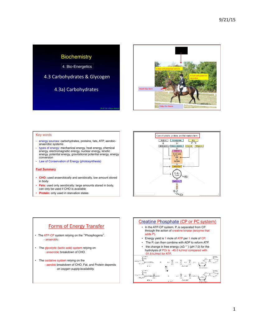
9/21/15 1 Biochemistry 4.3 Carbohydrates & Glycogen 4.3a) Carbohydrates Prof. Dr. Klaus Heese 4. Bio-Energetics Eddy the Horse Insert hay here A fuel cell powered vehicle… A fuel cell powered operator - energy sources: carbohydrates, proteins, fats, ATP, aerobic- anaerobic systems - types of energy: mechanical energy, heat energy, chemical energy, electromagnetic energy, nuclear energy, kinetic energy, potential energy, gravitational potential energy, energy conversion - Law of Conservation of Energy (photosynthesis) Fuel Summary • CHO: used anaerobically and aerobically, low amount stored in body • Fats: used only aerobically: large amounts stored in body, can only be used if CHO is available • Protein: only used in starvation states Key words Forms of Energy Transfer • The ATP-CP system relying on the “Phosphogens”. - anaerobic • The glycolytic (lactic acid) system relying on - anaerobic breakdown of CHO. • The oxidative system relying on the - aerobic breakdown of CHO, Fat, and Protein depends on oxygen supply/availability. • In the ATP-CP system, Pi is separated from CP through the action of creatine kinase (enzyme that adds P). • Energy yield is 1 mole of ATP per 1 mole of CP . • The Pi can then combine with ADP to reform ATP. • the change in free energy (ΔG °’ ) (pH 7.0) for the hydrolysis of PCr is -45.0 kJ/mol compared with -31.8 kJ/mol for ATP , C reatine P hosphate (CP or PC system)
Transcript of Biochemistry - Hanyang
9/21/15
1
Biochemistry
4.3 Carbohydrates & Glycogen
4.3a) Carbohydrates
Prof. Dr. Klaus Heese
4. Bio-Energetics
Eddy the Horse
Insert hay here
A fuel cell powered vehicle…
A fuel cell powered operator
- energy sources: carbohydrates, proteins, fats, ATP, aerobic-anaerobic systems#
- types of energy: mechanical energy, heat energy, chemical energy, electromagnetic energy, nuclear energy, kinetic energy, potential energy, gravitational potential energy, energy conversion#
- Law of Conservation of Energy (photosynthesis)##Fuel Summary!!• CHO: used anaerobically and aerobically, low amount stored
in body#• Fats: used only aerobically: large amounts stored in body,
can only be used if CHO is available#• Protein: only used in starvation states#
Key words#
Forms of Energy Transfer • The ATP-CP system relying on the “Phosphogens”.#
- anaerobic#
• The glycolytic (lactic acid) system relying on # - anaerobic breakdown of CHO.#
• The oxidative system relying on the # - aerobic breakdown of CHO, Fat, and Protein depends # on oxygen supply/availability.!
• In the ATP-CP system, Pi is separated from CP through the action of creatine kinase (enzyme that adds P).#
• Energy yield is 1 mole of ATP per 1 mole of CP.#• The Pi can then combine with ADP to reform ATP. #• the change in free energy (ΔG °’ ) (pH 7.0) for the
hydrolysis of PCr is -45.0 kJ/mol compared with -31.8 kJ/mol for ATP,#
Creatine Phosphate (CP or PC system)
9/21/15
2
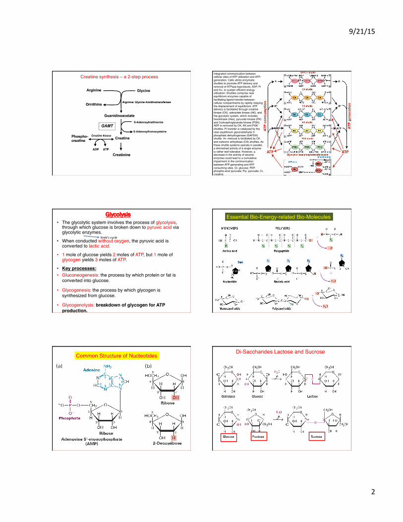
Creatine synthesis – a 2-step process Integrated communication between cellular sites of ATP utilization and ATP-generation. Cells utilize enzymatic shuttles to promote ATP delivery and removal of ATPase byproducts, ADP, Pi and H+, to sustain efficient energy utilization. Shuttles comprise near equilibrium enzymes capable of facilitating ligand transfer between cellular compartments by rapidly relaying the displacement of equilibrium. ATP delivery is facilitated through creatine kinase (CK), adenylate kinase (AK), and the glycolytic system, which includes hexokinase (Hex), pyruvate kinase (PK) and 3-phosphoglycerate kinase (PGK). ADP is removed by CK, AK and PGK shuttles. Pi transfer is catalyzed by the near-equilibrium glyceraldehyde 3-phosphate dehydrogenase (GAPDH) shuttle. H+ removal is facilitated by CK and carbonic anhydrase (CA) shuttles. As these shuttle systems operate in parallel, a diminished activity of a single enzyme is rather well tolerated. However, a decrease in the activity of several enzymes could lead to a cumulative impairment in the communication between ATP-generating and ATP consuming sites. Gl, glucose; PEP, phospho-enol pyruvate; Pyr, pyruvate; Cr, creatine.#
Glycolysis • The glycolytic system involves the process of glycolysis,
through which glucose is broken down to pyruvic acid via glycolytic enzymes. #
• When conducted without oxygen, the pyruvic acid is converted to lactic acid. #
• 1 mole of glucose yields 2 moles of ATP, but 1 mole of glycogen yields 3 moles of ATP.#
• Key processes:!• Gluconeogenesis: the process by which protein or fat is
converted into glucose.#
• Glycogenesis: the process by which glycogen is synthesized from glucose.##
• Glycogenolysis: breakdown of glycogen for ATP production.!
Kreb’s cycle
Essential Bio-Energy-related Bio-Molecules#
Common Structure of Nucleotides#Di-Saccharides Lactose and Sucrose#
9/21/15
3
As for all parts of this lecture:#It’s not about human physiology – but, to know the systems in order to be able to make use of it (‘translate’ and apply) from the bioengineering point of view !##Without its knowledge, no innovative and creative ideas for its potential bioengineering applications !#
Carbohydrates
Carbohydrates
• glucose provides energy for the brain and ½ of energy for muscles and Mssues
• glycogen is stored glucose • glucose is immediate energy • glycogen is reserve energy
Carbohydrates
• all plant food • milk
• carbohydrates are not equal – simple carbohydrates – complex carbohydrates
Simple Carbohydrates
• sugars – monosaccharides – single sugars – disaccharides – 2 monosaccharides
Complex Carbohydrates
• starches and fibers • polysaccharides
– chains of monosaccharides
9/21/15
4
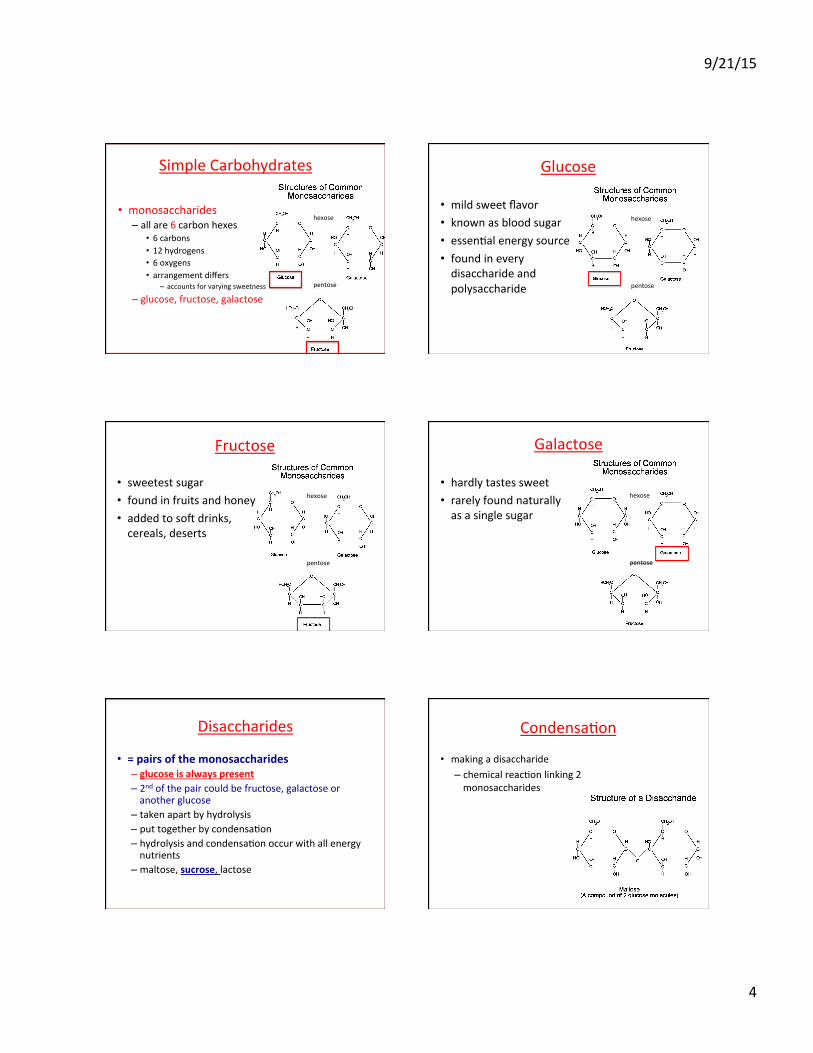
Simple Carbohydrates
• monosaccharides – all are 6 carbon hexes
• 6 carbons • 12 hydrogens • 6 oxygens • arrangement differs
– accounts for varying sweetness – glucose, fructose, galactose
hexose pentose
Glucose
• mild sweet flavor • known as blood sugar • essenMal energy source • found in every disaccharide and polysaccharide
hexose pentose
Fructose
• sweetest sugar • found in fruits and honey • added to soY drinks, cereals, deserts
hexose pentose
Galactose
• hardly tastes sweet • rarely found naturally as a single sugar
hexose pentose
Disaccharides
• = pairs of the monosaccharides – glucose is always present – 2nd of the pair could be fructose, galactose or another glucose
– taken apart by hydrolysis – put together by condensaMon – hydrolysis and condensaMon occur with all energy nutrients
– maltose, sucrose, lactose
CondensaMon
• making a disaccharide – chemical reacMon linking 2 monosaccharides
9/21/15
5
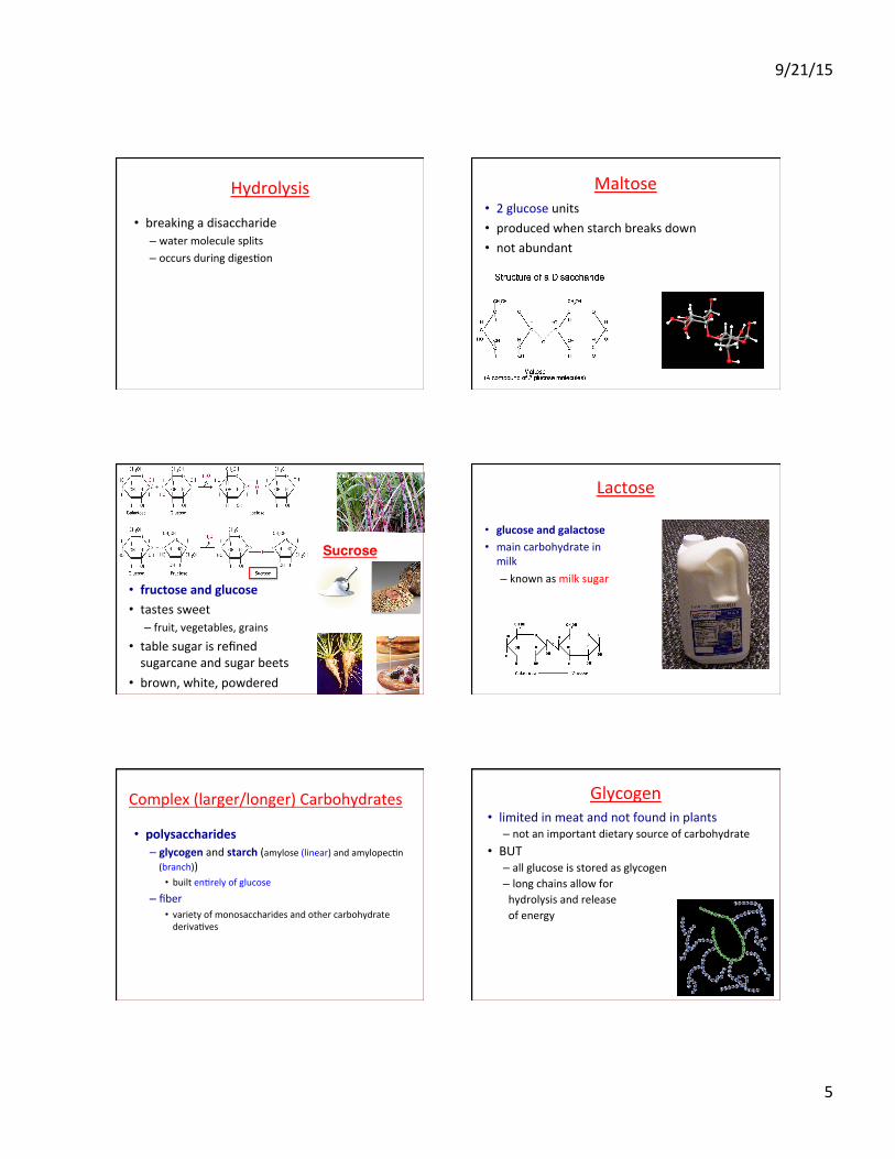
Hydrolysis
• breaking a disaccharide – water molecule splits – occurs during digesMon
Maltose
• 2 glucose units • produced when starch breaks down • not abundant
• fructose and glucose • tastes sweet
– fruit, vegetables, grains • table sugar is refined sugarcane and sugar beets
• brown, white, powdered
Sucrose!
Lactose
• glucose and galactose • main carbohydrate in milk – known as milk sugar
Complex (larger/longer) Carbohydrates
• polysaccharides – glycogen and starch (amylose (linear) and amylopecMn (branch))
• built enMrely of glucose – fiber
• variety of monosaccharides and other carbohydrate derivaMves
Glycogen • limited in meat and not found in plants
– not an important dietary source of carbohydrate • BUT
– all glucose is stored as glycogen – long chains allow for hydrolysis and release of energy
9/21/15
6
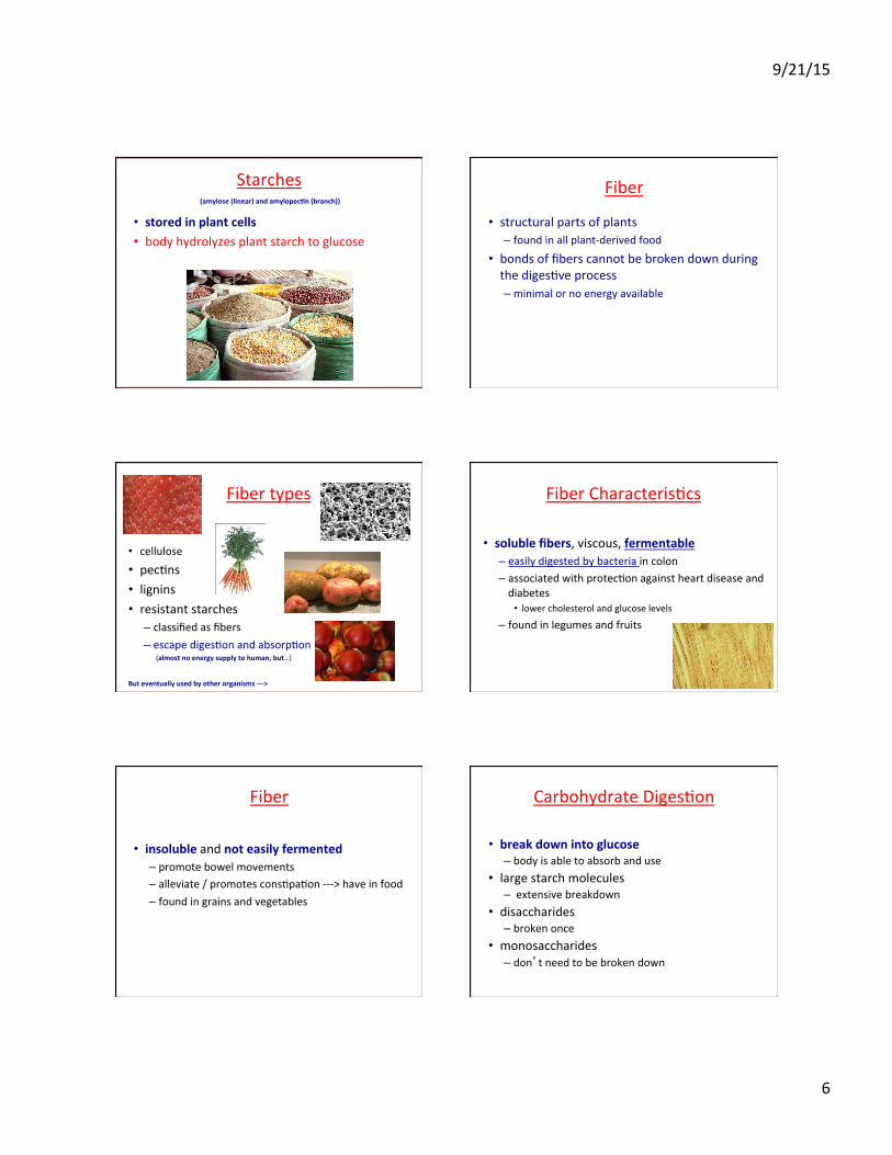
Starches
• stored in plant cells • body hydrolyzes plant starch to glucose
(amylose (linear) and amylopec8n (branch)) Fiber
• structural parts of plants – found in all plant-‐derived food
• bonds of fibers cannot be broken down during the digesMve process – minimal or no energy available
Fiber types
• cellulose • pecMns • lignins • resistant starches
– classified as fibers – escape digesMon and absorpMon (almost no energy supply to human, but…)
But eventually used by other organisms -‐-‐-‐>
Fiber CharacterisMcs
• soluble fibers, viscous, fermentable – easily digested by bacteria in colon – associated with protecMon against heart disease and diabetes
• lower cholesterol and glucose levels – found in legumes and fruits
Fiber
• insoluble and not easily fermented – promote bowel movements – alleviate / promotes consMpaMon -‐-‐-‐> have in food – found in grains and vegetables
Carbohydrate DigesMon
• break down into glucose – body is able to absorb and use
• large starch molecules – extensive breakdown
• disaccharides – broken once
• monosaccharides – don’t need to be broken down
9/21/15
7
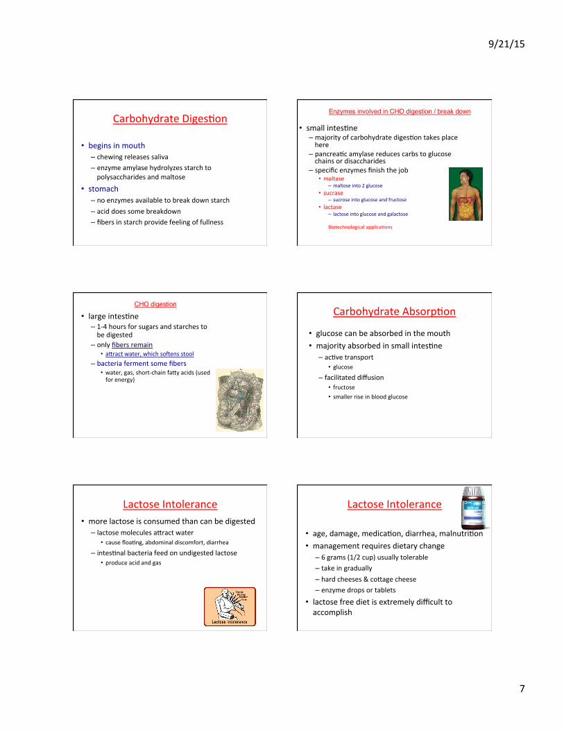
Carbohydrate DigesMon
• begins in mouth – chewing releases saliva – enzyme amylase hydrolyzes starch to polysaccharides and maltose
• stomach – no enzymes available to break down starch – acid does some breakdown – fibers in starch provide feeling of fullness
• small intesMne – majority of carbohydrate digesMon takes place here
– pancreaMc amylase reduces carbs to glucose chains or disaccharides
– specific enzymes finish the job • maltase
– maltose into 2 glucose • sucrase
– sucrose into glucose and fructose • lactase
– lactose into glucose and galactose
Biotechnological applicaMons
Enzymes involved in CHO digestion / break down#
• large intesMne – 1-‐4 hours for sugars and starches to be digested
– only fibers remain • aeract water, which soYens stool
– bacteria ferment some fibers • water, gas, short-‐chain faey acids (used for energy)
CHO digestion#Carbohydrate AbsorpMon
• glucose can be absorbed in the mouth • majority absorbed in small intesMne
– acMve transport • glucose
– facilitated diffusion • fructose • smaller rise in blood glucose
Lactose Intolerance • more lactose is consumed than can be digested
– lactose molecules aeract water • cause floaMng, abdominal discomfort, diarrhea
– intesMnal bacteria feed on undigested lactose • produce acid and gas
Lactose Intolerance
• age, damage, medicaMon, diarrhea, malnutriMon • management requires dietary change
– 6 grams (1/2 cup) usually tolerable – take in gradually – hard cheeses & coeage cheese – enzyme drops or tablets
• lactose free diet is extremely difficult to accomplish

9/21/15
8
Carbohydrate Metabolism • 1/3 of body’s glycogen is stored in liver
– released as glucose to bloodstream 1. eat – intake glucose 2. liver condenses extra glucose to glycogen 3. blood glucose falls 4. liver hydrolyzes glycogen to glucose Glycogen is bulky, so we store only so much: short
term energy supply Fat is the long term energy supply.
Glucose for Energy
• enzymes break apart glucose – yielding energy • inadequate supply of carbohydrates
– ketone bodies (fat fragments) are an alternate energy source during starvaMon
– excess ketones can lead to ketosis: imbalance of acids in body
• minimum of 50 – 100 grams of carbs/day are needed to avoid ketosis
Glucose Homeostasis
• maintaining an even balance of glucose is controlled by insulin and glucagon – insulin
• moves glucose into the blood (ensures proper usage/uptake of glucose by the various cells/Mssues/organs – if -‐-‐-‐> diabetes)
– glucagon • brings glucose out of storage
• maintaining balance – balanced meals at regular intervals
• fiber and some fat slow the digesMve process down • glucose gets into the blood slow and steady
3 or 5, doesn’t maeer -‐-‐-‐ but energy input ~ energy output
Maintaining Blood Glucose
Homeostasis
Intes8ne When a person eats, blood glucose rises.
1
2
Insulin s8mulates the uptake of glucose into cells and storage as glycogen in the liver and muscles. Insulin also s8mulates the conversion of excess glucose into fat for storage.
3
4
5
6
7 Blood glucose begins to rise.
a The stress hormone epinephrine and other hormones also bring glucose out of storage.
Glucose Insulin Glucagon Glycogen
Glucagon s8mulates liver cells to break down glycogen and release glucose into the blood.a
Liver
Low blood glucose s8mulates the pancreas to release glucagon into the bloodstream.
As the body's cells use glucose, blood levels decline.
Glucagon
Pancreas
Fat cell
Liver
Muscle
High blood glucose s8mulates the pancreas to release insulin.
Pancreas
Insulin
9/21/15
9
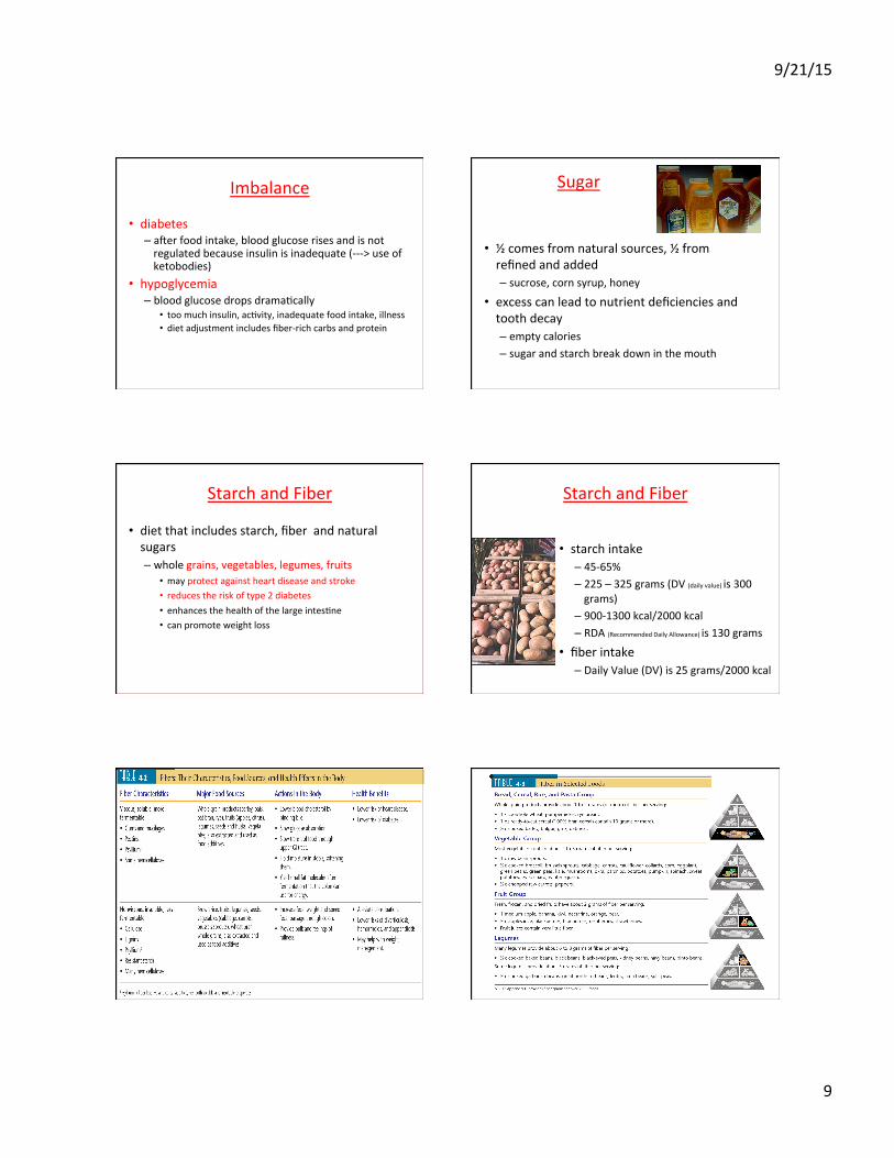
Imbalance
• diabetes – aYer food intake, blood glucose rises and is not regulated because insulin is inadequate (-‐-‐-‐> use of ketobodies)
• hypoglycemia – blood glucose drops dramaMcally
• too much insulin, acMvity, inadequate food intake, illness • diet adjustment includes fiber-‐rich carbs and protein
Sugar
• ½ comes from natural sources, ½ from refined and added – sucrose, corn syrup, honey
• excess can lead to nutrient deficiencies and tooth decay – empty calories – sugar and starch break down in the mouth
Starch and Fiber
• diet that includes starch, fiber and natural sugars – whole grains, vegetables, legumes, fruits
• may protect against heart disease and stroke • reduces the risk of type 2 diabetes • enhances the health of the large intesMne • can promote weight loss
Starch and Fiber
• starch intake – 45-‐65% – 225 – 325 grams (DV (daily value) is 300 grams)
– 900-‐1300 kcal/2000 kcal – RDA (Recommended Daily Allowance) is 130 grams
• fiber intake – Daily Value (DV) is 25 grams/2000 kcal

9/21/15
10
Groceries
• grains: 1 serving = 15 grams • vegetables
– ½ cup starchy = 15 grams – ½ cup nonstarchy = 5 grams
• fruit: 1 serving = 15 grams • milk: 1 cup = 12 grams • meat: none or liele • legumes: ½ cup = 15 grams
ArMficial Sweeteners
• help keep sugar and energy intake down • anything we eat has FDA (food and drug administraMon) approval – saccharin – aspartame – acesulfame potassium – sucralose – neotame
Sugar Replacers
• sugar alcohols – provide bulk and sweetness
• cookies, gum, candy, jelly
– do contain minimal kcal – low glycemic response
• absorbed slowly – do not cause dental caries
Biochemistry
4.3b) Glycogen Metabolism
Prof. Dr. Klaus Heese
Glycogen is a polymer of glucose residues linked by w α(1à4) glycosidic bonds, mainly w α(1à6) glycosidic bonds, at branch points. Glycogen chains & branches are longer than shown.
Glucose is stored as glycogen predominantly in liver and muscle cells.
H O
OHH
OHH
OH
CH2OH
HO H
H
OHH
OH
CH2OH
H
O
HH H O
OH
OHH
OH
CH2
HH H O
H
OHH
OH
CH2OH
H
OH
HH O
OH
OHH
OH
CH2OH
H
O
H
O
1 4
6
H O
H
OHH
OH
CH2OH
HH H O
H
OHH
OH
CH2OH
HH
O1
OH
3
4
5
2
glycogen
9/21/15
11
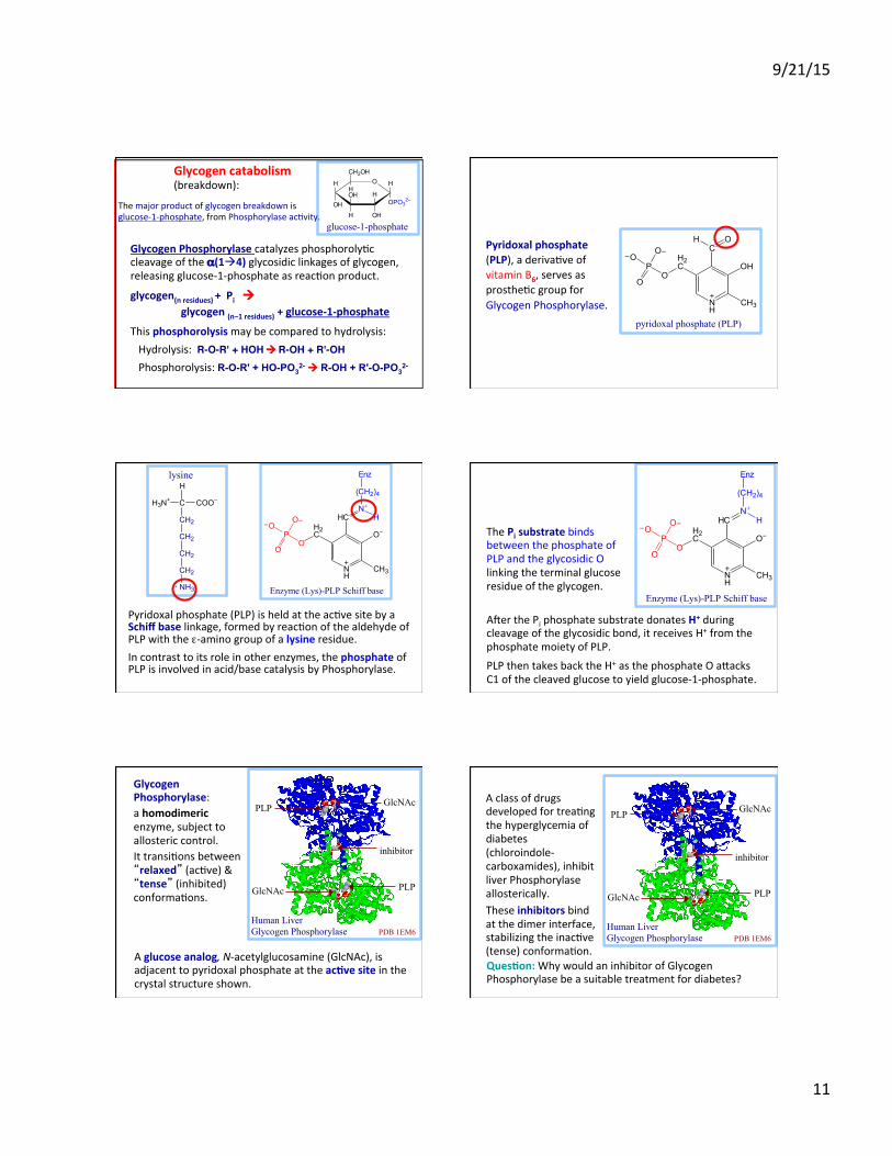
Glycogen Phosphorylase catalyzes phosphorolyMc cleavage of the α(1à4) glycosidic linkages of glycogen, releasing glucose-‐1-‐phosphate as reacMon product.
glycogen(n residues) + Pi ! glycogen (n–1 residues) + glucose-‐1-‐phosphate
This phosphorolysis may be compared to hydrolysis: Hydrolysis: R-O-R' + HOH ! R-OH + R'-OH
Phosphorolysis: R-O-R' + HO-PO32- ! R-OH + R'-O-PO3
2-
glucose-1-phosphate
H O
OHH
OHH
OH
CH2OH
H
OPO32−
HGlycogen catabolism (breakdown):
The major product of glycogen breakdown is glucose-‐1-‐phosphate, from Phosphorylase acMvity.
Pyridoxal phosphate (PLP), a derivaMve of vitamin B6, serves as prostheMc group for Glycogen Phosphorylase.
pyridoxal phosphate (PLP)
NH
CO
P
O−O
O
OH
CH3
CH O
−
+
H2
Pyridoxal phosphate (PLP) is held at the acMve site by a Schiff base linkage, formed by reacMon of the aldehyde of PLP with the ε-‐amino group of a lysine residue. In contrast to its role in other enzymes, the phosphate of PLP is involved in acid/base catalysis by Phosphorylase.
NH
CO
P
O−O
O
O−
CH3
HC−
+
H2
N
(CH2)4
Enz
H+
Enzyme (Lys)-PLP Schiff base
H3N+ C COO−
CH2
CH2
CH2
CH2
NH3
H
+
lysine
The Pi substrate binds between the phosphate of PLP and the glycosidic O linking the terminal glucose residue of the glycogen.
NH
CO
P
O−O
O
O−
CH3
HC−
+
H2
N
(CH2)4
Enz
H+
Enzyme (Lys)-PLP Schiff base
AYer the Pi phosphate substrate donates H+ during cleavage of the glycosidic bond, it receives H+ from the phosphate moiety of PLP. PLP then takes back the H+ as the phosphate O aeacks C1 of the cleaved glucose to yield glucose-‐1-‐phosphate.
A glucose analog, N-‐acetylglucosamine (GlcNAc), is adjacent to pyridoxal phosphate at the ac8ve site in the crystal structure shown.
Glycogen Phosphorylase: a homodimeric enzyme, subject to allosteric control. It transiMons between “relaxed” (acMve) & “tense” (inhibited) conformaMons.
PLP
PLP GlcNAc
GlcNAc
inhibitor
Human Liver Glycogen Phosphorylase PDB 1EM6
Ques8on: Why would an inhibitor of Glycogen Phosphorylase be a suitable treatment for diabetes?
A class of drugs developed for treaMng the hyperglycemia of diabetes (chloroindole-‐carboxamides), inhibit liver Phosphorylase allosterically. These inhibitors bind at the dimer interface, stabilizing the inacMve (tense) conformaMon.
PLP
PLP GlcNAc
GlcNAc
inhibitor
Human Liver Glycogen Phosphorylase PDB 1EM6
9/21/15
12
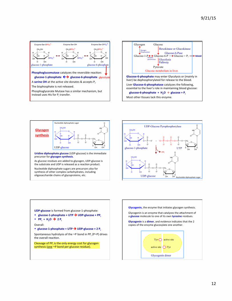
Phosphoglucomutase catalyzes the reversible reacMon: glucose-‐1-‐phosphate "! glucose-‐6-‐phosphate A serine OH at the acMve site donates & accepts Pi. The bisphosphate is not released. Phosphoglycerate Mutase has a similar mechanism, but instead uses His for Pi transfer.
glucose-1-phosphate glucose-6-phosphate
H O
OHH
OHH
OH
CH2OH
H
OPO32−
H H O
OHH
OHH
OH
CH2OPO32−
H
OH
HH O
OHH
OHH
OH
CH2OPO32−
H
OPO32−
H
Enzyme-Ser-OPO32− Enzyme-Ser-OPO3
2−Enzyme-Ser-OH
glycolysis# Glucose-‐6-‐phosphate may enter Glycolysis or (mainly in liver) be dephosphorylated for release to the blood. Liver Glucose-‐6-‐phosphatase catalyzes the following, essenMal to the liver's role in maintaining blood glucose: glucose-‐6-‐phosphate + H2O à glucose + Pi Most other Mssues lack this enzyme.
Glycogen Glucose Hexokinase or Glucokinase Glucose-6-Pase Glucose-1-P Glucose-6-P Glucose + Pi Glycolysis Pathway Pyruvate Glucose metabolism in liver.
blood!
Glycogen Phosphorylase
Phospho glucomutase
Uridine diphosphate glucose (UDP-‐glucose) is the immediate precursor for glycogen synthesis. As glucose residues are added to glycogen, UDP-‐glucose is the substrate and UDP is released as a reacMon product. NucleoMde diphosphate sugars are precursors also for synthesis of other complex carbohydrates, including oligosaccharide chains of glycoproteins, etc.
OO
OHOH
HH
H
CH2
H
HN
N
O
O
OP
O
O−
P
O
O−
H O
OH
H
OHH
OH
CH2OH
H
O
H
UDP-glucose
Glycogen synthesis
NucleoMde diphosphate sugar
OO
OHOH
HH
H
CH2
H
HN
N
O
O
OP
O
O−
P
O
O−
H O
OH
H
OHH
OH
CH2OH
H
O
H
O−P
O
O−
H O
OH
H
OHH
OH
CH2OH
H
O
H
OO
OHOH
HH
H
CH2
H
HN
N
O
O
OP
O
O−
P
O
O−
OP−O
O
O−
PPi
+
UDP-glucose
glucose-1-phosphate UTP
UDP-Glucose Pyrophosphorylase
NucleoMde diphosphate sugar
UDP-‐glucose is formed from glucose-‐1-‐phosphate: w glucose-‐1-‐phosphate + UTP ! UDP-‐glucose + PPi w PPi + H2O ! 2 Pi
Overall: w glucose-‐1-‐phosphate + UTP ! UDP-‐glucose + 2 Pi
Spontaneous hydrolysis of the ~P bond in PPi (P~P) drives the overall reacMon.
Cleavage of PPi is the only energy cost for glycogen synthesis (one ~P bond per glucose residue).
Glycogenin, the enzyme that iniMates glycogen synthesis.
Glycogenin is an enzyme that catalyzes the aeachment of a glucose molecule to one of its own tyrosine residues.
Glycogenin is a dimer, and evidence indicates that the 2 copies of the enzyme glucosylate one another.
Tyr− active site
active site −Tyr
Glycogenin dimer

9/21/15
13
A glycosidic bond is formed between the anomeric C1 of the glucose moiety derived from UDP-‐glucose and the hydroxyl oxygen of a tyrosine side-‐chain of Glycogenin. UDP is released as a product.
H O
OH
H
OHH
OH
CH2OH
HO H
H
OHH
OH
CH2OH
H
O
HHC
CH
NH
CH2
O
O
H O
OH
H
OHH
OH
CH2OH
HH
C
CH
NH
CH2
O
O1
5
4
3 2
6
H O
OH
H
OHH
OH
CH2OH
HH
O1
5
4
3 2
6
P O P O Uridine
O
O−
O
O−
C
CH
NH
CH2
HO
O
tyrosine residue of Glycogenin
O-linked glucose residue
+ UDP
UDP-glucose
Glycogenin then catalyzes glucosylaMon at C4 of the aeached glucose (UDP-‐glucose again the donor), to yield an O-‐linked disaccharide with α(1→4) glycosidic linkage. This is repeated unMl a short linear glucose polymer with α(1→4) glycosidic linkages is built up on Glycogenin.
H O
OH
H
OHH
OH
CH2OH
HO H
H
OHH
OH
CH2OH
H
O
HHC
CH
NH
CH2
O
O
H O
OH
H
OHH
OH
CH2OH
HH
C
CH
NH
CH2
O
O1
5
4
3 2
6
H O
OH
H
OHH
OH
CH2OH
HH
O1
5
4
3 2
6
P O P O Uridine
O
O−
O
O−
C
CH
NH
CH2
HO
O
UDP-glucose
O-linked glucose residue
α(1à4) linkage
+ UDP
+ UDP
Answer: Most of the Glycogenin is found associated with glycogen par8cles (branched glycogen chains) in the cytoplasm.
Glycogen Synthase then catalyzes elonga8on of glycogen chains iniMated by Glycogenin.
Ques8on: Where would you expect to find Glycogenin within a cell?
Glycogen Synthase catalyzes transfer of the glucose moiety of UDP-‐glucose to the hydroxyl at C4 of the terminal residue of a glycogen chain to form an α(1→ 4) glycosidic linkage:
glycogen(n residues) + UDP-‐glucose ! glycogen(n +1 residues) + UDP
A branching enzyme transfers a segment from the end of a glycogen chain to the C6 hydroxyl of a glucose residue of glycogen to yield a branch with an α(1→6) linkage.
Both synthesis & breakdown of glycogen are spontaneous. If both pathways were acMve simultaneously in a cell, there would be a "fu8le cycle" with cleavage of one ~P bond per cycle (in forming UDP-‐glucose). To prevent such a fuMle cycle, Glycogen Synthase and Glycogen Phosphorylase are reciprocally regulated, by allosteric effectors and by phosphorylaMon.
Glycogen Synthesis
UTP UDP + 2 Pi
glycogen(n) + glucose-1-P glycogen(n + 1) Glycogen Phosphorylase Pi
Glycogen Phosphorylase in muscle is subject to allosteric regulaMon by AMP, ATP, and glucose-‐6-‐phosphate. A separate isozyme of Phosphorylase expressed in liver is less sensiMve to these allosteric controls.
w AMP (present significantly when ATP is depleted) ac8vates Phosphorylase, promoMng the relaxed conformaMon.
w ATP & glucose-‐6-‐phosphate, which both have binding sites that overlap that of AMP, inhibit Phosphorylase, promoMng the tense conformaMon.
w Thus glycogen breakdown is inhibited when ATP and glucose-‐6-‐phosphate are plenMful.
9/21/15
14
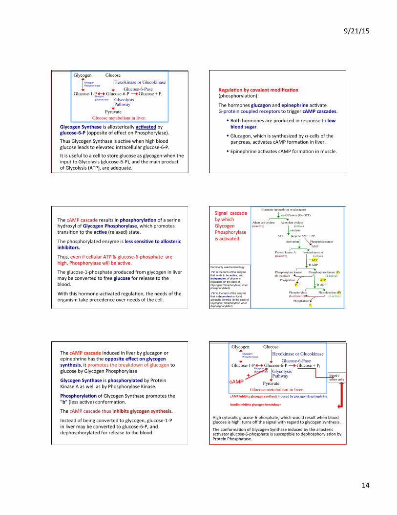
Glycogen Synthase is allosterically ac8vated by glucose-‐6-‐P (opposite of effect on Phosphorylase). Thus Glycogen Synthase is acMve when high blood glucose leads to elevated intracellular glucose-‐6-‐P. It is useful to a cell to store glucose as glycogen when the input to Glycolysis (glucose-‐6-‐P), and the main product of Glycolysis (ATP), are adequate.
Glycogen Glucose Hexokinase or Glucokinase Glucose-6-Pase Glucose-1-P Glucose-6-P Glucose + Pi Glycolysis Pathway Pyruvate Glucose metabolism in liver.
Phospho glucomutase
Glycogen Phosphorylase
Regula8on by covalent modifica8on (phosphorylaMon):
The hormones glucagon and epinephrine acMvate G-‐protein coupled receptors to trigger cAMP cascades.
w Both hormones are produced in response to low blood sugar.
w Glucagon, which is synthesized by α-‐cells of the pancreas, acMvates cAMP formaMon in liver.
w Epinephrine acMvates cAMP formaMon in muscle.
The cAMP cascade results in phosphoryla8on of a serine hydroxyl of Glycogen Phosphorylase, which promotes transiMon to the ac8ve (relaxed) state.
The phosphorylated enzyme is less sensi8ve to allosteric inhibitors.
Thus, even if cellular ATP & glucose-‐6-‐phosphate are high, Phosphorylase will be acMve.
The glucose-‐1-‐phosphate produced from glycogen in liver may be converted to free glucose for release to the blood.
With this hormone-‐acMvated regulaMon, the needs of the organism take precedence over needs of the cell.
Signal cascade by which Glycogen Phosphorylase is acMvated.
Hormone (epinephrine or glucagon)
via G Protein (Gα-GTP)
Adenylate cyclase Adenylate cyclase (inactive) (active) catalysis
ATP cyclic AMP + PPi
Activation Phosphodiesterase AMP
Protein kinase A Protein kinase A (inactive) (active) ATP
ADP
Phosphorylase kinase Phosphorylase kinase (P) (b-inactive) (a-active) Phosphatase ATP Pi ADP Phosphorylase Phosphorylase (P) (b-allosteric) (a-active) Phosphatase
Pi
Commonly used terminology:
w "a" is the form of the enzyme that tends to be active, and independent of allosteric regulators (in the case of Glycogen Phosphorylase, when phosphorylated).
w "b" is the form of the enzyme that is dependent on local allosteric controls (in the case of Glycogen Phosphorylase when dephosphorylated).
The cAMP cascade induced in liver by glucagon or epinephrine has the opposite effect on glycogen synthesis, it promotes the breakdown of glucogen to glucose by Glycogen Phosphorylase
Glycogen Synthase is phosphorylated by Protein Kinase A as well as by Phosphorylase Kinase.
Phosphoryla8on of Glycogen Synthase promotes the "b" (less acMve) conformaMon.
The cAMP cascade thus inhibits glycogen synthesis.
Instead of being converted to glycogen, glucose-‐1-‐P in liver may be converted to glucose-‐6-‐P, and dephosphorylated for release to the blood.
High cytosolic glucose-‐6-‐phosphate, which would result when blood glucose is high, turns off the signal with regard to glycogen synthesis.
The conformaMon of Glycogen Synthase induced by the allosteric acMvator glucose-‐6-‐phosphate is suscepMble to dephosphorylaMon by Protein Phosphatase.
Glycogen Glucose Hexokinase or Glucokinase Glucose-6-Pase Glucose-1-P Glucose-6-P Glucose + Pi Glycolysis Pathway Pyruvate Glucose metabolism in liver.
Glycogen Phosphorylase
Phospho glucomutase
cAMP#+ blood /
other cells
cAMP inhibits glycogen synthesis induced by glucagon & epinephrine. Insulin inhibits glycogen breakdown
9/21/15
15
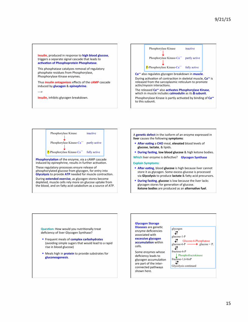
Insulin, produced in response to high blood glucose, triggers a separate signal cascade that leads to ac8va8on of Phosphoprotein Phosphatase.
This phosphatase catalyzes removal of regulatory phosphate residues from Phosphorylase, Phosphorylase Kinase enzymes.
Thus insulin antagonizes effects of the cAMP cascade induced by glucagon & epinephrine.
-‐-‐-‐>
Insulin, inhibits glycogen breakdown.
Ca++ also regulates glycogen breakdown in muscle. During acMvaMon of contracMon in skeletal muscle, Ca++ is released from the sarcoplasmic reMculum to promote acMn/myosin interacMons. The released Ca++ also ac8vates Phosphorylase Kinase, which in muscle includes calmodulin as its δ subunit. Phosphorylase Kinase is partly acMvated by binding of Ca++ to this subunit.
Phosphorylase Kinase inactive
Phosphorylase Kinase-Ca++ partly active
P-Phosphorylase Kinase-Ca++ fully active
Phosphoryla8on of the enzyme, via a cAMP cascade induced by epinephrine, results in further acMvaMon. These regulatory processes ensure release of phosphorylated glucose from glycogen, for entry into Glycolysis to provide ATP needed for muscle contracMon. During extended exercise, as glycogen stores become depleted, muscle cells rely more on glucose uptake from the blood, and on faey acid catabolism as a source of ATP.
Phosphorylase Kinase inactive
Phosphorylase Kinase-Ca++ partly active
P-Phosphorylase Kinase-Ca++ fully active
A gene8c defect in the isoform of an enzyme expressed in liver causes the following symptoms: w Acer ea8ng a CHO meal, elevated blood levels of glucose, lactate, & lipids.
w During fas8ng, low blood glucose & high ketone bodies. Which liver enzyme is defecMve?
Explain Symptoms: w Acer ea8ng, blood glucose is high because liver cannot store it as glycogen. Some excess glucose is processed via Glycolysis to produce lactate & faey acid precursors.
w During fas8ng, glucose is low because the liver lacks glycogen stores for generaMon of glucose. Ketone bodies are produced as an alterna8ve fuel.
Glycogen Synthase
Ques8on: How would you nutriMonally treat deficiency of liver Glycogen Synthase?
w Frequent meals of complex carbohydrates (avoiding simple sugars that would lead to a rapid rise in blood glucose)
w Meals high in protein to provide substrates for gluconeogenesis.
Glycogen Storage Diseases are geneMc enzyme deficiencies associated with excessive glycogen accumula8on within cells.
Some enzymes whose deficiency leads to glycogen accumulaMon are part of the inter-‐connected pathways shown here.
glycogen glucose-1-P Glucose-6-Phosphatase glucose-6-P glucose + Pi fructose-6-P Phosphofructokinase fructose-1,6-bisP Glycolysis continued
9/21/15
16
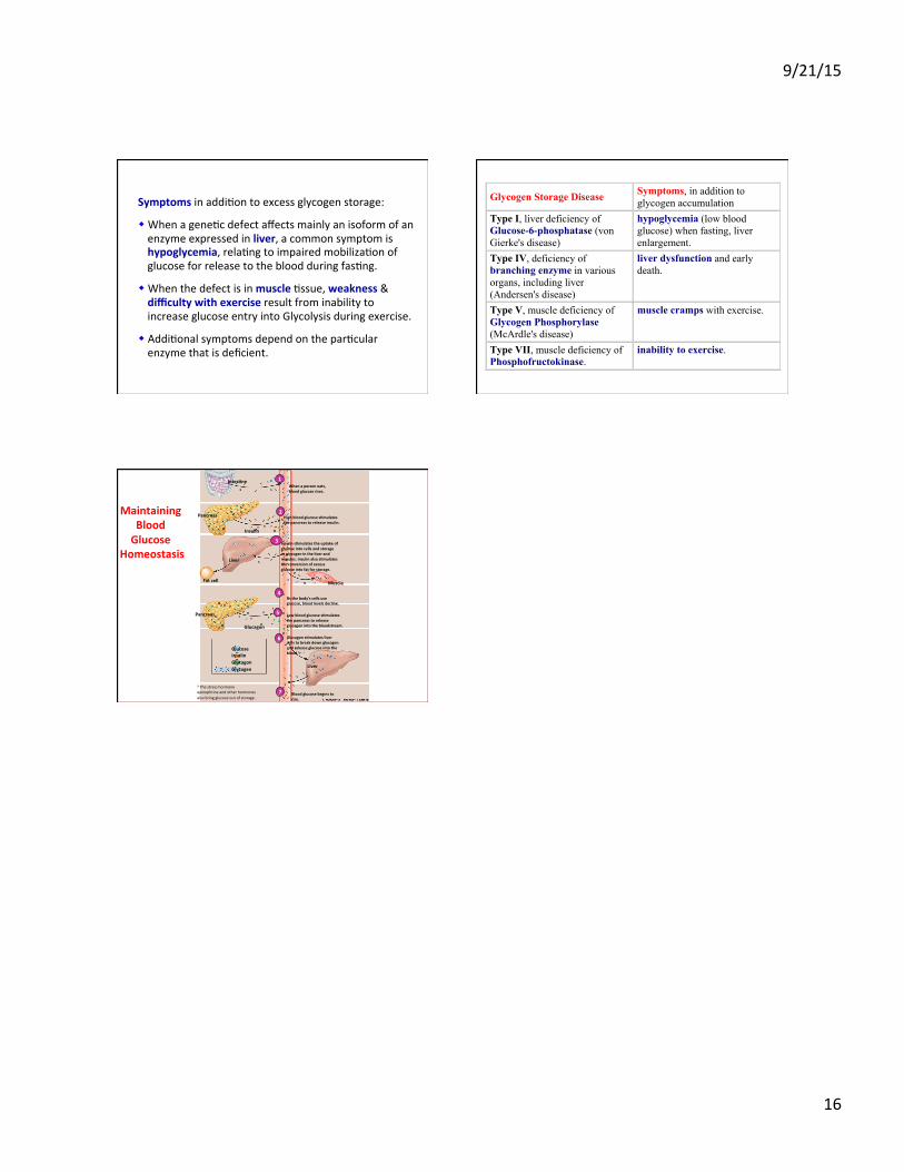
Symptoms in addiMon to excess glycogen storage:
w When a geneMc defect affects mainly an isoform of an enzyme expressed in liver, a common symptom is hypoglycemia, relaMng to impaired mobilizaMon of glucose for release to the blood during fasMng.
w When the defect is in muscle Mssue, weakness & difficulty with exercise result from inability to increase glucose entry into Glycolysis during exercise.
w AddiMonal symptoms depend on the parMcular enzyme that is deficient.
Glycogen Storage Disease Symptoms, in addition to glycogen accumulation
Type I, liver deficiency of Glucose-6-phosphatase (von Gierke's disease)
hypoglycemia (low blood glucose) when fasting, liver enlargement.
Type IV, deficiency of branching enzyme in various organs, including liver (Andersen's disease)
liver dysfunction and early death.
Type V, muscle deficiency of Glycogen Phosphorylase (McArdle's disease)
muscle cramps with exercise.
Type VII, muscle deficiency of Phosphofructokinase.
inability to exercise.
Maintaining Blood Glucose
Homeostasis
Intes8ne When a person eats, blood glucose rises.
1
2
Insulin s8mulates the uptake of glucose into cells and storage as glycogen in the liver and muscles. Insulin also s8mulates the conversion of excess glucose into fat for storage.
3
4
5
6
7 Blood glucose begins to rise.
a The stress hormone epinephrine and other hormones also bring glucose out of storage.
Glucose Insulin Glucagon Glycogen
Glucagon s8mulates liver cells to break down glycogen and release glucose into the blood.a
Liver
Low blood glucose s8mulates the pancreas to release glucagon into the bloodstream.
As the body's cells use glucose, blood levels decline.
Glucagon
Pancreas
Fat cell
Liver
Muscle
High blood glucose s8mulates the pancreas to release insulin.
Pancreas
Insulin