BiochemicalBasisofAsthma Therapy - The Journal of ... the airway smooth muscle of asthmatic...
8
Biochemical Basis of Asthma Therapy * Published, JBC Papers in Press, July 28, 2011, DOI 10.1074/jbc.R110.206466 Peter J. Barnes 1 From the National Heart and Lung Institute, Imperial College, London SW3 6LY, United Kingdom Current therapy for asthma is highly effective. 2 -Adrenergic receptor ( 2 AR) agonists are the most effective bronchodilators and relax airway smooth muscle cells through increased cAMP concentrations and directly opening large conductance Ca 2 channels. 2 AR may also activate alternative signaling pathways that may have detrimental effects in asthma. Glucocorticoids are the most effective anti-inflammatory treatments and switch off multiple activated inflammatory genes through recruitment of histone deacetylase-2, activating anti-inflammatory genes, and through increasing mRNA stability of inflammatory genes. There are beneficial molecular interactions between 2 AR and glucocorticoid-activated pathways. Understanding these signal- ing pathways may lead to even more effective therapies in the future. Asthma has become one of the most prevalent diseases and is increasing throughout the world, particularly in developing countries. It is characterized by variable airflow obstruction that is secondary to an allergic pattern of inflammation in the airways, which involves infiltration with inflammatory cells (eosinophils and T-helper 2 (T H 2) 2 cells) and the activation of resident mast cells and dendritic cells by allergens (1, 2). Struc- tural cells, such as airway epithelial cells and smooth muscle cells, are important sources of inflammatory mediators as well as activated inflammatory cells. Chronic inflammation may lead to structural changes in the airways, including increased airway smooth muscle cells, fibrosis, angiogenesis, and hyper- plasia of mucus-secreting cells. Multiple inflammatory media- tors are involved, and many cytokines and chemokines orches- trate this complex chronic inflammation (3). Although asthma is a complex inflammatory disease, current therapies (if taken correctly) are very effective in the majority of patients (4). There is now a good understanding of how current asthma therapies work at a biochemical level, and this has formed the basis for a search for new treatments (5). Drugs used to treat asthma include bronchodilators, which act mainly by reversing airway smooth muscle contraction, and anti-inflam- matory drugs, which suppress inflammation in the airways. The inflammation of asthma is confined to the airways, so inhala- tional therapy has been found to be the most effective treatment modality and largely avoids systemic side effects. 2 -Adrenergic Agonists 2 -Agonists are by far the most effective bronchodilators for asthma, as they act as functional antagonists and relax airway smooth muscle cells whatever the constricting stimulus. Long-acting 2 -agonists (LABA), such as salmeterol and for- moterol, have a 12-h duration of action, but once-daily drugs, including indacaterol, vilanterol, and olodaterol, have been developed recently (6). LABA should always be used in combination with a corticosteroid, as they are potentially dangerous if used alone because they do not effectively treat the underlying inflammation. Bronchodilator Mechanisms—Occupation of 2 -adrenergic receptors ( 2 AR) by agonists results in the activation of ade- nylyl cyclase via the stimulatory G-protein (G s ). This increases intracellular cAMP, leading to activation of PKA, which phos- phorylates several target proteins within the cell, leading to activation of myosin light chain phosphatase and inhibition of myosin light chain kinase and thus relaxation of airway smooth muscle (Fig. 1). In addition, 2 -agonists open large conduc- tance calcium-activated potassium channels (BK Ca ), which repolarize the smooth muscle cell so that Ca 2 is sequestrated into intracellular stores. 2 AR are also directly coupled to K Ca via G s , so relaxation of airway smooth muscle may occur inde- pendently of an increase in cAMP. Some actions of 2 -agonists are mediated via other cAMP-regulated proteins, such as EPAC (exchange protein activated by cAMP) (7). For example, the inhibition of airway smooth muscle cell proliferation by 2 -agonists appears to be dependent on EPAC rather than PKA (8). Airway smooth muscle shows resistance to 2 AR desensiti- zation, and this may be due to a large receptor reserve and a very low level of expression of the enzyme GRK2 (G-protein recep- tor kinase-2), which phosphorylates and inactivates occupied 2 -receptors (9). IL-1 uncouples pulmonary 2 -receptors in rats in vivo by increasing GRK2/5 activity and expression and thus reducing responsiveness to 2 -agonists (10). However, uncertainty remains as to whether 2 AR signaling is abnormal in the airway smooth muscle of asthmatic patients. Other Airway Effects— 2 -Agonists may have additional effects on airways, as 2 -receptors are localized to several dif- ferent cells types in the airways. 2 -Agonists may therefore cause bronchodilatation in vivo not only via a direct action on airway smooth muscle but also indirectly by inhibiting the release of bronchoconstrictor mediators from inflammatory cells and of neurotransmitters from airway nerves. For exam- ple, 2 -agonists inhibit mediator release from mast cells though closing an intermediate conductance Ca 2 -activated K chan- nel (K Ca 3.1) coupled to G s (11). Stem cell factor, which is secreted by epithelial cells in asthmatic patients, is an important * This is the fourth article in the Thematic Minireview Series on Molecular Bases of Disease: Asthma. This minireview will be reprinted in the 2011 Minireview Compendium, which will be available in January, 2012. 1 To whom correspondence should be addressed. E-mail: p.j.barnes@ imperial.ac.uk. 2 The abbreviations used are: T H 2, T-helper 2; LABA, long-acting 2 -agonist(s); 2 AR, 2 -adrenergic receptor(s); PLC, phospholipase C; ACh, acetylcholine; ICS, inhaled corticosteroid(s); GR, glucocorticoid receptor(s); GRE, gluco- corticoid response element(s); SLPI, secretory leukoprotease inhibitor; COPD, chronic obstructive pulmonary disease; PDE, phosphodiesterase(s); HDAC, histone deacetylase(s); CysLT, cysteinyl leukotriene(s); 5-LOX, 5-li- poxygenase; cPLA 2 , cytosolic phospholipase A 2 . THE JOURNAL OF BIOLOGICAL CHEMISTRY VOL. 286, NO. 38, pp. 32899 –32905, September 23, 2011 © 2011 by The American Society for Biochemistry and Molecular Biology, Inc. Printed in the U.S.A. SEPTEMBER 23, 2011 • VOLUME 286 • NUMBER 38 JOURNAL OF BIOLOGICAL CHEMISTRY 32899 MINIREVIEW This paper is available online at www.jbc.org by guest on May 30, 2018 http://www.jbc.org/ Downloaded from
Transcript of BiochemicalBasisofAsthma Therapy - The Journal of ... the airway smooth muscle of asthmatic...

Biochemical Basis of AsthmaTherapy*Published, JBC Papers in Press, July 28, 2011, DOI 10.1074/jbc.R110.206466
Peter J. Barnes1
From the National Heart and Lung Institute, Imperial College,London SW3 6LY, United Kingdom
Current therapy for asthma is highly effective. �2-Adrenergicreceptor (�2AR) agonists are themost effective bronchodilatorsand relax airway smooth muscle cells through increased cAMPconcentrations and directly opening large conductance Ca2�
channels.�2ARmay also activate alternative signaling pathwaysthat may have detrimental effects in asthma. Glucocorticoidsare the most effective anti-inflammatory treatments and switchoff multiple activated inflammatory genes through recruitmentof histone deacetylase-2, activating anti-inflammatory genes,and through increasing mRNA stability of inflammatory genes.There are beneficial molecular interactions between �2AR andglucocorticoid-activated pathways.Understanding these signal-ing pathways may lead to even more effective therapies in thefuture.
Asthma has become one of themost prevalent diseases and isincreasing throughout the world, particularly in developingcountries. It is characterized by variable airflow obstructionthat is secondary to an allergic pattern of inflammation in theairways, which involves infiltration with inflammatory cells(eosinophils and T-helper 2 (TH2)2 cells) and the activation ofresident mast cells and dendritic cells by allergens (1, 2). Struc-tural cells, such as airway epithelial cells and smooth musclecells, are important sources of inflammatory mediators as wellas activated inflammatory cells. Chronic inflammation maylead to structural changes in the airways, including increasedairway smooth muscle cells, fibrosis, angiogenesis, and hyper-plasia of mucus-secreting cells. Multiple inflammatory media-tors are involved, and many cytokines and chemokines orches-trate this complex chronic inflammation (3).Although asthma is a complex inflammatory disease, current
therapies (if taken correctly) are very effective in themajority ofpatients (4). There is now a good understanding of how currentasthma therapies work at a biochemical level, and this hasformed the basis for a search for new treatments (5). Drugs usedto treat asthma include bronchodilators, which act mainly by
reversing airway smooth muscle contraction, and anti-inflam-matory drugs, which suppress inflammation in the airways. Theinflammation of asthma is confined to the airways, so inhala-tional therapy has been found to be themost effective treatmentmodality and largely avoids systemic side effects.
�2-Adrenergic Agonists
�2-Agonists are by far the most effective bronchodilators forasthma, as they act as functional antagonists and relax airwaysmooth muscle cells whatever the constricting stimulus.Long-acting �2-agonists (LABA), such as salmeterol and for-moterol, have a 12-h duration of action, but once-dailydrugs, including indacaterol, vilanterol, and olodaterol, havebeen developed recently (6). LABA should always be used incombination with a corticosteroid, as they are potentiallydangerous if used alone because they do not effectively treatthe underlying inflammation.Bronchodilator Mechanisms—Occupation of �2-adrenergic
receptors (�2AR) by agonists results in the activation of ade-nylyl cyclase via the stimulatory G-protein (Gs). This increasesintracellular cAMP, leading to activation of PKA, which phos-phorylates several target proteins within the cell, leading toactivation of myosin light chain phosphatase and inhibition ofmyosin light chain kinase and thus relaxation of airway smoothmuscle (Fig. 1). In addition, �2-agonists open large conduc-tance calcium-activated potassium channels (BKCa), whichrepolarize the smooth muscle cell so that Ca2� is sequestratedinto intracellular stores. �2AR are also directly coupled to KCavia Gs, so relaxation of airway smooth muscle may occur inde-pendently of an increase in cAMP. Some actions of �2-agonistsaremediated via other cAMP-regulated proteins, such as EPAC(exchange protein activated by cAMP) (7). For example, theinhibition of airway smooth muscle cell proliferation by�2-agonists appears to be dependent on EPAC rather than PKA(8).Airway smooth muscle shows resistance to �2AR desensiti-
zation, and thismay be due to a large receptor reserve and a verylow level of expression of the enzyme GRK2 (G-protein recep-tor kinase-2), which phosphorylates and inactivates occupied�2-receptors (9). IL-1� uncouples pulmonary �2-receptors inrats in vivo by increasing GRK2/5 activity and expression andthus reducing responsiveness to �2-agonists (10). However,uncertainty remains as to whether �2AR signaling is abnormalin the airway smooth muscle of asthmatic patients.Other Airway Effects—�2-Agonists may have additional
effects on airways, as �2-receptors are localized to several dif-ferent cells types in the airways. �2-Agonists may thereforecause bronchodilatation in vivo not only via a direct action onairway smooth muscle but also indirectly by inhibiting therelease of bronchoconstrictor mediators from inflammatorycells and of neurotransmitters from airway nerves. For exam-ple,�2-agonists inhibitmediator release frommast cells thoughclosing an intermediate conductance Ca2�-activated K� chan-nel (KCa3.1) coupled to Gs (11). Stem cell factor, which issecreted by epithelial cells in asthmatic patients, is an important
* This is the fourth article in the Thematic Minireview Series on MolecularBases of Disease: Asthma. This minireview will be reprinted in the 2011Minireview Compendium, which will be available in January, 2012.
1 To whom correspondence should be addressed. E-mail: [email protected].
2 The abbreviations used are: TH2, T-helper 2; LABA, long-acting �2-agonist(s);�2AR, �2-adrenergic receptor(s); PLC, phospholipase C; ACh, acetylcholine;ICS, inhaled corticosteroid(s); GR, glucocorticoid receptor(s); GRE, gluco-corticoid response element(s); SLPI, secretory leukoprotease inhibitor;COPD, chronic obstructive pulmonary disease; PDE, phosphodiesterase(s);HDAC, histone deacetylase(s); CysLT, cysteinyl leukotriene(s); 5-LOX, 5�-li-poxygenase; cPLA2, cytosolic phospholipase A2.
THE JOURNAL OF BIOLOGICAL CHEMISTRY VOL. 286, NO. 38, pp. 32899 –32905, September 23, 2011© 2011 by The American Society for Biochemistry and Molecular Biology, Inc. Printed in the U.S.A.
SEPTEMBER 23, 2011 • VOLUME 286 • NUMBER 38 JOURNAL OF BIOLOGICAL CHEMISTRY 32899
MINIREVIEW This paper is available online at www.jbc.org
by guest on May 30, 2018
http://ww
w.jbc.org/
Dow
nloaded from

factor in keeping mast cells at the airway surface in asthma (2)and counteracts this effect of �2-agonists (12). Whether�2-agonists have anti-inflammatory effects in asthma is contro-versial. The inhibitory effects of �2-agonists on mast cell medi-ator release and microvascular leakage are clearly anti-inflam-matory, suggesting that �2-agonists may modify acuteinflammation. However, �2-agonists do not have a significantinhibitory effect on the chronic inflammation of asthmatic air-ways, which is suppressed by glucocorticoids. This has nowbeen confirmed by several biopsy and bronchoalveolar lavagestudies in patients with asthma who are taking regular �2-ago-nists (including LABA), which demonstrate no significantreduction in the number or activation of inflammatory cells inthe airways, in contrast to resolution of the inflammation,which occurs with inhaled glucocorticoids (13). This is likely tobe related to the fact that �2-agonist effects on macrophages,eosinophils, and lymphocytes are rapidly desensitized due to alow density of �2-receptors on these cells and high expressionof GRK2 (9). Indeed, exposure to LABA increases the expres-sion of GRK2 and GRK5 in human peripheral lung (14).
�2-Receptor Polymorphisms—There are several single-nucle-otide polymorphisms and haplotypes of the human �2AR gene(ADRB2) that may affect �2AR function. The common variantsare G16R and Q27E, which have in vitro effects on receptordesensitization, but clinical studies have shown inconsistenteffects on the bronchodilator responses to short- and long-act-ing �2-agonists (15). Some studies have shown that patientswith the common homozygous Arg16/Arg variant have morefrequent adverse effects and a poorer response to short-acting�2-agonists than heterozygotes orGly16/Gly homozygotes (16),but, overall, these differences are small, and there appears to beno clinical value inmeasuringADRB2 genotype. No differences
have been found with responses to LABA between these geno-types (17).Alternative Signaling of �2-Receptors—It is now recognized
that, although �2AR are coupled through Gs to relax airwaysmooth muscle, they may activate alternative signaling path-ways that may have deleterious effects, such as increasinginflammation (Fig. 1). In �2AR-overexpressing mice, Gq cou-pled to phospholipase C�1 (PLC�1) is activated, resulting in anenhanced bronchoconstrictor response to mediators, such asACh and histamine, that signal throughGq (18).��-Subunits ofGs may also signal through PLC activation (PLC�2) (19). �-Ar-restin-1 and -2 are adaptor proteins involved in uncouplingphosphorylated �2-receptors from Gs, leading to internaliza-tion by clathrin-coated pits. �-Arrestins determine whether�2-receptors are degraded within the cell by endocytosis or arerecycled to the cell membrane (20). Interaction of �2-receptorswith �-arrestin-2 leads to ubiquitination of each protein andsubsequent proteasomal destruction (21). As well as terminat-ing receptor function, �-arrestins act as a scaffold to allowreceptors to enhance other signaling pathways, such as MAPKand PI3K, independently of G-proteins and therefore allow�2AR to regulate different responses in the cell (22, 23). Thismay contribute to the adverse effects of LABA that have beenreported (24). Inverse agonists, such as nadolol and carvedilol,block �2AR and inhibit the signaling of constitutively active�2AR and paradoxically have been found to have beneficialeffects inmurinemodels of asthma. Furthermore,�2AR knock-outmice are protected from development of asthma (25).�2ARantagonists without inverse agonist activity, such as alprenolol,fail to reverse the asthma phenotype, however. A pilot study inasthma patients showed that, although nadolol reduced lungfunction in the short-term, there was a reduction in airwayhyper-responsiveness after 9 weeks of therapy (26). Deletion ofthe�-arrestin-2 gene prevents the recruitment of inflammatorycells and airway hyper-responsiveness in mice sensitized andexposed to allergen (27), and both structural and inflammatorycells are involved (28). It is possible that �2-receptor activationin epithelial cells activates �-arrestin-1/2, leading to activationof proinflammatory kinase pathways, such as p38 MAPK, withthe activation of proinflammatory genes (29). Biased �2-ago-nists that favor Gs signaling pathways rather than �-arrestinrecruitment may prove to be more effective as bronchodilatorsin the future (20).
Anticholinergics
Anticholinergics are antagonists of muscarinic receptors,and their only action at therapeutic doses is to block the effectsof endogenous acetylcholine (ACh). In animals and humans,there is a small degree of resting bronchomotor tone due totonic vagal nerve impulses that release ACh in the vicinity ofairway smooth muscle because it can be blocked by anticholin-ergic drugs. AChmay also be released from other airway cells,including epithelial and inflammatory cells (30). Airway epi-thelial cells contain all of the machinery needed for AChsynthesis and release (31, 32). Tiotropium bromide is a once-daily inhaled anticholinergic that dissociates slowly frommuscarinic M1- and M3-receptors and more rapidly fromM2-receptors. Although �2-agonists are the most effective
FIGURE 1. �2-Agonist signaling pathways. In the classical pathway, �2-ago-nists bind to �2AR, which are coupled via a stimulatory G-protein (Gs) toadenylyl cyclase (AC), resulting in formation of cAMP. cAMP activates PKA,which phosphorylates myosin light chain kinase (MLCK) in airway smoothmuscle cells, resulting in relaxation. Increased cAMP may also activate EPACto mediate effects such as inhibition of cell proliferation. �2AR are also cou-pled via Gs to a large conductance calcium-activated potassium channels(BK), leading to decreased intercellular Ca2� and inhibition of myosin lightchain kinase activation. Alternative signaling by �2AR may activate ��-sub-units of G� and Gq and via G�q, resulting in activation of PLC. �2AR alsointeract with �-arrestin-2, which interacts with p38 MAPK and PI3K. Thesealternative signaling pathways may increase the expression of inflammatoryproteins and therefore may have a deleterious effect in asthma.
MINIREVIEW: Biochemical Basis of Asthma Therapy
32900 JOURNAL OF BIOLOGICAL CHEMISTRY VOLUME 286 • NUMBER 38 • SEPTEMBER 23, 2011
by guest on May 30, 2018
http://ww
w.jbc.org/
Dow
nloaded from

bronchodilators in asthma, anticholinergics may have someadditive bronchodilator effect, particularly in patients withsevere disease (33). M3-receptors via Gq and the activation ofPLC result in hydrolysis of phosphatidylinositol 4,5-bispho-sphate and generation of inositol 1,4,5 trisphosphate, whichreleases Ca2� from intracellular stores, thus resulting in con-traction of airway smooth muscle cells and mucus secretion.M3-receptors may also be involved in the structural remod-eling that occurs in some patients with asthma, as theincrease in airway smooth muscle that occurs after chronicallergen exposure in mice is prevented by tiotropium (34).M2-receptors are also highly expressed in airway smoothmuscle cells and inhibit adenylyl cyclase via Gi, thus coun-teracting the bronchodilator effect of �2-agonists. M2-re-ceptors also counteract �2-agonists by inhibiting KCa in tra-cheal smoothmuscle cells via G�� subunits (35). PresynapticM2-receptors that limit ACh release are defective in animalmodels of asthma, and this may be secondary to eotaxinrelease from parasympathetic nerves with neural recruit-ment of eosinophils that release basic proteins to causeM2-receptor dysfunction (36). There is indirect evidencethat presynaptic M2-receptor function is also impaired inasthmatic patients (37).
Glucocorticoids
Glucocorticoids are by far themost effective therapy for con-trolling asthma, and inhaled corticosteroids (ICS) have becomethemainstay of treatment for all patients with persistent symp-toms. There has been major progress in understanding the cel-lular and molecular mechanisms involved in their anti-inflam-matory effects in asthma (38).Anti-inflammatory Mechanisms—Glucocorticoids diffuse
across the cell membrane and bind to glucocorticoid recep-
tors (GR) in the cytoplasm (39, 40). Upon ligand binding, GRare activated and released from chaperone proteins (heatshock protein 90 and others) and rapidly translocate to thenucleus, where they exert their molecular effects. The mech-anism of nuclear translocation involves the nuclear importproteins importin-� (karyopherin-�) and importin-13 (41,42). There is only one form of GR that binds glucocorticoids,termed GR�. GR� is an alternatively spliced form of GR thatinteracts with DNA but not with glucocorticoids, so it maytheoretically act as a dominant-negative inhibitor of gluco-corticoid action by interfering with the binding of GR toDNA (43).GR homodimerize and bind to glucocorticoid response ele-
ments (GRE) in the promoter region of glucocorticoid-respon-sive genes, and this interaction switches on (or occasionallyswitches off) gene transcription (Fig. 2). Activation of glucocor-ticoid-responsive genes occurs via an interaction between theDNA-bound GR and transcriptional coactivator molecules,such as CBP (cAMP-responsive element-binding protein-bind-ing protein), which have intrinsic histone acetyltransferaseactivity and cause acetylation of core histones (particularly his-tone 4). This tags histones to recruit chromatin-remodelingengines, such as SWI/SNF, and subsequent association withRNA polymerase II, resulting in gene activation (44, 45). Genesthat are switched on by glucocorticoids include genes encoding�2AR and the anti-inflammatory proteins secretory leukopro-tease inhibitor (SLPI) and MKP-1 (MAPK phosphatase-1),which inhibits MAPK pathways. These effects may contributeto the anti-inflammatory actions of glucocorticoids (46, 47). GRinteraction with negative GRE or with GRE that cross the tran-scriptional start site may suppress gene transcription, and thismay be important in mediating many of the side effects of glu-
FIGURE 2. Anti-inflammatory effects of glucocorticoids. Glucocorticoids cross the cell membrane and bind to GR� in the cytoplasm, which translocates tothe nucleus. GR homodimers bind to GRE in glucocorticoid-responsive genes, which may trans-activate genes encoding anti-inflammatory proteins, such asSLPI, MKP-1, and glucocorticoid-induced leucine zipper (GILZ). GR also interacts with coactivator molecules, such as CBP, which have been activated byproinflammatory transcription factors, such as NF-�B. GR recruits HDAC2, which deacetylates core histones to suppress inflammatory gene transcription. GRalso has post-translational effects by increasing the expression of tristetraprolin (TTP), which binds to the AU-rich untranslated ends of mRNAs of someinflammatory cytokines, resulting in destabilization and thus reduced expression of these cytokines.
MINIREVIEW: Biochemical Basis of Asthma Therapy
SEPTEMBER 23, 2011 • VOLUME 286 • NUMBER 38 JOURNAL OF BIOLOGICAL CHEMISTRY 32901
by guest on May 30, 2018
http://ww
w.jbc.org/
Dow
nloaded from

cocorticoids, such as inhibition of osteocalcin, which isinvolved in bone synthesis (48).The major action of glucocorticoids is to switch off multiple
activated inflammatory genes that code for cytokines, chemo-kines, adhesion molecules, inflammatory enzymes, and recep-tors (49). These genes are switched on in the airways by proin-flammatory transcription factors, such as NF-�B and AP-1(activator protein-1), both of which are usually activated at sitesof inflammation in asthma and chronic obstructive pulmonarydisease (COPD), resulting in the switching on of multipleinflammatory genes. These genes are activated through inter-actions with transcriptional coactivator molecules in a mannersimilar to that described above for GR-mediated gene tran-scription (38).Activated GR interact with corepressor molecules to attenu-
ate NF-�B-associated coactivator activity, thus reducing his-tone acetylation, chromatin remodeling, and RNA polymeraseII actions (38, 44). More importantly, reduction of histoneacetylation occurs through the specific recruitment of HDAC2(histone deacetylase-2) to the activated inflammatory genecomplex by activated GR, thereby resulting in effective sup-pression of activated inflammatory genes within the nucleus(Fig. 2). GR becomes acetylated upon ligand binding, allowing itto bind to GRE, and HDAC2 can target acetylated GR, therebyallowing it to associatewith theNF-�B complex (50). The site ofacetylation of GR is the lysine-rich region �495 to �492 withthe sequence KKTK. Site-directed mutagenesis of Lys494 andLys495 prevents GR acetylation and reduces activation of theSLPI gene by glucocorticoids, whereas repression of NF-�B isunaffected.Additional mechanisms are also important in the anti-in-
flammatory actions of glucocorticoids. Glucocorticoids havepotent inhibitory effects onMAPK signaling pathways throughthe induction of MKP-1, and this may inhibit the expression ofmultiple inflammatory genes (Fig. 2) (46, 47). An importanteffect of glucocorticoids in the treatment of asthma is throughsuppression of TH2 cells and TH2 cytokines (IL-4, IL-5, andIL-13), and thismay bemediated via inhibition of the transcrip-tion factor GATA3, which regulates the transcription of TH2cytokine genes (51). This is controlled by translocation ofGATA3 from the cytoplasm to the nucleus via importin-� afterphosphorylation by p38 MAPK. Glucocorticoids potentlyinhibit GATA3 nuclear translocation, as GR competes fornuclear import via importin-� and also induces MKP-1 toreverse the phosphorylation of GATA3 by p38 MAPK (52). Afurther immunosuppressive effect of glucocorticoids is throughenhanced activity and expression of indoleamine 2,3-dioxyge-nase, a tryptophan-degrading enzyme that plays a key role inthe regulation of T lymphocyte function in allergic diseasesthough increased secretion of the anti-inflammatory cytokineIL-10 (53). Interestingly, this effect of glucocorticoids onindoleamine 2,3-dioxygenase is further enhanced by statins(54).Post-transcriptional Effects—Some proinflammatory genes,
such as TNF-�, have unstable mRNA that is rapidly degradedby certain RNases but stabilized when cells are stimulated byinflammatory mediators. Glucocorticoids reverse this effect,resulting in rapid degradation of mRNA and reduced inflam-
matory protein secretion (55). This may be mediated throughthe increased gene expression of proteins that destabilizemRNAs of inflammatory proteins, such as the zinc finger pro-tein tristetraprolin, which binds to theAU-rich 3�-untranslatedregion of mRNAs of certain cytokine mRNAs (Fig. 2) (56).Interaction with �2-Adrenergic Receptors—Inhaled �2-ago-
nists and glucocorticoids are frequently used together (usuallyas a fixed-combination inhaler of a glucocorticoid with aLABA), and it is now recognized that there are important bio-chemical interactions between these two classes of drug (57,58). Glucocorticoids increase transcription of the �2-receptorgene, resulting in increased expression of cell surface receptors.This has been demonstrated in human lung in vitro (59) andnasalmucosa in vivo (60) after topical application of a glucocor-ticoid. In this way, glucocorticoids protect against the down-regulation of �2-receptors after long-term administration (61).This may be important for the non-bronchodilator effects of�2-agonists, such as mast cell stabilization. Glucocorticoidsmay also enhance the coupling of �2-receptors to Gs, thusenhancing �2-agonist effects and reversing the uncoupling of�2-receptors thatmay occur in response to inflammatorymedi-ators, such as IL-1�, through a stimulatory effect onGRK2 (14).
There is increasing evidence that �2-agonists may affect GRfunction and thus enhance the anti-inflammatory effects of glu-cocorticoids. LABA increase the translocation of GR from thecytoplasm to the nucleus after activation by glucocorticoids(62). This effect has been demonstrated in sputum macro-phages of asthmatic patients after treatment with an inhaledglucocorticoid and inhaled LABA (63). This suggests thatLABA and glucocorticoids enhance each other’s beneficialeffects in asthma therapy, and thismay contribute to the greaterefficacy of combination inhalers compared with increaseddoses of ICS in clinical trials (64).Glucocorticoid Resistance Pathways—Patients with severe
asthma and asthmatics who smoke have a poor response toglucocorticoids, which necessitates the need for high doses,and a few patients are completely resistant (65). Several bio-chemical mechanisms have now been identified to accountfor glucocorticoid resistance. In smoking asthmatics andpatients with severe asthma, there is a reduction in HDAC2activity and expression, which prevents glucocorticoidsfrom switching off activated inflammatory genes (66, 67).This reduction in HDAC2may be secondary to oxidative andnitrative stress and the generation of peroxynitrite, whichnitrates critical tyrosine residues on HDAC2, leading to itsubiquitination and proteasomal degradation (Fig. 3) (68).Oxidative stress activates PI3K�, which results in subse-quent HDAC2 phosphorylation and inactivation (69).Hypoxia reduces transcription of the HDAC2 gene via acti-vation of hypoxia-inducible factor-1�, which binds to thepromoter region, leading to transcriptional repression (70).Other mechanisms may also contribute to glucocorticoidinsensitivity, including reduced translocation of GR as aresult of phosphorylation by p38MAPK (71) and JNK, whichphosphorylates GR at Ser226 (72). Some asthmatic patientswith severe glucocorticoid resistance show abnormal his-tone acetylation patterns (73). Another proposed mecha-nism is that increased GR� may prevent GR� binding to
MINIREVIEW: Biochemical Basis of Asthma Therapy
32902 JOURNAL OF BIOLOGICAL CHEMISTRY VOLUME 286 • NUMBER 38 • SEPTEMBER 23, 2011
by guest on May 30, 2018
http://ww
w.jbc.org/
Dow
nloaded from
DNA, but the amounts of GR� appear to be too low (74).Although glucocorticoids do not bind to GR�, it is transcrip-tionally active, and the GR antagonist mifepristone (RU-486)binds to GR�, making it translocate to the nucleus, but theendogenous ligand of GR� is currently unidentified (75).
Theophylline
Methylxanthines, such as theophylline, which are related tocaffeine, have been used in the treatment of asthma since 1930,and theophylline is still widely used in developing countriesbecause it is inexpensive. However, the frequency of side effectsand the relatively low efficacy of theophylline have recently ledto reduced usage in many countries because inhaled �2-ago-nists aremore effective as bronchodilators and ICS have greateranti-inflammatory effects. In patientswith severe asthma, theo-phylline still remains a very useful add-on therapy, and there isincreasing evidence that it has anti-inflammatory effects andmay enhance the anti-inflammatory effects of glucocorticoids(76).The mechanism of action of theophylline is still uncertain.
The bronchodilator effect seen at high plasma concentrations(10–20 mg/liter) is due to inhibition of phosphodiesterases(PDE) in airway smooth muscle, particularly PDE3, whichresults in increased cAMP concentrations. Inhibition of PDE4accounts for the common side effects of nausea, diarrhea, andheadaches. At therapeutic concentrations, theophylline antag-onizes adenosine receptors, particularly A2B-receptors, onmast cells to inhibit adenosine-mediated mediator release (77).Antagonism of A1-receptors may account for the serious sideeffects of cardiac arrhythmias and seizures. Theophylline pre-vents the translocation of NF-�B into the nucleus, thus poten-tially reducing the expression of inflammatory genes in asthma,and this appears to be due to a protective effect against thedegradation of the inhibitory protein I�B� (78). However, theseeffects are seen only at high concentrations and may be medi-ated by inhibition of PDE. Theophylline promotes apoptosis ineosinophils and neutrophils in vitro. This is associated with areduction in the anti-apoptotic protein Bcl-2 (79). This effect is
not mediated via PDE inhibition but, in neutrophils, may bemediated by antagonism of adenosine A2A-receptors (80).
Theophylline is an activator of histone deacetylases (HDAC)at low therapeutic concentrations (1–5 mg/liter), thus enhanc-ing the anti-inflammatory effects of glucocorticoids in vitro andin animal models in vivo (81, 82). This mechanism is indepen-dent of PDE inhibition or adenosine antagonism and appears tobe mediated in vitro and in vivo by direct inhibition of oxidant-activated PI3K�, which is activated by oxidative stress (69, 83).The anti-inflammatory effects of theophylline are inhibited bythe HDAC inhibitor trichostatin A. Low doses of theophyllineincrease HDAC activity in bronchial biopsies of asthmaticpatients and correlate with reduction in eosinophils in the air-way wall (81).
Leukotriene Modifiers
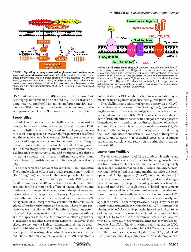
Cysteinyl leukotrienes (CysLT) are produced in asthma andhave potent effects on airway function, inducing bronchocon-striction, plasma exudation, and mucus secretion, and possiblyon eosinophilic inflammation (84). Blocking leukotriene path-ways may be beneficial in asthma, and this has led to the devel-opment of 5�-lipoxygenase (5-LOX) enzyme inhibitors (ofwhich zileuton is the only drug marketed) and several antago-nists of the CysLT1 receptor, including montelukast, zafirlu-kast, and pranlukast. Although there are clinical improvementsin symptoms and lung function and reduced exacerbations,these drugs are significantly less effective than low doses of ICSin asthma but are used because they are effective orally andappear to be safe. The pathways involved inCysLT synthesis arestrictly compartmentalized within the cell. Ca2� stimulates thebinding of type IVAcytosolic phospholipaseA2 (cPLA2�) to thecell membrane, with release of arachidonic acid, and the bind-ing of 5-LOX to the nuclear membrane, where it co-localizeswith FLAP (five-LOX-activating protein) and converts arachi-donic acid to LTA4 (Fig. 4). In certain cells that express LTC4synthase (mast cells and eosinophils), 5-LOX also co-localizeswith these enzymes to generate CysLT from LTA4 (85). FLAP,LTC4 synthase, and PLA2 inhibitors are now in development as
FIGURE 3. Signaling pathways involved in glucocorticoid resistance insevere asthma and smoking asthmatics. Oxidative and nitrative stress gen-erates peroxynitrite, which nitrates specific tyrosine residues (NO-Tyr) onHDAC2, resulting in its ubiquitination (Ub) and proteasomal degradation. Oxi-dative stress also activates PI3K�, which also leads to subsequent phos-phorylation (P) and ubiquitination of HDAC2, resulting in glucocorticoidresistance.
FIGURE 4. Leukotriene modifiers. Cell activation increases intracellular Ca2�
ions, leading to association of cPLA2� with the nuclear membrane, generat-ing arachidonic acid. This activates 5-LOX, which is also located in the nuclearmembrane bound to FLAP. This generates LTA4, which is converted by mem-brane-bound LTC4 synthase (LTC4S) in cells, such as mast cells and eosino-phils, into LTC4, LTD4, and LTE4, which then activate CysLT1 receptors on tar-get cells, such as airway smooth muscle cells. This pathway may be blocked byvarious types of drugs shown in green boxes.
MINIREVIEW: Biochemical Basis of Asthma Therapy
SEPTEMBER 23, 2011 • VOLUME 286 • NUMBER 38 JOURNAL OF BIOLOGICAL CHEMISTRY 32903
by guest on May 30, 2018
http://ww
w.jbc.org/
Dow
nloaded from

potential asthma therapies. Secreted PLA2 enzymes cooperatewith cPLA2; secreted PLA2-X is increased in the airways ofasthmatic patients and potently stimulates CysLT release fromeosinophils, which is inhibited by p38 MAPK and JNK inhibi-tors (86).
Concluding Remarks
There is now a relatively good understanding of how drugsused to treat asthma work in terms of their biochemical mech-anisms, providing opportunities to improve existing treatmentsand to discover novel therapies in the future (5). For example,once-daily �2-agonists, such as indacaterol and vilanterol, thatdissociatemore slowly from�2ARhave beendeveloped (6). Therecognition that �2-agonists may have potentially adverseeffects through activating inflammatory pathways via interac-tion with �-arrestins might lead to the development of biased�2-agonists that are less likely to have this activity and, at thesame time, are less likely to result in tolerance (20). Severalonce-daily anticholinergics have been developed for treatingCOPD, but they may also be useful in treating patients withsevere asthma (33). There are additive effects between �2-ago-nists and anticholinergics, which may be explained by cross-talk between signaling pathways, such that inhibition ofM2-re-ceptors may enhance the stimulatory effect of �2-agonists onadenylyl cyclase, and inhibition of phosphatidylinositol hydro-lysis via M3-receptors may also increase signaling effects of�2-agonists. This has led to the development of fixed-combina-tion inhalers that contain a once-daily �2-agonist and anticho-linergic (87).Understanding the biochemical pathways involved in sup-
pression of inflammation by glucocorticoids and the molecularmechanisms of glucocorticoid resistance has led to new thera-peutic approaches. Nonsteroidal selective glucocorticoidreceptor agonists (so-called dissociated steroids) that target thetrans-repression pathway linked to inhibition of NF-�B-in-duced inflammatory genes with relative sparing of trans-acti-vation pathways that involve DNA binding should theoreticallyreduce the side effects of glucocorticoids that appear to bemediatedmainly via trans-activation (88). In reality, it has beendifficult to demonstrate marked dissociation of beneficial andadverse effects with the compounds currently in development.Approaches to treating glucocorticoid resistance in asthmainclude alternative anti-inflammatory treatments such asMAPK inhibitors and drugs that activateHDAC2, such as theo-phylline and other PI3K� inhibitors. PDE4 inhibitors (nowapproved in Europe for severe COPD) may be useful in severeasthma (89). Although leukotriene receptor antagonists havebeen disappointing in the treatment of asthma, drugs actinghigher up the leukotriene synthesis pathway, such as 5-LOXand subtype-selective PLA2 inhibitors, might be more effectivethrough inhibiting the synthesis of more inflammatory media-tors (90). It has so far proved difficult to development novelanti-inflammatory treatments for asthma that are as safe oreffective as ICS, but perhaps a better understanding of theinflammatory signaling pathways in asthmatic airway cellsmight lead to more effective novel classes of therapy in thefuture.
REFERENCES1. Barnes, P. J. (2008) Nat. Immunol. Rev. 8, 183–1922. Hamid, Q., and Tulic, M. (2009) Annu. Rev. Physiol. 71, 489–5073. Barnes, P. J. (2008) J. Clin. Invest. 118, 3546–35564. Bateman, E. D., Hurd, S. S., Barnes, P. J., Bousquet, J., Drazen, J. M.,
FitzGerald, M., Gibson, P., Ohta, K., O’Byrne, P., Pedersen, S. E., Piz-zichini, E., Sullivan, S. D., Wenzel, S. E., and Zar, H. J. (2008) Eur. Respir. J.31, 143–178
5. Barnes, P. J. (2010) Trends Pharmacol. Sci. 31, 335–3436. Cazzola, M., and Matera, M. G. (2009) Eur. Respir. J. 34, 757–7697. Holz, G. G., Kang, G., Harbeck, M., Roe, M. W., and Chepurny, O. G.
(2006) J. Physiol. 577, 5–158. Kassel, K. M., Wyatt, T. A., Panettieri, R. A., Jr., and Toews, M. L. (2008)
Am. J. Physiol. Lung Cell. Mol. Physiol. 294, L131–L1389. McGraw, D. W., and Liggett, S. B. (1997) J. Biol. Chem. 272, 7338–734410. Mak, J. C., Hisada, T., Salmon, M., Barnes, P. J., and Chung, K. F. (2002)
Br. J. Pharmacol. 135, 987–99611. Duffy, S. M., Cruse, G., Lawley,W. J., and Bradding, P. (2005) FASEB J. 19,
1006–100812. Cruse, G., Yang,W., Duffy, S. M., Chachi, L., Leyland, M., Amrani, Y., and
Bradding, P. (2010) J. Allergy Clin. Immunol. 125, 257–26313. Howarth, P. H., Beckett, P., and Dahl, R. (2000) Respir. Med. 94, S22–S2514. Mak, J. C., Chuang, T. T., Harris, C. A., and Barnes, P. J. (2002) Eur.
J. Pharmacol. 436, 165–17215. Hawkins, G. A.,Weiss, S. T., and Bleecker, E. R. (2008) Pharmacogenomics
9, 349–35816. Israel, E., Chinchilli, V.M., Ford, J. G., Boushey,H.A., Cherniack, R., Craig,
T. J., Deykin, A., Fagan, J. K., Fahy, J. V., Fish, J., Kraft, M., Kunselman, S. J.,Lazarus, S. C., Lemanske, R. F., Jr., Liggett, S. B., Martin, R. J., Mitra, N.,Peters, S. P., Silverman, E., Sorkness, C. A., Szefler, S. J., Wechsler, M. E.,Weiss, S. T., and Drazen, J. M. (2004) Lancet 364, 1505–1512
17. Bleecker, E. R., Postma, D. S., Lawrance, R. M., Meyers, D. A., Ambrose,H. J., and Goldman, M. (2007) Lancet 370, 2118–2125
18. McGraw, D.W., Almoosa, K. F., Paul, R. J., Kobilka, B. K., and Liggett, S. B.(2003) J. Clin. Invest. 112, 619–626
19. Zhou, Y., Sondek, J., and Harden, T. K. (2008) Biochemistry 47,4410–4417
20. Violin, J. D., and Lefkowitz, R. J. (2007) Trends Pharmacol. Sci. 28,416–422
21. Shenoy, S. K., McDonald, P. H., Kohout, T. A., and Lefkowitz, R. J. (2001)Science 294, 1307–1313
22. Schmid, C. L., and Bohn, L. M. (2009) Pharmacol. Ther. 121, 285–29323. DeWire, S.M., Ahn, S., Lefkowitz, R. J., and Shenoy, S. K. (2007)Annu. Rev.
Physiol. 69, 483–51024. Nelson, H. S., and Dorinsky, P. M. (2006)Ann. Intern. Med. 145, 706–71025. Nguyen, L. P., Lin, R., Parra, S., Omoluabi,O., Hanania,N.A., Tuvim,M. J.,
Knoll, B. J., Dickey, B. F., and Bond, R. A. (2009) Proc. Natl. Acad. Sci.U.S.A. 106, 2435–2440
26. Hanania, N. A., Singh, S., El-Wali, R., Flashner,M., Franklin, A. E., Garner,W. J., Dickey, B. F., Parra, S., Ruoss, S., Shardonofsky, F., O’Connor, B. J.,Page, C., and Bond, R. A. (2008) Pulm. Pharmacol. Ther. 21, 134–141
27. Walker, J. K., Fong, A.M., Lawson, B. L., Savov, J. D., Patel, D.D., Schwartz,D. A., and Lefkowitz, R. J. (2003) J. Clin. Invest. 112, 566–574
28. Hollingsworth, J. W., Theriot, B. S., Li, Z., Lawson, B. L., Sunday, M.,Schwartz, D. A., and Walker, J. K. (2010) Am. J. Respir. Cell Mol. Biol. 43,269–275
29. Gong, K., Li, Z., Xu, M., Du, J., Lv, Z., and Zhang, Y. (2008) J. Biol. Chem.283, 29028–29036
30. Wessler, I., and Kirkpatrick, C. J. (2008) Br. J. Pharmacol. 154, 1558–157131. Kummer, W., Lips, K. S., and Pfeil, U. (2008) Histochem. Cell Biol. 130,
219–23432. Bateman, E. D., Rennard, S., Barnes, P. J., Dicpinigaitis, P. V., Gosens, R.,
Gross, N. J., Nadel, J. A., Pfeifer, M., Racke, K., Rabe, K. F., Rubin, B. K.,Welte, T., and Wessler, I. (2009) Pulm. Pharmacol. Ther. 22, 533–542
33. Peters, S. P., Kunselman, S. J., Icitovic, N., Moore, W. C., Pascual, R.,Ameredes, B. T., Boushey, H. A., Calhoun, W. J., Castro, M., Cherniack,R. M., Craig, T., Denlinger, L., Engle, L. L., DiMango, E. A., Fahy, J. V.,
MINIREVIEW: Biochemical Basis of Asthma Therapy
32904 JOURNAL OF BIOLOGICAL CHEMISTRY VOLUME 286 • NUMBER 38 • SEPTEMBER 23, 2011
by guest on May 30, 2018
http://ww
w.jbc.org/
Dow
nloaded from

Israel, E., Jarjour, N., Kazani, S. D., Kraft, M., Lazarus, S. C., Lemanske,R. F., Jr., Lugogo, N., Martin, R. J., Meyers, D. A., Ramsdell, J., Sorkness,C. A., Sutherland, E. R., Szefler, S. J., Wasserman, S. I., Walter, M. J.,Wechsler, M. E., Chinchilli, V. M., and Bleecker, E. R. (2010) N. Engl.J. Med. 363, 1715–1726
34. Bos, I. S., Gosens, R., Zuidhof, A. B., Schaafsma, D., Halayko, A. J., Meurs,H., and Zaagsma, J. (2007) Eur. Respir. J. 30, 653–661
35. Zhou, X. B., Wulfsen, I., Lutz, S., Utku, E., Sausbier, U., Ruth, P., Wieland,T., and Korth, M. (2008) J. Biol. Chem. 283, 21036–21044
36. Fryer, A. D., Stein, L. H., Nie, Z., Curtis, D. E., Evans, C.M., Hodgson, S. T.,Jose, P. J., Belmonte, K. E., Fitch, E., and Jacoby, D. B. (2006) J. Clin. Invest.116, 228–236
37. Minette, P. A., Lammers, J. W., Dixon, C. M., McCusker, M. T., andBarnes, P. J. (1989) J. Appl. Physiol. 67, 2461–2465
38. Barnes, P. J. (2006) Br. J. Pharmacol. 148, 245–25439. Rhen, T., and Cidlowski, J. A. (2005) N. Engl. J. Med. 353, 1711–172340. Nicolaides, N. C., Galata, Z., Kino, T., Chrousos, G. P., and Charmandari,
E. (2010) Steroids 75, 1–1241. Goldfarb, D. S., Corbett, A. H.,Mason, D. A., Harreman,M. T., andAdam,
S. A. (2004) Trends Cell Biol. 14, 505–51442. Tao, T., Lan, J., Lukacs, G. L., Hache, R. J., and Kaplan, F. (2006) Am. J.
Respir. Cell Mol. Biol. 35, 668–68043. Lewis-Tuffin, L. J., and Cidlowski, J. A. (2006) Ann. N.Y. Acad. Sci. 1069,
1–944. Ito, K., Barnes, P. J., and Adcock, I. M. (2000) Mol. Cell. Biol. 20,
6891–690345. John, S., Sabo, P. J., Johnson, T. A., Sung, M. H., Biddie, S. C., Lightman,
S. L., Voss, T. C., Davis, S. R.,Meltzer, P. S., Stamatoyannopoulos, J. A., andHager, G. L. (2008)Mol. Cell 29, 611–624
46. Barnes, P. J. (2006) Eur. Respir. J. 27, 413–42647. Clark, A. R. (2003) J. Endocrinol. 178, 5–1248. Dostert, A., and Heinzel, T. (2004) Curr. Pharm. Des. 10, 2807–281649. Barnes, P. J., and Adcock, I. M. (2003) Ann. Intern. Med. 139, 359–37050. Ito, K., Yamamura, S., Essilfie-Quaye, S., Cosio, B., Ito,M., Barnes, P. J., and
Adcock, I. M. (2006) J. Exp. Med. 203, 7–1351. Maneechotesuwan, K., Xin, Y., Ito, K., Jazrawi, E., Lee, K. Y., Usmani, O. S.,
Barnes, P. J., and Adcock, I. M. (2007) J. Immunol. 178, 2491–249852. Maneechotesuwan, K., Yao, X., Ito, K., Jazrawi, E., Usmani, O. S., Adcock,
I. M., and Barnes, P. J. (2009) PLoS Med. 6, e100007653. Maneechotesuwan, K., Supawita, S., Kasetsinsombat, K.,Wongkajornsilp,
A., and Barnes, P. J. (2008) J. Allergy Clin. Immunol. 121, 43–5054. Maneechotesuwan, K., Ekjiratrakul, W., Kasetsinsombat, K., Wongka-
jornsilp, A., andBarnes, P. J. (2010) J. Allergy Clin. Immunol. 126, 754–76255. Bergmann, M. W., Staples, K. J., Smith, S. J., Barnes, P. J., and Newton, R.
(2004) Am. J. Respir. Cell Mol. Biol. 30, 555–56356. Smoak, K., and Cidlowski, J. A. (2006)Mol. Cell. Biol. 26, 9126–913557. Barnes, P. J. (2002) Eur. Respir. J. 19, 182–19158. Newton, R., Leigh, R., and Giembycz, M. A. (2010) Pharmacol. Ther. 125,
286–32759. Mak, J. C., Nishikawa, M., and Barnes, P. J. (1995) Am. J. Physiol. 268,
L41–L4660. Baraniuk, J. N., Ali, M., Brody, D., Maniscalco, J., Gaumond, E., Fitzgerald,
T., Wong, G., Yuta, A., Mak, J. C., Barnes, P. J., Bascom, R., and Troost, T.(1997) Am. J. Respir. Crit. Care Med. 155, 704–710
61. Mak, J. C., Nishikawa, M., Shirasaki, H., Miyayasu, K., and Barnes, P. J.(1995) J. Clin. Invest. 96, 99–106
62. Roth, M., Johnson, P. R., Rudiger, J. J., King, G. G., Ge, Q., Burgess, J. K.,Anderson, G., Tamm, M., and Black, J. L. (2002) Lancet 360, 1293–1299
63. Usmani, O. S., Ito, K., Maneechotesuwan, K., Ito, M., Johnson,M., Barnes,
P. J., and Adcock, I. M. (2005)Am. J. Respir. Crit. CareMed. 172, 704–71264. Gibson, P. G., Powell, H., and Ducharme, F. M. (2007) J. Allergy Clin.
Immunol. 119, 344–35065. Barnes, P. J., and Adcock, I. M. (2009) Lancet 373, 1905–191766. Hew, M., Bhavsar, P., Torrego, A., Meah, S., Khorasani, N., Barnes, P. J.,
Adcock, I., and Chung, K. F. (2006) Am. J. Respir. Crit. Care Med. 174,134–141
67. Barnes, P. J. (2009) Annu. Rev. Physiol. 71, 451–46468. Osoata, G. O., Yamamura, S., Ito, M., Vuppusetty, C., Adcock, I. M.,
Barnes, P. J., and Ito, K. (2009) Biochem. Biophy. Res. Commun. 384,366–371
69. To, Y., Ito, K., Kizawa, Y., Failla, M., Ito, M., Kusama, T., Elliott, W. M.,Hogg, J. C., Adcock, I. M., and Barnes, P. J. (2010) Am. J. Resp. Crit. CareMed. 182, 897–904
70. Charron, C. E., Chou, P. C., Coutts, D. J., Kumar, V., To, M., Akashi, K.,Pinhu, L., Griffiths,M., Adcock, I.M., Barnes, P. J., and Ito, K. (2009) J. Biol.Chem. 284, 36047–36054
71. Irusen, E., Matthews, J. G., Takahashi, A., Barnes, P. J., Chung, K. F., andAdcock, I. M. (2002) J. Allergy Clin. Immunol. 109, 649–657
72. Ismaili, N., and Garabedian, M. J. (2004) Ann. N.Y. Acad. Sci. 1024,86–101
73. Matthews, J. G., Ito, K., Barnes, P. J., and Adcock, I. M. (2004) J. AllergyClin. Immunol. 113, 1100–1108
74. Pujols, L., Mullol, J., and Picado, C. (2007) Curr. Allergy Asthma Rep. 7,93–99
75. Lewis-Tuffin, L. J., Jewell, C. M., Bienstock, R. J., Collins, J. B., and Ci-dlowski, J. A. (2007)Mol. Cell. Biol. 27, 2266–2282
76. Barnes, P. J. (2003) Am. J. Respir. Crit. Care Med. 167, 813–81877. Wilson, C. N. (2008) Br. J. Pharmacol. 155, 475–48678. Ichiyama, T., Hasegawa, S., Matsubara, T., Hayashi, T., and Furukawa, S.
(2001) Naunyn-Schmiedebergs Arch. Pharmacol. 364, 558–56179. Chung, I. Y., Nam-Kung, E. K., Lee, N. M., Chang, H. S., Kim, D. J., Kim,
Y. H., and Park, C. S. (2000) Cell. Immunol. 203, 95–10280. Yasui, K., Agematsu, K., Shinozaki, K., Hokibara, S., Nagumo, H., Naka-
zawa, T., and Komiyama, A. (2000) J. Leukocyte Biol. 67, 529–53581. Ito, K., Lim, S., Caramori, G., Cosio, B., Chung, K. F., Adcock, I. M., and
Barnes, P. J. (2002) Proc. Natl. Acad. Sci. U.S.A. 99, 8921–892682. Cosio, B. G., Tsaprouni, L., Ito, K., Jazrawi, E., Adcock, I. M., and Barnes,
P. J. (2004) J. Exp. Med. 200, 689–69583. Marwick, J. A., Caramori, G., Stevenson, C. S., Casolari, P., Jazrawi, E.,
Barnes, P. J., Ito, K., Adcock, I. M., Kirkham, P. A., and Papi, A. (2009)Am. J. Respir. Crit. Care Med. 179, 542–548
84. Peters-Golden, M., and Henderson, W. R., Jr. (2007) N. Engl. J. Med. 357,1841–1854
85. Newcomer, M. E., and Gilbert, N. C. (2010) J. Biol. Chem. 285,25109–25114
86. Lai, Y., Oslund, R. C., Bollinger, J. G., Henderson, W. R., Jr., Santana, L. F.,Altemeier, W. A., Gelb, M. H., and Hallstrand, T. S. (2010) J. Biol. Chem.285, 41491–41500
87. vanNoord, J. A., Buhl, R., Laforce, C.,Martin, C., Jones, F., Dolker,M., andOverend, T. (2010) Thorax 65, 1086–1091
88. Schacke, H., Berger,M., Rehwinkel, H., andAsadullah, K. (2007)Mol. Cell.Endocrinol. 275, 109–117
89. Hatzelmann, A., Morcillo, E. J., Lungarella, G., Adnot, S., Sanjar, S.,Beume, R., Schudt, C., and Tenor, H. (2010) Pulm. Pharmacol. Ther. 23,235–256
90. Garcia-Garcia, H. M., and Serruys, P. W. (2009) Curr. Opin. Lipidol. 20,327–332
MINIREVIEW: Biochemical Basis of Asthma Therapy
SEPTEMBER 23, 2011 • VOLUME 286 • NUMBER 38 JOURNAL OF BIOLOGICAL CHEMISTRY 32905
by guest on May 30, 2018
http://ww
w.jbc.org/
Dow
nloaded from

Peter J. BarnesBiochemical Basis of Asthma Therapy
doi: 10.1074/jbc.R110.206466 originally published online July 28, 20112011, 286:32899-32905.J. Biol. Chem.
10.1074/jbc.R110.206466Access the most updated version of this article at doi:
Alerts:
When a correction for this article is posted•
When this article is cited•
to choose from all of JBC's e-mail alertsClick here
http://www.jbc.org/content/suppl/2011/09/15/R110.206466.DCAuthor_profileRead an Author Profile for this article at
http://www.jbc.org/content/286/38/32899.full.html#ref-list-1
This article cites 90 references, 24 of which can be accessed free at
by guest on May 30, 2018
http://ww
w.jbc.org/
Dow
nloaded from