Clinical diagnostic biochemistry - 15 Dr. Maha Al-Sedik 2015 CLS 334.
Upload
russell-caseyCategory
view
238download
4
Biochemical instrumental analysis-7
Dr. Maha Al-Sedik
Electrophoresis
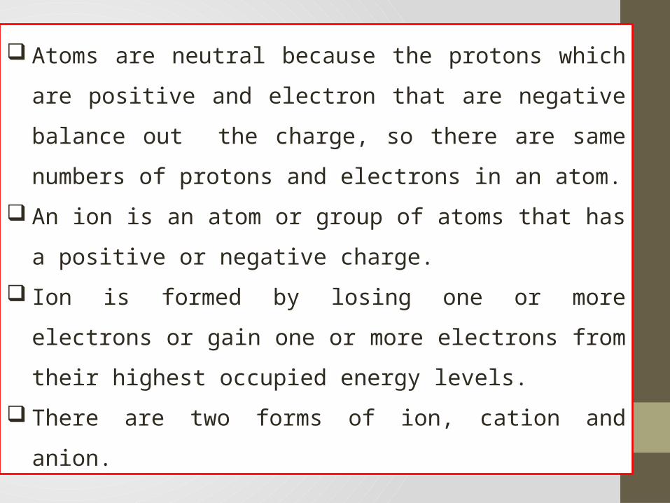
Atoms are neutral because the protons which are positive
and electron that are negative balance out the charge, so
there are same numbers of protons and electrons in an
atom.
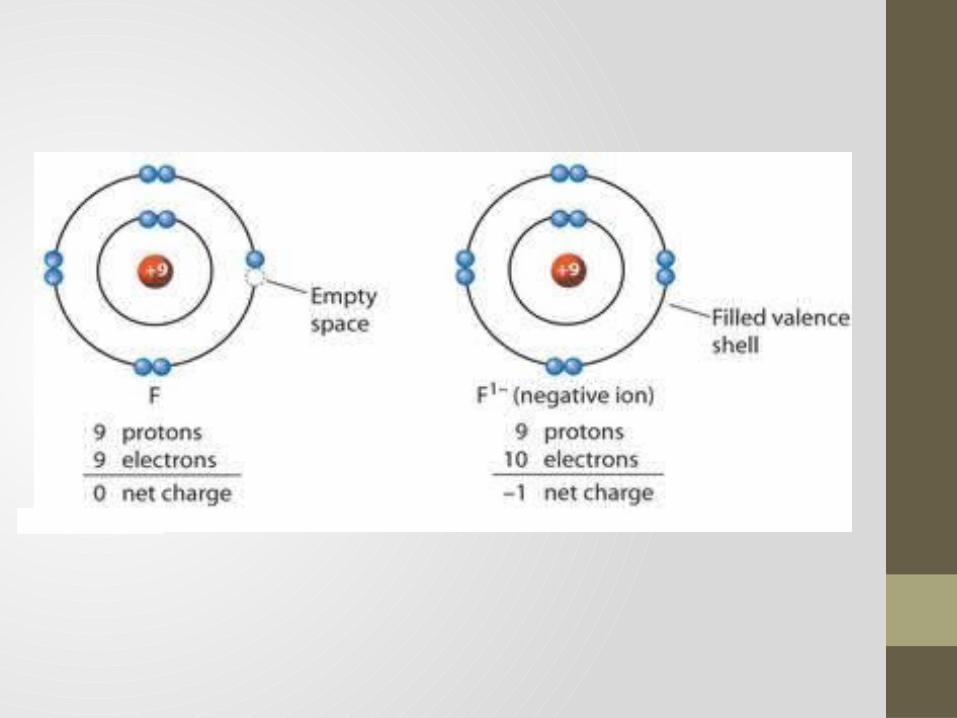
An ion is an atom or group of atoms that has a positive or
negative charge.
Ion is formed by losing one or more electrons or gain one or
more electrons from their highest occupied energy levels.
There are two forms of ion, cation and anion.
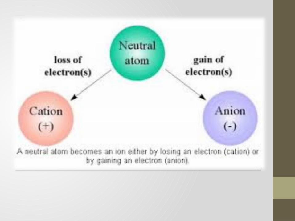
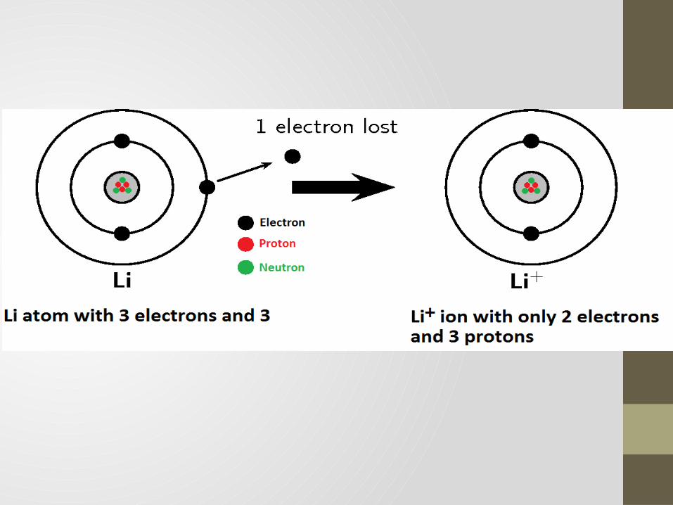
CATION Cation is a positive ion.
Cations are formed by losing electrons in the highest
occupied energy levels.
By losing electrons, the number of protons and electrons are
not balanced, there are more protons than electron so the
atom became positively charged.
The charge for the cation is written as the charge followed
by a plus sign.
ANION Anion is a negative charged ion.
Anions are formed by gaining electron to make the highest
occupied energy level full.
By gaining electrons, there are more numbers of electrons than
protons in an atom so the atom became negative charged.
The charge for the anion is written as the charge followed by a
minus sign.

Practice problems Magnesium have the atomic number of 12, if it forms an ion, is it
going to form a cation or an anion?
How many electrons will it gain/lose?
(Magnesium have two valence electron on its outer energy level,
so it will become a cation by losing two electrons).
Ions are atoms which have
gained or lost one or more
electrons giving the ion a net
positive or negative charge.
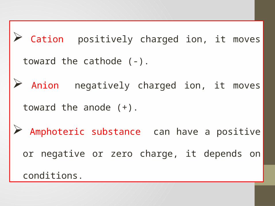
Cation positively charged ion, it moves toward the
cathode (-).
Anion negatively charged ion, it moves toward the anode
(+).
Amphoteric substance can have a positive or negative or
zero charge, it depends on conditions.
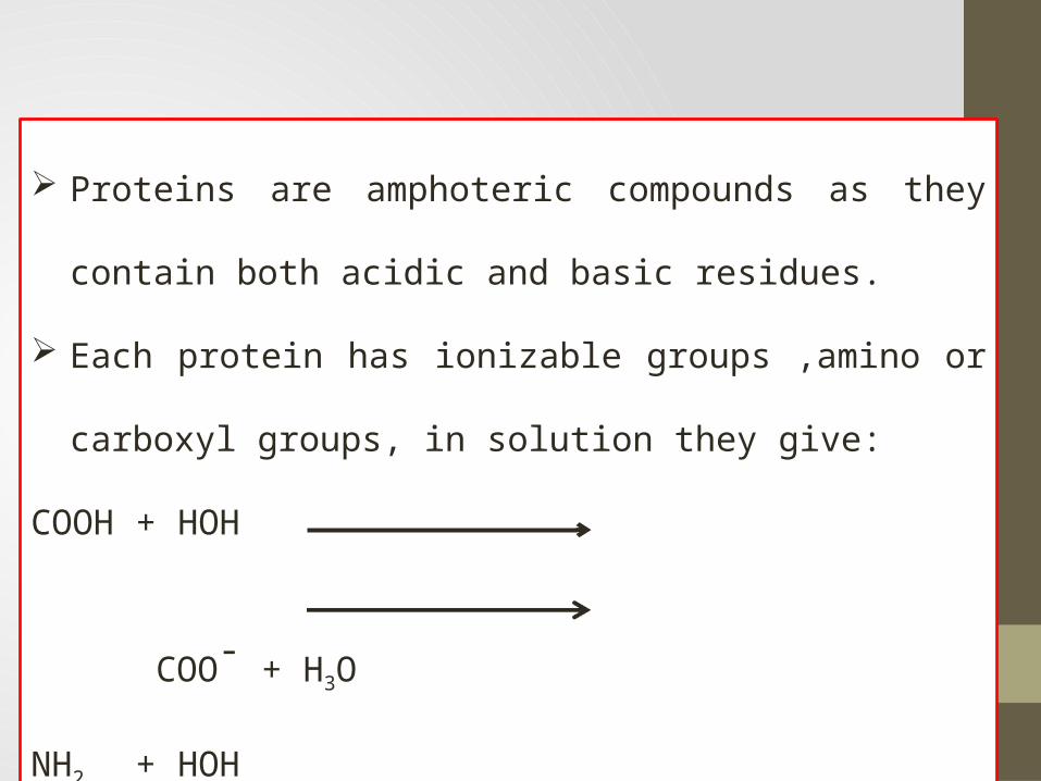
Proteins are amphoteric compounds as they contain both
acidic and basic residues.
Each protein has ionizable groups ,amino or carboxyl
groups, in solution they give:
COOH + HOH COO- + H3O
NH2 + HOH NH3+ + OH
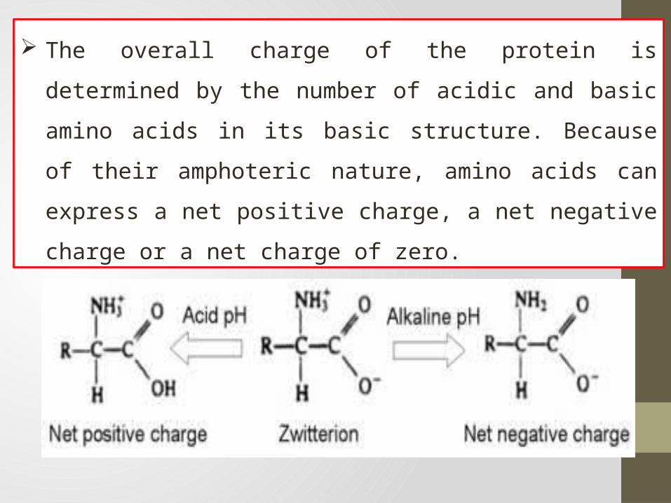
The overall charge of the protein is determined by the
number of acidic and basic amino acids in its basic structure.
Because of their amphoteric nature, amino acids can express
a net positive charge, a net negative charge or a net charge
of zero.
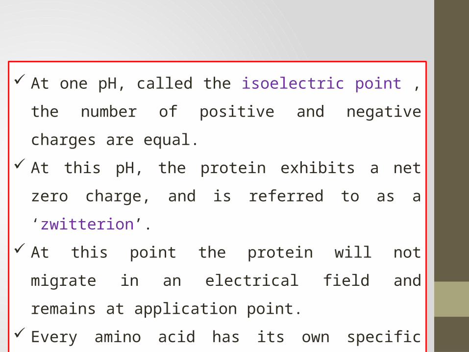
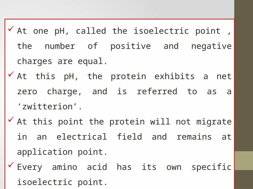
At one pH, called the isoelectric point , the number of
positive and negative charges are equal.
At this pH, the protein exhibits a net zero charge, and is
referred to as a ‘zwitterion’.
At this point the protein will not migrate in an electrical field
and remains at application point.
Every amino acid has its own specific isoelectric point.
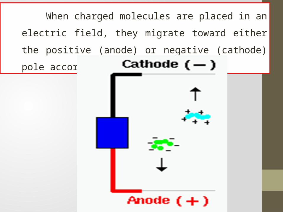
When charged molecules are placed in an electric field,
they migrate toward either the positive (anode) or negative
(cathode) pole according to their charge.
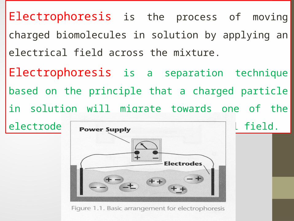
Electrophoresis is the process of moving charged
biomolecules in solution by applying an electrical field across
the mixture.
Electrophoresis is a separation technique based on the principle
that a charged particle in solution will migrate towards one of the
electrodes when placed in an electrical field.
Electrophoresis is a technique used in clinical laboratories to
separate particles (proteins) from each other:
Proteins in body fluids: serum, urine, CSF.
Proteins in erythrocytes: hemoglobin.
Nucleic acids: DNA, RNA.
Factors influenced electrophoresis mobility:
The electric field.
Net charge of the molecule.
Size and shape.
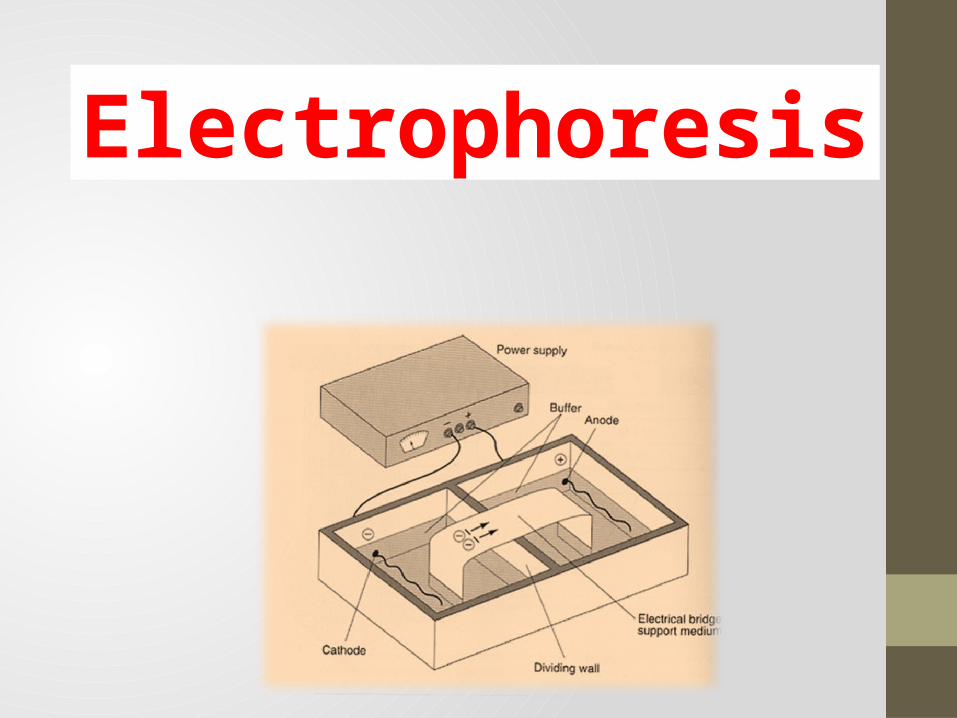
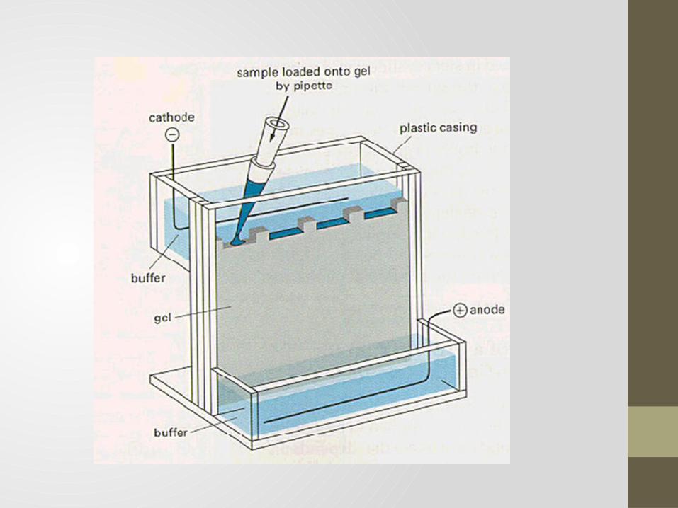
Instrumentation and reagents: Two buffer boxes contain the buffer used in the process.
Each buffer box contains an electrode made of either
platinum or carbon, the polarity of which is determined by the
mode of connection to the power supply.
The electrophoresis support on which separation takes place
may contact the buffer directly, or by means of wicks.
The entire apparatus is covered to minimize evaporation and
protect the system.
The power supply to provide electrical power.
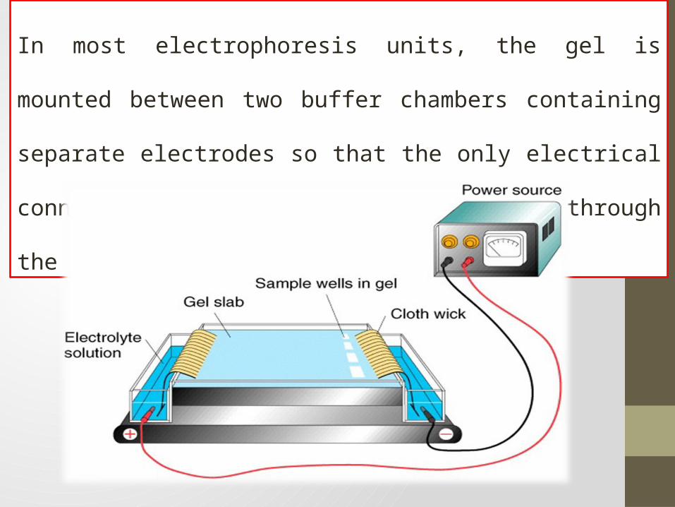
In most electrophoresis units, the gel is mounted between two buffer
chambers containing separate electrodes so that the only electrical
connection between the two chambers is through the gel.
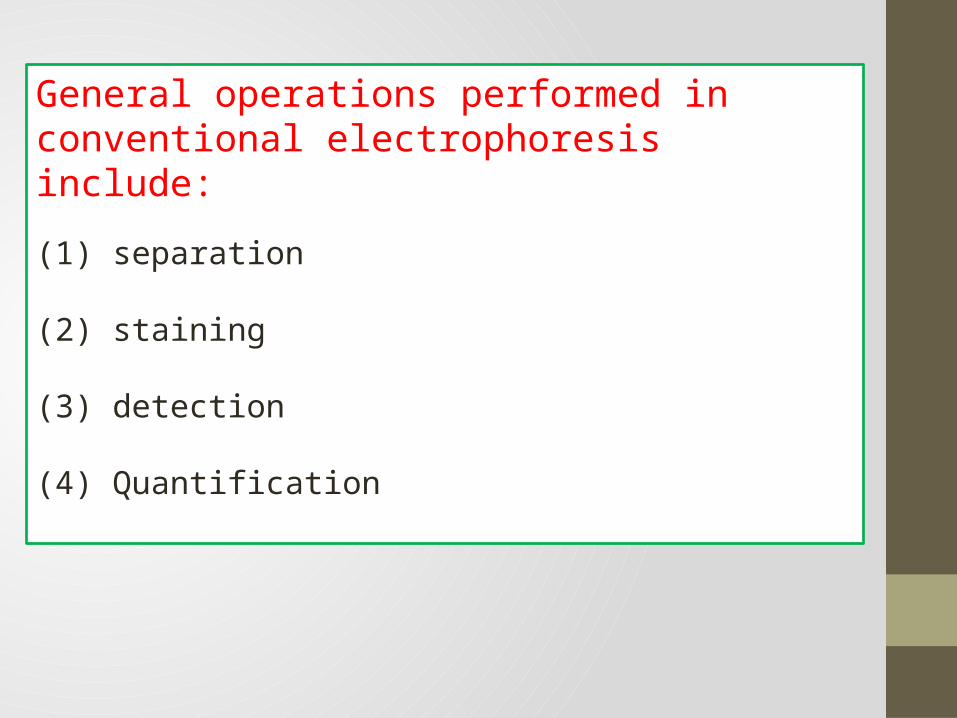
General operations performed in conventional electrophoresis include:
(1) separation
(2) staining
(3) detection
(4) Quantification
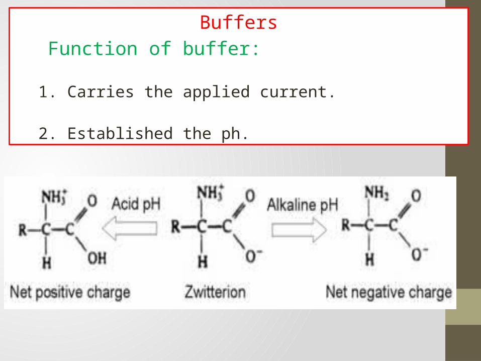
Buffers
Function of buffer:
1. Carries the applied current.
2. Established the ph.
At one pH, called the isoelectric point , the number of positive
and negative charges are equal.
At this pH, the protein exhibits a net zero charge, and is
referred to as a ‘zwitterion’.
At this point the protein will not migrate in an electrical field
and remains at application point.
Every amino acid has its own specific isoelectric point.
At a pH above its isoelectric point, the proteins will have a net
negative charge and will migrate towards the anode.
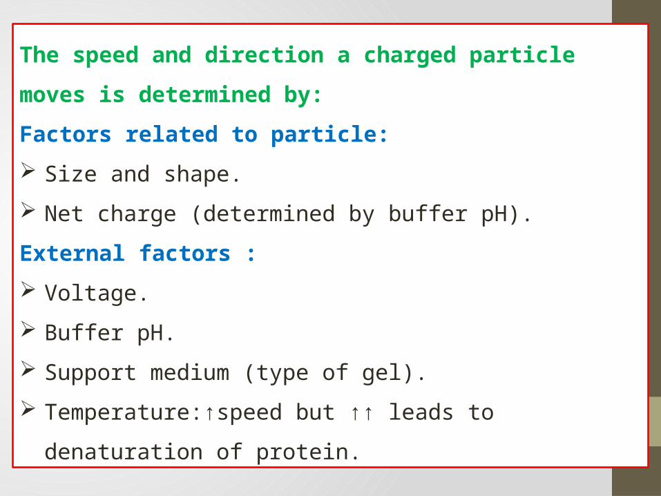
The speed and direction a charged particle moves is determined by:
Factors related to particle:
Size and shape.
Net charge (determined by buffer pH).
External factors :
Voltage.
Buffer pH.
Support medium (type of gel).
Temperature:↑speed but ↑↑ leads to denaturation of protein.
Electrophoresis of plasma protein
The proteins found in plasma have amino acids as their subunits,
and each protein has its own specific isoelectric point.
Because of their different isoelectric points, each protein will
move at a different rate when placed in an electrical field.
Proteins with similar isoelectric points will migrate to a similar
area in an electrical field.
o Normal total plasma protein concentration: 6 – 8.5 gm / dl.
o Electrophoresis separates serum Proteins into 5 distinct zones or
bands at barbital buffer at pH 8.6 :
Albumin.
Alpha globulin ( Alpha 1 and Alpha 2 ).
Beta globulin ( Beta 1 and Beta 2 ).
Gamma globulin: IgG, IgA, IgM, IgD, IgE and C-reactive protein.
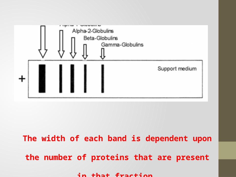
The width of each band is dependent upon the number of
proteins that are present in that fraction.
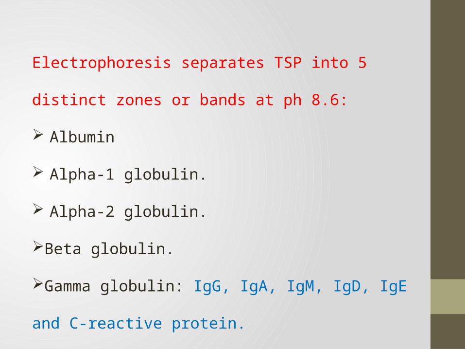
Electrophoresis separates TSP into 5 distinct zones or bands
at ph 8.6:
Albumin
Alpha-1 globulin.
Alpha-2 globulin.
Beta globulin.
Gamma globulin: IgG, IgA, IgM, IgD, IgE and C-reactive
protein.
Q – WHAT is the difference
between serum proteins and
plasma proteins in electrophoresis ?
Serum:
is a clear yellowish fluid that remains from blood plasma after
clotting factors (fibrinogen, prothrombin ect.) that have been used in
the formation of a clot.
Plasma:
is a clear yellowish fluid that still contains all of the clotting factors
and have not been solidified into clot.
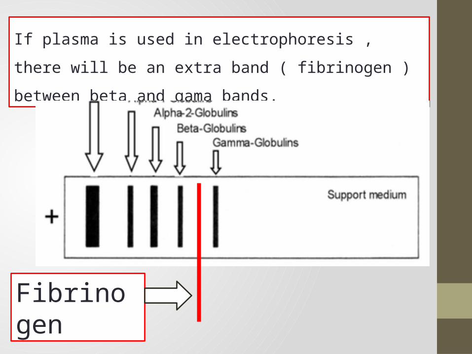
If plasma is used in electrophoresis , there will be an extra band
( fibrinogen ) between beta and gama bands.
Fibrinogen
Basic Procedure:
Sample is applied to an agarose gel.
Gel is placed into electrophoresis cell.
containing barbital buffer at pH 8.6.
Power is applied creating an electrical field and the proteins are
separated.
Proteins are fixed to the gel and stained.
Separated proteins on gel are scanned.

THANK YOU