
2D vs. 3D Squirrel Eiserloh Director, TrueThought [email protected].

Biochemical Features of the Grey Squirrel LensSeymour Zigman, Tereso Paxhia, and William Waldron
The ocular lens of the grey squirrel (Sciurus carolinensis) is an excellent model for studies of eye-light interactions that apply to the human system. In this diurnal animal, lens size, shape, yellowpigmentation, and light absorption properties have important similarities to those of young children.This article describes the observations of soluble to insoluble protein conversion with chronologicalaging, and the loss of heavier lens crystallins in the internal as compared to the external layers ofthe lens. Such changes are related to aging, as the older lens material is present in the nuclear core,while the younger lens material is superficial. It describes the subunit peptides of the solublecrystallins and of the extrinsic and instrinsic proteins associated with fiber cell membranes. Squirrellens fiber membranes release most of their extrinsic peptides in 8 M urea, as do those of other younganimals. Due to the presence of near-UV absorbing species of low molecular weight, the squirrel lenshas great potential for studies of the effects of near-UV radiation on the lens. Invest Ophthalmol VisSci 26:1075-1082, 1985
Attention has previously been drawn to certainfeatures of grey squirrel (Sciurus carolinensis) lensesthat are similar to those of humans. Cooper andRobson1 observed the presence of dialyzable yellowpigment with a 365-nm absorption peak that resem-bles a young human lens pigment with similar features.Van Heyningen2 indicated the presence in the squirrellens of N-acetyl-3-OH-L-kynurenine shown to bepresent in human lenses. Rafferty et al3 have shownthe presence of electronmicroscopically observableintermediate filaments in the most anterior portionof the disc-shaped squirrel lens, but not in the sphericallenses of rodents, whose function is to participate inthe accomodative process.
This study reports our initial observations of themorphologic and biochemical features of grey squirrellenses, which emphasize their usefulness as modelsfor human lenses. In addition to the features men-tioned above, the squirrel is a diurnal animal, as isman, while the rodents often used in eye research arenearly exclusively nocturnal. Squirrel and humanlenses interact with radiant energy similarly, due tothe presence of yellow pigments, which improve thequality of cone (photopic) vision by eliminating chro-matic aberration, serve as blue and UV filters toprotect the retina from light damage.4 This reportshows that the size, weight, chromophores, and pro-
From the Departments of Ophthalmology and Biochemistry,University of Rochester School of Medicine and Dentistry, Roch-ester, New York.
Supported by National Eye Institute (NIH) EY-00459 and Re-search to Prevent Blindness, Inc.
Submitted for publication: June 11, 1984.Reprint requests: Dr. Seymour Zigman, Ophthalmology Research
Laboratory (Box 314), University Rochester Medical Center, 601Elmwood Avenue, Rochester, NY 14642.
teins of grey squirrel lenses have much in commonwith those of man.
Materials and MethodsGrey squirrels (Sciurus carolinensis) were obtained
under a scientific collector's license. They were placedand maintained in our animal facility using humanetreatment and careful handling. Procedures usingsquirrels conform fully to the ARVO Resolution onthe Use of Animals in Research. Squirrels were killedwith Nembutal overdosing, and their eyes and lenseswere carefully removed. Body weights of the animalsand fresh lens weights were measured, and lens lighttransmission was observed by using a Cary 14 spec-trophotometer (Applied Physics Corp; Monrovia,CA).5 Some lenses were frozen in glass vials on dryice and stored at -50°C for future use.
For biochemical studies, fresh or frozen individuallenses (average weight 100 mg) were decapsulatedand homobenized in 4.0 ml of 0.1 M PO4 (pH 7)buffer. Some lenses were separated into concentriclayers by soaking them in buffer and removing ap-proximately 0.5-mm thick portions with a micropi-pe,tte and spatulas. All solutions were maintained oncrushed ice and were protected from ambient lightexposure. Centrifugation at 13,000 X g (at 10°C) sed-imented the insoluble (INS) protein plus membranefraction; subsequent centrifugation at 100,000 Xgwas employed to sediment the high molecular weightaggregated protein fraction (HM), leaving the totalsoluble protein (TSP) in the supernatant. Since theyellow pigments of the squirrel lenses are low molec-ular weight water-soluble entities, they were separatedfrom the lens proteins by pressure dialysis, using YM2 (Amicon) filter discs with molecular weight cutoffof 1,000 daltons.
1075
Downloaded From: http://iovs.arvojournals.org/pdfaccess.ashx?url=/data/journals/iovs/933124/ on 06/25/2018

1076 INVESTIGATIVE OPHTHALMOLOGY & VISUAL SCIENCE / Augusr 1985 Vol. 26
LENSWEIGHTCMG)
140. T
1 10. •
90.
70.
50.
A—A-
250. 350.
BODY WT CGM)
450 569
Fig. I. Increase in fresh lens weight vs body weight of greysquirrels (Sciurus carolinensis). Each point on the graph representsthe average of the lenses of two animals. No greater than a 10%difference between the weights of the two lenses of each animalwas found.
Proteins were estimated by the Lowry procedure.For polyacrylamide gel electrophoresis, the conditionsused were already described by Zigman et al.6 Detailsof the procedure will also be found in the legends offigures related to polyacrylamide gel electrophoresis.HPLC (TSK 3000 columns, 30 cm) analyses of theTSP fractions of various layers were done with thefollowing conditions: buffer, 0.05 M PO4 (dibasic)+ 0.1 M Na2 SO4, pH 7.0; pressure, 200 psi; elutionrate, 0.4 ml/min; wavelength, 280 nm.
Absorption spectra were measured with a Cary 14recording spectrophotometer; fluorescence spectrawith a Perkin-Elmer 512 spectrofluometer; mass spec-tra were obtained using the following conditions:High and low resolution Dupont Inst. electron impactCEC-110B; source temperature, 200°C at 70 ev;acceleration, 9 KV; field desorption, varian MATinstrument; field ionization, FE/FI/EI source at 8
Table 1. Protein content of grey squirrel lensesrelative to age and weight
Lens weight(mg)
70.778.692.0
117.3138.0152.2
Total protein(mg)
24.226.329.138.744.447.6
%Protein
34.232.831.632.632.033.2
Age(mo)
38
10184270
KV, potential of 3-6 KV; temperature, 100°C. Carbondendrite emitters at 1000 resolution were used.
Results
The squirrel lenses obtained and used in the bio-chemical studies presented below ranged in weightfrom 71 to 152 mg. These were removed fromanimals whose body weights ranged from 292 g to552 g, and the relationship between lens and bodyweights is shown in Figure 1. The wet weights, proteincontents, and ages in this series of squirrel lenses aregiven in Table 1. From data published by Fisher andPerry7 on the age of Sciurus carolinensis relative tolens dry weight, it was possible to estimate theapproximate ages of these animals, which range from3 mo (70 mg lens) to 6 yr (152 mg lens).
The change in distribution of protein fractions inthis series of squirrels is presented in Figure 2. Aconstant decline in percent total soluble protein (TSP)and increases in the high molecular weight colloidal(urea soluble) and insoluble fractions (SDS and SDS+ DTT soluble) were observed. In a study of thedistribution of protein fractions in concentric layersof the lens, as shown in Figure 3, TSP is present inthe outer cortex at a level twice that in the nuclear
100. T
% oftotalprote i nCTSP)
60. 80. 100. 120.Lens Weight Cmg)
-0- TSPCR) -»- HMWCR) INS
50.
% oftotalprote i nCHMW
andINS)
Fig. 2. Changes in the dis-tribution of total soluble,high molecular weight col-loidal (HMW), and total wa-ter-insoluble (INS) proteinfraction vs increasing greysquirrel lens weight. Eachpoint represents the averageof the lenses of two animals.
Downloaded From: http://iovs.arvojournals.org/pdfaccess.ashx?url=/data/journals/iovs/933124/ on 06/25/2018
No. 8 DIQCHEMI5TRY OF GREY SQUIRREL LENS / Zigmon er ol. 1077
TSP US SDS SDS+DTT
>f
> •
V-
% oftotalprolei nin layer
tea.
88.
60.
40.
20. -
0 .
50. T
40. -•
30. -•
20. ••
1 0 . -•
5 . T
4. --
3 . • •
2 . •
1 . - •
0 .
5. -I
4. -
3.
2. •
I . • •
bid ocLTD] ic
ON
IN
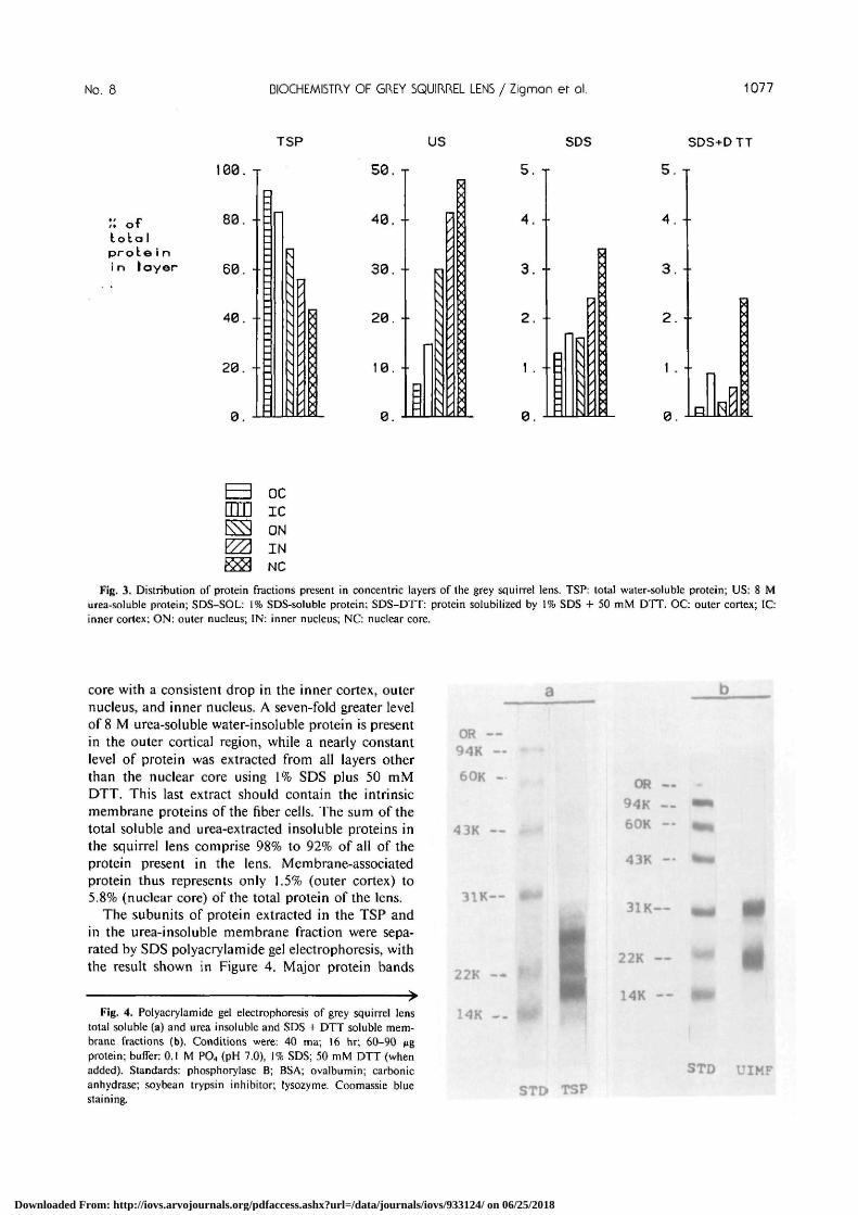
Fig. 3. Distribution of protein fractions present in concentric layers of the grey squirrel lens. TSP: total water-soluble protein; US: 8 Murea-soluble protein; SDS-SOL: 1% SDS-soluble protein; SDS-DTT: protein solubilized by 1% SDS + 50 mM DTT. OC: outer cortex; IC:inner cortex; ON: outer nucleus; IN: inner nucleus; NC: nuclear core.
core with a consistent drop in the inner cortex, outernucleus, and inner nucleus. A seven-fold greater levelof 8 M urea-soluble water-insoluble protein is presentin the outer cortical region, while a nearly constantlevel of protein was extracted from all layers otherthan the nuclear core using 1% SDS plus 50 mMDTT. This last extract should contain the intrinsicmembrane proteins of the fiber cells. The sum of thetotal soluble and urea-extracted insoluble proteins inthe squirrel lens comprise 98% to 92% of all of theprotein present in the lens. Membrane-associatedprotein thus represents only 1.5% (outer cortex) to5.8% (nuclear core) of the total protein of the lens.
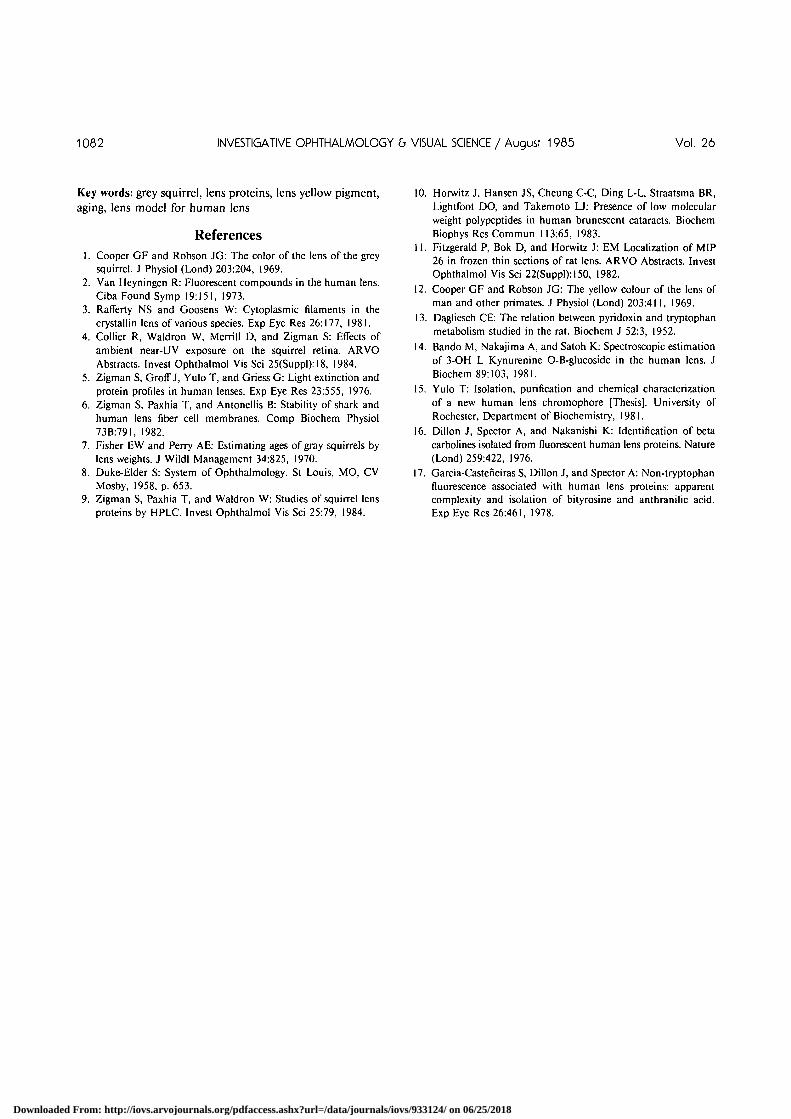
The subunits of protein extracted in the TSP andin the urea-insoluble membrane fraction were sepa-rated by SDS polyacrylamide gel electrophoresis, withthe result shown in Figure 4. Major protein bands
Fig. 4. Polyacrylamide gel electrophoresis of grey squirrel lenstotal soluble (a) and urea insoluble and SDS + DTT soluble mem-brane fractions (b). Conditions were: 40 ma; 16 hr; 60-90 ^gprotein; buffer: 0.1 M PO4 (pH 7.0), 1% SDS; 50 mM DTT (whenadded). Standards: phosphorylase B; BSA; ovalbumin; carbonicanhydrase; soybean trypsin inhibitor; lysozyme. Coomassie bluestaining.
a
OR —94K —60K -
43K --
31K--
22K —
OR —94K —60K —
43K —
31K—
22K —
14K —
STD UIMFSTD TSP
Downloaded From: http://iovs.arvojournals.org/pdfaccess.ashx?url=/data/journals/iovs/933124/ on 06/25/2018

1078 INVESTIGATIVE OPHTHALMOLOGY & VISUAL SCIENCE / Augusr 1985 Vol. 26
V 09 40 22 12 NP16
V 89 40 22 12 NP
,
V 69 40 22 12 NP
V 69 40 22 12 NP
Fig. 5. HPLC separationof the total soluble proteinspresent in concentric layersof the grey squirrel lens.Conditions for preparationand HPLC analyses are de-tailed in the methods. Ab-breviations as in Figure 3.Molecular weights X 1000are shown across the top ofthe profiles. V: void volumeprotein; NP: nonprotein.
with molecular weights of 24, 21, and 18K werefound in the TSP, while major bands of 32K and22K were found in the urea insoluble membranefraction (UIMF) when DTT was included in the SDSbuffer. Without DTT in the buffer, UIMF also con-tained some material that did not enter the gel andanother band of 60K dalton molecular weight.
Squirrel lens TSP was also subjected to high per-formance liquid chromatography (HPLC) after beingextracted from 0.5 mm concentric layers, with theresults shown in Figure 5. In the outer layers, a majorprotein band appears in the void volume, with othermajor bands having approximately 69K, 40K, 22K,and 12K dalton molecular weights. As analyses ofthe more inner layers were examined, it was clearthat the void volume protein level diminishes mark-edly, until it is nearly totally absent from the nuclearcore. Even the 69K, and 40K bands are consistentlylower in layers located further toward the centralregion of the lens. Another striking change is thebuildup of peptide material with a 16K molecularweight. The material eluting from the HPLC column
in the final position represents low molecular weightnonprotein material, some of which is a water-solublelow molecular weight pigment.
Figure 6 exhibits the visible light absorption spec-trum of a young adult squirrel lens (ie, lens weightof 100 mg). Strong absorption begins as light with awavelength of 475 nm enters the lens; as the wave-length reaches 420 nm, total absorption is observed.This strong blue visible absorption imparts a yellowcolor to the whole squirrel lens. The change inpigment density as the lens grows is demonstrated inTable 2A. The optical density at 365 nm (peakpigment absorption) per unit protein in the lensincreases substantially with aging. The distribution ofyellow pigment among concentric layers of squirrellenses was studied by separating the layers and esti-mating the 365 nm optical density per unit proteinin the layers. Table 2B indicates that there is a gradualdecrease in pigment density toward the inner nuclearregion of the lens.
When the chemical entities responsible for thisblue and near-UV energy absorption were separated
Downloaded From: http://iovs.arvojournals.org/pdfaccess.ashx?url=/data/journals/iovs/933124/ on 06/25/2018

No. 8 BIOCHEMISTRY OF GREY SQUIRREL LENS / Zigmon er ol. 1079
2.0
WAVELENGTH ( n m )
Fig. 6. Absorption spectrum of a fresh 100 mg grey squirrel lensas measured with a Cary 14 spectrophotometer and using a speciallydesigned cuvette (see Zigman et al5).
from the TSP fraction by pressure dialysis (1000dalton cutoff), and their spectral absorptions weremeasured, the curves in Figure 7 were obtained. Thetwo chromophores were separated by thin layer chro-
Table 2. Lens pigment densities
A. Change in 365 nm absorbing pigment density with lens growth
Lens Weight (mg)Optical densityper mg protein
70110120
0.0130.0200.032
B. Change in 365 nm absorbing pigment due to position withinthe lens
LayerOptical density per mg
total protein in each layer
Outer cortexInner cortexOuter nucleusInner nucleus
0.0150.0130.0120.014
matography, and their chemical properties were fur-ther analyzed (see Table 3). The 365 nm absorbingchromophore (compound 1) has properties in com-mon with N-acetyl-3-OH-kynurenine such as absorp-tion spectra and fluorescence. It differs from 3-hy-droxykynurenine as shown in Table 3 and by HPLCon Cig porasil columns with 50% methanol as solvent.Squirrel pigment elutes in an earlier position than 3-OH kynurenine. The mass spectrum of compound 1is shown in Figure 8, which indicates molecularweight products of 135 and 149 daltons.
A precursor.product relationship seems to existbetween compound 1 (pale yellow) and compound 2(bright yellow) that is mediated by exposure to air.
A (nm) X (nm)
Fig. 7. Absorption spectra of the low molecular weight water-soluble chromophores of the squirrel lens as purified by thin layerchromatography on silica gel plates with methanol:water of 9:1 and eluted after fluorescent detection in distilled water. Other properties areindicated in Table 3.
Downloaded From: http://iovs.arvojournals.org/pdfaccess.ashx?url=/data/journals/iovs/933124/ on 06/25/2018

1080 INVESTIGATIVE OPHTHALMOLOGY & VISUAL SCIENCE / Augusr 1985 Vol. 26
Table 3. Properties of grey squirrel lens water-soluble dialyzed chromophores after biogel P-2and silica gel TLC purification
RFColor
Fluorescentappearance
Ninhydrinreaction
Absorptionmaxima
Fluorescencewavelengths
Solvent
Spot I
0.66Yellow
Dull yellow
Dull yellow
225 nm265 nm365 nm
285 nm excited345 nm emittedMeOH:H2O(9:l)
3-OHKynurenine
0.71Yellow
Bright yellow
Yellow
230 nm270 nm370 nm
287 nm317 nm
Spot 2
0.52Yellow-
orangeBright-
orangeOrange
235 nmtr 320 nm425 nm445 nm
Figure 9 illustrates the change with time in theabsorption spectrum of a dialyzate of TSP fromsquirrel lens. The original spectrum is that of thepale yellow compound 1, and while standing at roomtemperature, either in light or dark, a shift to theabsorption spectrum of the darker yellow compound2 takes place. Compound 2 in squirrel lens extracts
is artifactually produced by the oxidation of com-pound 1.
Discussion
The lens of the diurnal grey squirrel is more similarto the human lens than that of the other commonlyused rodents, including the nocturnal rabbit, mouse,rat, and guinea pig. Squirrel lenses have flattenedanterior and nearly spherical posterior surfaces, andare known to accomodate by 1.5 to 2.0 diopters.3-8
They also contain a yellow pigment that filters lightwith short blue and long-UV wavelengths. Squirrellenses are large, from 70 to 150 mg in wet weight,and reach 6 mm in diameter and 5 mm in anteriorto posterior surface over a long lifespan. The grossfeatures of the lens of the squirrel recommend it as amodel for the human lens. Certain disadvantages,however, also apply to the use of the squirrel.
Squirrels are wild animals that must be trapped,and, therefore, they are not genetically pure as arethe other rodents commonly used in research. Thustheir exact chronological age is not known, and oldanimals (ie, 10 yr and older) are seldom available forstudy. Squirrels need to be handled with greater carethan other research rodents, as they are very quickand more intelligent than the other rodents. The
I T I I J « O ? - O C t - < !
•a
•a.
Mass
Spectra
SQUIRREL
•
MOL. W T ,
Fig. 8. Mass spectral analysis of the purified low molecular weight water-soluble chromophore 1 after purification by Biogel P2chromatography. The conditions of the runs are detailed in the Methods. (Chromophore 1 = spot 1 = compound 1).
Downloaded From: http://iovs.arvojournals.org/pdfaccess.ashx?url=/data/journals/iovs/933124/ on 06/25/2018

No. 8 BIOCHEMISTRY OF GREY SQUIRREL LENS / Zigmon er ol. 1081
lenses of squirrels are composed of thinner fiber cellsand contain mostly water soluble low molecularweight pigments, as are found only in young humans.Human pigments seem to be concentrated in thenucleus, while squirrel lens pigments are predominantin the cortex.
The results of these studies have shown that thedistribution of lens protein between the soluble andinsoluble plus membrane associated fractions changewith age similarly to those of other animals andhumans. The insoluble fraction increases at the ex-pense of the soluble fraction. Squirrel lens proteinsare represented by components that resemble alpha,beta, and gamma crystallins of other animals inmolecular weight and electrophoretic mobilities, andthey cross react immunologically with those of hu-mans.9
Similar to the findings of Horwitz et al10 for humanlens proteins, two major age-related changes in thecrystallins of squirrel lenses were observed in layersremoved consecutively from outer cortex to nuclearcore. There is a decrease of all crystallins with molec-ular weights greater than 20K daltons, and an accu-mulation of lower molecular weight peptide material(ie, approximately 16K). Peptide material of thisapproximate molecular weight has been observedusing polyacrylamide gel electrophoresis. These ob-servations suggest that both aggregation and proteol-ysis of lens crystallins occur with aging, as establishedby position within the lens of the sample.
With regard to the ease of extractibility of wholesquirrel lens insoluble protein, 98% of this fractionwas dissolved in 8 M urea. Only 1.8% of the waterinsoluble protein of the whole lens remains to bedissolved in 1% SDS, and therefore 0.3% remains tobe dissolved in SDS containing the reducing agentSDS (50 mM). The squirrel lens urea insoluble proteinis thus found to be present at much lower levels thanit is in other animals. Low levels of insoluble proteinare often found in young lenses. The intrinsic mem-brane polypeptides that are finally dissolved in SDS+ DTT have molecular weights of 32 and 22K,which differ slightly from the 27 or 26K and 24 and22K peptides found in comparable preparations ofother animal lenses."
The water-soluble pigment of the squirrel lens isquite similar to that of young human lenses in opticalproperties, as already shown by Cooper and Rob-son.112 This squirrel pigment was reported to be N-acetyl-3-OH-kynurenine by Van Heyningen.2 Thesquirrel pigment described above has the exact spectralproperties of pure N-acetyl-3-OH-kynurenine as re-ported by Dalgliesh.13 The mass spectrum revealsseveral decomposition products of the 365-nm ab-sorbing compound whose combined molecular weights
i
5 -
4 -
3 "
2 -
.1 -
0 -
/Ml
. 1iV-;V V />
300
-1
r-I
ii
/
s\ ^̂
400
J 1
»
\\\v
500^ 1 k—
2 5 0 3 5 0 4 5 0
WAVELENGTH (nm)
Fig. 9. Conversion of grey squirrel lens chromophore 1 spectralcurve into that of chromophore 2 due to autoxidation. The sameresult occurs both in light and dark conditions. : time 0; :time, 5 hr; : time, 19 hr.
result in a mass close to that of N-acetyl-3-OH-kynurenine.
Bando et al14 confirmed that 3-OH-L-kynurenineglucoside is the low molecular weight water-solublepigment of the human lens, which decreased withaging from age 40 on. While in the human, thequantity of the water-soluble pigment diminishesmarkedly with age, as also described by Yulo andZigman,15 the quantity of the squirrel pigment in-creases. Squirrel pigment seems to increase slightlyin the nucleus of the lens. Oxidation causes a dark-ening of the pigment, and as will be described in asubsequent report, the dialyzed pigment is bleachedby exposure to near-UV light. While it appears thatthe human lens pigment becomes bound to lensproteins with aging,1617 the squirrel lens pigmentremains free.
The above findings support the conclusion that thesquirrel lens is a superior model for studies of eye-light interactions. A subsequent paper deals withnear-UV radiation-induced photochemical reactionsof the squirrel lens involving the pigment and thecrystallins.
Downloaded From: http://iovs.arvojournals.org/pdfaccess.ashx?url=/data/journals/iovs/933124/ on 06/25/2018
1082 INVESTIGATIVE OPHTHALMOLOGY & VISUAL SCIENCE / Augusr 1985 Vol. 26
Key words: grey squirrel, lens proteins, lens yellow pigment,aging, lens model for human lens
References1. Cooper GF and Robson JG: The color of the lens of the grey
squirrel. J Physiol (Lond) 203:204, 1969.2. Van Heyningen R: Fluorescent compounds in the human lens.
Ciba Found Symp 19:151, 1973.3. Rafferty NS and Goosens W: Cytoplasmic filaments in the
crystallin lens of various species. Exp Eye Res 26:177, 1981.4. Collier R, Waldron W, Merrill D, and Zigman S: Effects of
ambient near-UV exposure on the squirrel retina. ARVOAbstracts. Invest Ophthalmol Vis Sci 25(Suppl):18, 1984.
5. Zigman S, Groff J, Yulo T, and Griess G: Light extinction andprotein profiles in human lenses. Exp Eye Res 23:555, 1976.
6. Zigman S, Paxhia T, and Antonellis B: Stability of shark andhuman lens fiber cell membranes. Comp Biochem Physiol73B:791, 1982.
7. Fisher EW and Perry AE: Estimating ages of gray squirrels bylens weights. J Wildl Management 34:825, 1970.
8. Duke-Elder S: System of Ophthalmology. St Louis, MO, CVMosby, 1958, p. 653.
9. Zigman S, Paxhia T, and Waldron W: Studies of squirrel lensproteins by HPLC. Invest Ophthalmol Vis Sci 25:79, 1984.
10. Horwitz J, Hansen JS, Cheung C-C, Ding L-L, Straatsma BR,Lightfoot DO, and Takemoto LJ: Presence of low molecularweight polypeptides in human brunescent cataracts. BiochemBiophys Res Commun 113:65, 1983.
11. Fitzgerald P, Bok D, and Horwitz J: EM Localization of MIP26 in frozen thin sections of rat lens. ARVO Abstracts. InvestOphthalmol Vis Sci 22(Suppl):150, 1982.
12. Cooper GF and Robson JG: The yellow colour of the lens ofman and other primates. J Physiol (Lond) 203:411, 1969.
13. Dagliesch CE: The relation between pyridoxin and tryptophanmetabolism studied in the rat. Biochem J 52:3, 1952.
14. Bando M, Nakajima A, and Satoh K: Spectroscopic estimationof 3-OH L Kynurenine O-B-glucoside in the human lens. JBiochem 89:103, 1981.
15. Yulo T: Isolation, purification and chemical characterizationof a new human lens chromophore [Thesis]. University ofRochester, Department of Biochemistry, 1981.
16. Dillon J, Spector A, and Nakanishi K: Identification of betacarbolines isolated from fluorescent human lens proteins. Nature(Lond) 259:422, 1976.
17. Garcia-Casteneiras S, Dillon J, and Spector A: Non-tryptophanfluorescence associated with human lens proteins: apparentcomplexity and isolation of bityrosine and anthranilic acid.Exp Eye Res 26:461, 1978.
Downloaded From: http://iovs.arvojournals.org/pdfaccess.ashx?url=/data/journals/iovs/933124/ on 06/25/2018


