Biochem Kit : 24 Assays (Absorbance Based) Cat. # BK127 S
16
Protocol Cytoskeleton, Inc. The Protein Experts cytoskeleton.com Phone: (303) 322.2254 Fax: (303) 322.2257 Customer Service: [email protected] Technical Support: [email protected] V 11.0 G-LISA ® Cdc42 Activation Assay Biochem Kit ™ : 24 Assays (Absorbance Based) Cat. # BK127-S
Transcript of Biochem Kit : 24 Assays (Absorbance Based) Cat. # BK127 S

Protocol
Cytoskeleton, Inc.
The Protein
Experts
cytoskeleton.com Phone: (303) 322.2254 Fax: (303) 322.2257
Customer Service: [email protected]
Technical Support: [email protected]
V 11.0
G-LISA® Cdc42 Activation Assay
Biochem Kit™: 24 Assays
(Absorbance Based)
Cat. # BK127-S

cytoskeleton.com Page 2

cytoskeleton.com Page 3
Section I: Introduction
Assay Principle -------------------------------------------------------------------------- 4
Section II: Kit Contents --------------------------------------------------------------------------- 5-6
Section III: Reconstitution and Storage of Components ------------------------------ 7
Section IV: G-LISA® Assay
G-LISA® Grow Cells and Prepare Lysates --------------------------------------- 8-10
G-LISA® Assay Preparation ---------------------------------------------------------- 11
G-LISA® Assay Protocol -------------------------------------------------------------- 12-13
Section V: Technical Support and Purchaser Notification
Troubleshooting ------------------------------------------------------------------------- 14
Limited Use Statement ---------------------------------------------------------------- 15
Manual Contents

cytoskeleton.com Page 4
Assay Principle
The Cdc42 G-LISA® kit contains a Cdc42-GTP-binding protein linked to the wells of a 96 well
plate. Active, GTP-bound Cdc42 in cell/tissue lysates will bind to the wells while inactive GDP-
bound Cdc42 is removed during washing steps. The bound active Cdc42 is detected with a
Cdc42 specific antibody. The degree of Cdc42 activation is determined by comparing readings
from activated lysates versus non-activated lysates. Inactivation of Cdc42 is generally achieved
in tissue culture by a serum starvation step (see G-LISA Technical Guide). A basic schematic of
the steps involved in the G-LISA® is shown in Figure 1. Typical G-LISA® results are shown in
Figure 2.
Figure 1: Simple and Quick Protocol Figure 2: Typical G-LISA® Results
I: Introduction
Cdc42 measurement using the BK127 (or BK127-S)
G-LISA®.format. Control protein or cell lysates were
incubated in separate wells of the G-LISA® plate.
The wells were probed with anti-Cdc42 monoclonal
antibody and a secondary antibody. Finally, the plate
was developed with a colorimetric substrate and the
absorbance was read at 490 nm. 2.5 ng control;
constitutively active Cdc42 (Part # CDCA), CN04
activated; Swiss 3T3 lysates of cells activated with
bacterial cytotoxic necrotizing factor toxin, serum
starved; Swiss 3T3 lystaes of serum starved cells.
Background values for this assay are 0.1-0.4. Data
shown has been background subtracted.
cytoskeleton.com Page 5
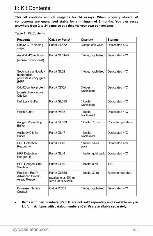
This kit contains enough reagents for 24 assays. When properly stored, kit
components are guaranteed stable for a minimum of 6 months. You can assay
anywhere from 2 to 24 samples at a time for your own convenience.
Table 1: Kit Contents
• Items with part numbers (Part #) are not sold separately and available only in
kit format. Items with catalog numbers (Cat. #) are available separately.
Reagents Cat. # or Part # * Quantity Storage
Cdc42-GTP binding strips
Part # GL572 3 strips of 8 wells Desiccated 4°C
Anti-Cdc42 antibody
(mouse monoclonal)
Part # GL514B
1 tube, lyophilized Desiccated 4°C
Secondary antibody - horseradish peroxidase conjugate (HRP)
Part # GL02 1 tube, lyophilized Desiccated 4°C
Cdc42 control protein
(constitutively active Cdc42)
Part # CDCA
3 tubes, lyophilized
Desiccated 4°C
Cell Lysis Buffer Part # GL535 1 bottle, lyophilized
Desiccated 4°C
Wash Buffer Part # PE38 1 bottle, lyophilized
Desiccated 4°C
Antigen Presenting Buffer
Part # GL539 1 bottle, 10 ml Room temperature
Antibody Dilution Buffer
Part # GL47 1 bottle, lyophilized
Desiccated 4°C
HRP Detection Reagent A
Part # GL43 1 tablet, silver pack
Desiccated 4°C
HRP Detection Reagent B
Part # GL44 1 tablet, gold pack Desiccated 4°C
HRP Reagent Stop Solution
Part # GL80 1 bottle, 8 ml 4°C
Precision Red™ Advanced Protein Assay Reagent
Part # GL550
(available as 500 ml size Cat. # ADV02)
1 bottle, 30 ml Room temperature
Protease Inhibitor Cocktail
Cat. # PIC02 1 tube, lyophilized Desiccated 4°C
II: Kit Contents
cytoskeleton.com Page 6
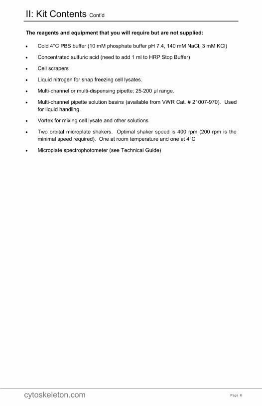
The reagents and equipment that you will require but are not supplied:
• Cold 4°C PBS buffer (10 mM phosphate buffer pH 7.4, 140 mM NaCl, 3 mM KCl)
• Concentrated sulfuric acid (need to add 1 ml to HRP Stop Buffer)
• Cell scrapers
• Liquid nitrogen for snap freezing cell lysates.
• Multi-channel or multi-dispensing pipette; 25-200 µl range.
• Multi-channel pipette solution basins (available from VWR Cat. # 21007-970). Used
for liquid handling.
• Vortex for mixing cell lysate and other solutions
• Two orbital microplate shakers. Optimal shaker speed is 400 rpm (200 rpm is the
minimal speed required). One at room temperature and one at 4°C
• Microplate spectrophotometer (see Technical Guide)
II: Kit Contents Cont’d

cytoskeleton.com Page 7
Many of the kit components are provided in lyophilized form. Prior to beginning the
assay, you will need to reconstitute several components as shown in Table 2.
Table 2: Component Storage and Reconstitution
III: Reconstitution and Storage of Components
Kit Component Reconstitution Storage
Cdc42-GTP binding strips
Keep the strips in the sealed bag with desiccant at all times. Reconstitution is not necessary prior to the start of the assay. If detached, pellets should be tapped to the bottom of the well prior to resuspension.
Store desiccated at 4°C
Anti-Cdc42 antibody Centrifuge briefly to collect the pellet to the bottom of the tube. Dissolve the powder in 100 µl of PBS.
Store at 4°C
Secondary antibody HRP
Centrifuge briefly to collect the pellet to the bottom of the tube. Dissolve the powder in 80 µl of PBS. Do not use sodium azide in combination with this antibody as it will inactivate the HRP.
Store at 4°C
Cdc42 control protein
This is resuspended in 500 ul of lysis buffer just prior to use. See Table 4. Each tube is good for one experiment.
Use within 15 min after reconstitution
Cell Lysis Buffer Reconstitute in 30 ml of Milli-Q water.
This solution may take 5-10 min to resuspend. Use a 10 ml pipette to thoroughly resuspend the buffer.
Store at 4°C
Wash Buffer Reconstitute in 1 L of Milli-Q water.
This solid will take 45-60 min to resuspend. A magnetic stir bar and stir plate can be used to help resuspension.
Store at room temperature
Antigen Presenting Buffer
No reconstitution necessary. Store at room temperature
Antibody Dilution Buffer
Reconstitute in 15 ml of Milli-Q water. Store at 4°C
HRP Detection Reagent A
Resuspend tablet in 10 ml sterile distilled water.
Aliquot into 12 x 0.8 ml volumes.
Store at -70°C
NOTE: Do not store at –20°C
HRP Detection Reagent B
Resuspend tablet in 10 ml sterile distilled water.
Aliquot into 12 x 0.8 ml volumes.
Store at -70°C
NOTE: Do not store at –20°C
HRP Stop Solution Carefully add 1 ml of concentrated sulfuric acid (18 M) to HRP Stop Solution. Check the box on the top of the bottle to indicate acid has been added.
Store at 4°C
Precision Red™
Advanced Protein
Assay Reagent
No reconstitution necessary. Store at room temperature
Protease Inhibitor
Cocktail Reconstitute in 1 ml of dimethyl sulfoxide (DMSO) for
100X stock. Store at 4°C. The cocktail will freeze at 4°C

cytoskeleton.com Page 8
First time users should read the G-LISA Technical Guide, especially the description of
lysate preparation which is critical for the success of the G-LISA assay. The G-LISA® kit
uses 50 µl of lysate (0.25-1 mg/ml lysate protein concentration) per assay. To keep the
assay in the linear range, we highly recommend using a lysate concentration of 0.25 - 1
mg/ml for the Cdc42 G-LISA®.
A) Growth and Treatment of Cell Lines
Cells should be plated and grown to desired confluency in appropriate culture conditions
(consult literature for particular cell line). Confluent cells can be used experimentally,
including for transfection, RNA interference, or serum-starvation, if appropriate. Prior to
Cdc42 stimulation, cells should be kept in a “controlled state” via serum starvation so that
basal Cdc42 activity is low. Optimal confluency prior to serum starvation and GTPase
activation varies by cell line and should be determined empirically. Upon stimulation,
Cdc42 proteins are generally activated very rapidly and transiently (30 s to 30 min).
B) Rapid Processing of Cells to Prepare Lysates
GTP bound (active) Cdc42 is a labile entity and the bound GTP is susceptible to
hydrolysis during and after cell lysis, resulting in Cdc42 inactivation. Rapid processing
(<10 min) on ice is essential for accurate and reproducible results. The following
guidelines should be followed (See Table 3 for preparing reagents needed for cell lysate
preparation).
Washing
1. Retrieve culture dish from incubator, immediately place on ice, aspirate off media,
and wash cells with ice-cold PBS to remove serum proteins.
2. Aspirate off all PBS buffer. This is essential so that the lysis buffer is not diluted.
Cell Lysis
To make lysate at a concentration between 0.25 to 1.0 mg/ml, adjust the amount of lysis
buffer depending on cell and plate type. Empirically determine the exact lysis volumes for
any given cell line. The time period between cell lysis and snap-freezing of lysates is
critically important (no more than 10 min on ice). Take the following precautions:
1. Keep solutions and lysates embedded in ice so that the temperature is below 4°C.
2. Lyse cells in an appropriate volume of ice-cold cell lysis buffer.
3. Immediately harvest cell lysates with a cell scraper.
4. Transfer lysates into pre-labeled and pre-chilled 1.5 ml microfuge tubes on ice.
5. Immediately clarify lysates by centrifugation at 10,000 x g , 4°C for 1 min.
6. Save at least 20 ul of lysate on ice for protein quantification and 50-200 µl for Cdc42
quantification by western blotting.
IV: How to Grow Cells and Prepare Lysates

cytoskeleton.com Page 9
7. Snap-freeze 120 µl aliquots of all cell lysates in liquid nitrogen immediately after
harvest and clarification. Store at -70°C. Lysates can be stored at -70°C for no
longer than 30 days.
C) Measure Lysate Protein Concentration
1. Add 20 µl of each lysate or lysis buffer into disposable 1 ml cuvettes.
2. Add 1 ml of Precision RedTM Advanced Protein Assay Reagent (Part # GL550) to
each cuvette.
3. Incubate for 1 min at room temperature.
4. Blank spectrophotometer with the lysis buffer at 600 nm.
5. Read absorbance of lysates samples.
6. Multiply the absorbance by 5 to obtain the protein concentration in mg/ml (See
Technical Guide).
7. Calculate how much ice-cold lysis buffer is needed to equalize the cell extracts to
give identical protein concentrations in each sample between 0.25 - 1.0 mg/ml. It is
not necessary to equalize protein concentration if the sample variation is less than
10%.
The volume of cold cell lysis buffer to be added to the more concentrated samples can be
calculated as follows:
where A is the higher concentration lysates (mg/ml) and B is the concentration of the
most dilute sample (mg/ml).
Technical Tip
Once an optimal protein concentration for the Cdc42 assay has been determined, it is
easier to equalize all cell lysates to the optimal concentration at the time of lysate
preparation and before freezing lysate aliquots. This eliminates the need to equalize
frozen lysate samples immediately prior to performing the assay and lysates can simply
be thawed and used in the G-LISA.
IV: How to Grow Cells and Prepare Lysates Cont’d
A – B
——— x (volume of A) = __________________ µl
B

cytoskeleton.com Page 10
IV: How to Grow Cells and Prepare Lysates Cont’d
✓ Reagent Preparation
Precision Red™ Advanced
Protein Assay Reagent
Place on the bench and use at room temperature.
1.5 ml microfuge tubes, labeled
and chilled
Use for aliquoting lysates.
Ice buckets Use to pre-chill reagents and scrape cells.
Protease Inhibitor Cocktail Resuspend in 1 ml of dimethyl sulfoxide (DMSO) and keep at
room temperature.
Lysis buffer + protease
inhibitors, ice-cold
a. Empirically determine volume of Lysis Buffer needed per
culture vessel (see Technical Guide). We recommend a
final lysate concentration between 0.25-1.0 mg/ml for initial
experiments.
b. Determine total volume of Lysis Buffer needed by
multiplying the lysis volume per culture vessel (µl) by
number of vessels x 1.3.
c. Aliquot this volume of Lysis Buffer into a clearly labeled tube
and place in ice.
d. Add 10 µl of protease inhibitor cocktail per ml of aliquoted
Lysis Buffer.
e. Mix well and leave on ice.
f. Lysis Buffer needs to be ice cold.
PBS pH 7.2, ice-cold Phosphate-buffered saline is not provided in the kit. It should
be prepared prior to the assay and placed on ice for at least 30
min to ensure that it is ice cold.
Cell scrapers Use to harvest cells.
Liquid nitrogen Use to snap-freeze lysate aliquots.
Table 3: Reagents Needed for Lysate Preparation

cytoskeleton.com Page 11
First time users should read the G-LISA Technical Guide, especially the description of
lysate preparation which is critical for the success of the G-LISA assay. The G-LISA® kit
uses 50 µl of lysate (0.25-1 mg/ml protein concentration) per assay. To keep the assay in
the linear range, we highly recommend using a lysate concentration of 0.25 - 1 mg/ml for
the Cdc42 G-LISA®. The reagents and equipment listed in Table 4 should be prepared
prior to performing the assay.
Table 4: Assay Preparation for G-LISA®
IV: G-LISA Assay Preparation
Reagent Preparation ✓ Samples to be
assayed All experimental samples should be prepared prior to G-LISA assay preparation. The following instructions assume that frozen lysates are being used for the assay. Lysates should remain frozen until indicated in G-LISA assay protocol.
Cdc42-GTP binding strips
Remove strips from 4°C and keep in its protective bag. Place on your bench at room temperature for 30 min.
Do not remove the strips from the bag until immediately prior to the experiment.
Milli-Q water 30 ml placed on ice.
Protease Inhibitor Cocktail
Resuspend in 1 ml of dimethyl sulfoxide (DMSO) and keep at room temperature.
Lysis Buffer Required for blank samples (60 µl per sample) and sample dilutions (if
necessary).
Add 10 µl of protease inhibitor cocktail per ml of Lysis Buffer.
Mix well and leave on ice.
Cdc42 control protein
Dissolve one tube in 500 μl Lysis Buffer and leave on ice. Use within 15
minutes. Discard any unused stock.
Anti-Cdc42 antibody
Have primary antibody stock ready on ice. For each 8-well strip, you will need to mix 25 µl antibody with 500 µl Antibody Dilution Buffer (1:20 dilution). This dilution step should be performed just prior to use as detailed in assay protocol.
Secondary antibody
Have secondary antibody stock ready on ice. For each 8-well strip, you will need to mix 8 µl antibody with 500 µl Antibody Dilution Buffer (1:62.5 dilution). This dilution step should be performed just prior to use as detailed in assay protocol.
Antibody Dilution Buffer
Place reconstituted buffer on the bench and use at room temperature.
Wash Buffer Place on the bench and use at room temperature.
Antigen Presenting Buffer
Place on the bench and use at room temperature.
HRP Detection Reagents A and B
The 0.8 ml aliquots of these reagents can remain at -70°C until secondary antibody addition as detailed in the assay protocol.
HRP Stop Solution
Make sure that the box on top of the bottle is checked, indicating sulfuric acid has been added to the solution.
Place the bottle on your bench and allow to warm to room temperature.
Vortex Used for mixing reagents, it is helpful to keep one close to the assay area.

cytoskeleton.com Page 12
1. Aliquot 120 µl of Lysis Buffer into a labeled microfuge tube. Place on ice. This is the
buffer blank.
2. Mix 30 µl of CDCA (Cdc42 Control Protein) with 90 µl of ice-cold Lysis Buffer in a
labeled microfuge tube. Place on ice. This is the positive control sample. The
positive control is at 2.5ng per well and should be 1-1.2 after background
subtraction. Do not re-use.
3. Remove the number of Cdc42-GTP strips required, place in strip holder, and place
on ice. Return remaining strips to storage.
4. Keep the plate on ice and dissolve the powder in the wells with 100 l ice-cold water.
Detachment of the whilte powder pellet will not affect assay performance. Tap pellets
to the bottom of the wells prior to resuspension.
5. Thaw the snap frozen cell lysates in a ROOM TEMPERATURE water bath.
Immediately place on ice after thawing.
6. If not already equalized, add required amount of ice-cold Lysis Buffer to equalize all
lysate concentrations. Calculate dilution factors required BEFORE thawing lysates.
7. Completely remove the water from the microplate wells as follows:
Vigorously flick the plate to remove solution from each well, followed by a series of 5-
7 vigorous pats onto paper towels. The complete removal of solution from wells
between steps of the G-LISA® is very important to avoid high background readings.
At an absorbance of 490 nm, buffer-only wells should read between 0.10 – 0.4 and
positive control wells should read between 1-1.2 (after subtraction of blank).
8. Return plate back to ice. Immediately add 50 µl of equalized cell lysate to replicate
wells.
9. Pipette 50 µl of buffer blank control into duplicate wells.
10. Pipette 50 µl of Cdc42 positive control into duplicate wells.
11. Immediately place the plate on a cold orbital microplate shaker (400 rpm
recommended, 200 rpm minimum) at 4°C for exactly 15 min.
12. During the incubation, dilute the anti-Cdc42 primary antibody to 1/20 in Antibody
Dilution Buffer (add 25 µl of antibody to every 500 µl Antibody Dilution Buffer). Note:
The final volume of 500 µl is adequate for one strip (8 wells).
13. After 15 min, remove the solution from the wells and wash twice with 200 µl Wash
Buffer at room temperature using a multi-channel pipettor. Do not leave plate
unattended at this time. Vigorously remove the Wash Buffer after each wash as
described in step 7.
14. Place plate on the bench.
IV: G-LISA Assay Protocol

cytoskeleton.com Page 13
15. Immediately pipette 200 µl of room temperature Antigen Presenting Buffer into
each well using a multi-channel pipettor and incubate at room temperature for
exactly 2 min.
16. Vigorously flick out the Antigen Presenting Buffer as described in step 7.
17. Immediately wash the wells three times with 200 µl of room temperature Wash
Buffer, removing Wash Buffer as described in step 7.
18. Add 50 µl of diluted anti-Cdc42 primary antibody to each well and leave the plate on
the orbital microplate shaker (200-400 rpm) at room temperature for 30 min.
19. During primary antibody incubation, dilute secondary HRP labeled antibody to 1/62.5
in Antibody Dilution Buffer (add 8 µl of antibody to every 500 µl Antibody Dilution
Buffer). Note: The final volume of 500 µl is adequate for one strip (8 wells).
20. Vigorously flick out the anti-Cdc42 primary antibody as described in step 7.
21. Immediately wash the wells three times with 200 µl of room temperature Wash
Buffer, removing Wash Buffer as described in step 7.
22. Add 50 µl of diluted Secondary antibody to each well and leave the plate on a
microplate shaker (200–400 rpm) at room temperature for 30 min.
23. During secondary antibody incubation, thaw an aliquot of HRP detection reagents A
and B in a room temperature water bath and remove as soon as they are thawed.
Do not mix.
24. Immediately prior to the end of the secondary antibody incubation, mix HRP
detection reagents A and B in equal volumes (70 µl of A/B mixture per well is
needed). Protect mixture from light. Discard unused solution.
25. Vigorously flick out the secondary antibody as described in step 7.
26. Wash the wells three times with 200 µl of room temperature Wash Buffer as
described in step 7.
27. Pipette 70 µl of the mixed HRP detection reagent into each well and incubate at
37°C for 15 min.
28. Add 140 µl of HRP Stop Buffer to each well.
29. Check that the wells are free of bubbles; if not, remove before continuing.
30. Immediately read the signal by measuring absorbance at 490 nm using a microplate
spectrophotometer. Designate Lysis Buffer only wells as the assay Blank.
IV: Assay Protocol Cont’d

cytoskeleton.com Page 14
V: Troubleshooting
Observation Possible cause Remedy
Weak or no signal in all wells.
1. Slow processing of samples or processing > 4oC.
2. Wells dried out during experiment.
3. Strips became damp during storage. Well contents will appear sticky and opaque.
4. A step or component of the assay was omitted.
5. Insufficient HRP reaction time.
1. Process samples quickly on ice. Snap freeze aliquots.
2. Do not remove the solution in the wells unless the next solution is ready.
3. Store the strips in the desiccant bag with the bag securely sealed. If wells appear sticky and opaque, the strips can no longer be used.
4. Confirm with checklist that all reagents were added.
5. When Cdc42 signal is very low, allow longer HRP development times for a stronger signal. HRP Stop Solution should be added prior to reading at 490 nm.
High signal in all wells.
1. Antibody concentration is too high.
2. Washes were insufficient.
3. HRP reagents stored incorrectly
1. Follow the recommended dilution of antibodies in the manual. If still too high, an antibody titration is necessary to optimize your results.
2. Follow the instructions for washing in the manual.
3. Aliquot HRP solutions and store at –70°C. Do not reuse aliquots once thawed.
Background readings are high (>0.40).
1. Inefficient removal of solutions from
G-LISA ® wells.
2. HRP reagents stored incorrectly
1. Background should read between 0.10 – 0.40. Vigorous flicking and patting of the inverted plate is required to completely remove solutions from the wells after each step is complete. See G-LISA instructional video for details.
2. Aliquot HRP solutions and store at –70°C. Do not reuse aliquots once thawed
Induced sample does not give significant signal increase.
1. Poor inducer activity.
2. Technique not rapid or cold enough.
3. The endogenous GTP-Cdc42 level is too high.
4. Tissue culture cells not correctly serum starved.
5. Temperature of lysis and incubation is not 4oC.
1. Purchase a fresh vial of inducer.
2. Confirm instructions were followed using the Experiment Record Sheet (see Technical Guide).
3. Titrate down the amount of extract to be added.
4. See Technical Guide and references therein for guidance on serum starvation. It is a good idea to stain cells with phalloidin to qualitatively determine success of serum starvation and induction.
5. Lyse cells on ice, keep cell lysis buffer and distilled water on ice.
Significant variation between replicate samples.
1. Incorrect volume of solutions for each step added in the wells.
2. Inaccurate pipetting.
1. Follow the instruction for recommended volume in the manual.
2. A multi-channel pipettor is recommended.
Positive control not working.
1. Positive control protein was stored incorrectly after reconstitution.
2. Positive control resuspended incorrectly
1. Store at 4°C.
2. Use CDCA reconstitution buffer for resuspension.
cytoskeleton.com Page 15
Limited Use Statement
The G-LISA® kits are based on patented technology developed at Cytoskeleton Inc.
(Patent# 7,763,418 B2). The purchase of this product conveys to the buyer the non-
transferable right to use the purchased amount of product and components of product in
research conducted by the buyer. The buyer cannot sell or otherwise transfer this
product or any component thereof to a third party or otherwise use this product or its
components for commercial purposes. Commercial purposes include, but are not limited
to: use of the product or its components in manufacturing; use of the product or its
components to provide a service; resale of the product or its components.
The terms of this Limited Use Statement apply to all buyers including academic and for-
profit entities. If the purchaser is not willing to accept the conditions of this Limited Use
Statement, Cytoskeleton Inc. is willing to accept return of the unused product with a full
refund.
V: Limited Use Statement

cytoskeleton.com Phone: (303) 322.2254 Fax: (303) 322.2257
Customer Service: [email protected]
Technical Support: [email protected]

![Biochem [Gluconeogenesis]](https://static.fdocuments.in/doc/165x107/577c82b31a28abe054b1e4af/biochem-gluconeogenesis.jpg)


![Research Article EVALUATION OF ANTI-INFLAMMATORY … · Percentage inhibition= [(absorbance of blank – absorbance of sample)/(absorbance of blank)]×100 1 In-vitro anti-inflammatory](https://static.fdocuments.in/doc/165x107/5e832a1607bd17145979ab05/research-article-evaluation-of-anti-inflammatory-percentage-inhibition-absorbance.jpg)