BIOACTIVITY-GUIDED ISOLATION OF -...
34
BIOACTIVITY-GUIDED ISOLATION OF AILANTHUS MALABARICA FOR TOXOPLASMA GONDII AND CHOLINESTERASE INHIBITORY ACTIVITIES by LEE YUAN-E Thesis submission in fulfillment of the requirements for the Degree of Master of Science (Pharmacy) March 2017
Transcript of BIOACTIVITY-GUIDED ISOLATION OF -...
BIOACTIVITY-GUIDED ISOLATION OF AILANTHUS MALABARICA FOR TOXOPLASMA GONDII AND CHOLINESTERASE INHIBITORY
ACTIVITIES
by
LEE YUAN-E
Thesis submission in fulfillment of the requirements
for the Degree of
Master of Science (Pharmacy)
March 2017
ii
ACKNOWLEDGEMENT
Firstly, I would like to express my sincere gratitude to my main supervisor
Prof. Chan Kit Lam for the continuous support of my master study and related
research, for his patience, motivation, and immense knowledge. His guidance helped
me in all the time of research and writing of this thesis. I could not have imagined
having a better advisor and mentor for my master study.
Besides that, I would like to express my special thanks to my co-supervisor, Dr.
Vikneswaran Murugaiyah, who has given me generous amount of guidance despite
his busy schedule. Without his support, I would not have fulfilled this dissertation. I
would also like to show my deepest gratitude to Prof. Rahmah Noordin, who as my
co-supervisor has given me full support in completing my research. I would like to
also thank the members of Prof Rahmah's laboratory (Pn. Ezzati Zahidah and En.
Hafiz) who have given me generous help during my research at INFORMM.
I would also like to acknowledge School of Pharmaceutical Sciences and Institute of
Postgraduate Studies (IPS) for giving me the opportunities to pursue my graduate
studies. Besides, I would like to express my gratitude to Graduate Assistance Scheme
(GA), MyBrain 15 from ministry of higher education and BIDP programme from
IPHARM for funding me throughout this research.
Last but not the least, I would like to thank my family: my parents and to my
brothers and friends for supporting me spiritually throughout writing this thesis and
my life in general.
iii
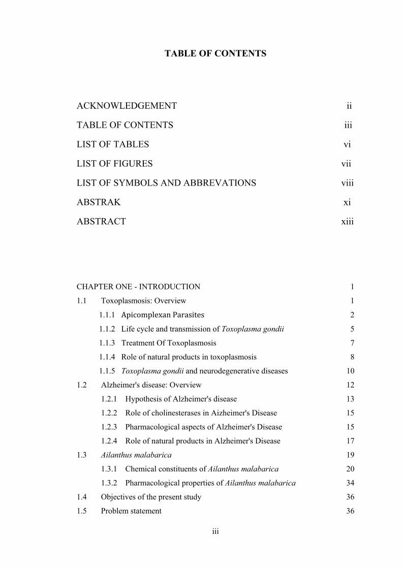
TABLE OF CONTENTS
ACKNOWLEDGEMENT ii
TABLE OF CONTENTS iii
LIST OF TABLES vi
LIST OF FIGURES vii
LIST OF SYMBOLS AND ABBREVATIONS viii
ABSTRAK xi
ABSTRACT xiii
CHAPTER ONE - INTRODUCTION 1
1.1 Toxoplasmosis: Overview 1
1.1.1 ApicomplexanParasites 2
1.1.2 Life cycle and transmission of Toxoplasma gondii 5
1.1.3 Treatment Of Toxoplasmosis 7
1.1.4 Role of natural products in toxoplasmosis 8
1.1.5 Toxoplasma gondii and neurodegenerative diseases 10
1.2 Alzheimer's disease: Overview 12
1.2.1 Hypothesis of Alzheimer's disease 13
1.2.2 Role of cholinesterases in Aizheimer's Disease 15
1.2.3 Pharmacological aspects of Alzheimer's Disease 15
1.2.4 Role of natural products in Alzheimer's Disease 17
1.3 Ailanthus malabarica 19
1.3.1 Chemical constituents of Ailanthus malabarica 20
1.3.2 Pharmacological properties of Ailanthus malabarica 34
1.4 Objectives of the present study 36
1.5 Problem statement 36
iv
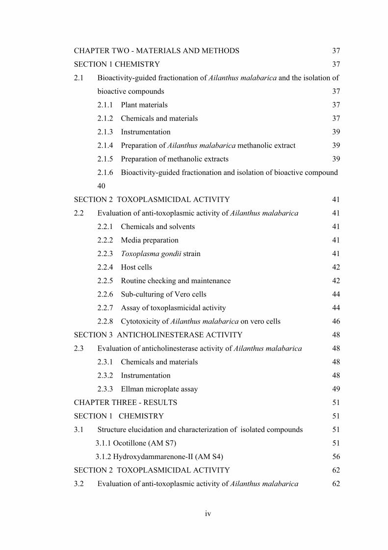
CHAPTER TWO - MATERIALS AND METHODS 37
SECTION 1 CHEMISTRY 37
2.1 Bioactivity-guided fractionation of Ailanthus malabarica and the isolation of
bioactive compounds 37
2.1.1 Plant materials 37
2.1.2 Chemicals and materials 37
2.1.3 Instrumentation 39
2.1.4 Preparation of Ailanthus malabarica methanolic extract 39
2.1.5 Preparation of methanolic extracts 39
2.1.6 Bioactivity-guided fractionation and isolation of bioactive compound
40
SECTION 2 TOXOPLASMICIDAL ACTIVITY 41
2.2 Evaluation of anti-toxoplasmic activity of Ailanthus malabarica 41
2.2.1 Chemicals and solvents 41
2.2.2 Media preparation 41
2.2.3 Toxoplasma gondii strain 41
2.2.4 Host cells 42
2.2.5 Routine checking and maintenance 42
2.2.6 Sub-culturing of Vero cells 44
2.2.7 Assay of toxoplasmicidal activity 44
2.2.8 Cytotoxicity of Ailanthus malabarica on vero cells 46
SECTION 3 ANTICHOLINESTERASE ACTIVITY 48
2.3 Evaluation of anticholinesterase activity of Ailanthus malabarica 48
2.3.1 Chemicals and materials 48
2.3.2 Instrumentation 48
2.3.3 Ellman microplate assay 49
CHAPTER THREE - RESULTS 51
SECTION 1 CHEMISTRY 51
3.1 Structure elucidation and characterization of isolated compounds 51
3.1.1 Ocotillone (AM S7) 51
3.1.2 Hydroxydammarenone-II (AM S4) 56
SECTION 2 TOXOPLASMICIDAL ACTIVITY 62
3.2 Evaluation of anti-toxoplasmic activity of Ailanthus malabarica 62
v
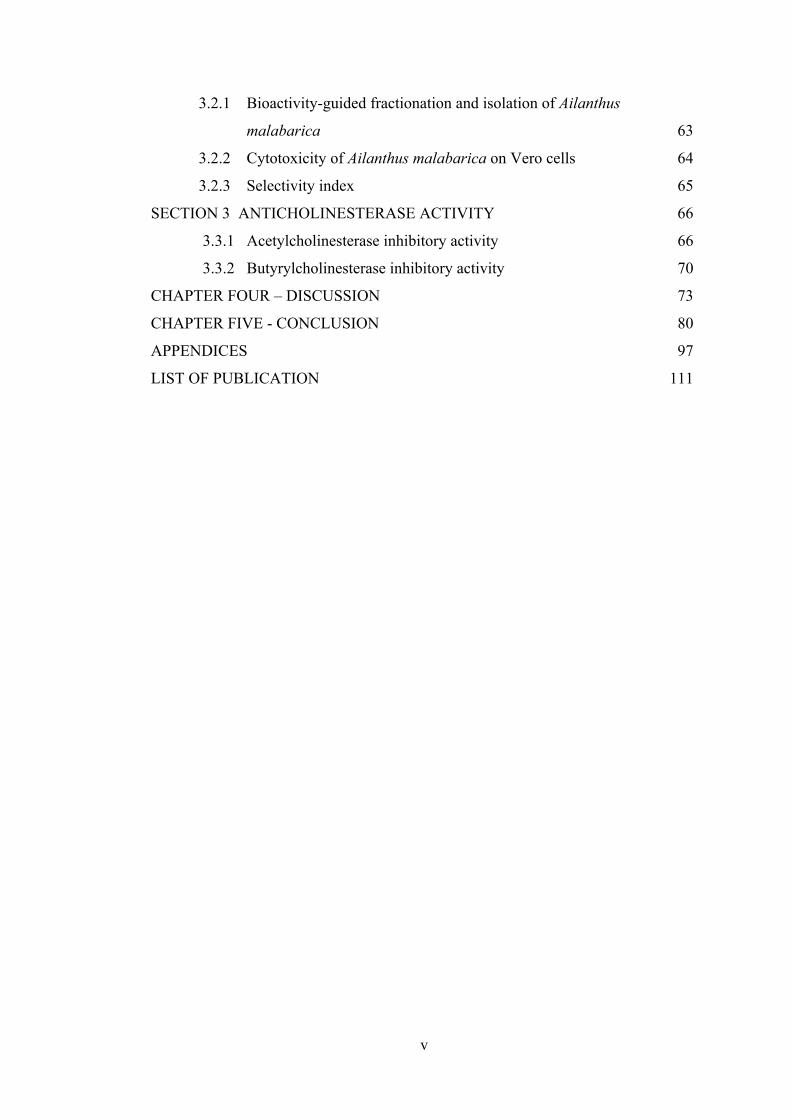
3.2.1 Bioactivity-guided fractionation and isolation of Ailanthus
malabarica 63
3.2.2 Cytotoxicity of Ailanthus malabarica on Vero cells 64
3.2.3 Selectivity index 65
SECTION 3 ANTICHOLINESTERASE ACTIVITY 66
3.3.1 Acetylcholinesterase inhibitory activity 66
3.3.2 Butyrylcholinesterase inhibitory activity 70
CHAPTER FOUR – DISCUSSION 73
CHAPTER FIVE - CONCLUSION 80
APPENDICES 97
LIST OF PUBLICATION 111
vi
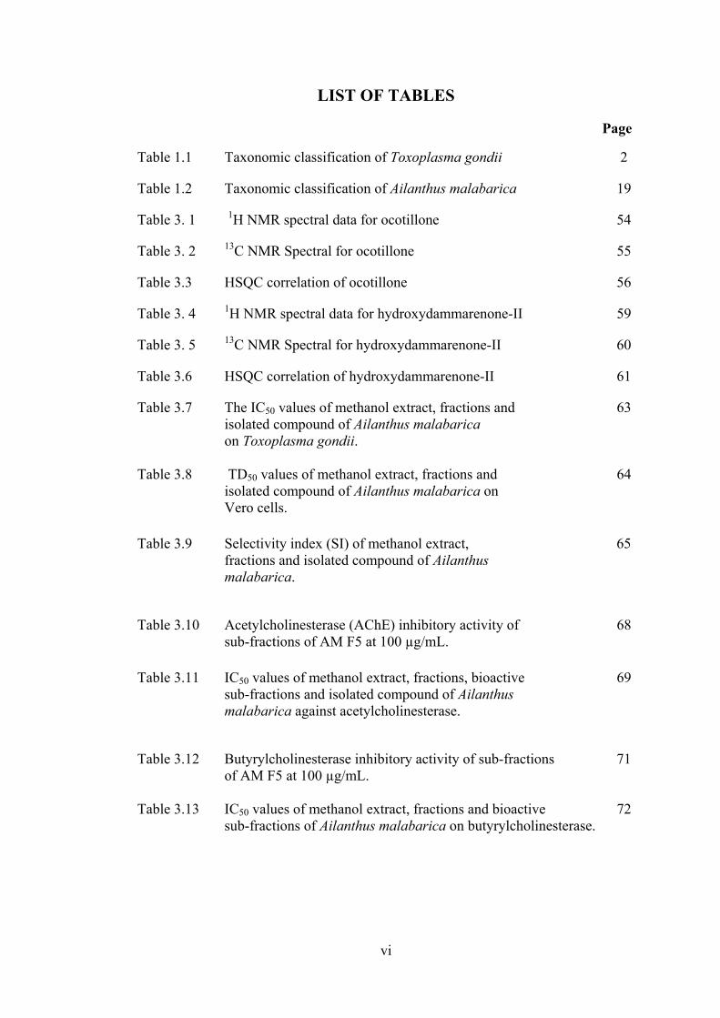
LIST OF TABLES
Page
Table 1.1 Taxonomic classification of Toxoplasma gondii 2
Table 1.2 Taxonomic classification of Ailanthus malabarica 19
Table 3. 1 1H NMR spectral data for ocotillone 54
Table 3. 2 13C NMR Spectral for ocotillone 55
Table 3.3 HSQC correlation of ocotillone 56
Table 3. 4 1H NMR spectral data for hydroxydammarenone-II 59
Table 3. 5 13C NMR Spectral for hydroxydammarenone-II 60
Table 3.6 HSQC correlation of hydroxydammarenone-II 61
Table 3.7 The IC50 values of methanol extract, fractions and 63 isolated compound of Ailanthus malabarica on Toxoplasma gondii.
Table 3.8 TD50 values of methanol extract, fractions and 64 isolated compound of Ailanthus malabarica on Vero cells. Table 3.9 Selectivity index (SI) of methanol extract, 65 fractions and isolated compound of Ailanthus malabarica.
Table 3.10 Acetylcholinesterase (AChE) inhibitory activity of 68 sub-fractions of AM F5 at 100 µg/mL. Table 3.11 IC50 values of methanol extract, fractions, bioactive 69 sub-fractions and isolated compound of Ailanthus malabarica against acetylcholinesterase.
Table 3.12 Butyrylcholinesterase inhibitory activity of sub-fractions 71 of AM F5 at 100 µg/mL. Table 3.13 IC50 values of methanol extract, fractions and bioactive 72 sub-fractions of Ailanthus malabarica on butyrylcholinesterase.
vii
LIST OF FIGURES
Page
Figure 1.1 The morphology of apicomplexan parasites. There are a 4 collection of organelles found specifically in this phylum. The apicoplast is located next to the Golgi body. Figure 1.1 Life cycle of Toxoplasma gondii. The figure was adapted 7 from Hill & Dubey, 2002 Figure 2.1 Isolation scheme from Ailanthus malabarica 38 Figure 2.2 Outline of cell passage for Vero cells 43 43 Figure 2.3 Outline of anti-toxoplasmicidal activity of Ailanthus 46 malabarica on Vero cells 46 Figure 3. 1 Chemical structure of ocotillone 53 Figure 3. 2 Chemical structure of hydroxydammarenone-II 58 Figure 3. 3 Percentage of inhibition of methanol extract of 62 Ailanthus malabarica and clindamycin on Toxoplasma gondii. Figure 3.4: Acetylcholinesterase (AChE) inhibitory activity 66 of Ailanthus malabarica methanol extract and its fractions at 100 µg/mL. Figure 3.5 Butyrylcholinesterase (BuChE) inhibitory activity 70 of Ailanthus malabarica methanol extract and its fractions at 100 µg/mL.
viii
LIST OF SYMBOLS AND ABBREVATION
% Percentage
°C Degree celcius
α Alpha
β Beta
µ Micro
Aβ Amyloid beta
ACh Acetylcholine
AChE Acetylcholinesterase
Abs Absorbance
AD Alzheimer's disease
AIDS Acquired immunodeficiency syndrome
BuChE Butyrylcholinesterase
DEPTQ Distorsionless enhancement by polarization transfer including
the detection of quaternary nuclei
DMEM Dulbecco's Modified Eagle Medium
DMSO Dimethyl sulfoxide
DTNB 5,5'-Dithiobis(2-nitrobenzoic acid)
ESI Electrospray ionization
ix
FBS Fetal bovine serum
FDA Food and Drug Administration
g Gram
GM Growth medium
h Hour
H Hydrogen
HMBC Heteronuclear Multiple Bond Coherence
HSQC Heteronuclear Single Quantum Coherence
IC50 Inhibitory concentration of 50 %
IR Infrared
mg Milligram
mg/mL Milligram per millilitre
mins Minutes
mL Millilitre
MS Mass spectrometry
MTS 3-(4,5-Dimethythiazol-2-yl)-5-(3-carboxymethoxyphenyl)-2-
sulfophenyl)-2H-tetrazolium
nm Nanometer
NMR Nuclear magnetic resonance
x
PBS Phosphate buffered saline
Rf Retention factor
SEM Standard error of means
SI Selectivity index
TLC Thin layer chromatography
WHO World Health Organization
xi
PEMENCILAN BERPANDU BIO-AKTIVITI AILANTHUS MALABARICA
UNTUK AKTIVITI PERENCATAN TOXOPLASMA GONDII DAN
KOLINESTERASE
ABSTRAK
Tumbuh-tumbuhan daripada Ailanthus genus diedarkan secara meluas di
seluruh Asia dan memainkan peranan penting sebagai tumbuhan perubatan. Aktiviti-
aktiviti biologi spesies Ailanthus telah dikaji secara meluas dan didapati mempunyai
aktiviti seperti anti-protozoa, anti-virus, anti-bakteria dan anti-fungal. Kajian ini
bertujuan untuk menilai perencatan aktiviti Toxoplasma gondii dan enzim
kolinesterase, serta analisis fitokimia untuk ekstrak metanol Ailanthus malabarica.
Dalam kajian ini, ekstrak metanol Ailanthus malabarica disiasat bagi aktiviti-aktiviti
anti-parasit terhadap Toxoplasma gondii. IC50 daripada anti-parasit aktiviti Ailanthus
malabarica adalah 11.12 μg/mL. Ailanthus malabarica diperingkatkan dengan
mengguna kromatografi resin untuk menghasilkan lima fraksi. Fraksi yang paling
aktif ialah AM F5 dengan IC50 9.98 μg/mL. Selepas itu, AM F5 diperingkatkan
menggunai kromatografi silika gel untuk menghasilkan ocotillone. IC50 untuk
ocotillone adalah 16.18 μg/mL. Ailanthus malabarica kemudian diuji pada sel Vero
dalam keadaan in vitro dengan menggunakan MTS assay untuk mengaji tahap toksik.
Ekstrak dan fraksi telah didapati tidak toksik kepada sel-sel (LC50> 20 μg / mL).
AM F3, AM F4 dan ocotillone menunjukkan tahap toksik yang rendah pada nilai
LC50 247 μg/mL, 238 μg/mL dan 223 μg/mL masing-masing. Tahap toksik AM F5
adalah tinggi berbanding dengan fraksi lain dengan nilai 49.87 μg/mL. Indeks
pemilihan (SI) daripada ocotillone menunjukkan pemilihan tertinggi iaitu 13.83.
xii
Disebabkan Toxoplasma gondii adalah parasit neurotropic, jangkitan telah dibukti
akan menyebabkan perubahan tingkah laku dan gejala neurocognitive dalam
mamalia. Dalam kajian berikutnya, Ailanthus malabarica telah diuji untuk
perencatan enzim kolinesterase. Pemeringkatan berulang AM F5 menghasilkan
sebatian lain, AM S4 (hydroxydammarenone-II). Ekstra dan fraksi telah diuji untuk
perencatan kedua-dua acetylcholinesterase (AChE) dan butyrylcholinesterase
(BuChE). Pada 100 μg/mL, AM S4 (hydroxydammarenone-II), AM S6 dan AM S8
daripada AM F5 menunjukkan perencatan melebihi 50 %. IC50 daripada ketiga-tiga
pecahan ini adalah 12.54 μg/mL, 10.00 μg/mL dan 76.05 μg/mL masing-masing.
Sebaliknya, untuk perencatan BuChE aktiviti, AM S10 dan AM S14 menunjukkan
perencatan yang melebihi 50% apabila diuji di kepekatan 100 μg/mL. IC50 AM S10
dan AM S14 pada BuChE adalah 2.98 μg/mL dan 25.31 μg/mL masing-masing.
AM F5 yang memiliki kesan anti- toxoplasmicidal tertinggi dinilai kepada aktiviti
anti- cholinesterase . Tetapi, perencatan enzim kolinesterase AM F5 adalah kira-kira
13 %. Ocotillone yang didapati merencat pertumbuhan Toxoplasma gondii
menunjukkan perencatan di acetylcholinesterase pada 34 % dan 18 % pada
butyrylcholinesterase . Peratusan perencatan adalah agak rendah berbanding dengan
lain-lain pecahan. Struktur AM S4 dan AM S7 telah ditentukan sebagai
hydroxydammarenone-II dan ocotillone masing-masing dengan menggunakan NMR ,
IR dan spektroskopi jisim. Sebagai kesimpulan, hanya AM S4
(hydroxydammarenone -II) menunjukkan aktiviti anti-acetylcholinesterase yang aktif
pada 12 μg/mL. Bagaimanapun , sub-fraksi ini perlu diuji ke atas aktiviti anti-
toxoplasma untuk menentukan kesan terhadap Toxoplasma gondii.
xiii
BIOACTIVITY-GUIDED ISOLATION OF AILANTHUS MALABARICA FOR
TOXOPLASMA GONDII AND CHOLINESTERASE INHIBITORY
ACTIVITY
ABSTRACT
The plants of the genus Ailanthus are distributed widely over Asia and play a
significant role as medicinal plant. Biological activities of Ailanthus species has been
studied extensively and found to possess activities like anti-protozoal, anti-viral, anti-
bacterial and anti-fungal. The present study aimed to evaluate the anti-toxoplasma
and cholinesterase inhibitory activity, as well as phytochemical analysis of
methanolic extract of Ailanthus malabarica. In this work, the methanolic extract of
Ailanthus malabarica was investigated for the anti-parasitic activities against
Toxoplasma gondii. The IC50 of Ailanthus malabarica on the anti-parasitic activity
was 11.12 µg/mL. Ailanthus malabarica was further fractionated using resin column
chromatography to yield five fractions. The most active fraction was AM F5 with
IC50 of 9.98 µg/mL. AM F5 was then further subjected to silica gel column
chromatography to yield ocotillone. The IC50 of ocotillone was 16.18 µg/mL.
Ailanthus malabarica was then tested on Vero cell line in an in vitro MTS assay to
study for the cytotoxicity of the plant. The crude extract as well as the sub-fractions
were not toxic to the cells (TD50 > 20 µg/mL). AM F3, AM F4 and ocotillone
showed little cytotoxicity at TD50 value of 247 µg/mL, 238 μg/mL and 223 μg/mL
respectively. AM F5 was the most toxic at TD50 of 49.87 µg/mL. The selectivity
index (SI) of ocotillone showing the highest selectivity was 13.83. As Toxoplasma
gondii was a neurotropic parasite, the infection has been shown to cause behavioral
xiv
changes and neurocognitive symptoms in mamalian hosts. In subsequent study,
Ailanthus malabarica was tested for cholinesterase enzyme inhibition. Fraction AM
F5 which was found active in anti-toxoplasmic activity was subjected to anti-
cholinesterase inhibitory assay. Repeated fractionation of AM F5 yielded another
compound, AM S4 (hydroxydammarenone-II). The crude extract and the sub-
fractions were tested for inhibition of both acetylcholinesterase (AChE) and
butyrylcholinesterase (BuChE). At 100 µg/mL, AM S4 (hydroxydammarenone-II),
AM S6 and AM S8 from AM F5 showed above 50 % of inhibition on AChE. The
IC50 of these three sub-fractions were 12.54 µg/mL, 10.00 µg/mL and 76.05 µg/mL
respectively. On the other hand, for inhibition of BuChE, AM S10 and AM S14
showed inhibition above 50 % when tested at the concentration of 100 µg/mL. The
IC50 of AM S10 and AM S14 on BuChE were 2.98 µg/mL and 25.31 µg/mL
respectively. AM F5 which possessed highest anti-toxoplasmicidal effect was
evaluated on the anti-cholinesterase activity. However, the inhibition on
cholinesterase enzymes of AM F5 was about 13 %. Ocotillone which was found to
inhibit the growth of Toxoplasma gondii showed inhibition at acetylcholinesterase at
34 % and 18 % on butyrylcholinesterase. The percentage of inhibition was
comparatively lower when compared to other sub-fractions. AM S4 and AM S7 were
structurally elucidated as hydroxydammarenone-II and ocotillone respectively by
using NMR, IR and mass spectrometer. Only AM S4 (hydroxydammarenone-II)
showed potent anti-acetylcholinesterase activity at 12 µg/mL. However, this sub-
fraction was not tested on anti-toxoplasmicidal activity due to laboratory limitations.
In conclusion, AM F5 is potent against anti-toxoplasmic and cholinesterase
inhibitory activity, however further study should be evaluated on the potency of
hydroxydamarenone-II on anti-toxoplasmic activity to further confirm that it is the

xv
compound which is responsible to inhibit both toxoplasma and cholinesterase
activities.
1
CHAPTER ONE
INTRODUCTION
1.1 Toxoplasmosis: Overview
Toxoplasmosis is a disease caused by an obligate intracellular parasite, Toxoplasma
gondii. The discovery of Toxoplasma gondii can be dated back to 100 years ago, by
scientists in North Africa and in Brazil. Toxoplasma gondii is capable of infecting all
warm-blooded animals including humans. This protozoan parasite infects up to a
third of the world's population and mainly through consumption of undercooked food
containing tissue cysts or by ingestion of food and water that is contaminated with
oocysts shed by cats (Bowie et al., 1997). Most of the infections of Toxoplasma in
humans are asymptomatic, however, there are times where the infections lead to
devastating disease. Encephalitis causes severe damage, making it the most
important manifestation of toxoplasmosis in immune suppressed patients. The
infection may be found in any organs, causing symptom-like headache,
disorientation and drowsiness. Apart from that, toxoplasmosis can be acquired
congenitally and post-natally. Sero-negative pregnant women may be at risk of
contracting Toxoplasma gondii infection. This congenital infection may cause the
fetuses to be infected and develop severe complications and sometimes causing death.
The prevalence varied from place to place where it is estimated that 16-40 % of the
population was infected in the United States and the United Kingdom (Hill & Dubey,
2002). In Malaysia, the epidemiological surveys indicate that Toxoplasma antibody
is found in all ethnic groups with Malays having the highest percentage at 33 %,
followed by Indians at 29 % and Chinese at 18 % (Tee et al., 1998).
2
1.1.1 Apicomplexan parasites
Parasites refer to organisms that depend on the hosts to survive and at the same time
causing diseases to the hosts. These pathogenic parasites can be harmful to human as
well as some economically important animal species. One of the most important
groups of parasites is the apicomplexan parasites (Table 1) that include Plasmodium
spp., the causative agent of malaria, Cryptosporidium, an opportunistic intestinal
pathogen and also Toxoplasma gondii. Infection by protozoan parasites of the
phylum Apicomplexa sometimes leads to mortality of human and livestock.
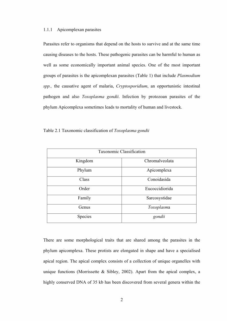
Table 2.1 Taxonomic classification of Toxoplasma gondii
There are some morphological traits that are shared among the parasites in the
phylum apicomplexa. These protists are elongated in shape and have a specialised
apical region. The apical complex consists of a collection of unique organelles with
unique functions (Morrissette & Sibley, 2002). Apart from the apical complex, a
highly conserved DNA of 35 kb has been discovered from several genera within the
Taxonomic Classification
Kingdom Chromalveolata
Phylum Apicomplexa
Class Conoidasida
Order Eucoccidiorida
Family Sarcosystidae
Genus Toxoplasma
Species gondii
3
phylum Apicomplexa including Toxoplasma (Beckers et al., 1995). This DNA
resides in an organelle called the apicoplast. This plastid-like organelle is found in
nearly all members of the phylum except for Cryptosporidium spp. (Zhu et al., 2000).
The apicoplast is believed to have its origins in a secondary endosymbiotic event.
This organelle has been investigated extensively since its discovery. It was found that
in Toxoplasma gondii, the inhibition of apicoplast DNA synthesis blocked the
replication of parasite. This suggests that apicoplast plays an essential role in the
survival of the parasites (Fichera & Roos, 1997). Apart from that, the chloroplast-
derived organelles were not found in humans as well as other mammals, thus
suggesting that the apicoplast might be useful in the study of parasite-specific targets
in the drug development against Apicomplexa. Toxoplasma gondii has been an
important model in the studies of apicomplast development and function.
4
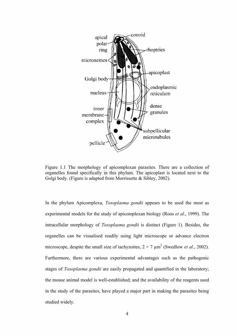
Figure 1.1 The morphology of apicomplexan parasites. There are a collection of organelles found specifically in this phylum. The apicoplast is located next to the Golgi body. (Figure is adapted from Morrissette & Sibley, 2002).
In the phylum Apicomplexa, Toxoplasma gondii appears to be used the most as
experimental models for the study of apicomplexan biology (Roos et al., 1999). The
intracellular morphology of Toxoplasma gondii is distinct (Figure 1). Besides, the
organelles can be visualised readily using light microscope or advance electron
microscope, despite the small size of tachyzoites, 2 × 7 µm2 (Swedlow et al., 2002).
Furthermore, there are various experimental advantages such as the pathogenic
stages of Toxoplasma gondii are easily propagated and quantified in the laboratory;
the mouse animal model is well-established; and the availability of the reagents used
in the study of the parasites, have played a major part in making the parasites being
studied widely.
5
1.1.2 Life cycle and transmission of Toxoplasma gondii
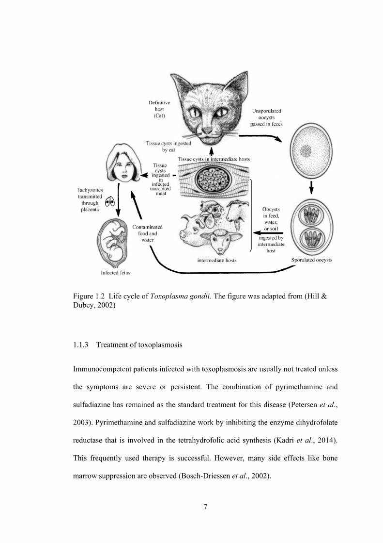
Toxoplasma gondii has a complex life cycle. Figure 2 illustrated the life cycle and
transmission route of Toxoplasma gondii. This parasite has a heteroxenous life cycle
and it can infect a wide range of warm-blooded host which includes human. An
example of a definitive host is the domestic cats. The infectious stages of the parasite
can be divided into three stages, namely the tachyzoites, bradyzoites and sporozoites.
Tachyzoites and bradyzoites are contained in tissue cysts while sporozoites are in
sporulated oocysts. All these three stages are infectious. (Hill & Dubey, 2002)
The sexual cycle occurs only in the intestine of cats. Replication occurs which leads
to the production of oocysts. This will then result in the shedding of oocysts in the
faeces of cats for 7-21 days. Cats are able to shed millions of oocysts even if only
one bradyzoite is ingested. Three infectious stages of Toxoplasma gondii ingested
will lead to shedding of oocysts in cats (Dubey et al., 1970). Sporulation will take
place and these infectious oocysts containing sporozoites will lead to tachyzoite stage
upon ingestion by mammals including human. Infection of the parasites on human
might also occur through the oral ingestion of tissue cysts found in raw or
undercooked meat.
Tachyzoites are oval or crescent in shape and are about 2-4 µm wide and 4-8 µm
long (Montoya & Liesenfeld, 2004). Parasites in this stage multiply rapidly and enter
the nucleated cells by active penetration to form cytoplasmic vacuole (Dobrowolski
& Sibley, 1996). Repeated replication of the tachyzoites will cause the disruption of
host cells. The tachyzoites will then enter and infect the central nervous system, the
6
eye, placental and skeletal tissues through the bloodstream. Replication of the
parasites will cause cell death and neighbouring cells will be invaded rapidly.
Inflammatory response and tissue destruction will be triggered in the tachyzoite stage,
which lead to clinical manifestations of diseases. Immune response triggered will act
as a pressure to transform tachyzoites into bradyzoites to form cysts.
The difference between the structure of bradyzoites and tachyzoites is that
bradyzoites have a nucleus situated toward the posterior end, while tachyzoites has a
nucleus found in the central (Dubey et al., 1998). Cysts are the infective stages for
both intermediate and definitive hosts. They are similar to tachyzoites
morphologically but multiply slowly and express their function differently.
Thousands of bradyzoites can be found in tissue cysts from brain cells, skeletal and
heart muscles. After ingestion of tissue cysts by the host, proteolytic enzymes in the
stomach and small intestine will dissolve the cyst wall. Bradyzoites released from
cysts will then enter the small intestine and initiate the generation of the parasites
(Dubey et al., 1998).
Infection of Toxoplasma gondii in human (Figure 2) often occurs through oral route.
Inappropriate handling of cat litters, eating undercooked meat and drinking of
contaminated water are a few common routes of transmission of the parasites to
human. Apart from that, infection might also occur congenitally. Congenital
infection occurs when a woman becomes infected during her pregnancy.
Transmission of Toxoplasma gondii might occur through blood transfusions and
organ transplants.
7
Figure 1.2 Life cycle of Toxoplasma gondii. The figure was adapted from (Hill & Dubey, 2002)
1.1.3 Treatment of toxoplasmosis
Immunocompetent patients infected with toxoplasmosis are usually not treated unless
the symptoms are severe or persistent. The combination of pyrimethamine and
sulfadiazine has remained as the standard treatment for this disease (Petersen et al.,
2003). Pyrimethamine and sulfadiazine work by inhibiting the enzyme dihydrofolate
reductase that is involved in the tetrahydrofolic acid synthesis (Kadri et al., 2014).
This frequently used therapy is successful. However, many side effects like bone
marrow suppression are observed (Bosch-Driessen et al., 2002).
8
Toxoplasma gondii infection can also be acquired by pregnant women. This may
happen during gestation and the infection can be transmitted to the foetus (Goldstein,
2008). Management of maternal and foetal infection varies between different
countries. Spiramycin or combination of pyrimethamine and sulfadiazine are often
used depending on the trimester of pregnancy (Rajapakse et al., 2013). Spiramycin is
a macrolide antibiotic and is often used during the first 18 weeks of gestation. It was
reported that this treatment is able to reduce the vertical transmission of the parasite
(Couvreur et al., 1988). However, spiramycin is not able to treat the infection in the
foetus due to the inability of the drug to cross the placenta. Combination of
pyrimethamine, sulfadiazine and folinic acid is used to treat pregnant women who
are infected after 18 weeks of gestation and also for those confirmed foetal infection.
Pyrimethamine should not be used during the first trimester of pregnancy due to the
teratogenic potential. The drug produces dose-related depression of the bone marrow
(Goldstein, 2008). This anti-toxoplasma treatment should be continued throughout
the pregnancy period. Consequently, the patients who received pyrimethamine
treatment should have their complete blood counts closely monitored.
1.1.4 Role of natural products in toxoplasmosis
Natural products continued to play a significant role in the drug discovery for the
treatment of various human diseases. Natural products are thought to contain the
desired properties of potency and often considered to have low toxicity. The use of
medicinal plants for the treatment of parasitic diseases is well documented since
ancient times. For example, Cinchona succiruba has been used to treat the infection
of malaria for centuries (Kaur et al., 2009).

9
Numerous natural products have been studied extensively for potential source of
new agents to treat toxoplasmosis. Among them, Artemisia annua L., an annual herb
belonging to the family Asteraceae, has been reported to inhibit Toxoplasma
replication in vitro (Jones-Brando, et al. 2006; Ke et al., 1990). The active compound,
artemisinin isolated from the plant was initially developed for anti-malaria activity
(de Ridder et al., 2008).
Medicinal herbs from South Korea were also tested for the anti-protozoal activity
against cultures of Toxoplasma gondii. In the study, it was found that the alcohol
extracts of Torilis japonica and Sophora flavescens demonstrated the best inhibition
of Toxoplasma gondii among all the medicinal herbs tested (Youn et al., 2003).
Oleuropein isolated from Fraxinus rhychophylla was investigated for its activity
against Toxoplasma gondii both in vitro and in vivo. It was reported that oleuropein
showed anti-toxoplasma activity in the infected mice (Jiang et al., 2008). Meanwhile
15 methanolic extracts of Korean traditional medicine were tested on the effect on
Toxoplasma gondii. Among them, it was reported that Zingiber officinale possesses
high anti-toxoplasma activity as well as low cytotoxicity (Choi et al., 2008).
Apart from that, maslinic acid (2α,3β-dihydroxyolean-12-en-28-oic acid) found in
the fruits of the olive (Olea europaea) was reported to have the ability of blocking
the entry of Toxoplasma gondii into the cells. This compound is present in many
plants and is found mainly in the leaves and fruits of Olea europaea (De Pablos et al.,
2010). Secrurinine produced by Securinega suffructicosa was evaluated against
Toxoplasma gondii. This widely used traditional Chinese medicine was found to
10
inhibit the growth of the parasite in vitro. This compound was also less toxic when
compared to pyrimethamine (Holmes et al., 2011). Another compound isolated from
the Chinese medicine was ginkgolic acid. This compound is found in Ginkgo biloba,
a chinese herb which was reported to possess anti-microbial properties and anti-
tumor properties (Yang et al., 2004). In the study, it was reported that ginkgolic acids
significantly inhibit Toxoplasma gondii DNA and protein synthesis at low
concentrations (Chen et al., 2008).
In Malaysia, crude methanolic extract and sub-fractions of Tinospora crispa were
investigated on against Toxoplasma gondii. This alkaloid-rich plant was reported to
show potential anti-toxoplasma activity (Lee et al., 2012). Eurycoma longifolia Jack
from the Simaroubaceae family has also been tested on the activity against
Toxoplasma gondii. It was reported that its extracts and sub-fractions significantly
inhibit the growth of Toxoplasma gondii at low concentrations (Kavitha et al., 2012).
1.1.5 Toxoplasma gondii and neurodegenerative diseases
Toxoplasma gondii, a neurotropic parasite, when infecting immunocompromised
hosts, often leads to severe encephalitis and neurocognitive sequelae (Daniels et al.,
2014). In year 1979, it was reported that infection of toxoplasmosis affects the
learning performance and memory of laboratory rats and mice. It was found that, in
the maze experiments and memory tests, infected rats and mice showed poor learning
performance and their memory was severely affected (Witting, 1979).
11
Antipsychotic medications have been shown to have anti-protozoal activities. In vitro
studies have evaluated the effect of antipsychotic medicines like phenothiazines,
which was found to inhibit the growth of Leishmania donovani (Pearson et al., 1984),
Plasmodium falciparum (Kristiansen & Jepsen, 1985) as well as Toxoplasma gondii
(Pezzella et al., 1997).
Various investigations evaluated the effects of drugs used in the treatment of
schizophrenia on the Toxoplasma gondii. Schizophrenia is known to cause deficits in
learning, memory as well as other cognitive functions. It still remain unclear how the
infection contributes to cognitive deficits in schizophrenia patients. Thus, with the
understanding of how latent infection cause deficits in learning and memory, new
therapeutic treatments can be developed (Daniels et al., 2014).
In year 2011, the possible association between toxoplasma infection and Alzheimer's
disease was investigated. The findings suggested that toxoplasma infection may be
involved in the pathogenesis mechanisms of Alzheimer's disease. This can serve as a
new approach for the management of this neurodegenerative disease (Kusbeci et al.,
2011).
12
1.2 Alzheimer's disease: Overview
Alzheimer's disease (AD) was first described more than 100 years ago. It is the most
common cause of dementia and was first reported by a German psychiatrist named
Alois Alzheimer in his study of a woman who suffered from progressive dementia.
This patient had suffered from a rapid loss of memory and she had become
disoriented in time and space. At the end of her life, she became bedridden and
incontinent and died four and a half years later. Post-mortem examination done on
her revealed the presence of amyloid plaques in her brain (Small & Cappai, 2006).
AD patients will present a gradual onset, showing a decline in cognition, behavioural
as well as motor functions. Patients suffer from AD will have their daily functioning
and quality of life affected greatly (Geldmacher & Whitehouse, 1996). At the early
stage of AD, symptoms of depression might be observed. Other cognitive symptoms
include loss of short-term memory, disorientation to time, place and people, and
language impairment.
Early diagnosis of AD can be done using improved diagnosis techniques and criteria.
Patients will can evaluated through some physical examinations, the detailed history
of patients and their mental state will be examined by using specific cognitive and
psychological tests (Alloul et al., 1998). However, at present, the definitive
diagnostic can only be done by histological examination of the brain tissue.
13
1.2.1 Hypothesis of Alzheimer's disease
Two major hypotheses have been proposed to explain the underlying molecular
mechanism of Alzheimer's disease. One of the hypotheses is the amyloid cascade
hypothesis. This hypothesis remains to be the best defined and most studied
framework for Alzheimer's disease even though the exact cause of the disease is still
being strongly debated. As described by Alois Alzheimer in his finding in year 1907,
the detection of amyloid plaques in patients were defined as the characteristic of
Alzheimer's disease. This hypothesis suggests that the deposition of amyloid beta
(Aβ) will lead to neuronal dysfunction and death in the brain (Ballard et al., 2011).
This extracellular deposits are found mostly in the neocortex and are comprised of 4-
kDa polypeptides (Glenner & Wong, 1984). Aβ is produced by the enzyme β-
secretase and subsequently by γ-secretase from a larger precursor known as the β-
amyloid protein precursor (APP) (Small & Cappai, 2006). This pathway is termed as
amyloidogenic pathway and produces both less toxic or more toxic and highly
aggregating Aβ depending on the cleavage sites. It was suggested that genetic and
environmental factors might contribute to the production of more toxic Aβ, of which
the mechanisms are not known (Tayeb et al., 2012). Accumulation of toxic Aβ leads
to a cascade of events including an inflammatory response, free radical formation,
oxidative stress and hyperphosphorylation of tau protein (Cummings, 2011).
On the other hand, intracellular neurofibrillary tangles (NFTs), according to amyloid
cascade hypothesis, is also another major component found in Alzheimer's disease. It
was said that the spread of NFTs from the hippocampus and enthorhinal cortex,
14
slowly to the rest of the brain matches the clinical profile of Alzheimer's disease
(Thal et al., 2000). NFTs are made up of hyperphosphorylated microtubule-
associated protein, tau. Tau is found widely in the brain and it belongs to the
microtubule associated protein family. The main function of this phosphoprotein is to
maintain the stability of microtubule (Guela et al., 1998). In Alzheimer's disease, this
function of stabilizing the microtubules is impaired and consequently, tangles will be
formed, leading to neurons dystrophy (Parihar & Hemnani, 2004).
The second hypothesis, known as the "cholinergic hypothesis" has gained attention
since the 1980's. Many studies supported the theory that age-related deficit in
acetylcholine (ACh) neurotransmission leads to cognitive impairments, which is
similar to the pathology of Alzheimer's disease. After conducting the post-mortem
and biopsy studies, it was found that acetylcholine synthesis and activity of choline
acetyltransferase were greatly reduced in Alzheimer's diseases patients. (Bowen et al.,
1977). In addition, the release and metabolism of acetylcholine were also reduced.
The cholinergic deficits were caused by the extensive cell loss in neocortex,
amygdala and hippocampus. This hypothesis was further supported by the findings
that cholinergic neurotransmission alters memory, learning and attention where
conditions such as senile plaques, choline acetyltransferase deficit and cognitive
impairment were found in Alzheimer's disease patients (Geula & Mesulam, 1995). It
was also found that when the activities of choline acetyltransferase and
acetylcholinesterase were reduced in the cortex, the amount of senile plaques
increased. Studies were done since the early 1980s to improve the cognitive abilities
of animals by manipulating the cholinergic systems and inducing learning and
memory disorders in hope to seek for potential drugs that are able to reverse this
15
alterations (Benzi & Moretti, 1998). This was done by prolonging the availability of
ACh released by inhibition of acetylcholinesterase (AChE) and butyrylcholinesterase
(BuChE) activity, which are enzymes that hydrolyze ACh in the brain.
1.2.2 Role of cholinesterases in Aizheimer's Disease
In patients of Alzheimer's disease, a decline in choline acetyltransferase and
acetylcholinesterase (ACh) has been reported in the cerebral cortex of the brain. Both
acetylcholinesterase (AChE) and butyrylcholinesterase (BuChE) are involved in the
breakdown of acetylcholine to choline and acetate and hence, terminating the
neurotransmitter’s function in the brain. Both enzymes are found in neurons and glial
and also neuritic plaques in Alzheimer's disease patients. Studies have suggested that
AChE plays the role of promoting the formation of amyloid beta plaques which will
lead to aggregation of this peptide into insoluble plaques (Rees et al., 2003). In the
brain of Alzheimer's disease patient, BuChE activity increases whereas AChE
activity remained unchanged or declines (Greig et al., 2002). BuChE may replace
AChE to hydrolyze brain ACh in advanced stage of Alzheimer's disease. With the
involvement of both AChE and BuChE in the breakdown of ACh in the brain, dual
inhibition of these enzymes might increase the efficacy of the treatment of
Alzheimer's disease.
1.2.3 Pharmacological aspects of Alzheimer's Disease
The major therapeutic approach to Alzheimer's disease is based on the cholinergic
hypothesis where the main action is to affect the cognitive decline by improving the
16
cholinergic neurotransmission (Benzi & Moretti, 1998). There are many available
strategies to enhance the cholinergic activity in the brain. However, cholinesterase
inhibitors (ChEIs) are the best-developed therapy and are used in most mild to
moderate stages of the disease. ChEIs work by blocking the breakdown of
neurotransmitter acetylcholine (ACh) by acetylcholinesterase (AChE) and hence,
prolonging the cholinergic transmission. Among many compounds developed,
tacrine and physostigmine were evaluated most extensively on their ability to inhibit
cholinesterase.
Generally, the available cholinesterase inhibitors can be divided into three main
classes based on the structure and mechanism. Tertiary amine compounds are the
reversible inhibitors where this class of compounds bind to the hydrophobic region
near the anionic α or β sites to trigger the inhibition. Drugs in this class are donepezil
(non-competitive inhibitor) and tacrine (mixed type). Second type of the compounds
are carbamates such as eptastigmine, which are pseudo-irreversible inhibitors and
third type are organophosphates such as dichlorvos that are irreversible inhibitors
(Benzi & Moretti, 1998).
Tacrine was the first drug approved by the Food and Drug Administration (FDA) for
the treatment of Alzheimer's disease in year 1993. However, adverse effects such as
hepatotoxicity and gastrointestinal upset were reported (Camps & Achab, 2000), thus
undermining its use as a drug. Galanthamine is a long acting, selective and reversible
AChE inhibitor. Another drug approved by FDA for the treatment of Alzheimer's
disease is the donepezil which is highly selective for AChE and lower affinity for

17
BuChE (Racchi et al., 2004). Recently, it was reported that combination therapy of
cholinesterase inhibitors with memantine has shown positive effect on the treatment
of Alzheimer's disease of moderate to severe cases (Matsunaga et al., 2014).
1.2.4 Role of natural products in Alzheimer's Disease
Natural products play a highly significant role in the drug discovery and
development process. In traditional practices, various plants have been used to treat
cognitive disorders. In Indian and Chinese traditional medicine system, Bacopa
monniera and Ginkgo biloba were used to improve cognitive disorders (Das et al.,
2002). In Korea, methanolic extracts of traditional herbs were studied for the
improvement of memory and cognition. It was found that Acorus calamus and
Epimedium koreanum possess cholinesterase inhibitory properties (Oh et al., 2004).
Several plants used in the traditional medicine remedies to treat forgetfulness and to
improve memory were studied on their AChE inhibitory activities. It was found that
the extracts from Stephania suberosa and Tabernaemontana divaricata showed
AChE inhibitory activity (Ingkaninan et al., 2003). In 2010, plants that are used in
traditional European medicine to treat different central nervous system disorders or
to improve memory were tested for their AChE as well as BuChE inhibitory effects.
The study showed that hexane or methanolic extracts of Arnica chamissonis, Ruta
graveolens and Hypericum perforatum showed inhibitory activity on cholinesterase
enzymes (Wszelaki et al., 2010).
Bioactive compounds were isolated from medicinal plants which contributed greatly
to the discovery of effective drugs to treat Alzheimer's disease. Physostigmine is an
18
indole alkaloid AChE inhibitor. It was isolated from Physostigma venenosum, which
was traditionally used as a ritual poison in Africa (Da-Yuan et al., 1996). The
discovery of physostigmine has led to the discovery of rivastigmine. Rivastigmine is
used in the UK for the treatment of mild to moderate Alzheimer's disease. This
AChE inhibitor works by inhibiting AChE in the cortex and hippocampus of the
brain (Farlow et al., 2000). Rivastigmine was tested in a group of clinically
characterised patients with Lewy-body dementia and results showed that patients
taking rivastigmine showed a 30% of improvement (McKeith et al., 2000).
Galanthus nivalis was traditionally used to treat neurological conditions. An alkaloid,
galantamine, isolated from this plant was reported to possess selective activity
against AChE than BuChE. The drug was well-tolerated when administered to
patients affected by Alzheimer's disease (López et al., 2002). In China, a moss which
is used traditionally to treat dementia was tested on the cholinesterase inhibitory
activity. The isolated huperzine A from this plant, Huperzia serrata was found to
improve memory retention processes in studies that used aged and adult rats (Raves
et al., 1997).
Tropical forest has a large repository of the medicinal plant species. This provides an
invaluable amount of materials for potential drug discovery. The search for
cholinesterase inhibitors from plant source has been the main focus since the last
decade. Natural-product based treatment on Alzheimer's disease has gained its
popularity due to the positive outcome of previous research studies. This serves as a
platform for future research and study to obtain effective cholinesterase inhibitors
from plant resources.
19
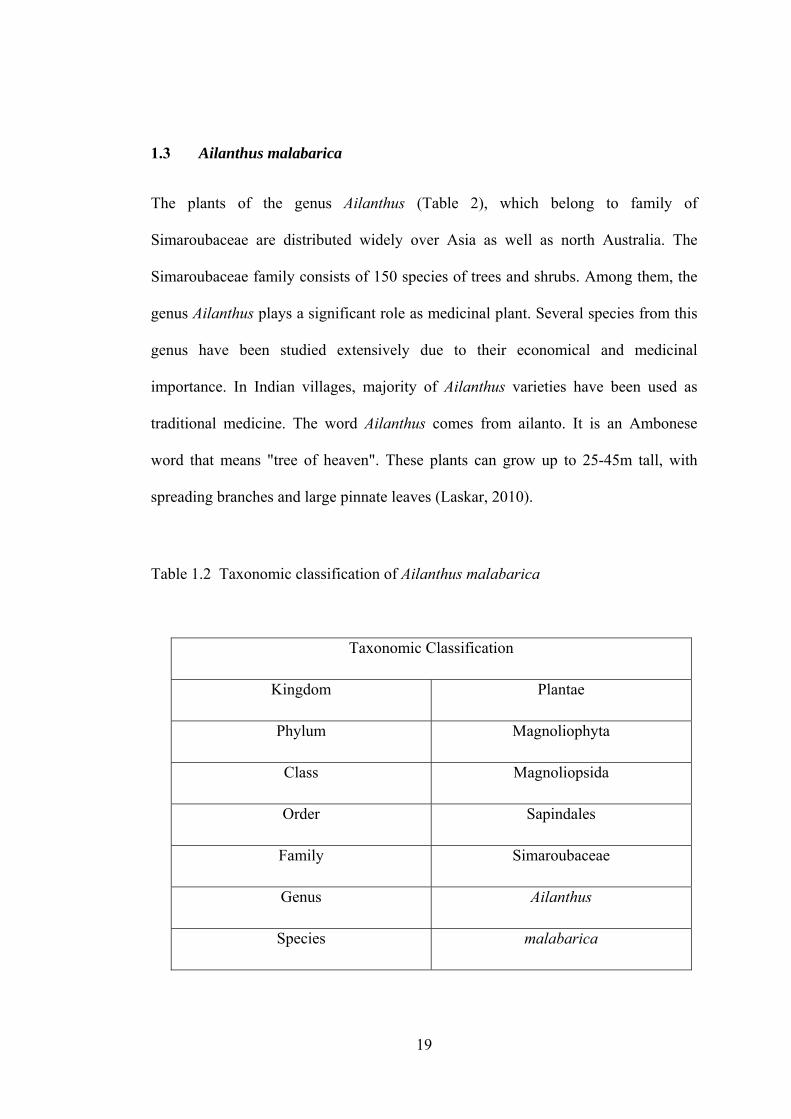
1.3 Ailanthus malabarica
The plants of the genus Ailanthus (Table 2), which belong to family of
Simaroubaceae are distributed widely over Asia as well as north Australia. The
Simaroubaceae family consists of 150 species of trees and shrubs. Among them, the
genus Ailanthus plays a significant role as medicinal plant. Several species from this
genus have been studied extensively due to their economical and medicinal
importance. In Indian villages, majority of Ailanthus varieties have been used as
traditional medicine. The word Ailanthus comes from ailanto. It is an Ambonese
word that means "tree of heaven". These plants can grow up to 25-45m tall, with
spreading branches and large pinnate leaves (Laskar, 2010).
Table 1.2 Taxonomic classification of Ailanthus malabarica
Taxonomic Classification
Kingdom Plantae
Phylum Magnoliophyta
Class Magnoliopsida
Order Sapindales
Family Simaroubaceae
Genus Ailanthus
Species malabarica