Bio-Electrospraying: A Potentially Safe Technique for ... for website/BAB.pdf · ARTICLE...
9
ARTICLE Bio-Electrospraying: A Potentially Safe Technique for Delivering Progenitor Cells Sambit Sahoo, 1,2 Wong Cheng Lee, 3 James Cho Hong Goh, 1,2,3 Siew Lok Toh 1,3,4 1 NUS Tissue Engineering Program, National University of Singapore, Singapore 2 Department of Orthopaedic Surgery, National University Hospital, National University of Singapore, Singapore 3 Tissue Repair Lab, Division of Bioengineering, National University of Singapore, EA-03-12, 9 Engineering Drive 1, Singapore 117576; telephone: 65-65162920; fax: 65-68723069; e-mail: [email protected] 4 Department of Mechanical Engineering, National University of Singapore, Singapore Received 18 November 2009; revision received 28 February 2010; accepted 1 March 2010 Published online 12 March 2010 in Wiley InterScience (www.interscience.wiley.com). DOI 10.1002/bit.22734 ABSTRACT: Bio-electrospraying is fast becoming an attrac- tive tool for in situ cell delivery into scaffolds for tissue engineering applications, with several cell types been suc- cessfully electrosprayed. Bone marrow derived mesenchymal progenitor/stem cells (BMSC), which are an important cell source for tissue engineering, have not been explored in detail and the effect of electrospraying on their ‘‘stemness’’ is not known. This study therefore investigates the effects of electrospraying on BMSC viability, proliferation, and multi- lineage differentiation potential. Electrospraying a BMSC suspension at flow rate of 6 mL/h and voltages of 7.5–15 kV could successfully generate a continuous, stable and linearly directed electrospray of cells. Morphological observation, trypan blue tests and alamar blue based metabolic assays revealed about 88% of these electrosprayed cells were viable, and proliferated at rates similar to native BMSCs. However, at higher voltages, electrospraying became unstable and reduced cell viability, possibly due to electrical or thermal damage to the cells. BMSCs electrosprayed at 7.5 kV also retained their multipotency and could be successfully dif- ferentiated into adipogenic, chondrogenic, and osteogenic lineages, demonstrating similar morphology and gene expression levels as induced native BMSCs. These results indicate that bio-electrospraying could be safely used as a progenitor/stem cell delivery technique for tissue engineer- ing and regenerative medicine applications. Biotechnol. Bioeng. 2010;106: 690–698. ß 2010 Wiley Periodicals, Inc. KEYWORDS: electrospraying; progenitor/stem cells; bone marrow stem cells; cell delivery; cell seeding; tissue engineering Introduction Tissue engineering approaches have conventionally relied upon separate sequential techniques for scaffold fabrication and subsequent cell seeding and population of the scaffolds. Such approaches face the limitation of poor cell infiltration and non-uniform cell distribution in three-dimensional (3- D) scaffolds. As the distribution of tissue formed within engineered constructs is related to the initial distribution of cells within the scaffold (Holy et al., 2000), several cell engineering techniques capable of directly delivering living cells in situ into simultaneously fabricated scaffolds have been attempted to overcome this limitation (Jayasinghe et al., 2006a; Yang and El Haj, 2006). In particular, the use of jet-based techniques to process cell suspensions has been gaining attention due to their ability to directly organize or ‘‘print’’ cells into 3-D architectures to create functional tissue constructs. Electrospraying or electrohydrodynamic jetting is one such jet-based technology wherein a liquid medium is charged to several thousand volts and passed through a large-bore needle to be fragmented into droplets (Eagles et al., 2006). ‘‘Bio-electrospraying’’ a suspension of living cells can form micro-droplets of cells without causing any apparent deleterious effect on the cells and can generate finer droplets and process denser cell suspensions than other jetting techniques such as ink-jet printing and aerosol delivery (Jayasinghe et al., 2006a,b). Recent studies have reported successful electrospraying of several mature cell types including smooth muscle cells (Stankus et al., 2006), neuronal cells (Eagles et al., 2006), kidney cells (Kwok et al., 2008), lymphocytes (Kempski et al., 2008), and even multicellular organisms (Clarke and Jayasinghe, 2008; Odenwalder et al., 2007). While more than 70% of the Sambit Sahoo and Wong Cheng Lee contributed equally to this work. Correspondence to: S.L. Toh Contract grant sponsor: National Medical Research Council, Singapore 690 Biotechnology and Bioengineering, Vol. 106, No. 4, July 1, 2010 ß 2010 Wiley Periodicals, Inc.
Transcript of Bio-Electrospraying: A Potentially Safe Technique for ... for website/BAB.pdf · ARTICLE...
ARTICLE
Bio-Electrospraying: A Potentially Safe Techniquefor Delivering Progenitor Cells
Sambit Sahoo,1,2 Wong Cheng Lee,3 James Cho Hong Goh,1,2,3 Siew Lok Toh1,3,4
1NUS Tissue Engineering Program, National University of Singapore, Singapore2Department of Orthopaedic Surgery, National University Hospital,
National University of Singapore, Singapore3Tissue Repair Lab, Division of Bioengineering, National University of Singapore,
EA-03-12, 9 Engineering Drive 1, Singapore 117576; telephone: 65-65162920;
fax: 65-68723069; e-mail: [email protected] of Mechanical Engineering, National University of Singapore,
Singapore
Received 18 November 2009; revision received 28 February 2010; accepted 1 March 2010
Published online 12 March 2010 in Wiley InterScience (www.interscience.wiley.com)
. DOI 10.1002/bit.22734ABSTRACT: Bio-electrospraying is fast becoming an attrac-tive tool for in situ cell delivery into scaffolds for tissueengineering applications, with several cell types been suc-cessfully electrosprayed. Bone marrow derived mesenchymalprogenitor/stem cells (BMSC), which are an important cellsource for tissue engineering, have not been explored indetail and the effect of electrospraying on their ‘‘stemness’’ isnot known. This study therefore investigates the effects ofelectrospraying on BMSC viability, proliferation, and multi-lineage differentiation potential. Electrospraying a BMSCsuspension at flow rate of 6 mL/h and voltages of 7.5–15 kVcould successfully generate a continuous, stable and linearlydirected electrospray of cells. Morphological observation,trypan blue tests and alamar blue based metabolic assaysrevealed about 88% of these electrosprayed cells were viable,and proliferated at rates similar to native BMSCs. However,at higher voltages, electrospraying became unstable andreduced cell viability, possibly due to electrical or thermaldamage to the cells. BMSCs electrosprayed at 7.5 kV alsoretained their multipotency and could be successfully dif-ferentiated into adipogenic, chondrogenic, and osteogeniclineages, demonstrating similar morphology and geneexpression levels as induced native BMSCs. These resultsindicate that bio-electrospraying could be safely used as aprogenitor/stem cell delivery technique for tissue engineer-ing and regenerative medicine applications.
Biotechnol. Bioeng. 2010;106: 690–698.
� 2010 Wiley Periodicals, Inc.
KEYWORDS: electrospraying; progenitor/stem cells; bonemarrow stem cells; cell delivery; cell seeding; tissueengineering
Sambit Sahoo and Wong Cheng Lee contributed equally to this work.
Correspondence to: S.L. Toh
Contract grant sponsor: National Medical Research Council, Singapore
690 Biotechnology and Bioengineering, Vol. 106, No. 4, July 1, 2010
Introduction
Tissue engineering approaches have conventionally reliedupon separate sequential techniques for scaffold fabricationand subsequent cell seeding and population of the scaffolds.Such approaches face the limitation of poor cell infiltrationand non-uniform cell distribution in three-dimensional (3-D) scaffolds. As the distribution of tissue formed withinengineered constructs is related to the initial distribution ofcells within the scaffold (Holy et al., 2000), several cellengineering techniques capable of directly delivering livingcells in situ into simultaneously fabricated scaffolds havebeen attempted to overcome this limitation (Jayasingheet al., 2006a; Yang and El Haj, 2006).
In particular, the use of jet-based techniques to processcell suspensions has been gaining attention due to theirability to directly organize or ‘‘print’’ cells into 3-Darchitectures to create functional tissue constructs.Electrospraying or electrohydrodynamic jetting is one suchjet-based technology wherein a liquid medium is charged toseveral thousand volts and passed through a large-boreneedle to be fragmented into droplets (Eagles et al., 2006).‘‘Bio-electrospraying’’ a suspension of living cells can formmicro-droplets of cells without causing any apparentdeleterious effect on the cells and can generate finer dropletsand process denser cell suspensions than other jettingtechniques such as ink-jet printing and aerosol delivery(Jayasinghe et al., 2006a,b). Recent studies have reportedsuccessful electrospraying of several mature cell typesincluding smooth muscle cells (Stankus et al., 2006),neuronal cells (Eagles et al., 2006), kidney cells (Kwok et al.,2008), lymphocytes (Kempski et al., 2008), and evenmulticellular organisms (Clarke and Jayasinghe, 2008;Odenwalder et al., 2007). While more than 70% of the
� 2010 Wiley Periodicals, Inc.
electrosprayed mature cells have been shown to remainviable in these studies during short-term culture over 1 week(Stankus et al., 2006), few studies have attempted to bio-electrospray stem cells (Mongkoldhumrongkul et al., 2009).Bio-electrosprayed stem cell viability has been demonstratedover a period of 72 h, but a detailed assessment of their fateafter extended culture periods, particularly their multi-lineage differentiation potential, has not been reported yet.
The multilineage differentiation potential of stem cells hasbeen explored for engineering and regeneration of variousmesenchymal tissues such as bone, cartilage, muscle,ligament, tendon, and adipose tissue (Banfi et al., 2000;Muraglia et al., 2000; Pittenger et al., 1999; Sahoo et al.,2006). The bone marrow is a rich source of hematopoieticstem cells and non-hematopoietic mesenchymal stem/progenitor cells. A subset of these mesenchymal cells thatare isolated by preferential adhesion on tissue culture plasticare termed as bone marrow stromal cells (BMSCs)(Prockop, 1997). BMSCs have been frequently used as seedcells in tissue engineering applications due to their ease ofisolation, capacity of in vitro self-renewal and expansion,and multipotency.
Bio-electrospraying could be a useful technique to deliverthese cells in situ into 3-D scaffolds, so as to improve cellinfiltration and distribution in the scaffolds. However, theeffects of bio-electrospraying, which involves exposing thecells to high electric fields, on BMSC viability, proliferationand multilineage differentiation potential is yet unknown.For the successful application of electrospraying in stem cellhandling, it is essential that the electrosprayed stem cells beinterrogated thoroughly to ensure that their viability andbio-functionality are not adversely affected. This study aimsto optimize bio-electrospraying of BMSCs and investigatethe effect of bio-electrospraying on BMSC viability,proliferation and multipotency.
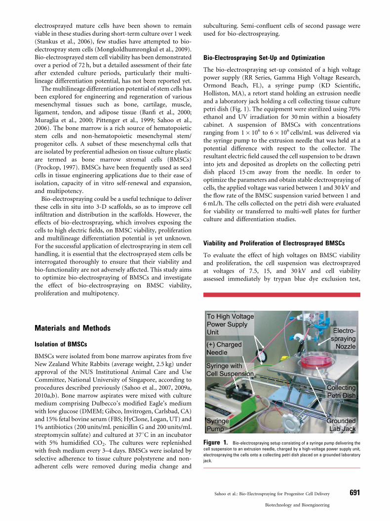
Figure 1. Bio-electrospraying setup consisting of a syringe pump delivering the
cell suspension to an extrusion needle, charged by a high-voltage power supply unit,
electrospraying the cells onto a collecting petri dish placed on a grounded laboratory
jack.
Materials and Methods
Isolation of BMSCs
BMSCs were isolated from bone marrow aspirates from fiveNew Zealand White Rabbits (average weight, 2.5 kg) underapproval of the NUS Institutional Animal Care and UseCommittee, National University of Singapore, according toprocedures described previously (Sahoo et al., 2007, 2009a,2010a,b). Bone marrow aspirates were mixed with culturemedium comprising Dulbecco’s modified Eagle’s mediumwith low glucose (DMEM; Gibco, Invitrogen, Carlsbad, CA)and 15% fetal bovine serum (FBS; HyClone, Logan, UT) and1% antibiotics (200 units/mL penicillin G and 200 units/mLstreptomycin sulfate) and cultured at 378C in an incubatorwith 5% humidified CO2. The cultures were replenishedwith fresh medium every 3–4 days. BMSCs were isolated byselective adherence to tissue culture polystyrene and non-adherent cells were removed during media change and
subculturing. Semi-confluent cells of second passage wereused for bio-electrospraying.
Bio-Electrospraying Set-Up and Optimization
The bio-electrospraying set-up consisted of a high voltagepower supply (RR Series, Gamma High Voltage Research,Ormond Beach, FL), a syringe pump (KD Scientific,Holliston, MA), a retort stand holding an extrusion needleand a laboratory jack holding a cell collecting tissue culturepetri dish (Fig. 1). The equipment were sterilized using 70%ethanol and UV irradiation for 30 min within a biosafetycabinet. A suspension of BMSCs with concentrationsranging from 1� 106 to 6� 106 cells/mL was delivered viathe syringe pump to the extrusion needle that was held at apotential difference with respect to the collector. Theresultant electric field caused the cell suspension to be drawninto jets and deposited as droplets on the collecting petridish placed 15 cm away from the needle. In order tooptimize the parameters and obtain stable electrospraying ofcells, the applied voltage was varied between 1 and 30 kV andthe flow rate of the BMSC suspension varied between 1 and6 mL/h. The cells collected on the petri dish were evaluatedfor viability or transferred to multi-well plates for furtherculture and differentiation studies.
Viability and Proliferation of Electrosprayed BMSCs
To evaluate the effect of high voltages on BMSC viabilityand proliferation, the cell suspension was electrosprayedat voltages of 7.5, 15, and 30 kV and cell viabilityassessed immediately by trypan blue dye exclusion test,
Sahoo et al.: Bio-Electrospraying for Progenitor Cell Delivery 691
Biotechnology and Bioengineering
using non-electrosprayed BMSCs (cell suspension passedthrough the electrospraying set-up at 0 kV, keeping all otherparameters unchanged) as control (n¼ 4). 0.1 mL of thesuspension of electrosprayed BMSCs in DMEM was mixedwith an equal volume of 0.4% trypan blue (Gibco) and thenumber of stained cells as well as the total number of cellscounted using a hemocytometer within 5 min.
Electrosprayed BMSCs (3� 105 BMSCs/well) were trans-ferred into separate wells of a 6-well culture plate (NUNC,Roskilde, Denmark) and cultured for 2 weeks. Cell viabilityand proliferation was monitored using alamar blue dyereduction assay by incubating the scaffolds (n¼ 3) incomplete medium containing 10% alamar blue for 3 h andmeasuring absorbance values of the media at 570 and600 nm using a 96-well microplate reader. The percentagereduction in dye, which is proportional to the cell viability inthe sample, was estimated following the vendor’s protocol(BioSource International, Camarillo, CA).
Multilineage Differentiation of Electrosprayed BMSCs
Based on the stability of the electrospraying processand results from the viability and proliferation tests, avoltage of 7.5 kV and flow rate of 6 mL/h were chosenfor electrospraying BMSCs for characterization oftheir multilineage differentiation potential. ElectrosprayedBMSCs were transferred into separate 6-well culture plates(3� 105 BMSCs/well) for osteogenic and adipogenic differ-entiation and 6� 105 cells were grown in a pellet culture forchondrogenic differentiation. The cultured cells weredifferentiated into adipogenic, osteogenic, and chondro-genic lineages using established protocols (Indrawattanaet al., 2004; Jaiswal et al., 1997; Pittenger et al., 1999;Sahoo et al., 2010a). In brief, adipogenic differentiation wasinduced by three cycles of induction (using DMEMsupplemented with 15% FBS, 0.5 mM 1-methyl-3-isobu-tylxanthine, 1mM dexamethasone, 10mg/mL insulin,0.2 mM indomethacin and antibiotics) and maintenancetreatment (using DMEM supplemented with 15% FBS,10mg/mL insulin and antibiotics). Differentiation was thendetected by Oil Red O staining for neutral lipid dropletsfound within adipocytes. Osteogenic differentiationof BMSCs was induced under the influence of 10 mMb-glycerophosphate, 0.1mM dexamethasone, 50mg/mLL-ascorbic acid 2-phosphate and 10mg/mL insulin, andAlizarin Red staining was carried out to detect orange-redcalcium deposits after 3 weeks. Chondrogenic differentia-tion was induced by culturing the BMSC pellet in serum-freeDMEM with 10 ng/mL TGF-b1, 50mg/mL L-ascorbic acid 2-phosphate, 1.25 mg/mL BSA, 0.1mM dexamethasone, 1�ITS (insulin–transferrin–selenium) and antibiotics supple-ments. After 3 weeks, the cell pellet was sectioned andstained with alcian blue for cartilage matrix-specific sulfatedmucosubstances. All the induction reagents, except ITS(Gibco) and TGF-b1 (R&D Systems, Minneapolis, MN),were from Sigma-Aldrich, St. Louis, MO.
692 Biotechnology and Bioengineering, Vol. 106, No. 4, July 1, 2010
As two separate controls, native BMSCs were eithersimilarly induced into the three lineages (positive control)or maintained in an undifferentiated condition usingexpansion medium (negative control).
Quantitative RT-PCR Analysis of BMSC Differentiation
In order to confirm BMSC differentiation, gene expressionof BMSC markers and several lineage-specific proteins wasstudied. The gene expression levels in differentiated bio-electrosprayed BMSCs were compared with two separatecontrols: native BMSCs that were either similarly inducedinto the three lineages (positive controls) or maintained inan undifferentiated condition using expansion media(negative controls). The BMSC markers used wereconnective tissue growth factor (CTGF) and matrix glaprotein (MGP) (Igarashi et al., 2007). The tissue-specificgenes analyzed were fatty acid binding protein-4 (FABP4)and peroxisome proliferator-activated receptor (PPARg2)for adipogenesis (Chawla et al., 1994; Noer et al., 2007), runtrelated transcription factor 2 (Runx2), osteopontin (OP)and osteonectin (ON) for osteogenesis (Guillot et al., 2008;Zou et al., 2008), and sox9 and aggrecan (Agg) forchondrogenesis (Indrawattana et al., 2004; Zou et al.,2008). Total RNA was extracted from the induced and non-induced control BMSCs using RNeasy Mini Kit (Qiagen,Valencia, CA), and quantitative reverse transcriptase-mediated PCR (Q-RT-PCR) was performed using SYBR-green chemistry using glyceraldehyde 3-phosphate dehy-drogenase (GAPDH) as the reference gene. Primersequences were designed, using Primer-3 software, fromrabbit gene sequences obtained from the NCBI GenBankand RefSeq databases, and synthesized by ResearchBiolabs, Singapore (Table I). cDNA synthesis and PCRexpansion (using iScript and iQ SYBR Green Supermix,Bio-Rad Laboratories, Hercules, CA) were performed in aiCycler iQ detection system (Bio-Rad Laboratories). Datawere analyzed for relative expression using the DDCT
method, and normalized against the basal expression profileof undifferentiated BMSCs (n¼ 3, for each group).
Statistical Analysis
All data were expressed as mean� standard error. Dueto small sample sizes, statistical analyses were performedby non-parametric tests using SPSS Statistics 17.0 statisticalsoftware package. Multiple groups (in trypan blue testsand Q RT-PCR results for each gene) were comparedby Kruskal–Wallis tests and post hoc Mann–Whitneytests with Bonferroni correction. Multiple group com-parisons involving repeated measures (in alamar blueassays) were performed by Friedman’s test and post hocWilcoxon tests. P-values less than 0.05 were consideredsignificant.
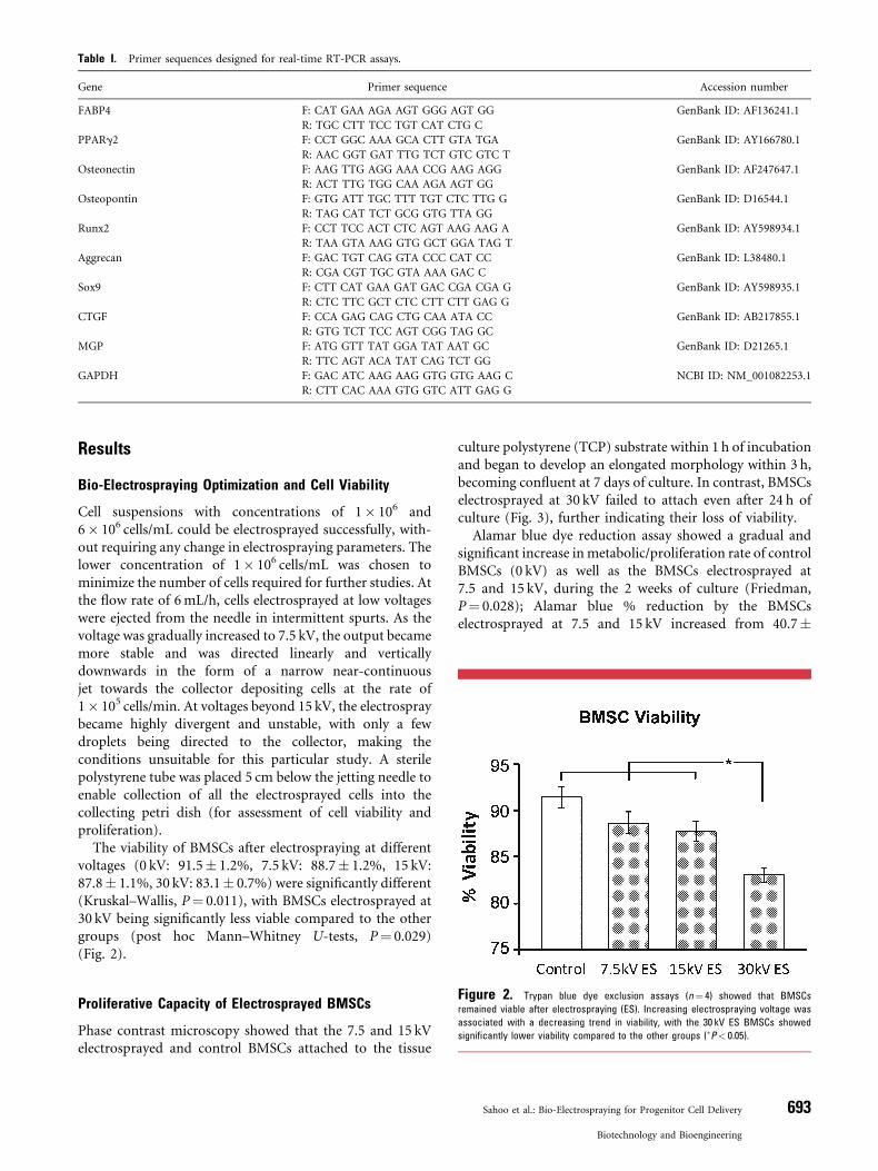
Table I. Primer sequences designed for real-time RT-PCR assays.
Gene Primer sequence Accession number
FABP4 F: CAT GAA AGA AGT GGG AGT GG GenBank ID: AF136241.1
R: TGC CTT TCC TGT CAT CTG C
PPARg2 F: CCT GGC AAA GCA CTT GTA TGA GenBank ID: AY166780.1
R: AAC GGT GAT TTG TCT GTC GTC T
Osteonectin F: AAG TTG AGG AAA CCG AAG AGG GenBank ID: AF247647.1
R: ACT TTG TGG CAA AGA AGT GG
Osteopontin F: GTG ATT TGC TTT TGT CTC TTG G GenBank ID: D16544.1
R: TAG CAT TCT GCG GTG TTA GG
Runx2 F: CCT TCC ACT CTC AGT AAG AAG A GenBank ID: AY598934.1
R: TAA GTA AAG GTG GCT GGA TAG T
Aggrecan F: GAC TGT CAG GTA CCC CAT CC GenBank ID: L38480.1
R: CGA CGT TGC GTA AAA GAC C
Sox9 F: CTT CAT GAA GAT GAC CGA CGA G GenBank ID: AY598935.1
R: CTC TTC GCT CTC CTT CTT GAG G
CTGF F: CCA GAG CAG CTG CAA ATA CC GenBank ID: AB217855.1
R: GTG TCT TCC AGT CGG TAG GC
MGP F: ATG GTT TAT GGA TAT AAT GC GenBank ID: D21265.1
R: TTC AGT ACA TAT CAG TCT GG
GAPDH F: GAC ATC AAG AAG GTG GTG AAG C NCBI ID: NM_001082253.1
R: CTT CAC AAA GTG GTC ATT GAG G
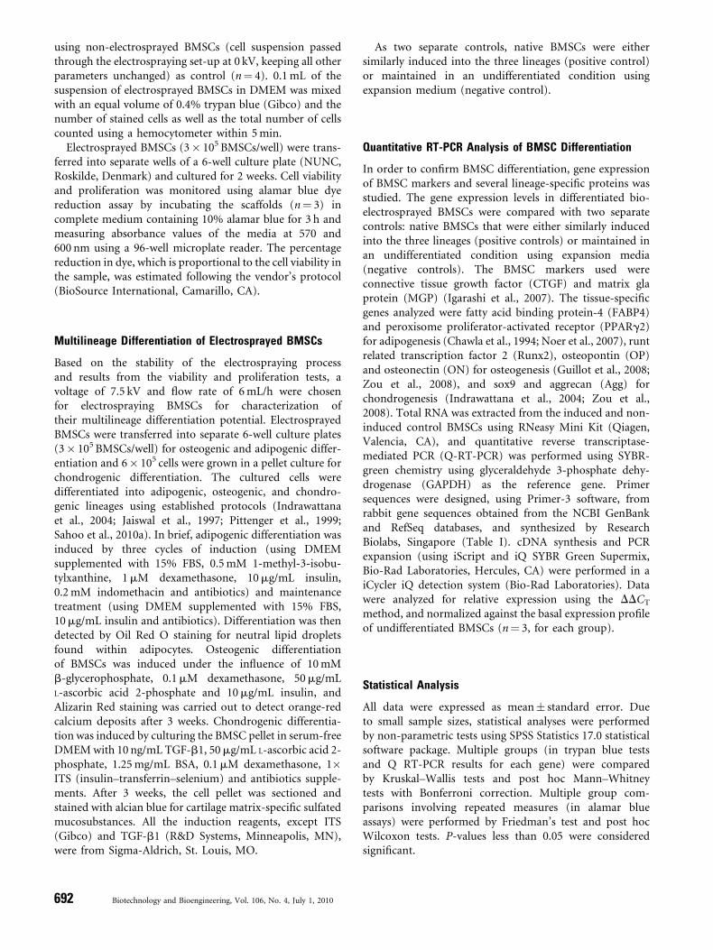
Figure 2. Trypan blue dye exclusion assays (n¼ 4) showed that BMSCs
remained viable after electrospraying (ES). Increasing electrospraying voltage was
associated with a decreasing trend in viability, with the 30 kV ES BMSCs showed
significantly lower viability compared to the other groups (�P< 0.05).
Results
Bio-Electrospraying Optimization and Cell Viability
Cell suspensions with concentrations of 1� 106 and6� 106 cells/mL could be electrosprayed successfully, with-out requiring any change in electrospraying parameters. Thelower concentration of 1� 106 cells/mL was chosen tominimize the number of cells required for further studies. Atthe flow rate of 6 mL/h, cells electrosprayed at low voltageswere ejected from the needle in intermittent spurts. As thevoltage was gradually increased to 7.5 kV, the output becamemore stable and was directed linearly and verticallydownwards in the form of a narrow near-continuousjet towards the collector depositing cells at the rate of1� 105 cells/min. At voltages beyond 15 kV, the electrospraybecame highly divergent and unstable, with only a fewdroplets being directed to the collector, making theconditions unsuitable for this particular study. A sterilepolystyrene tube was placed 5 cm below the jetting needle toenable collection of all the electrosprayed cells into thecollecting petri dish (for assessment of cell viability andproliferation).
The viability of BMSCs after electrospraying at differentvoltages (0 kV: 91.5� 1.2%, 7.5 kV: 88.7� 1.2%, 15 kV:87.8� 1.1%, 30 kV: 83.1� 0.7%) were significantly different(Kruskal–Wallis, P¼ 0.011), with BMSCs electrosprayed at30 kV being significantly less viable compared to the othergroups (post hoc Mann–Whitney U-tests, P¼ 0.029)(Fig. 2).
Proliferative Capacity of Electrosprayed BMSCs
Phase contrast microscopy showed that the 7.5 and 15 kVelectrosprayed and control BMSCs attached to the tissue
culture polystyrene (TCP) substrate within 1 h of incubationand began to develop an elongated morphology within 3 h,becoming confluent at 7 days of culture. In contrast, BMSCselectrosprayed at 30 kV failed to attach even after 24 h ofculture (Fig. 3), further indicating their loss of viability.
Alamar blue dye reduction assay showed a gradual andsignificant increase in metabolic/proliferation rate of controlBMSCs (0 kV) as well as the BMSCs electrosprayed at7.5 and 15 kV, during the 2 weeks of culture (Friedman,P¼ 0.028); Alamar blue % reduction by the BMSCselectrosprayed at 7.5 and 15 kV increased from 40.7�
Sahoo et al.: Bio-Electrospraying for Progenitor Cell Delivery 693
Biotechnology and Bioengineering
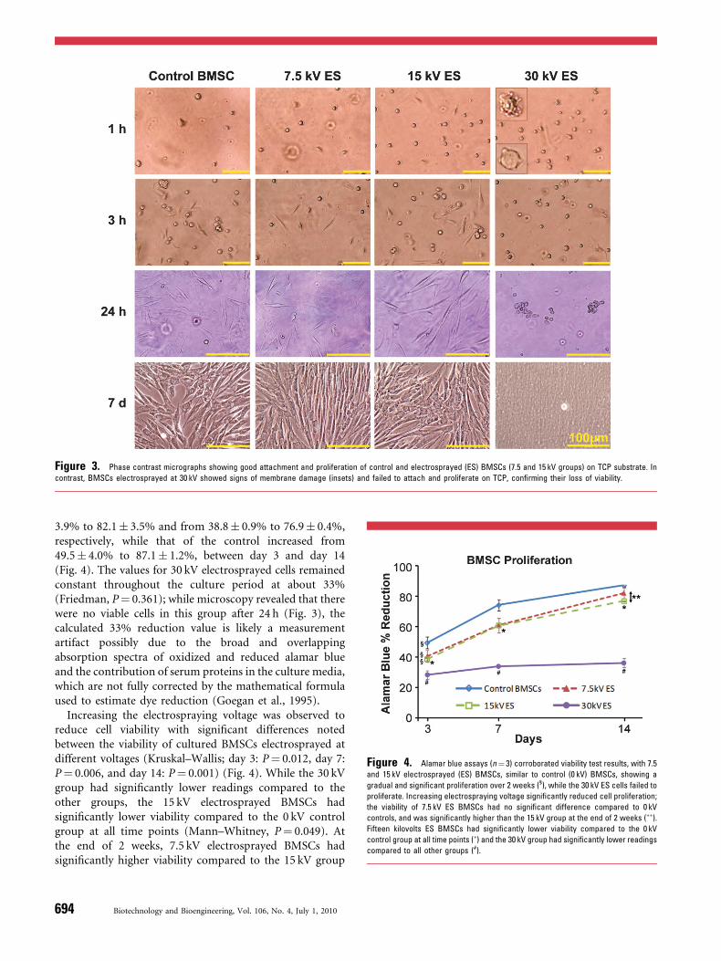
Figure 3. Phase contrast micrographs showing good attachment and proliferation of control and electrosprayed (ES) BMSCs (7.5 and 15 kV groups) on TCP substrate. In
contrast, BMSCs electrosprayed at 30 kV showed signs of membrane damage (insets) and failed to attach and proliferate on TCP, confirming their loss of viability.
Figure 4. Alamar blue assays (n¼ 3) corroborated viability test results, with 7.5
and 15 kV electrosprayed (ES) BMSCs, similar to control (0 kV) BMSCs, showing a
gradual and significant proliferation over 2 weeks (§), while the 30 kV ES cells failed to
proliferate. Increasing electrospraying voltage significantly reduced cell proliferation;
the viability of 7.5 kV ES BMSCs had no significant difference compared to 0 kV
controls, and was significantly higher than the 15 kV group at the end of 2 weeks (��).
Fifteen kilovolts ES BMSCs had significantly lower viability compared to the 0 kV
control group at all time points (�) and the 30 kV group had significantly lower readings
compared to all other groups (#).
3.9% to 82.1� 3.5% and from 38.8� 0.9% to 76.9� 0.4%,respectively, while that of the control increased from49.5� 4.0% to 87.1� 1.2%, between day 3 and day 14(Fig. 4). The values for 30 kV electrosprayed cells remainedconstant throughout the culture period at about 33%(Friedman, P¼ 0.361); while microscopy revealed that therewere no viable cells in this group after 24 h (Fig. 3), thecalculated 33% reduction value is likely a measurementartifact possibly due to the broad and overlappingabsorption spectra of oxidized and reduced alamar blueand the contribution of serum proteins in the culture media,which are not fully corrected by the mathematical formulaused to estimate dye reduction (Goegan et al., 1995).
Increasing the electrospraying voltage was observed toreduce cell viability with significant differences notedbetween the viability of cultured BMSCs electrosprayed atdifferent voltages (Kruskal–Wallis; day 3: P¼ 0.012, day 7:P¼ 0.006, and day 14: P¼ 0.001) (Fig. 4). While the 30 kVgroup had significantly lower readings compared to theother groups, the 15 kV electrosprayed BMSCs hadsignificantly lower viability compared to the 0 kV controlgroup at all time points (Mann–Whitney, P¼ 0.049). Atthe end of 2 weeks, 7.5 kV electrosprayed BMSCs hadsignificantly higher viability compared to the 15 kV group
694 Biotechnology and Bioengineering, Vol. 106, No. 4, July 1, 2010
(Mann–Whitney, P¼ 0.049), with no significant differencecompared to the 0 kV controls (Mann–Whitney, P¼ 0.275).
Multilineage Differentiation of Electrosprayed BMSCs
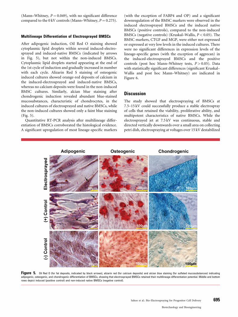
After adipogenic induction, Oil Red O staining showedcytoplasmic lipid droplets within several induced-electro-sprayed and induced-native BMSCs (indicated by arrowsin Fig. 5), but not within the non-induced BMSCs.Cytoplasmic lipid droplets started appearing at the end ofthe 1st cycle of induction and gradually increased in numberwith each cycle. Alizarin Red S staining of osteogenicinduced cultures showed orange-red deposits of calcium inthe induced-electrosprayed and induced-native BMSCs,whereas no calcium deposits were found in the non-inducedBMSC cultures. Similarly, alcian blue staining afterchondrogenic induction revealed abundant blue-stainedmucosubstances, characteristic of chondrocytes, in theinduced cultures of electrosprayed and native BMSCs, whilethe non-induced cultures showed only a faint blue staining(Fig. 5).
Quantitative RT-PCR analysis after multilineage differ-entiation of BMSCs corroborated the histological evidence.A significant upregulation of most lineage-specific markers
Figure 5. Oil Red O (for fat deposits, indicated by black arrows), alizarin red (for c
adipogenic, osteogenic, and chondrogenic differentiation of BMSCs, showing that electros
rows depict induced (positive control) and non-induced native BMSCs (negative control).
(with the exception of FABP4 and OP) and a significantdownregulation of the BMSC markers were observed in theinduced electrosprayed BMSCs and the induced nativeBMSCs (positive controls), compared to the non-inducedBMSCs (negative controls) (Kruskal–Wallis, P< 0.05). TheBMSC markers, CTGF and MGP, were either not expressedor expressed at very low levels in the induced cultures. Therewere no significant differences in expression levels of thelineage-specific genes (with the exception of aggrecan) inthe induced-electrosprayed BMSCs and the positivecontrols (post hoc Mann–Whitney tests, P> 0.05). Datawith statistically significant differences (significant Kruskal–Wallis and post hoc Mann–Whitney) are indicated inFigure 6.
Discussion
The study showed that electrospraying of BMSCs at7.5–15 kV could successfully produce a stable electrosprayof cells that retained the viability, proliferative ability, andmultipotent characteristics of native BMSCs. While theelectrosprayed jet at 7.5 kV was continuous, stable anddirected vertically downwards over a small area on collectingpetri dish, electrospraying at voltages over 15 kV destabilized
alcium deposits) and alcian blue staining (for sulfated mucosubstances) indicating
prayed BMSCs retained their multilineage differentiation potential. Middle and bottom
Sahoo et al.: Bio-Electrospraying for Progenitor Cell Delivery 695
Biotechnology and Bioengineering
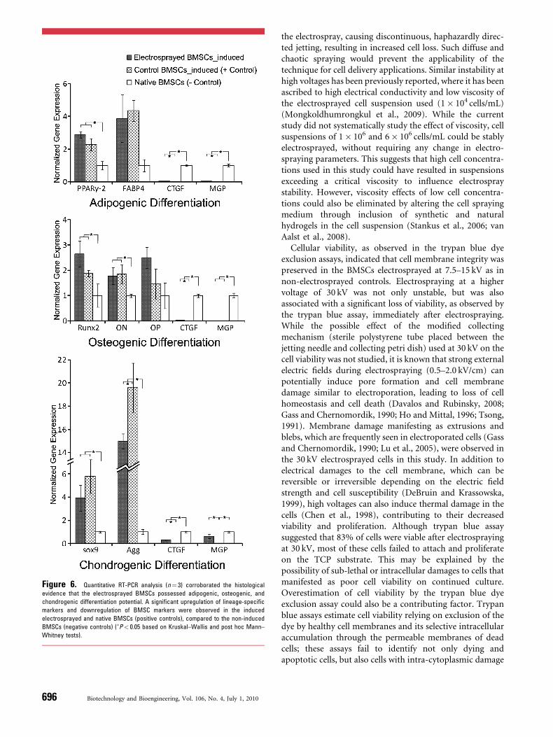
Figure 6. Quantitative RT-PCR analysis (n¼ 3) corroborated the histological
evidence that the electrosprayed BMSCs possessed adipogenic, osteogenic, and
chondrogenic differentiation potential. A significant upregulation of lineage-specific
markers and downregulation of BMSC markers were observed in the induced
electrosprayed and native BMSCs (positive controls), compared to the non-induced
BMSCs (negative controls) (�P< 0.05 based on Kruskal–Wallis and post hoc Mann–
Whitney tests).
696 Biotechnology and Bioengineering, Vol. 106, No. 4, July 1, 2010
the electrospray, causing discontinuous, haphazardly direc-ted jetting, resulting in increased cell loss. Such diffuse andchaotic spraying would prevent the applicability of thetechnique for cell delivery applications. Similar instability athigh voltages has been previously reported, where it has beenascribed to high electrical conductivity and low viscosity ofthe electrosprayed cell suspension used (1� 104 cells/mL)(Mongkoldhumrongkul et al., 2009). While the currentstudy did not systematically study the effect of viscosity, cellsuspensions of 1� 106 and 6� 106 cells/mL could be stablyelectrosprayed, without requiring any change in electro-spraying parameters. This suggests that high cell concentra-tions used in this study could have resulted in suspensionsexceeding a critical viscosity to influence electrospraystability. However, viscosity effects of low cell concentra-tions could also be eliminated by altering the cell sprayingmedium through inclusion of synthetic and naturalhydrogels in the cell suspension (Stankus et al., 2006; vanAalst et al., 2008).
Cellular viability, as observed in the trypan blue dyeexclusion assays, indicated that cell membrane integrity waspreserved in the BMSCs electrosprayed at 7.5–15 kV as innon-electrosprayed controls. Electrospraying at a highervoltage of 30 kV was not only unstable, but was alsoassociated with a significant loss of viability, as observed bythe trypan blue assay, immediately after electrospraying.While the possible effect of the modified collectingmechanism (sterile polystyrene tube placed between thejetting needle and collecting petri dish) used at 30 kV on thecell viability was not studied, it is known that strong externalelectric fields during electrospraying (0.5–2.0 kV/cm) canpotentially induce pore formation and cell membranedamage similar to electroporation, leading to loss of cellhomeostasis and cell death (Davalos and Rubinsky, 2008;Gass and Chernomordik, 1990; Ho and Mittal, 1996; Tsong,1991). Membrane damage manifesting as extrusions andblebs, which are frequently seen in electroporated cells (Gassand Chernomordik, 1990; Lu et al., 2005), were observed inthe 30 kV electrosprayed cells in this study. In addition toelectrical damages to the cell membrane, which can bereversible or irreversible depending on the electric fieldstrength and cell susceptibility (DeBruin and Krassowska,1999), high voltages can also induce thermal damage in thecells (Chen et al., 1998), contributing to their decreasedviability and proliferation. Although trypan blue assaysuggested that 83% of cells were viable after electrosprayingat 30 kV, most of these cells failed to attach and proliferateon the TCP substrate. This may be explained by thepossibility of sub-lethal or intracellular damages to cells thatmanifested as poor cell viability on continued culture.Overestimation of cell viability by the trypan blue dyeexclusion assay could also be a contributing factor. Trypanblue assays estimate cell viability relying on exclusion of thedye by healthy cell membranes and its selective intracellularaccumulation through the permeable membranes of deadcells; these assays fail to identify not only dying andapoptotic cells, but also cells with intra-cytoplasmic damage
such as that incurred by thermal injury, and generallyoverestimate viability in such cell populations (Altman et al.,1993; Mascotti et al., 2000).
Since electrospraying at 7.5 kV was stable and did notnegatively affect cell viability and proliferation, 7.5 kVelectrosprayed cells were chosen for further characterizationof BMSCs for demonstration of their multipotency.Histochemical staining as well as gene expression analysis(upregulation of lineage-specific markers and downregula-tion of BMSC markers) showed that electrosprayed BMSCscould be successfully differentiated into adipogenic,chondrogenic, and osteogenic lineages. No significantdifferences could be observed in expression levels of mostlineage-specific genes after induction of electrosprayed andcontrol BMSCs, indicating that the multipotency of theprogenitor cells was preserved after electrospraying. It wasobserved that while induced native BMSCs stoppedexpressing the BMSC markers, the induced bio-electro-sprayed BMSCs still expressed them at low levels; inaddition, bio-electrosprayed BMSCs after chondrogenicinduction expressed aggrecan at a significantly lower levelthan the induced native BMSCs. This suggests the possibilitythat some of the bio-electrosprayed BMSCs might haveretained their BMSC phenotype instead of differentiatingalong terminal lineages. Further investigation using humanmesenchymal stem cells (MSCs; as compared to theheterogeneous population of primary rabbit BMSCs usedin this study), including additional differentiation markersand larger sample sizes (to allow parametric data analysis)would be required to obtain a better understanding of thestemness of bio-electrosprayed stem cells.
A recent study has reported the successful electrosprayingof immortalized human MSCs: a viability of more than 98%(based on propidium iodide staining and flow cytometry)without any apoptotic cell death was reported, and the cellsproliferated at rates similar to control cells over 3 days(Mongkoldhumrongkul et al., 2009). While the observeddifference in cell viability (98%, compared to 88% observedin this study) could be due several factors such as differencesin the electrospraying conditions, varying cell susceptibilityto damages during electrospraying, as well as differentsensitivity of the viability test used, both studies indicate thesafety and potential of use of electrospraying technique forstem cell delivery. Additionally, the current study demon-strates cell proliferation being maintained normally over alonger period, and more importantly, demonstrates thepreservation of multipotency of the BMSCs.
Bio-electrospraying could be used in combination withthe closely related technology of electrospinning to generatenanofibrous scaffolds seeded in situ with a uniform 3-Ddistribution of cells. While conventionally seeded electro-spun scaffolds have slow and restricted cell infiltration intothe depths of the scaffolds, simultaneous electrospinning ofnanofibers and bio-electrospraying of cells can improve cellinfiltration and distribution in these scaffolds (Sahoo et al.,2009b; Stankus et al., 2006, 2007). In order to make thetechnique applicable in tissue engineering, a moving
mechanism such as a X–Y translating or a rotating collector(in place of the static collecting petri dish used in this study)could be incorporated to achieve spatial control on celldelivery into the scaffold (Stankus et al., 2006, 2007).Nanofibrous scaffolds thus rapidly and uniformly seededwith multipotent BMSCs, which can be differentiated alongany desired lineage, would have the potential for use indifferent tissue engineering applications.
Conclusion
This study shows that electrospraying of BMSCs at 7.5–15 kV could successfully generate a stable, linearly directedelectrospray of progenitor cells. The preserved viability,proliferative ability, and multilineage differentiationpotential of such electrosprayed BMSCs indicate that bio-electrospraying could be safely used as a progenitor/stem celldelivery technique for tissue engineering. When combinedwith the technology of electrospinning, bio-electrosprayinghas the potential to create nanofibrous scaffolds with auniform 3-D distribution of multipotent BMSCs for usein various tissue engineering and regenerative medicineapplications.
References
Altman SA, Randers L, Rao G. 1993. Comparison of trypan blue dye
exclusion and fluorometric assays for mammalian cell viability deter-
minations. Biotechnol Prog 9(6):671–674.
Banfi A, Muraglia A, Dozin B, Mastrogiacomo M, Cancedda R, Quarto R.
2000. Proliferation kinetics and differentiation potential of ex vivo
expanded human bone marrow stromal cells: Implications for their use
in cell therapy. Exp Hematol 28(6):707–715.
Chawla A, Schwarz EJ, Dimaculangan DD, Lazar MA. 1994. Peroxisome
proliferator-activated receptor (PPAR) gamma: Adipose-predominant
expression and induction early in adipocyte differentiation. Endocri-
nology 135(2):798–800.
Chen W, Han Y, Chen Y, Xie JT. 1998. Field-induced electroconformational
damages in cell membrane proteins: A new mechanism involved in
electrical injury. Bioelectrochem Bioenerg 47(2):237–245.
Clarke JD, Jayasinghe SN. 2008. Bio-electrosprayed multicellular zebrafish
embryos are viable and develop normally. Biomed Mater 3(1):11001.
Davalos RV, Rubinsky B. 2008. Temperature considerations during irre-
versible electroporation. Int J Heat Mass Transf 51(23–24):5617–55622.
DeBruin KA, Krassowska W. 1999. Modeling electroporation in a single cell.
I. Effects Of field strength and rest potential. Biophys J 77(3):1213–
1224.
Eagles PA, Qureshi AN, Jayasinghe SN. 2006. Electrohydrodynamic jetting
of mouse neuronal cells. Biochem J 394(Pt 2):375–378.
Gass GV, Chernomordik LV. 1990. Reversible large-scale deformations in
the membranes of electrically-treated cells: Electroinduced bleb for-
mation. Biochim Biophys Acta 1023(1):1–11.
Goegan P, Johnson G, Vincent R. 1995. Effects of serum protein and colloid
on the alamarBlue assay in cell cultures. Toxicol In Vitro 9(3):257–266.
Guillot PV, De Bari C, Dell’Accio F, Kurata H, Polak J, Fisk NM. 2008.
Comparative osteogenic transcription profiling of various fetal and
adult mesenchymal stem cell sources. Differentiation 76(9):946–957.
Ho SY, Mittal GS. 1996. Electroporation of cell membranes: A review. Crit
Rev Biotechnol 16(4):349–362.
Holy CE, Shoichet MS, Davies JE. 2000. Engineering three-dimensional
bone tissue in vitro using biodegradable scaffolds: Investigating initial
Sahoo et al.: Bio-Electrospraying for Progenitor Cell Delivery 697
Biotechnology and Bioengineering
cell-seeding density and culture period. J Biomed Mater Res 51(3):376–
382.
Igarashi A, Segoshi K, Sakai Y, Pan H, Kanawa M, Higashi Y, Sugiyama M,
Nakamura K, Kurihara H, Yamaguchi S, Tsuji K, Kawamoto T, Kato Y.
2007. Selection of common markers for bone marrow stromal cells
from various bones using real-time RT-PCR: Effects of passage number
and donor age. Tissue Eng 13(10):2405–2417.
Indrawattana N, Chen G, Tadokoro M, Shann LH, Ohgushi H, Tateishi T,
Tanaka J, Bunyaratvej A. 2004. Growth factor combination for chon-
drogenic induction from human mesenchymal stem cell. Biochem
Biophys Res Commun 320(3):914–919.
Jaiswal N, Haynesworth SE, Caplan AI, Bruder SP. 1997. Osteogenic
differentiation of purified, culture-expanded human mesenchymal
stem cells in vitro. J Cell Biochem 64(2):295–312.
Jayasinghe SN, Eagles PA, Qureshi AN. 2006a. Electric field driven jetting:
An emerging approach for processing living cells. Biotechnol J 1(1):
86–94.
Jayasinghe SN, Qureshi AN, Eagles PA. 2006b. Electrohydrodynamic jet
processing: An advanced electric-field-driven jetting phenomenon for
processing living cells. Small 2(2):216–219.
Kempski H, Austin N, Roe A, Chatters S, Jayasinghe SN. 2008. Pilot study to
investigate the possibility of cytogenetic and physiological changes in
bio-electrosprayed human lymphocyte cells. Regen Med 3(3):343–
349.
Kwok A, Arumuganathar S, Irvine S, McEwan JR, Jayasinghe SN. 2008.
A hybrid bio-jetting approach for directly engineering living cells.
Biomed Mater 3(2):25008.
Lu H, Schmidt MA, Jensen KF. 2005. A microfluidic electroporation device
for cell lysis. Lab Chip 5(1):23–29.
Mascotti K, McCullough J, Burger SR. 2000. HPC viability measurement:
Trypan blue versus acridine orange and propidium iodide. Transfusion
40(6):693–696.
Mongkoldhumrongkul N, Flanagan JM, Jayasinghe SN. 2009. Direct jetting
approaches for handling stem cells. Biomed Mater 4(1):15018.
Muraglia A, Cancedda R, Quarto R. 2000. Clonal mesenchymal progenitors
from human bone marrow differentiate in vitro according to a hier-
archical model. J Cell Sci 113(Pt 7):1161–11616.
Noer A, Boquest AC, Collas P. 2007. Dynamics of adipogenic promoter
DNA methylation during clonal culture of human adipose stem cells to
senescence. BMC Cell Biol 8:18.
Odenwalder PK, Irvine S, McEwan JR, Jayasinghe SN. 2007. Bio-electro-
sprays: A novel electrified jetting methodology for the safe handling and
deployment of primary living organisms. Biotechnol J 2(5):622–630.
698 Biotechnology and Bioengineering, Vol. 106, No. 4, July 1, 2010
Pittenger MF, Mackay AM, Beck SC, Jaiswal RK, Douglas R, Mosca JD,
Moorman MA, Simonetti DW, Craig S, Marshak DR. 1999. Multi-
lineage potential of adult human mesenchymal stem cells. Science
284(5411):143–147.
Prockop DJ. 1997. Marrow stromal cells as stem cells for nonhematopoietic
tissues. Science 276(5309):71–74.
Sahoo S, Ouyang H, Goh JC, Tay TE, Toh SL. 2006. Characterization of a
novel polymeric scaffold for potential application in tendon/ligament
tissue engineering. Tissue Eng 12(1):91–99.
Sahoo S, Cho-Hong JG, Siew-Lok T. 2007. Development of hybrid polymer
scaffolds for potential applications in ligament and tendon tissue
engineering. Biomed Mater 2(3):169–173.
Sahoo S, Ang LT, Goh JCH, Toh SL. 2009a. Growth factor delivery through
electrospun nanofibers in scaffolds for tissue engineering applications.
J Biomed Mater Res A. doi:10.1002/jbm.a.32645.
Sahoo S, Lee WC, Goh JCH, Toh SL. 2009b. Electrosprayed progenitor
cells in electrospun scaffolds for tissue engineering applications,
2nd TERMIS World Congress, Seoul, South Korea.
Sahoo S, Ang LT, Goh JCH, Toh SL. 2010a. Bioactive nanofibers for
fibroblastic differentiation of mesenchymal precursor cells for liga-
ment/tendon tissue engineering applications. Differentiation 79(2):
102–110.
Sahoo S, Toh SL, Goh JCH. 2010b. A bFGF-releasing silk/PLGA-based
biohybrid scaffold for ligament/tendon tissue engineering using
mesenchymal progenitor cells. Biomaterials 31(11):2990–2998.
Stankus JJ, Guan J, Fujimoto K, Wagner WR. 2006. Microintegrating
smooth muscle cells into a biodegradable, elastomeric fiber matrix.
Biomaterials 27(5):735–744.
Stankus JJ, Soletti L, Fujimoto K, Hong Y, Vorp DA, Wagner WR. 2007.
Fabrication of cell microintegrated blood vessel constructs through
electrohydrodynamic atomization. Biomaterials 28(17):2738–2746.
Tsong TY. 1991. Electroporation of cell membranes. Biophys J 60(2):297–
306.
van Aalst JA, Reed CR, Han L, Andrady T, Hromadka M, Bernacki S,
Kolappa K, Collins JB, Loboa EG. 2008. Cellular incorporation into
electrospun nanofibers: Retained viability, proliferation, and function
in fibroblasts. Ann Plast Surg 60(5):577–583.
Yang Y, El Haj AJ. 2006. Biodegradable scaffolds—Delivery systems for cell
therapies. Expert Opin Biol Ther 6(5):485–498.
Zou L, Zou X, Chen L, Li H, Mygind T, Kassem M, Bunger C. 2008.
Multilineage differentiation of porcine bone marrow stromal cells
associated with specific gene expression pattern. J Orthop Res 26(1):
56–64.